Embed Size (px)
Citation preview
Newsdesk
336 http://neurology.thelancet.com Vol 4 June 2005
Putative neurotoxin ecologically widespread
Results of a new study by JamesConner (Department of Neurosciencesat the University of California at SanDiego, CA, USA) and colleaguesindicate that depletion of cholinergicfunction in the basal forebraincompletely abolishes the corticalplasticity needed to mediate functionalrecovery after brain injury.
Working with colleagues AndreaChiba and Mark Tuszynski (Depart-ment of Cognitive Science), Connerused rats that had been taught to reachfor food pellets with their forepaws toinvestigate the role of the cholinergicsystem in rehabilitation from braininjury (Neuron 2005; 46: 173–79). Therats were divided into two groups; onegroup was given a toxin to specificallyeliminate cholinergic inputs to the
motor and sensory regions of thecortex, whereas the other groupreceived sham injection. Both groupsof rats were then given small lesions inthe brain to cause deficits in limbmovement. After rehabilitationtraining, the control group recoveredabout 55% of the deficit in theirreaching ability, whereas the rats withdamaged cholinergic systemsrecovered about 18%. Moreimportantly, the reorganisation ofcortical circuitry thought responsiblefor mediating the functional recoverywas abolished in the rats lackingcholinergic input to the cortex.
“The reason these results are ofinterest is that this particular brainsystem—the basal forebrain cholin-ergic system—has been documented
to undergo degeneration or dys-function in several instances”,explains Conner. Studies havesuggested that cholinergic function ispartly compromised during normalageing in rats, monkeys, and humanbeings and can be severelycompromised in patients withAlzheimer’s disease. Also, variousstudies in rats and human beingshave suggested that the basalforebrain cholinergic system may alsobecome dysfunctional after traumaticbrain injury—eg, after a car accidentor another type of head impact. “Inthe latter case, one can imagine that adeficit in brain cholinergic functionmay impair the critical plasticitymechanisms required to recover fromthe damage caused by the head
Brain-injury recovery may need cholinergic system
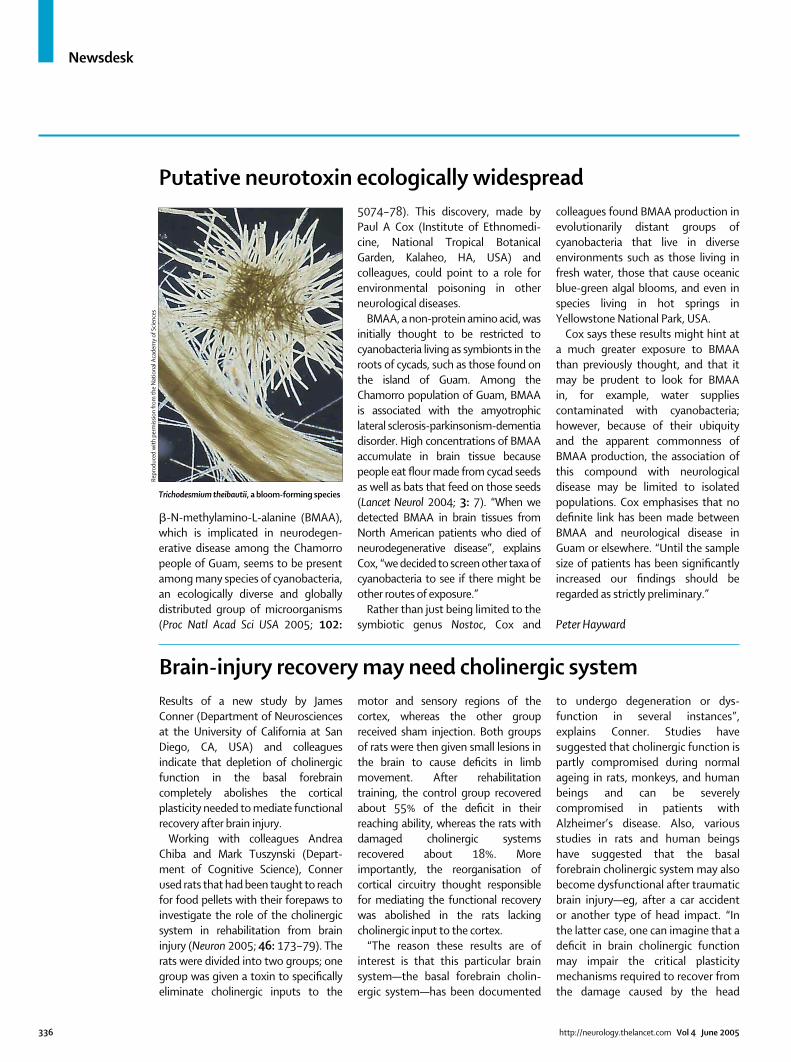
�-N-methylamino-L-alanine (BMAA),which is implicated in neurodegen-erative disease among the Chamorropeople of Guam, seems to be presentamong many species of cyanobacteria,an ecologically diverse and globallydistributed group of microorganisms(Proc Natl Acad Sci USA 2005; 102:
5074–78). This discovery, made byPaul A Cox (Institute of Ethnomedi-cine, National Tropical BotanicalGarden, Kalaheo, HA, USA) andcolleagues, could point to a role forenvironmental poisoning in otherneurological diseases.
BMAA, a non-protein amino acid, wasinitially thought to be restricted tocyanobacteria living as symbionts in theroots of cycads, such as those found onthe island of Guam. Among theChamorro population of Guam, BMAAis associated with the amyotrophiclateral sclerosis-parkinsonism-dementiadisorder. High concentrations of BMAAaccumulate in brain tissue becausepeople eat flour made from cycad seedsas well as bats that feed on those seeds(Lancet Neurol 2004; 3: 7). “When wedetected BMAA in brain tissues fromNorth American patients who died ofneurodegenerative disease”, explainsCox, “we decided to screen other taxa ofcyanobacteria to see if there might beother routes of exposure.”
Rather than just being limited to thesymbiotic genus Nostoc, Cox and
colleagues found BMAA production inevolutionarily distant groups ofcyanobacteria that live in diverseenvironments such as those living infresh water, those that cause oceanicblue-green algal blooms, and even inspecies living in hot springs inYellowstone National Park, USA.
Cox says these results might hint ata much greater exposure to BMAAthan previously thought, and that itmay be prudent to look for BMAAin, for example, water suppliescontaminated with cyanobacteria;however, because of their ubiquityand the apparent commonness ofBMAA production, the association ofthis compound with neurologicaldisease may be limited to isolatedpopulations. Cox emphasises that nodefinite link has been made betweenBMAA and neurological disease inGuam or elsewhere. “Until the samplesize of patients has been significantlyincreased our findings should beregarded as strictly preliminary.”
Peter Hayward
Trichodesmium theibautii, a bloom-forming species
Repr
oduc
ed w
ith
perm
issi
on fr
om th
e N
atio
nal A
cade
my
of S
cien
ces
Newsdesk
injury. In a sense, the accident is aone-two punch, whereby the injurydamages the brain and also impairsthe brain’s inherent mechanisms forrecovering from the damage”, Connercontinues.
According to Conner, more studiesare needed to further elucidate theprecise mechanisms involved, and his
group is considering various animalmodels in which cholinergic deficitshave been documented, includingmodels of ageing and traumatic braininjury. “Eventually, our hope is toassess whether enhancing cholinergicfunction, either with FDA approvedcholinesterase inhibitors, like donepezilor tacrine, or with cholinergic-specific
trophic factors, such as nerve growthfactor, will improve recovery. Nervegrowth factor is presently beingevaluated in human clinical trials fortreating the cholinergic deficitsassociated with Alzheimer’s disease”,he reports.
Kathryn Senior
http://neurology.thelancet.com Vol 4 June 2005 337
Researchers have isolated infectiousprion proteins that, when injected intothe brain, make hamsters displaysymptoms of transmissible spongiformencephalopathies (TSEs). This workhelps to resolve a long-standing debateon the cause of these degenerativebrain diseases, and may aid thedevelopment of speedy diagnostictests.
TSEs—such as Creutzfeldt-Jakobdisease and fatal familial insomnia inhuman beings and bovine spongiformencephalopathy in cattle—can occurwhen food contaminated withabnormal prion proteins is eaten. Theinfectious prions make their way to thebrain where they boost their numbersby twisting healthy prion proteins intotheir own shape.
This “prion hypothesis” earnedStanley Prusiner the 1997 Nobel Prizein Medicine, but controversy continuesover whether a protein alone cantrigger TSEs. Some researchers believethat the infectious agent must alsocontain genetic material, such as DNAor RNA, which instructs healthyproteins to turn bad.
To resolve the issue, Claudio Soto(University of Texas Medical Branch,Galveston, TX, USA) and colleaguesdecided to make infectious prionproteins in a test tube and use them toinfect healthy laboratory hamsters(Cell 2005; 121: 195–206), ruling outthe possibility that some other agentmight cause the disease.
The researchers developed a PCR-liketechnique, called protein misfoldingcyclic amplification (PMCA), which can
amplify a single molecule of infectiousprion protein up to 10 million times.Because their starting material wasinfected hamster brain, the researchersthen had to dilute out all of the originalinfectious protein leaving behind onlynewly synthesised prions. After a 1020
dilution, the team estimate theirlaboratory-made protein is 100% pure.
The new prion protein is almostidentical to its natural, infectiouscounterpart—structures and solubilitypatterns are similar, and high temp-eratures and protease enzymes canbe withstood. Critically, when thelaboratory-made protein was injectedinto the brains of healthy hamsters,they became ill and showed TSE-likesymptoms. These symptoms, whichincluded motor problems, muscleweakness, and weight loss, were similarto those of control animals inoculatedwith infectious brain material. In bothgroups of hamsters, their brains werefull of vacuoles and aggregates of theabnormal prion protein.
These results suggest that rogueprion proteins can single-handedlycause disease. “Nucleic acids can’treplicate in our cell free system”, saysSoto, “so it’s unlikely that thesemolecules contributed to thepathology.”
“The study takes the prion hypothesisto much more solid ground”, saysPierluigi Gambetti (National PrionDisease Pathology Surveillance Center,Case Western Reserve University,Cleveland, OH, USA), “although itdoesn’t prove it completely.” Gambettisuspects that other molecules may still
contribute to the prion conversionprocess and that Soto’s PMCA methodwill allow researchers to identify theseaccomplices. The next step, he says, isto make a synthetic prion protein fromscratch, amplify it via PMCA, and use itto infect animals. Researchers couldthen add RNA, DNA, or other moleculesto the system to assess their role.
Soto hopes that his method will alsoaid the development of TSE diagnostictests. At present, the diseases can onlybe confirmed through post-mortemhistology. PMCA could be used toamplify trace amounts of abnormalprion protein in the blood of peopleinfected with TSE long before theypresent with symptoms. Similarmethods could also aid the diagnosisof other disorders associated withprotein misfolding, such as Alzheimer’sdisease.
Helen Pilcher
Prion hypothesis proved?
New technique can amplify prion proteins in vitro
John
McL
ean/
Scie
nce
Phot
o Li
brar
y
Rights were notgranted to includethis image in elec-
tronic media. Pleaserefer to the printed
journal.