Embed Size (px)
Citation preview

Brain Imaging Quality Assurance: How to Acquire the BestBrain Images Possible
Barbara J. Grabher, BS, CNMT, RT(N)
Grabher Consulting and Specialty Services, Abingdon, Maryland, and Life Molecular Imaging, Inc., Boston, Massachusetts
CE credit: For CE credit, you can access the test for this article, as well as additional JNMT CE tests, online at https://www.snmmilearningcenter.org.Complete the test online no later than March 2022. Your online test will be scored immediately. You may make 3 attempts to pass the test and mustanswer 80% of the questions correctly to receive 1.0 CEH (Continuing Education Hour) credit. SNMMI members will have their CEH credit added to theirVOICE transcript automatically; nonmembers will be able to print out a CE certificate upon successfully completing the test. The online test is free to SNMMImembers; nonmembers must pay $15.00 by credit card when logging onto the website to take the test.
Multiple factors affect image quality regardless of the organ beingimaged. Every aspect of an image protocol must be followed toensure the highest image quality and to prevent misinterpretationof the findings and an incorrect final report, which could negativelyaffect patient management. It is important that nuclear medicinetechnologists understand what can affect brain image quality andlearn how to overcome challenges in performing any type of brainimaging, be it SPECT, dopamine transporter imaging, amyloid imag-ing, or 18F-FDG PET.
Key Words: brain imaging; image quality; patient positioning;technical; quality assurance
J Nucl Med Technol 2019; 47:13–20DOI: 10.2967/jnmt.118.211771
Optimal imaging of the brain using molecular imagingtechniques relies on multiple factors. The aim of this con-tinuing education article is to review the administrative, tech-nical, and interpretative factors that can affect image qualitywhen acquiring any type of nuclear medicine brain image.The article will help the nuclear medicine technologist(NMT) understand the relationship between proper patientpositioning and image quality regardless of the type of brainimaging being performed. These techniques are applicable toboth SPECT and PET. Examples are provided for dopaminetransporter (DaT) imaging, amyloid imaging, and brain18F-FDG PET; however, the positioning techniques in thisarticle can also be applied to perfusion imaging with 99mTctracers. The article also illustrates the importance of follow-ing the instructions in a radiopharmaceutical package insert,including acquisition and processing parameters, to maxi-mize image quality. Lastly, this article reviews how to make
postacquisition adjustments to improve image quality whenneeded. A recent report (1) stated that the compounded an-nualized growth rate for molecular imaging is expected to be7.5% between 2017 and 2027. This growth rate is beingfueled by the use of cutting-edge technology such as PET/CT and PET/MR and by the new molecular imaging radio-tracers being introduced into the market for such uses asmovement-disorder imaging, amyloid plaque imaging, andprostate imaging. It is imperative that NMTs and interpretingphysicians understand all factors that can affect the quality ofbrain imaging and how to improve scan quality when chal-lenges arise.
DEFINITION OF QUALITY
Quality can be defined and measured in different waysdepending on the industry involved. In manufacturing,quality is defined as ‘‘A measure of excellence or a stateof being free from defects, deficiencies and significantvariations’’ (2). Many manufacturing companies, such asToyota, General Electric, and Motorola, use Six Sigmaand Lean Six Sigma process improvement practices andtechniques to help measure and improve quality in theproducts they manufacture and the services they provide.A Six Sigma level is defined as 3.4 defects per 1 millionopportunities (3). Most average companies operate at 3 or 4sigma, whereas world-class companies operate at 5 or6 sigma (4). An efficiency level of 99.9% might seem excel-lent, but from a Six Sigma perspective, 99.9% efficiencyequates to the following (4):
• 50 newborn babies per day dropped at birth by doctors• 10 h of unsafe drinking water produced every month• Two unsafe plane landings per day at O’Hare, or 4,380per year
• 16,000 pieces of mail per hour lost by the post office,or over 140 million per year
• 500 incorrect surgical operations per week, or 26,000per year
• 22,000 checks per hour deducted from the wrong bankaccount, or over 192 million per year
• 32,000 missed heartbeats per person per year
Received Sep. 11, 2018; revision accepted Nov. 13, 2018.For correspondence or reprints contact: Barbara J. Grabher, Grabher
Consulting and Specialty Services, 3113 Pouska Rd., Abingdon, MD 21009.E-mail: [email protected] online Jan. 25, 2019.COPYRIGHT© 2019 by the Society of Nuclear Medicine and Molecular Imaging.
BRAIN QUALITY ASSURANCE • Grabher 13
These examples show that 99.9% is not as good as maybe perceived and that there is always room for improvementin any industry. Many Six Sigma experts have estimatedthat a company operating at between 3 and 4 sigma canexpect about a 10% loss in revenues from inefficiency (5).In today’s competitive business environment, no company,hospital, or nuclear medicine department can afford to loserevenue because of inefficiency.In health care, quality is measured and described in many
ways. According to Carolyn M. Clancy, who providedtestimony before the Committee on Finance, Subcommitteeon Health Care, United States Senate, in March 2009:‘‘Health care quality is getting the right care to the rightpatient at the right time—every time’’ (6). Failure in reachingthis goal costs organizations money. Additional labor, addi-tional materials, and dissatisfaction on the part of both thepatient and the physician result in loss of revenue, whichtakes away from the hospital’s profitability and the overallsuccess of the health-care system. In the past decade, manyhealth-care organizations have started using Six Sigma andLean Six Sigma practices and procedures to help ensure thehighest-quality and most efficient care, as well as continuedsuccess and profitability (4). Six Sigma methodologies areused to analyze and streamline routine processes and proce-dures to improve every aspect of their quality. Table 1 pro-vides some examples of Six Sigma projects that improvedthe profitability of the health-care organizations that imple-mented them. Table 2 provides some examples of projectsthat could be implemented in a nuclear medicine or radio-logy department to improve efficiency and quality. Other ex-amples of Six Sigma projects include reduction of noise at
night, reduction of hospital readmissions, reduction of can-cellations, improvement of pain management, improvementof call-bell response time, improvement of the discharge pro-cess, improvement of medication education, improvement ofpatient flow, and improvement of rehab-patient satisfaction.All such projects lead to improved quality of care forpatients and a stronger, more financially secure health-careorganization.
THREE AREAS OF QUALITY IN IMAGING
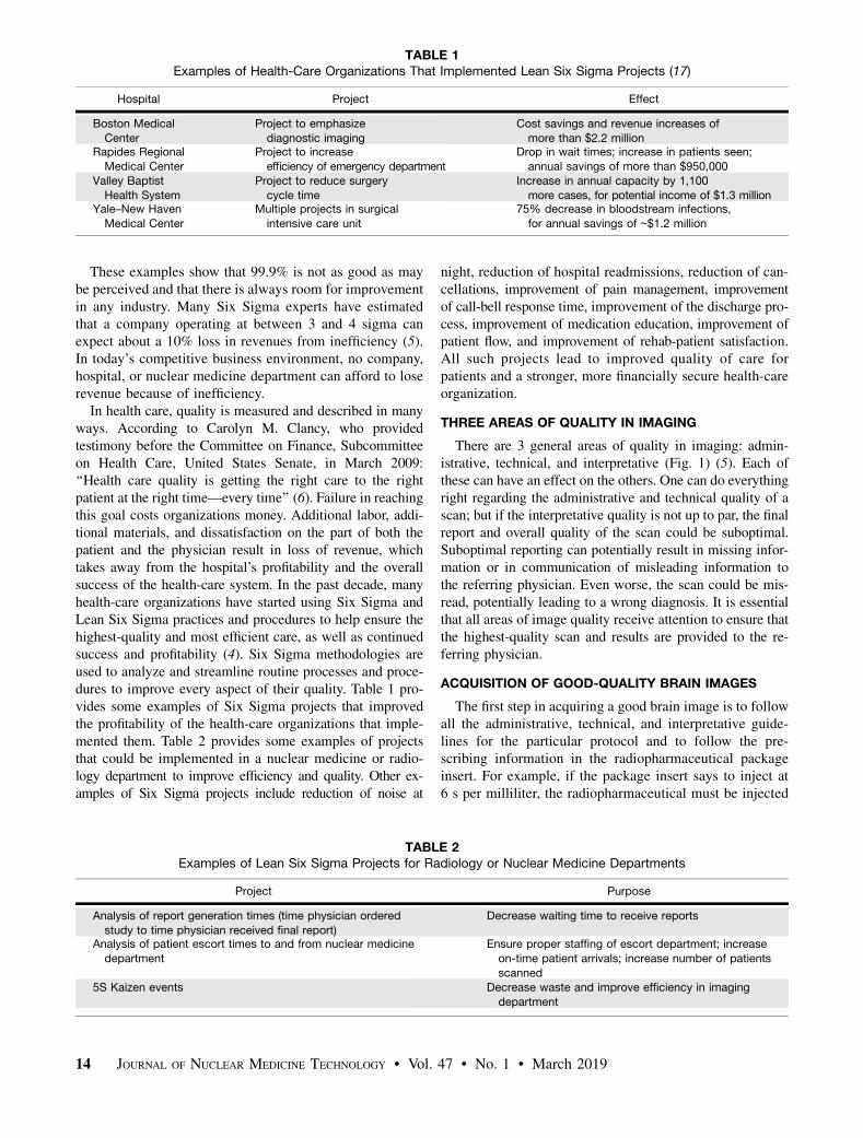
There are 3 general areas of quality in imaging: admin-istrative, technical, and interpretative (Fig. 1) (5). Each ofthese can have an effect on the others. One can do everythingright regarding the administrative and technical quality of ascan; but if the interpretative quality is not up to par, the finalreport and overall quality of the scan could be suboptimal.Suboptimal reporting can potentially result in missing infor-mation or in communication of misleading information tothe referring physician. Even worse, the scan could be mis-read, potentially leading to a wrong diagnosis. It is essentialthat all areas of image quality receive attention to ensure thatthe highest-quality scan and results are provided to the re-ferring physician.
ACQUISITION OF GOOD-QUALITY BRAIN IMAGES
The first step in acquiring a good brain image is to followall the administrative, technical, and interpretative guide-lines for the particular protocol and to follow the pre-scribing information in the radiopharmaceutical packageinsert. For example, if the package insert says to inject at6 s per milliliter, the radiopharmaceutical must be injected
TABLE 1Examples of Health-Care Organizations That Implemented Lean Six Sigma Projects (17)
Hospital Project Effect
Boston Medical
Center
Project to emphasize
diagnostic imaging
Cost savings and revenue increases of
more than $2.2 millionRapides Regional
Medical Center
Project to increase
efficiency of emergency department
Drop in wait times; increase in patients seen;
annual savings of more than $950,000Valley Baptist
Health SystemProject to reduce surgerycycle time
Increase in annual capacity by 1,100more cases, for potential income of $1.3 million
Yale–New Haven
Medical Center
Multiple projects in surgical
intensive care unit
75% decrease in bloodstream infections,
for annual savings of ∼$1.2 million
TABLE 2Examples of Lean Six Sigma Projects for Radiology or Nuclear Medicine Departments
Project Purpose
Analysis of report generation times (time physician ordered
study to time physician received final report)
Decrease waiting time to receive reports
Analysis of patient escort times to and from nuclear medicine
department
Ensure proper staffing of escort department; increase
on-time patient arrivals; increase number of patients
scanned5S Kaizen events Decrease waste and improve efficiency in imaging
department
14 JOURNAL OF NUCLEAR MEDICINE TECHNOLOGY • Vol. 47 • No. 1 • March 2019
at that rate. Instructions in a package insert are there for areason and should be followed for best results. However,there is one more thing NMTs can do to ensure the bestbrain images possible regardless of type of procedure:technical consistency. For the best image quality, techni-cal consistency is needed in patient preparation, patientpositioning, acquisition parameters, reconstruction param-eters, orientation of amyloid and 18F-FDG brain slices(transverse, sagittal, and coronal), orientation of 123I-ioflu-pane (DaTscan; GE Healthcare) images (especially coronaltilt), and display parameters. It is also important for the NMTto avoid overprocessing, making images look too smooth andpossibly causing the findings to be misinterpreted. Not beingtechnically consistent in how patients are positioned or im-ages processed may negatively affect the interpretation of thescan. If individual NMTs change processing parameters onthe basis of their eye, the physician may get inconsistentresults depending on which NMT did the processing. Beingtechnically consistent in all aspects of an imaging protocol iskey to achieving the highest-quality scan and highest-qualityreport. According to Sajdak et al., following standard oper-ating procedures helps ensure that nuclear medicine physi-cians and NMTs follow an imaging protocol helping to ensurereproducibility and consistency, which in turn are key in pro-ducing the highest-quality images (7).
PATIENT POSITIONING
Patient positioning is a critical technical step in the imagingprotocol. Proper positioning of the patient reduces the likeli-hood of artifacts due to motion, promotes patient comfort, and,most importantly, helps interpretation by providing standard-ized and artifact-free scans. The NMT should take the extratime to thoroughly explain the procedure to the patient andensure that the patient is comfortable and secure on the tableso there is less chance the patient will move and the scan willneed to be repeated.Before any type of brain scan, patients should use the
restroom and remove their eyeglasses, earrings, hair clips or
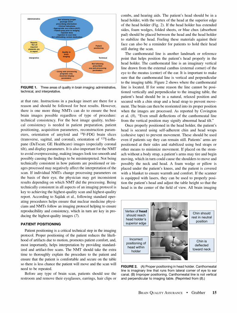
combs, and hearing aids. The patient’s head should be in ahead holder, with the vertex of the head at the superior edgeof the head holder (Fig. 2). If the head holder has extendedsides, foam wedges, folded sheets, or blue chux (absorbentpad) should be placed between the head and the head holderto stabilize the head. Feeling these materials against theirface can also be a reminder for patients to hold their headstill during the scan.
The canthomeatal line is another landmark or referencepoint that helps position the patient’s head properly in thehead holder. The canthomeatal line is an imaginary verticalline drawn from the external canthus (external corner) of theeye to the meatus (center) of the ear. It is important to makesure that the canthomeatal line is vertical and perpendicularto the imaging table. Figure 2 shows where the canthomeatalline is located. If for some reason the line cannot be posi-tioned vertically and perpendicular to the imaging table, thepatient’s head should be in a natural, relaxed position andsecured with a chin strap and a head strap to prevent move-ment. The brain can then be reoriented into its proper positionwhen the images are processed. As reported by Covingtonet al. (8), ‘‘Even small deflections of the canthomeatal linefrom the vertical position may signify abnormal head tilt.’’
Once properly positioned in the head holder, the patient’shead is secured using self-adherent chin and head wraps(cohesive tape) to prevent movement. These should be usedeven if patients say they can remain still. Patients’ arms arepositioned at their sides and stabilized using bed straps orother means to minimize movement. If placed on the stom-ach without a body strap, a patient’s arms may tire and beginmoving, which in turn could cause the shoulders to move andpossibly the neck and head. A foam wedge or pillow isplaced under the patient’s knees, and the patient is coveredwith a blanket to ensure warmth and comfort. If the scanneris equipped with lasers, they can be used to properly posi-tion the patient’s head and adjust the table height so that thehead is in the center of the field of view. All brain imaging
FIGURE 1. Three areas of quality in brain imaging: administrative,technical, and interpretative.
FIGURE 2. (A) Proper positioning in head holder. Canthomeatalline is imaginary line that runs from lateral corner of eye to earcanal. (B) Improper positioning. Canthomeatal line is not verticaland perpendicular to imaging table. (Reprinted from (8).)
BRAIN QUALITY ASSURANCE • Grabher 15
requires that the patient’s brain be in the center of the field ofview, with complete coverage of the cerebellum, especiallyfor amyloid or 18F-FDG brain imaging. Figure 3 shows im-ages of proper patient positioning on the imaging table usingthe head strap, chin strap, body strap, knee cushion, and laserlight system. The last step before the scan is to view the headposition from the end of the scanning table and correct forany signs of head rotation.Not all patients can lie flat and still on the imaging table,
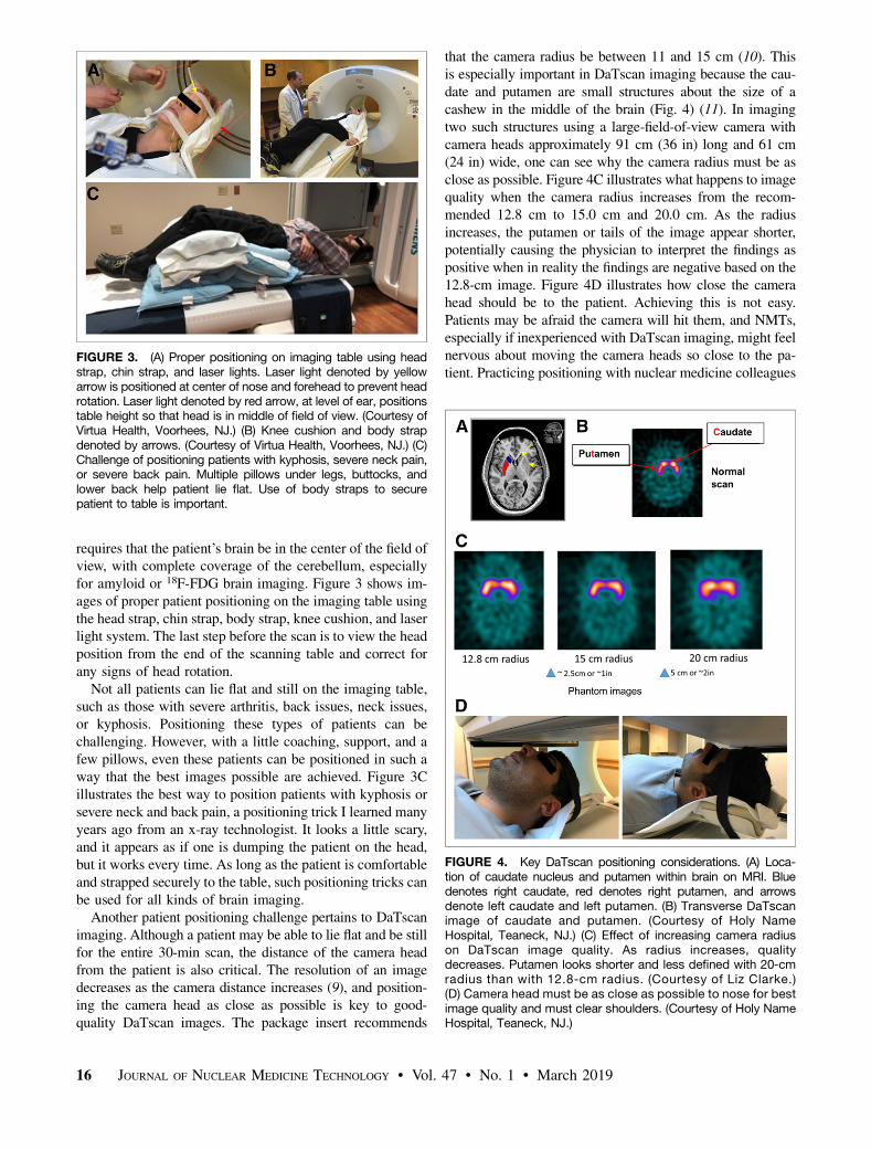
such as those with severe arthritis, back issues, neck issues,or kyphosis. Positioning these types of patients can bechallenging. However, with a little coaching, support, and afew pillows, even these patients can be positioned in such away that the best images possible are achieved. Figure 3Cillustrates the best way to position patients with kyphosis orsevere neck and back pain, a positioning trick I learned manyyears ago from an x-ray technologist. It looks a little scary,and it appears as if one is dumping the patient on the head,but it works every time. As long as the patient is comfortableand strapped securely to the table, such positioning tricks canbe used for all kinds of brain imaging.Another patient positioning challenge pertains to DaTscan
imaging. Although a patient may be able to lie flat and be stillfor the entire 30-min scan, the distance of the camera headfrom the patient is also critical. The resolution of an imagedecreases as the camera distance increases (9), and position-ing the camera head as close as possible is key to good-quality DaTscan images. The package insert recommends
that the camera radius be between 11 and 15 cm (10). Thisis especially important in DaTscan imaging because the cau-date and putamen are small structures about the size of acashew in the middle of the brain (Fig. 4) (11). In imagingtwo such structures using a large-field-of-view camera withcamera heads approximately 91 cm (36 in) long and 61 cm(24 in) wide, one can see why the camera radius must be asclose as possible. Figure 4C illustrates what happens to imagequality when the camera radius increases from the recom-mended 12.8 cm to 15.0 cm and 20.0 cm. As the radiusincreases, the putamen or tails of the image appear shorter,potentially causing the physician to interpret the findings aspositive when in reality the findings are negative based on the12.8-cm image. Figure 4D illustrates how close the camerahead should be to the patient. Achieving this is not easy.Patients may be afraid the camera will hit them, and NMTs,especially if inexperienced with DaTscan imaging, might feelnervous about moving the camera heads so close to the pa-tient. Practicing positioning with nuclear medicine colleagues
FIGURE 3. (A) Proper positioning on imaging table using headstrap, chin strap, and laser lights. Laser light denoted by yellowarrow is positioned at center of nose and forehead to prevent headrotation. Laser light denoted by red arrow, at level of ear, positionstable height so that head is in middle of field of view. (Courtesy ofVirtua Health, Voorhees, NJ.) (B) Knee cushion and body strapdenoted by arrows. (Courtesy of Virtua Health, Voorhees, NJ.) (C)Challenge of positioning patients with kyphosis, severe neck pain,or severe back pain. Multiple pillows under legs, buttocks, andlower back help patient lie flat. Use of body straps to securepatient to table is important.
FIGURE 4. Key DaTscan positioning considerations. (A) Loca-tion of caudate nucleus and putamen within brain on MRI. Bluedenotes right caudate, red denotes right putamen, and arrowsdenote left caudate and left putamen. (B) Transverse DaTscanimage of caudate and putamen. (Courtesy of Holy NameHospital, Teaneck, NJ.) (C) Effect of increasing camera radiuson DaTscan image quality. As radius increases, qualitydecreases. Putamen looks shorter and less defined with 20-cmradius than with 12.8-cm radius. (Courtesy of Liz Clarke.)(D) Camera head must be as close as possible to nose for bestimage quality and must clear shoulders. (Courtesy of Holy NameHospital, Teaneck, NJ.)
16 JOURNAL OF NUCLEAR MEDICINE TECHNOLOGY • Vol. 47 • No. 1 • March 2019
will increase the confidence of the NMT and therefore thecomfort of the patient. Shoulder clearance is also an impor-tant factor to consider in setting the radius.
HEAD ORIENTATION
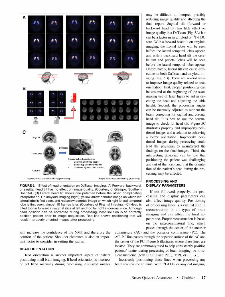
Head orientation is another important aspect of patientpositioning in all brain imaging. If head orientation is incorrector not fixed manually during processing, displayed images
may be difficult to interpret, possiblyreducing image quality and affecting thefinal report. Sagittal tilt (forward orbackward head tilt) has little effect onimage quality in a DaTscan (Fig. 5A) butcan be a factor in an amyloid or 18F-FDGscan. With a forward head tilt on amyloidimaging, the frontal lobes will be seenbefore the lateral temporal lobes appear,and with a backward head tilt the cere-bellum and parietal lobes will be seenbefore the lateral temporal lobes appear.Unfortunately, lateral tilt can cause diffi-culties in both DaTscan and amyloid im-aging (Fig. 5B). There are several waysto improve image quality related to headorientation. First, proper positioning canbe ensured at the beginning of the scan,making use of laser lights to aid in ori-enting the head and adjusting the tableheight. Second, the processing anglescan be manually adjusted to reorient thebrain, correcting for sagittal and coronalhead tilt. It is best to use the coronalimage to check for head tilt. Figure 5Cillustrates properly and improperly posi-tioned images and a solution to achievinga better orientation. Improperly posi-tioned images during processing couldlead the physician to misinterpret thefindings on the final images. Third, theinterpreting physician can be told thatpositioning the patient was challengingand out of the norm and that the orienta-tion of the patient’s head during the pro-cessing may be affected.
PROCESSING ANDDISPLAY PARAMETERS
If not followed properly, the pro-cessing and display parameters canalso affect image quality. Positioningof processing lines is a critical step inreconstruction in all types of brainimaging and can affect the final ap-pearance. Proper reconstruction is basedon the intercommissural line, whichpasses through the center of the anterior
commissure (AC) and the posterior commissure (PC). TheAC–PC line passes through the superior surface of the AC andthe center of the PC. Figure 6 illustrates where these lines arelocated. They are commonly used to help consistently positionpatients’ brains during processing of brain imaging, be it nu-clear medicine (both SPECT and PET), MRI, or CT (12).
Incorrectly positioning these lines when processing anybrain scan can be an issue. With 18F-FDG or amyloid imaging,
FIGURE 5. Effect of head orientation on DaTscan imaging. (A) Forward, backward,or sagittal head tilt has no effect on image quality. (Courtesy of Glasgow SouthernHospital.) (B) Lateral head tilt shows one putamen before the other, complicatinginterpretation. On amyloid imaging (right), yellow arrow denotes image on which leftlateral lobe is first seen, and red arrow denotes image on which right lateral temporallobe is first seen, almost 10 frames later. (Courtesy of Piramal Imaging.) (C) Head istilted too far forward in sagittal slice at left and too far right in coronal slice. Althoughhead position can be corrected during processing, best solution is to correctlyposition patient prior to image acquisition. Red line shows positioning that willresult in properly oriented images after processing.
BRAIN QUALITY ASSURANCE • Grabher 17
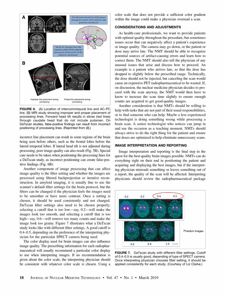
incorrect line placement can result in some regions of the brainbeing seen before others, such as the frontal lobes before thelateral temporal lobes. If lateral head tilt is not adjusted duringprocessing, poor image quality can also result (Fig. 5B). Specialcare needs to be taken when positioning the processing lines fora DaTscan study, as incorrect positioning can create false-pos-itive findings (Fig. 6B).Another component of image processing that can affect
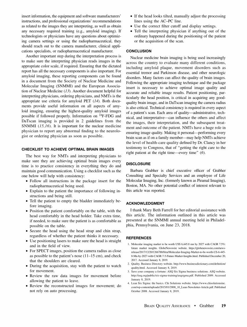
image quality is the filter setting and whether the images areprocessed using filtered backprojection or iterative recon-struction. In amyloid imaging, it is usually fine to use thescanner’s default filter settings for the brain protocol, but thefilters can be changed if the physician feels the images needto be smoother or have more contrast. Once a setting ischosen, it should be used consistently and not changed.DaTscan filter settings also need to be chosen properly;selecting a cutoff that is too low—say, 0.2—will make theimages look too smooth, and selecting a cutoff that is toohigh—say, 0.6—will remove too many counts and make theimage look too grainy. Figure 7 illustrates what a DaTscanstudy looks like with different filter settings. A good cutoff is0.4–0.5, depending on the preference of the interpreting phy-sician for the particular SPECT camera being used.The color display used for brain images can also influence
image quality. The prescribing information for each radiophar-maceutical will usually recommend a particular color displayto use when interpreting images. If no recommendation isgiven about the color scale, the interpreting physician shouldbe consistent with whatever color scale is chosen. Using a
color scale that does not provide a sufficient color gradientwithin the image could make a physician overread a scan.
CONSIDERATIONS AND ADJUSTMENTS
As health-care professionals, we want to provide patientswith optimal quality throughout the procedure, but sometimesissues occur that can negatively affect a patient’s experienceor image quality. The camera may go down, or the patient ordose may arrive late. The NMT should be able to recognizepotential sources of artifact-causing errors and learn how tocorrect them. The NMT should also tell the physician of anyunusual issues that arise and discuss how to proceed. Anexample is a patient who arrives late, so that the dose hasdropped to slightly below the prescribed range. Technically,the dose should not be injected, but canceling the scan wouldcause an expensive PET radiopharmaceutical to be wasted. If,on discussion, the nuclear medicine physician decides to pro-ceed with the scan anyway, the NMT would then have toknow to increase the scan time slightly to ensure enoughcounts are acquired to get good-quality images.
Another consideration is that NMTs should be willing tohelp with tasks that are not part of their usual responsibilities,or to find someone who can help. Maybe a less experiencedtechnologist is doing something wrong while processing abrain scan. A senior technologist who notices can jump inand use the occasion as a teaching moment. NMTs shouldalways strive to do the right thing for the patient and ensurethat doses are optimized to help eliminate unnecessary scans.
IMAGE INTERPRETATION AND REPORTING
Image interpretation and reporting is the final step in thequest for the best-quality brain images possible. NMTs can doeverything right on their end in positioning the patient andacquiring and displaying the best images, but if the interpret-ing physician misreads something or leaves something out ofa report, the quality of the scan will be affected. Interpretingphysicians should review the radiopharmaceutical package
FIGURE 6. (A) Location of intercommissural line and AC–PCline. (B) MRI study showing improper and proper placement ofprocessing lines. Forward head tilt results in slices (red lines)through caudate head that do not include putamen. OnDaTscan studies, false-positive findings can result from incorrectpositioning of processing lines. (Reprinted from (8).)
FIGURE 7. DaTscan study with different filter settings. Cutoffof 0.4–0.5 is usually good, depending of type of SPECT camera.Once interpreting physician chooses filter setting, it should beapplied consistently for each study. (Courtesy of Liz Clarke.)
18 JOURNAL OF NUCLEAR MEDICINE TECHNOLOGY • Vol. 47 • No. 1 • March 2019
insert information, the equipment and software manufacturers’instructions, and professional organizations’ recommendationsas related to the images they are interpreting, as well as obtainany necessary required training (e.g., amyloid imaging). Iftechnologists or physicians have any questions about optimiz-ing camera settings or using the radiopharmaceutical, theyshould reach out to the camera manufacturer, clinical appli-cations specialists, or radiopharmaceutical manufacturer.Another important step during the interpretation process is
to make sure the interpreting physician reads images in theappropriate color scale, if required. Ensuring that the dictatedreport has all the necessary components is also important. Foramyloid imaging, these reporting components can be foundin a document from the Society of Nuclear Medicine andMolecular Imaging (SNMMI) and the European Associa-tion of Nuclear Medicine (13). Another document helpful forinterpreting physicians, ordering physicians, and NMTs is theappropriate use criteria for amyloid PET (14). Both docu-ments provide useful information on all aspects of amy-loid imaging, ensuring the highest-quality amyloid imagespossible if followed properly. Information on 18F-FDG andDaTscan imaging is provided in 2 guidelines from theSNMMI (15,16). It is important for the nuclear medicinephysician to report any abnormal finding to the neurolo-gist or ordering physician as soon as possible.
CHECKLIST TO ACHIEVE OPTIMAL BRAIN IMAGES
The best way for NMTs and interpreting physicians tomake sure they are achieving optimal brain images everytime is to practice consistency in everything they do andmaintain good communication. Using a checklist such as theone below will help with consistency:• Follow all instructions in the package insert for theradiopharmaceutical being used.
• Explain to the patient the importance of following in-structions and being still.
• Tell the patient to empty the bladder immediately be-fore imaging.
• Position the patient comfortably on the table, with thehead comfortably in the head holder. Take extra time,if needed, to make sure the patient is as comfortable aspossible on the table.
• Secure the head using the head strap and chin strap,regardless of whether the patient thinks it necessary.
• Use positioning lasers to make sure the head is straightand in the field of view.
• For SPECT images, position the camera radius as closeas possible to the patient’s nose (11–15 cm), and checkthat the shoulders are cleared.
• During the acquisition, stay with the patient to watchfor movement.
• Review the raw data images for movement beforeallowing the patient to leave.
• Review the reconstructed images for movement; donot rely on auto processing.
• If the head looks tilted, manually adjust the processinglines using the AC–PC line.
• Use the correct filter cutoff and display settings.• Tell the interpreting physician if anything out of theordinary happened during the positioning of the patientor the acquisition of the scan.
CONCLUSION
Nuclear medicine brain imaging is being used increasinglyacross the country to evaluate many different conditions,including amyloid plaque, movement disorders such asessential tremor and Parkinson disease, and other neurologicdisorders. Many factors can affect the quality of brain images.Following the appropriate imaging technique and the packageinsert is necessary to achieve optimal image quality andaccurate and reliable image results. Patient positioning, par-ticularly the head position, is critical in acquiring any good-quality brain image, and in DaTscan imaging the camera radiusis also critical. Technical consistency is required in every aspectof a patient’s scan. Each area of quality—administrative, tech-nical, and interpretative—can influence the others and affectthe images, their interpretation, and the subsequent treat-ment and outcome of the patient. NMTs have a huge role inensuring image quality. Making it personal—performing everybrain scan as if on a family member—may help NMTs achievethe level of health-care quality defined by Dr. Clancy in hertestimony to Congress, that of ‘‘getting the right care to theright patient at the right time—every time’’ (6).
DISCLOSURE
Barbara Grabher is chief executive officer of GrabherConsulting and Specialty Services and an employee of LifeMolecular Imaging, Inc. (formerly known as Piramal Imaging),Boston, MA. No other potential conflict of interest relevant tothis article was reported.
ACKNOWLEDGMENT
I thank Mary Beth Farrell for her editorial assistance withthis article. The information outlined in this article waspresented at the SNMMI annual meeting held in Philadel-phia, Pennsylvania, on June 23, 2018.
REFERENCES
1. Molecular imaging market to be worth US$ 6,445.8 mn by 2027 with CAGR 7.5%:
future market insights. GlobeNewswire website. https://globenewswire.com/news-
release/2017/12/20/1266788/0/en/Molecular-Imaging-Market-to-be-worth-US-6-445-
8-Mn-by-2027-with-CAGR-7-5-Future-Market-Insights.html. Published December 20,
2017. Accessed January 8, 2019.
2. Quality. Business Directory website. http://www.businessdictionary.com/definition/
quality.html. Accessed January 8, 2019.
3. Save your company a fortune: ASQ Six Sigma business solutions. ASQ website.
http://asq.org/public/six-sigma-training/asqsigma.pdf. Published 2009. Accessed
January 8, 2019.
4. Lean Six Sigma: the basics. Chi Solutions website. https://www.chisolutionsinc.
com/wp-content/uploads/2015/01/2008_10_Lean-Newsletter-Article.pdf. Published
October 2008. Accessed January 8, 2019.
BRAIN QUALITY ASSURANCE • Grabher 19

5. Evans MH. Reaching for Six Sigma. Excellence in Financial Management
website. http://www.exinfm.com/board/reaching_for_six_sigma.htm. Accessed
on April 26, 2018.
6. What is health care quality and who decides? U.S. Senate Committee on Finance
website. https://www.finance.senate.gov/imo/media/doc/640431.pdf. Published
March 18, 2009. Accessed January 8, 2019.
7. Sajdak R, Trembath L, Thomas K. The importance of standard operating proce-
dures in clinical trials. J Nucl Med Technol. 2013;41:231–233.
8. Covington MF, McMillan NA, Avery RJ, Kuo PH. The semicolon sign: dopa-
mine transporter imaging artifact from head tilt. J Nucl Med Technol. 2013;41:
105–107.
9. Rosenthal MS, Cullom J, Hawkins W, Moore SC, Tsui BM, Yester M. Quanti-
tative SPECT imaging: A review and recommendations by the Focus Committee
of the Society of Nuclear Medicine Computer and Instrumentation Council.
J Nucl Med. 1995;36:1489–1515.
10. DaTscan [prescribing information]. Arlington Heights, IL: GE Healthcare; 2015.
11. Lanciego JL, Luquin N, Obeso JA. Functional neuroanatomy of the basal gan-
glia. Cold Spring Harb Perspect Med. 2012;2:a009621.
12. Trembath A, Opanowski A. PET imaging of the brain for technologists: CTN
webinar series 2016. IDEAS-Study website. https://www.ideas-study.org/wp-
content/uploads/2017/01/PET-Imaging-of-the-Brain-for-Technologists_04Apr16.pdf.
Published 2016. Accessed January 8, 2019.
13. Minoshima S, Drzezga AE, Barthel H, et al. SNMMI procedure standard/EANM
practice guideline for amyloid PET imaging of the brain 1.0. J Nucl Med. 2016;57:
1316–1322.
14. Johnson KA, Minoshima S, Bohnen NI, et al. Appropriate use criteria for am-
yloid PET: a report of the Amyloid Imaging Task Force, the Society of Nuclear
Medicine and Molecular Imaging, and the Alzheimer’s Association. J Nucl Med.
2013;54:476–490.
15. Djang DS, Janssen MJ, Bohnen N, et al. SNM practice guideline for dopamine
transporter imaging with 123I-ioflupane SPECT 1.0. J Nucl Med. 2012;53:154–163.
16. Waxman A, Herholz K, Lewis DA, et al. Society of Nuclear Medicine Procedure
Guideline for FDG PET brain imaging: version 1.0. SNMMI website. http://
snmmi.files.cms-plus.com/docs/Society%20of%20Nuclear%20Medicine%
20Procedure%20Guideline%20for%20FDG%20PET%20Brain%20Imaging.pdf.
Published February 8, 2009. Accessed January 8, 2019.
17. Mehrotra S. Lean Six Sigma methodologies: How do they improve health
care industry? GreyCampus website. https://www.greycampus.com/blog/
quality-management/health-care-industry-can-reduce-wastage-by-these-lean-
six-sigma-methodologies. Published December 1, 2017. Accessed January 8,
2019.
20 JOURNAL OF NUCLEAR MEDICINE TECHNOLOGY • Vol. 47 • No. 1 • March 2019