Embed Size (px)
Citation preview
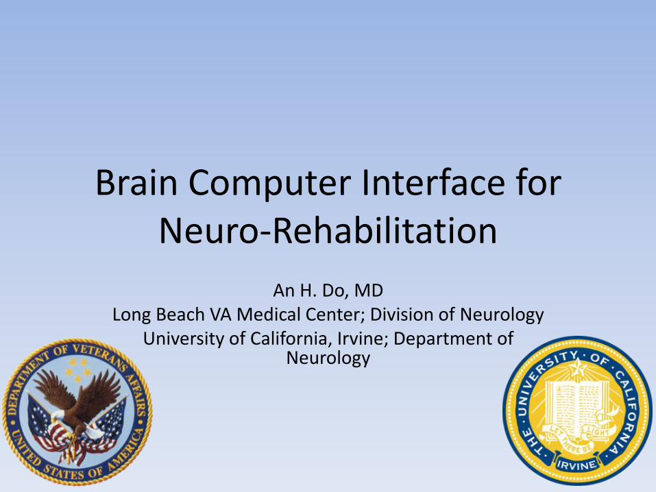
Brain Computer Interface for Neuro-Rehabilitation
An H. Do, MD Long Beach VA Medical Center; Division of Neurology
University of California, Irvine; Department of Neurology
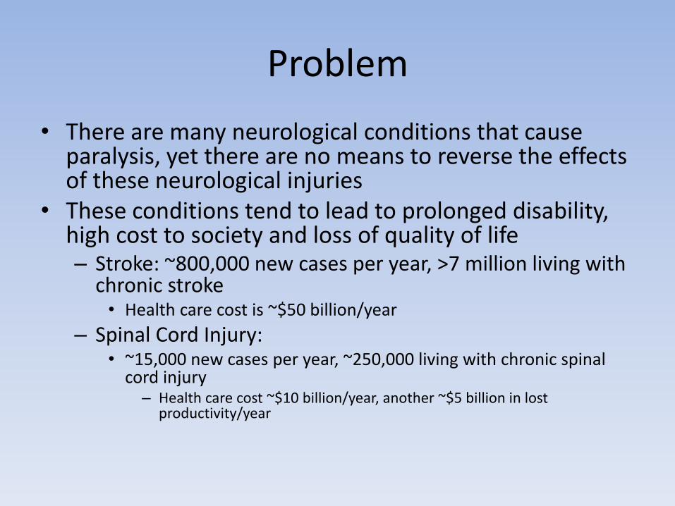
Problem
• There are many neurological conditions that cause paralysis, yet there are no means to reverse the effects of these neurological injuries
• These conditions tend to lead to prolonged disability, high cost to society and loss of quality of life – Stroke: ~800,000 new cases per year, >7 million living with
chronic stroke • Health care cost is ~$50 billion/year
– Spinal Cord Injury: • ~15,000 new cases per year, ~250,000 living with chronic spinal
cord injury – Health care cost ~$10 billion/year, another ~$5 billion in lost
productivity/year
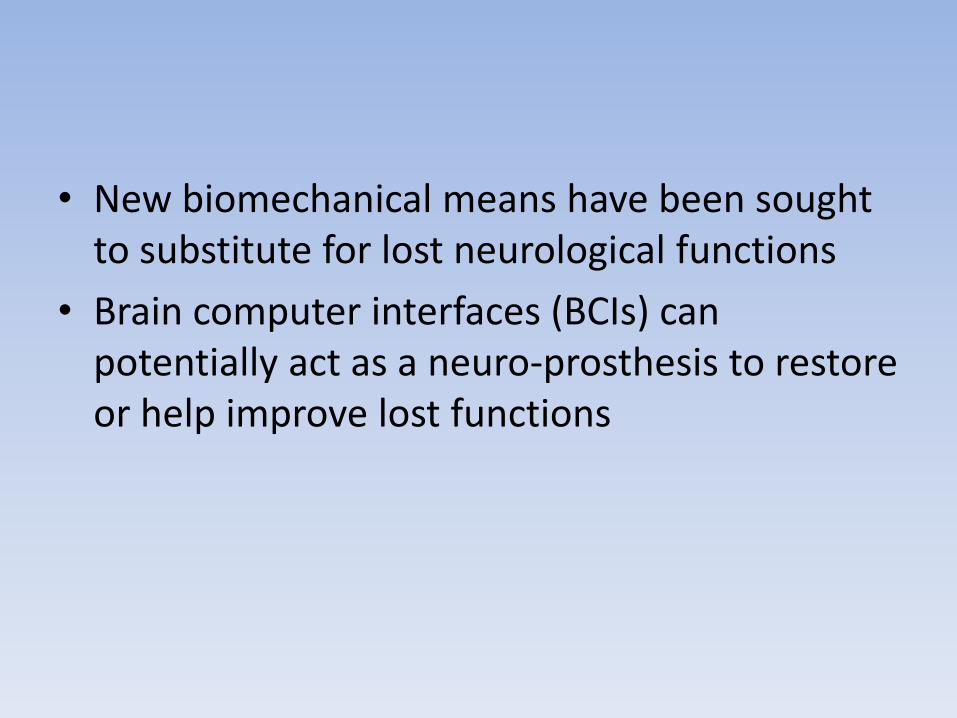
• New biomechanical means have been sought to substitute for lost neurological functions
• Brain computer interfaces (BCIs) can potentially act as a neuro-prosthesis to restore or help improve lost functions
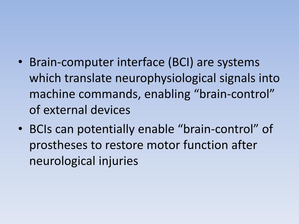
• Brain-computer interface (BCI) are systems which translate neurophysiological signals into machine commands, enabling “brain-control” of external devices
• BCIs can potentially enable “brain-control” of prostheses to restore motor function after neurological injuries
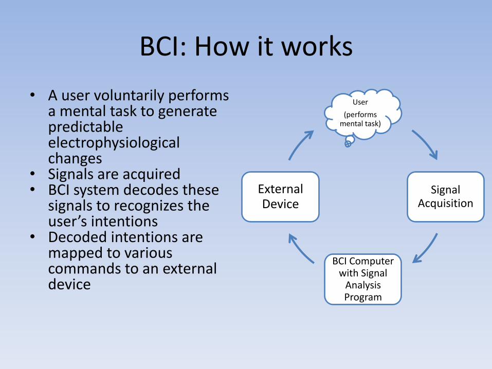
User
(performs mental task)
Signal Acquisition
BCI Computer with Signal
Analysis Program
External Device
BCI: How it works
• A user voluntarily performs a mental task to generate predictable electrophysiological changes
• Signals are acquired • BCI system decodes these
signals to recognizes the user’s intentions
• Decoded intentions are mapped to various commands to an external device
Outline
• Physiological basis of BCIs
• BCIs as Neuroprostheses
• BCIs as Neuro-rehabilitative tool
• Quick survey of BCI research community

PHYSIOLOGICAL BASIS OF BCIS
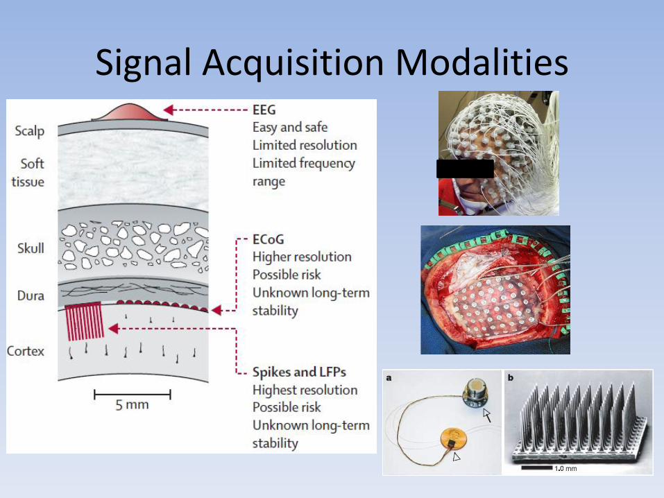
Signal Acquisition Modalities
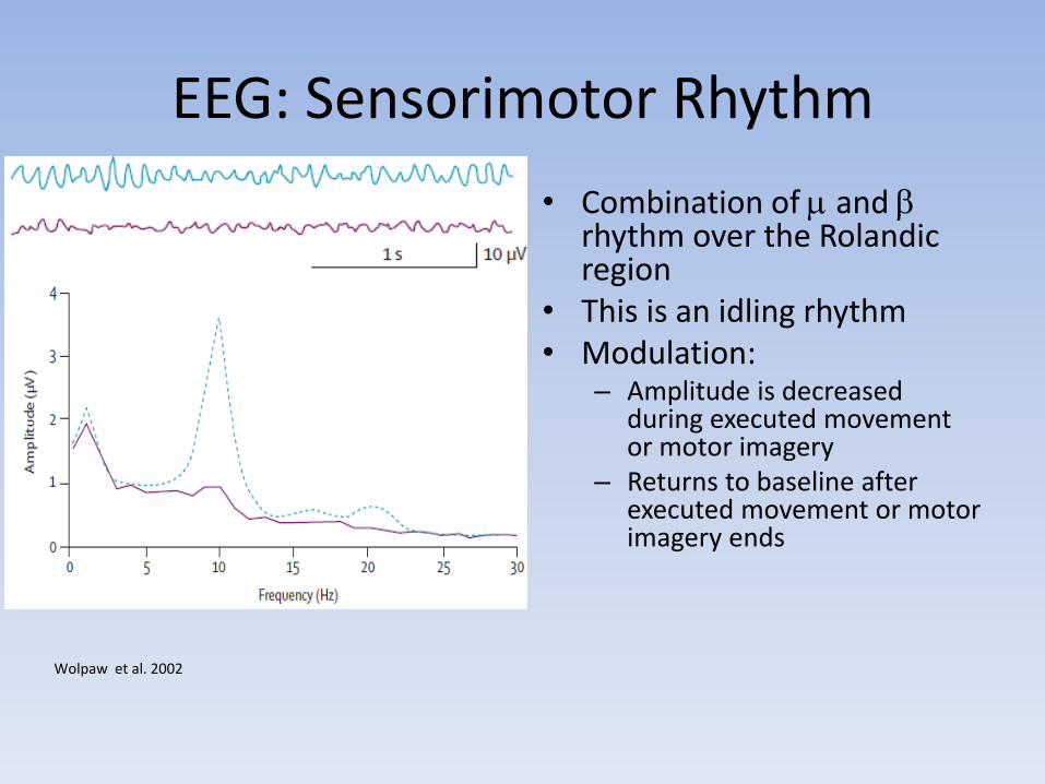
EEG: Sensorimotor Rhythm
• Combination of and rhythm over the Rolandic region
• This is an idling rhythm • Modulation:
– Amplitude is decreased during executed movement or motor imagery
– Returns to baseline after executed movement or motor imagery ends
Wolpaw et al. 2002
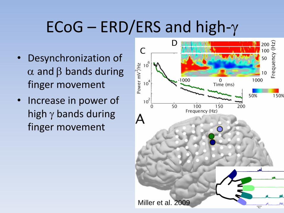
ECoG – ERD/ERS and high-
• Desynchronization of and bands during finger movement
• Increase in power of high bands during finger movement
Miller et al. 2009
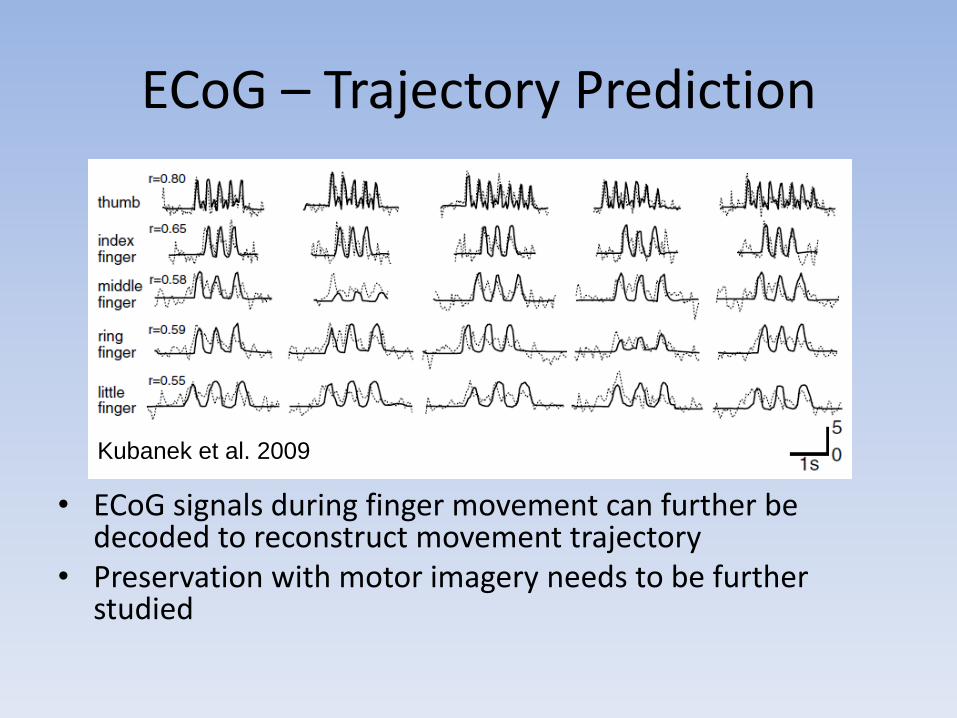
ECoG – Trajectory Prediction
• ECoG signals during finger movement can further be decoded to reconstruct movement trajectory
• Preservation with motor imagery needs to be further studied
Kubanek et al. 2009
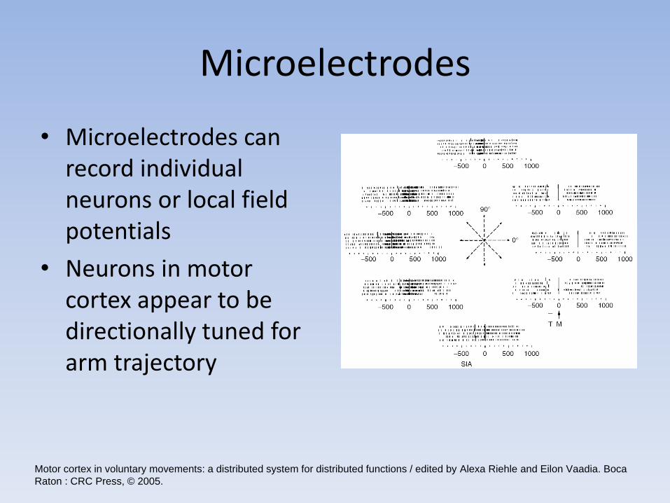
Microelectrodes
• Microelectrodes can record individual neurons or local field potentials
• Neurons in motor cortex appear to be directionally tuned for arm trajectory
Motor cortex in voluntary movements: a distributed system for distributed functions / edited by Alexa Riehle and Eilon Vaadia. Boca
Raton : CRC Press, © 2005.
BCI AS A NEURO-PROSTHESIS
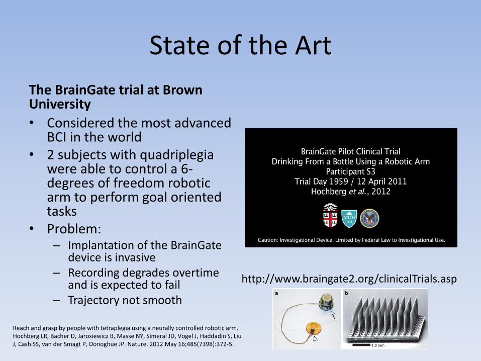
State of the Art
The BrainGate trial at Brown University
• Considered the most advanced BCI in the world
• 2 subjects with quadriplegia were able to control a 6-degrees of freedom robotic arm to perform goal oriented tasks
• Problem: – Implantation of the BrainGate
device is invasive – Recording degrades overtime
and is expected to fail – Trajectory not smooth
http://www.braingate2.org/clinicalTrials.asp
Reach and grasp by people with tetraplegia using a neurally controlled robotic arm. Hochberg LR, Bacher D, Jarosiewicz B, Masse NY, Simeral JD, Vogel J, Haddadin S, Liu J, Cash SS, van der Smagt P, Donoghue JP. Nature. 2012 May 16;485(7398):372-5.
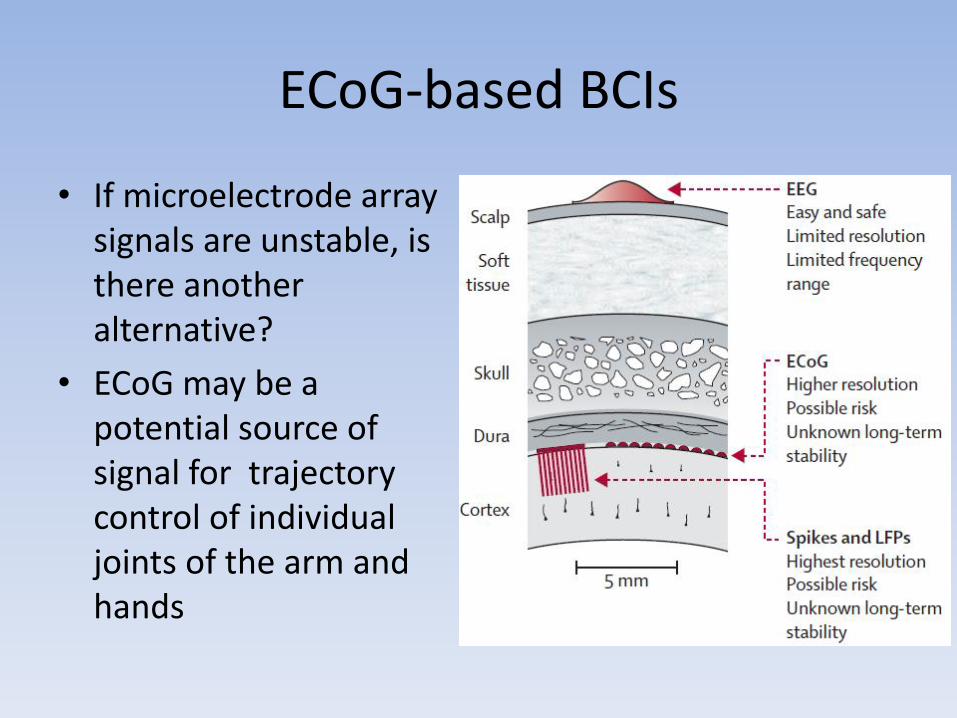
ECoG-based BCIs
• If microelectrode array signals are unstable, is there another alternative?
• ECoG may be a potential source of signal for trajectory control of individual joints of the arm and hands
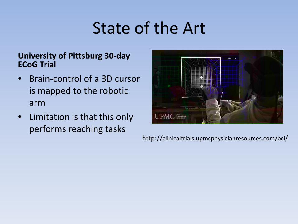
State of the Art
University of Pittsburg 30-day ECoG Trial
• Brain-control of a 3D cursor is mapped to the robotic arm
• Limitation is that this only performs reaching tasks
http://clinicaltrials.upmcphysicianresources.com/bci/
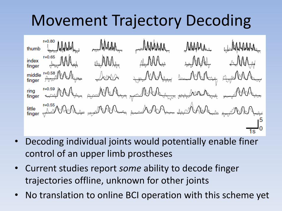
Movement Trajectory Decoding
• Decoding individual joints would potentially enable finer control of an upper limb prostheses
• Current studies report some ability to decode finger trajectories offline, unknown for other joints
• No translation to online BCI operation with this scheme yet
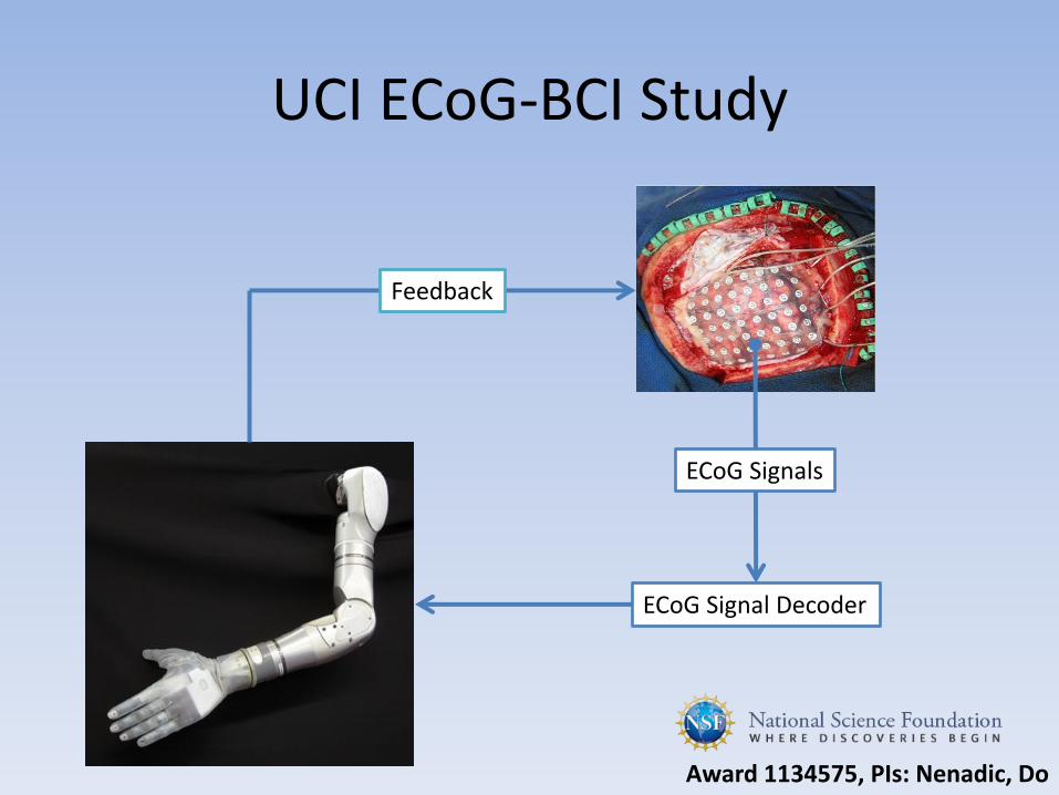
UCI ECoG-BCI Study
ECoG Signals
ECoG Signal Decoder
Feedback
Award 1134575, PIs: Nenadic, Do
BCI-Control of Ambulation
• Loss of gait function is typically seen after SCI
– Results in prolonged sitting and reduced physical activity, which leads to many medical co-morbidities
• Still no way to restore able-bodied-like ambulation after SCI
• BCIs can potentially restore able-bodied-like ambulation after SCI
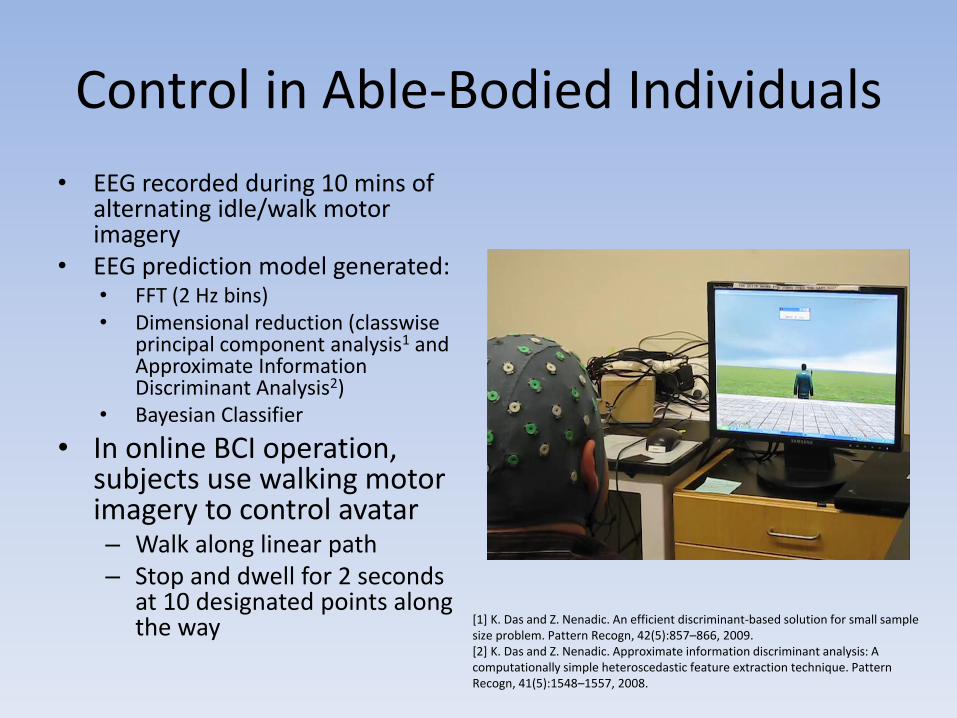
Control in Able-Bodied Individuals
• EEG recorded during 10 mins of alternating idle/walk motor imagery
• EEG prediction model generated: • FFT (2 Hz bins) • Dimensional reduction (classwise
principal component analysis1 and Approximate Information Discriminant Analysis2)
• Bayesian Classifier
• In online BCI operation, subjects use walking motor imagery to control avatar – Walk along linear path – Stop and dwell for 2 seconds
at 10 designated points along the way [1] K. Das and Z. Nenadic. An efficient discriminant-based solution for small sample
size problem. Pattern Recogn, 42(5):857–866, 2009. [2] K. Das and Z. Nenadic. Approximate information discriminant analysis: A computationally simple heteroscedastic feature extraction technique. Pattern Recogn, 41(5):1548–1557, 2008.
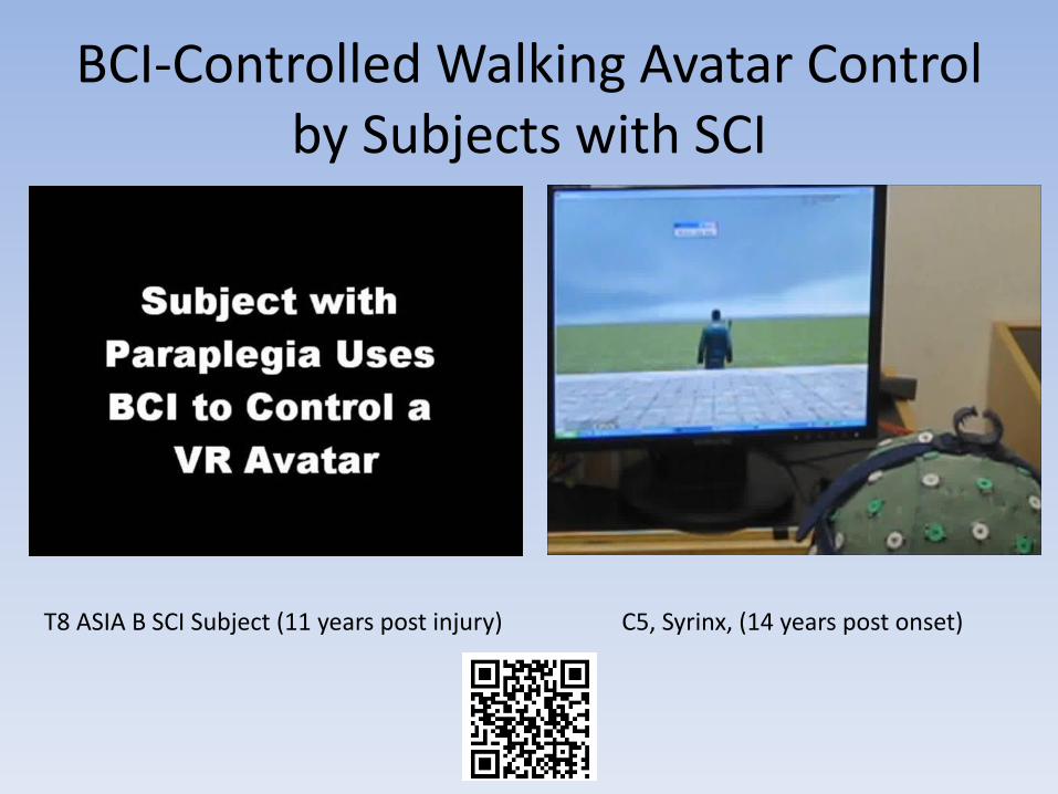
BCI-Controlled Walking Avatar Control by Subjects with SCI
T8 ASIA B SCI Subject (11 years post injury) C5, Syrinx, (14 years post onset)
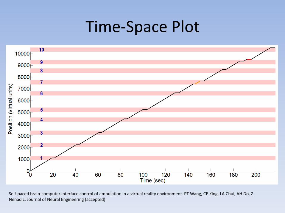
Time-Space Plot
Self-paced brain-computer interface control of ambulation in a virtual reality environment. PT Wang, CE King, LA Chui, AH Do, Z Nenadic. Journal of Neural Engineering (accepted).
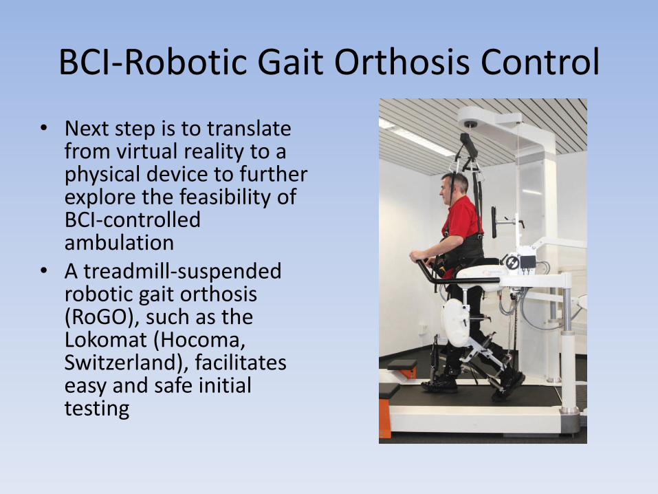
BCI-Robotic Gait Orthosis Control
• Next step is to translate from virtual reality to a physical device to further explore the feasibility of BCI-controlled ambulation
• A treadmill-suspended robotic gait orthosis (RoGO), such as the Lokomat (Hocoma, Switzerland), facilitates easy and safe initial testing
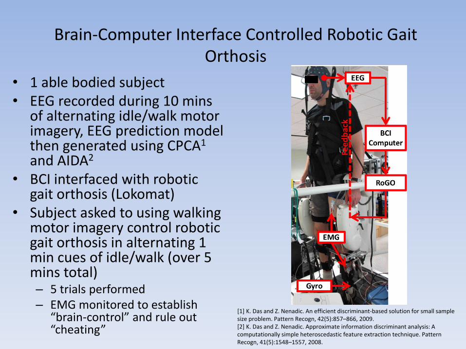
Brain-Computer Interface Controlled Robotic Gait
Orthosis
• 1 able bodied subject • EEG recorded during 10 mins
of alternating idle/walk motor imagery, EEG prediction model then generated using CPCA1 and AIDA2
• BCI interfaced with robotic gait orthosis (Lokomat)
• Subject asked to using walking motor imagery control robotic gait orthosis in alternating 1 min cues of idle/walk (over 5 mins total) – 5 trials performed – EMG monitored to establish
“brain-control” and rule out “cheating”
[1] K. Das and Z. Nenadic. An efficient discriminant-based solution for small sample size problem. Pattern Recogn, 42(5):857–866, 2009. [2] K. Das and Z. Nenadic. Approximate information discriminant analysis: A computationally simple heteroscedastic feature extraction technique. Pattern Recogn, 41(5):1548–1557, 2008.
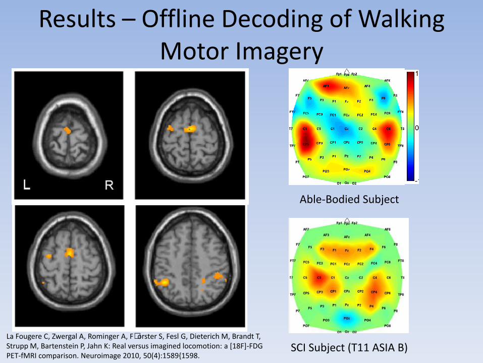
Results – Offline Decoding of Walking Motor Imagery
Able-Bodied Subject
SCI Subject (T11 ASIA B) La Fougere C, Zwergal A, Rominger A, F•orster S, Fesl G, Dieterich M, Brandt T, Strupp M, Bartenstein P, Jahn K: Real versus imagined locomotion: a [18F]-FDG PET-fMRI comparison. Neuroimage 2010, 50(4):1589{1598.
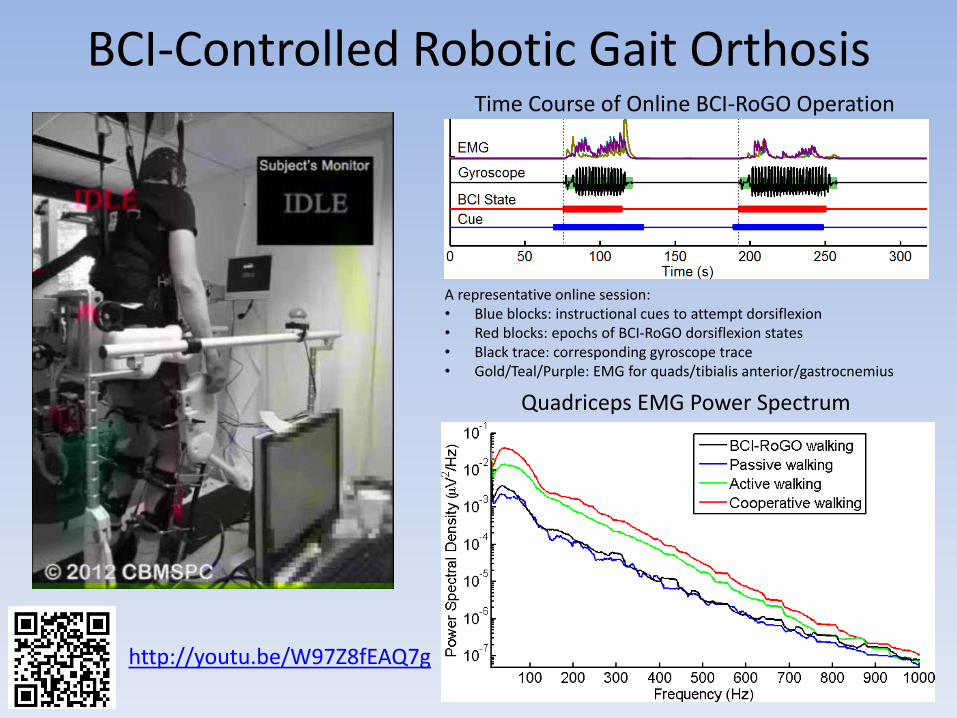
BCI-Controlled Robotic Gait Orthosis
http://youtu.be/W97Z8fEAQ7g
Quadriceps EMG Power Spectrum
Time Course of Online BCI-RoGO Operation
A representative online session: • Blue blocks: instructional cues to attempt dorsiflexion • Red blocks: epochs of BCI-RoGO dorsiflexion states • Black trace: corresponding gyroscope trace • Gold/Teal/Purple: EMG for quads/tibialis anterior/gastrocnemius
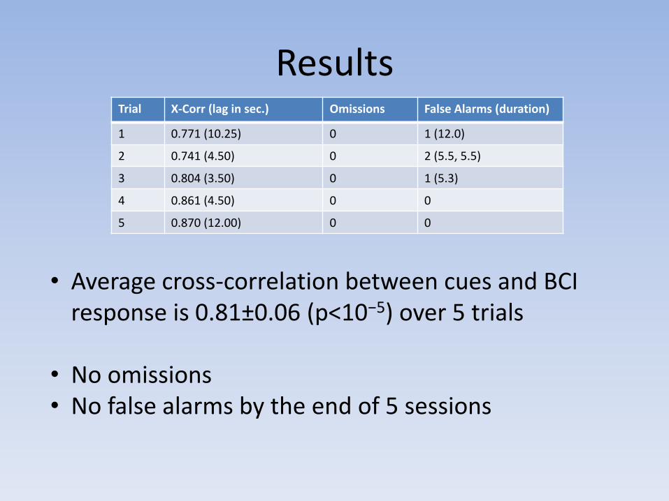
Results Trial X-Corr (lag in sec.) Omissions False Alarms (duration)
1 0.771 (10.25) 0 1 (12.0)
2 0.741 (4.50) 0 2 (5.5, 5.5)
3 0.804 (3.50) 0 1 (5.3)
4 0.861 (4.50) 0 0
5 0.870 (12.00) 0 0
• Average cross-correlation between cues and BCI response is 0.81±0.06 (p<10−5) over 5 trials
• No omissions • No false alarms by the end of 5 sessions
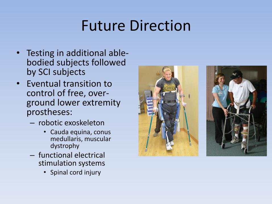
Future Direction
• Testing in additional able-bodied subjects followed by SCI subjects
• Eventual transition to control of free, over-ground lower extremity prostheses: – robotic exoskeleton
• Cauda equina, conus medullaris, muscular dystrophy
– functional electrical stimulation systems • Spinal cord injury
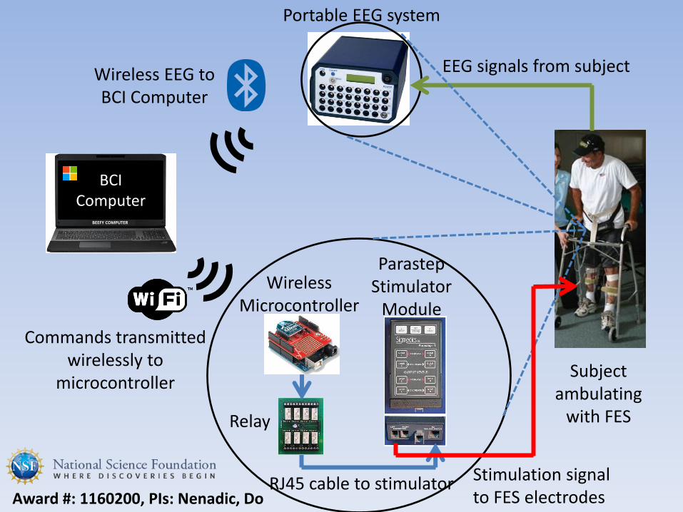
Wireless Microcontroller
Parastep Stimulator
Module
RJ45 cable to stimulator
Subject ambulating
with FES
Wireless EEG to BCI Computer
Commands transmitted wirelessly to
microcontroller
Portable EEG system
Stimulation signal to FES electrodes
EEG signals from subject
Relay
Award #: 1160200, PIs: Nenadic, Do
BCI Computer
BEEFY COMPUTER
BCI as Neuroprostheses- Conclusion
• Upper and lower extremity prostheses to restore basic movements after neurological injury may be possible
• Control is still very basic at this point, but continued research can potentially improve upon smoothness and increase degrees of freedom
BCI AS A REHABILITATIVE TOOL
Problem
• For the remainder of stroke, SCI, or TBI patients who are affected by partial paralysis, physiotherapy only provides a limited amount of neurological improvement
• Hence novel means must be designed to further improve neurological outcomes
• BCI-controlled functional electrical stimulation (FES) systems may be one such novel approach
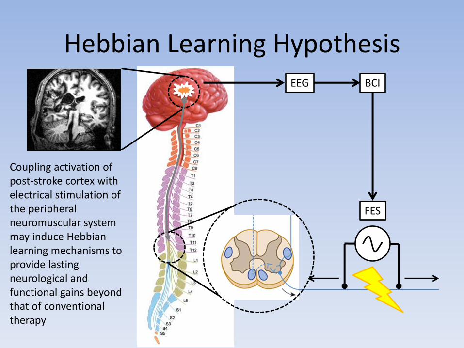
Hebbian Learning Hypothesis EEG BCI
FES
Coupling activation of post-stroke cortex with electrical stimulation of the peripheral neuromuscular system may induce Hebbian learning mechanisms to provide lasting neurological and functional gains beyond that of conventional therapy
BCI-FES for Neuro-rehabilitation
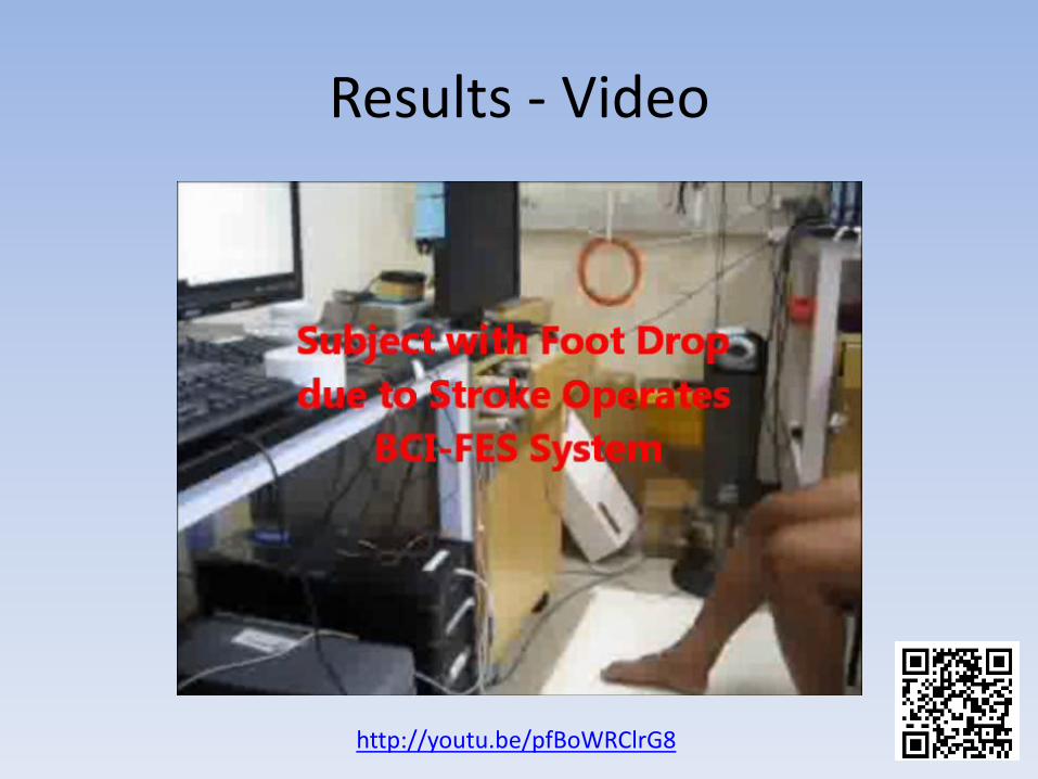
• This study describes the feasibility of such a system for foot drop due to stroke
• Stroke subjects with foot drop were recruited
• 10 minutes of training data acquired from 64 channel EEG during alternating epochs of attempted foot dorsiflexion and idling
EEG Decoding Model
• EEG decoding model generated using following algorithm – FFT (power calculated over 2 Hz bins) – Dimensional Reduction (with classwise Principal
Components Analysis1 and Approximate Information Discriminant Analysis2)
– Bayesian Classifier • Decoding algorithm will classify novel EEG as either Idling
or Dorsiflexion classes • Online, the decoding algorithm detects EEG changes
associated with attempted (but ineffective) foot dorsiflexion and the BCI system delivers electrical stimulation to elicit foot dorsiflexion
[1] K. Das and Z. Nenadic. An efficient discriminant-based solution for small sample size problem. Pattern Recogn, 42(5):857–866, 2009. [2] K. Das and Z. Nenadic. Approximate information discriminant analysis: A computationally simple heteroscedastic feature extraction technique. Pattern Recogn, 41(5):1548–1557, 2008.
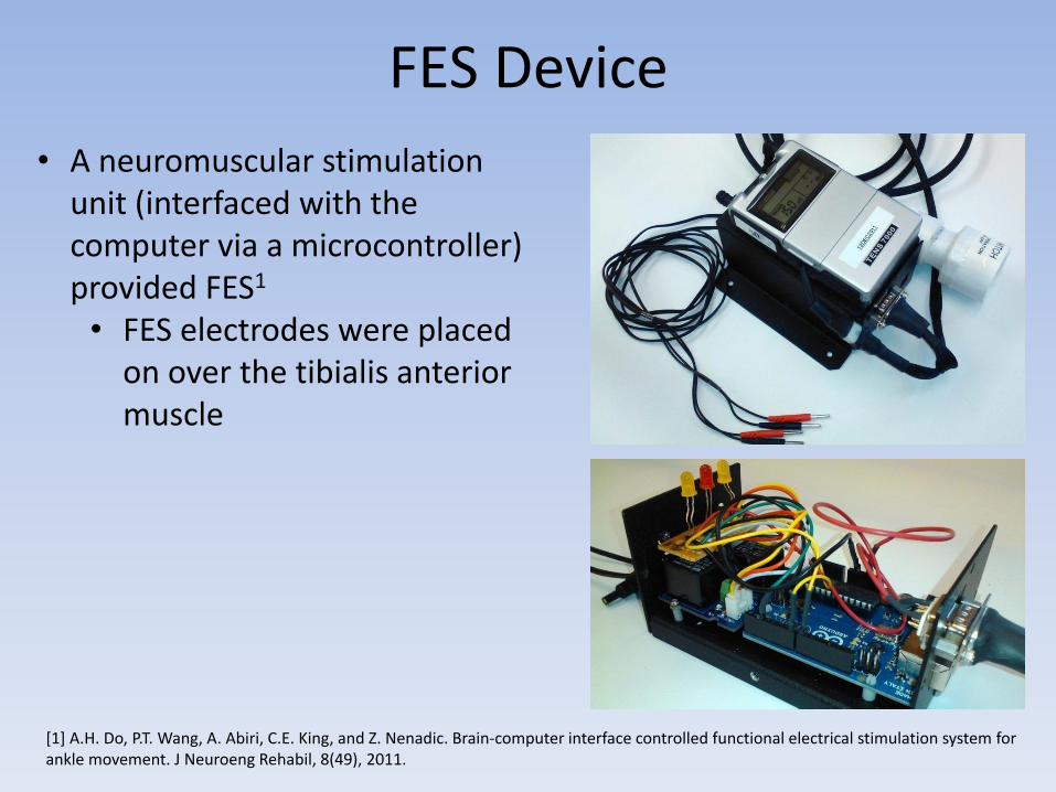
FES Device
[1] A.H. Do, P.T. Wang, A. Abiri, C.E. King, and Z. Nenadic. Brain-computer interface controlled functional electrical stimulation system for ankle movement. J Neuroeng Rehabil, 8(49), 2011.
• A neuromuscular stimulation unit (interfaced with the computer via a microcontroller) provided FES1 • FES electrodes were placed
on over the tibialis anterior muscle
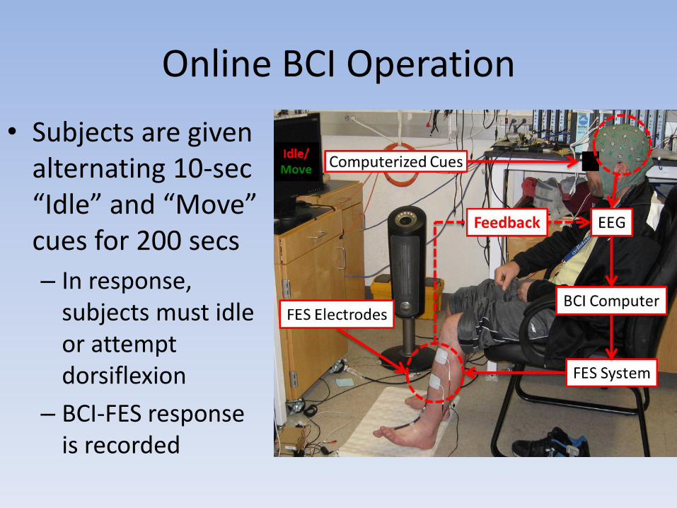
Online BCI Operation
• Subjects are given alternating 10-sec “Idle” and “Move” cues for 200 secs
– In response, subjects must idle or attempt dorsiflexion
– BCI-FES response is recorded
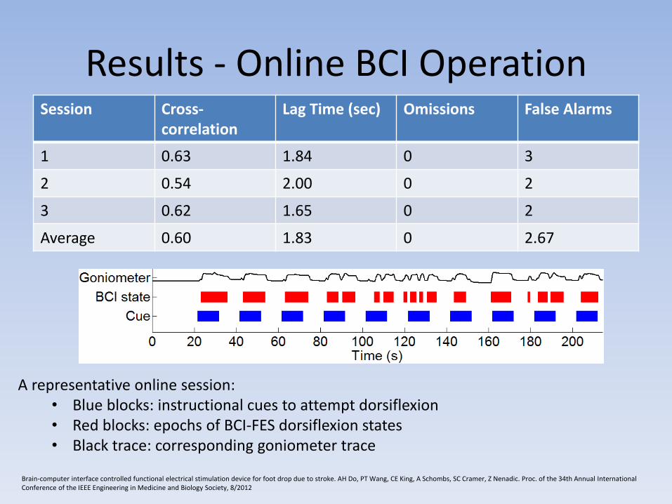
Results - Online BCI Operation Session Cross-
correlation Lag Time (sec) Omissions False Alarms
1 0.63 1.84 0 3
2 0.54 2.00 0 2
3 0.62 1.65 0 2
Average 0.60 1.83 0 2.67
A representative online session: • Blue blocks: instructional cues to attempt dorsiflexion • Red blocks: epochs of BCI-FES dorsiflexion states • Black trace: corresponding goniometer trace
Brain-computer interface controlled functional electrical stimulation device for foot drop due to stroke. AH Do, PT Wang, CE King, A Schombs, SC Cramer, Z Nenadic. Proc. of the 34th Annual International Conference of the IEEE Engineering in Medicine and Biology Society, 8/2012
BCIs as Rehabilitation Tool - Conclusion
• BCI-FES therapy is feasible in stroke patients, but will need to be formally tested in a stroke population to determine safety and efficacy
• Stroke patients may have non-classical motor representation, which necessitates data-driven BCI decoding approaches

SURVEY OF BCI RESEARCH COMMUNITY
Other BCI-FES Systems
Pfurtscheller et al. 2003
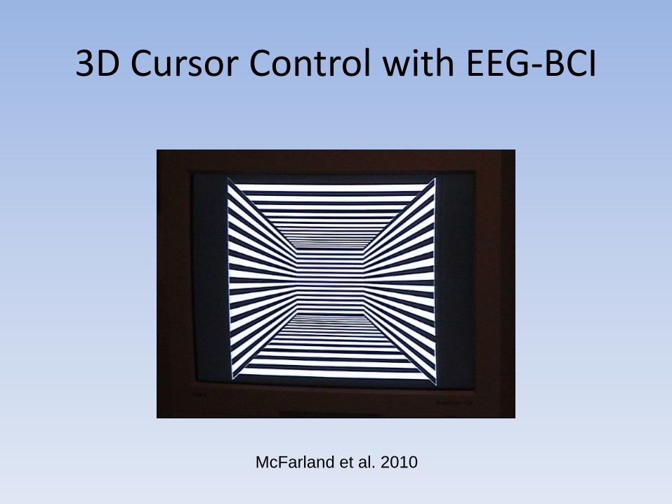
3D Cursor Control with EEG-BCI
McFarland et al. 2010
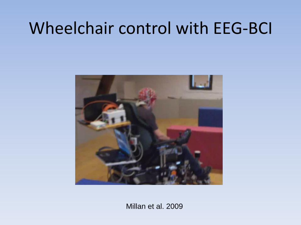
Wheelchair control with EEG-BCI
Millan et al. 2009
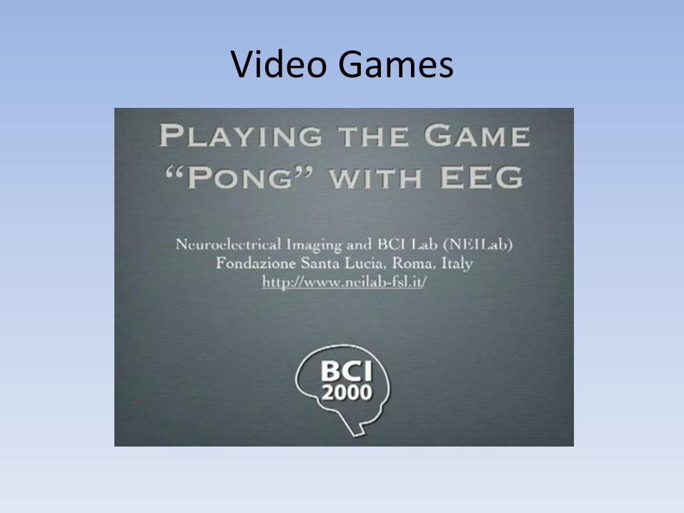
Video Games
Conclusion
• BCI technology may work as neuro-prostheses:
– Still need to define practical applications that will solve neurological problems and can potentially translate to the bedside
– Permanent solutions that work 24/7 and are stable over years
– Smooth and high accuracy control
Conclusion
• BCI technology may also work neuro-rehabilitative tools
– Still need to formally prove safety and efficacy
– Still need to implement in a practical manner
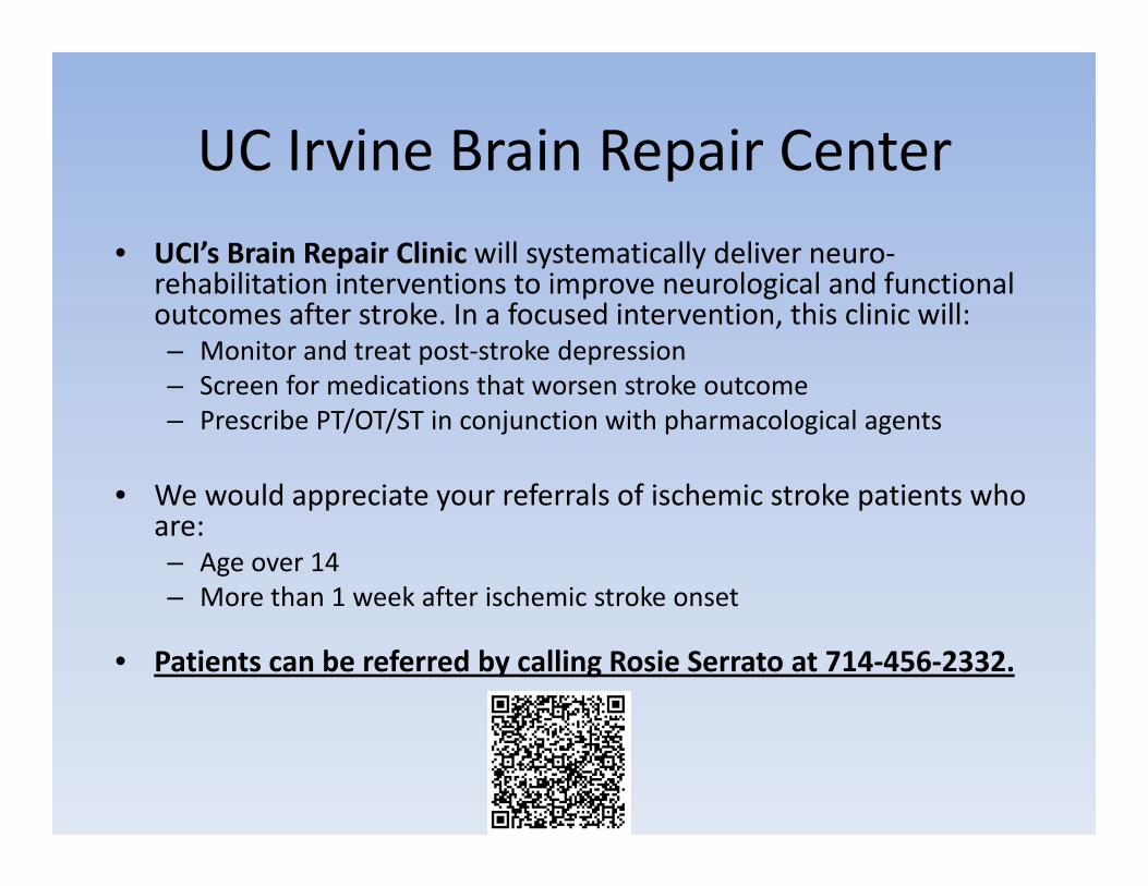
UC Irvine Brain Repair Center• UCI’s Brain Repair Clinic will systematically deliver neuro-
rehabilitation interventions to improve neurological and functional outcomes after stroke. In a focused intervention, this clinic will:– Monitor and treat post-stroke depression – Screen for medications that worsen stroke outcome – Prescribe PT/OT/ST in conjunction with pharmacological agents
• We would appreciate your referrals of ischemic stroke patients who are:– Age over 14– More than 1 week after ischemic stroke onset
• Patients can be referred by calling Rosie Serrato at 714-456-2332.

Acknowledgements
• UCI: – Zoran Nenadic, DSc – Steven Cramer, MD – Luis Chui, MD – Jack Lin, MD – Mona Sazgar, MD – Frank Hsu, MD, PHD – Po Wang – Christine King – Ahmad Abiri – Andrew Schombs – Lucy Der-Yaghiaian
• Long Beach VA: – Sophia Chun, MD
• UCLA: – John Stern, MD
• Rancho Los Amigos: – Susan Shaw, MD – Charles Liu, MD, PHD – David Millet, MD, PHD – Philip Requejo, PHD – Ann Nguyen
• CHOC: – Sharief Taraman, MD
And many others for their support

Acknowledgements
• Research funding provided by Department of Veterans Affairs, National Science Foundation, National Institute of Health (UCI ICTS), Roman Reed Spinal Cord Injury Research Fund of California, and American Brain Foundation.
![Brain Computer Interface for Neuro-rehabilitation With Deep … · neuro-rehabilitation,measuredontheFugl-MeyerAssessmentscale [30]. Besides, it can boost the interest, motivation](https://img.pdfslide.us/doc/110x75/5edf38a0ad6a402d666a91f7/brain-computer-interface-for-neuro-rehabilitation-with-deep-neuro-rehabilitationmeasuredonthefugl-meyerassessmentscale.jpg)