Embed Size (px)
DESCRIPTION
PPt
Citation preview

Brain anatomy: cerebral hemispheres

The cerebral hemispheres The cerebral cortex (consists of six lobes on
each side: frontal, parietal, temporal, occipital, insular, and limbic).
the underlying cerebral white matter, the basal ganglia: a complex of deep gray
matter masses.

Frontal lobe
Prefrontal: Personality And adaptation of the personality to
events and experiences Foresight and imagination Sense of self
frontal- main motor areas (originate movement that is co-ordinated elsewhere)

Parietal lobe
Principle sensory area Touch Proprioception Lesions cause sensory losses Involvement in cognition Receptive speech loss

Temporal lobe Cognition Emotion Memory Links to the hippocampus and the limbic
system are important to both of the above
Wernicke’s area (tempero-parietal) special role in auditory association and speech comprehension

What might be involved in speech processing?

Occipital lobe
Vision Visual processing and visual
association Involved in eye movement Hemianopia from damage
Think what might be involved in visual processing?

The cortex is particularly well developed in humans and is responsible for many higher brain functions, including manual dexterity (eg to move the fingers individually so as to play the piano); conscious, discriminative aspects of sensation; and cognitive activity, including language, reasoning, and many aspects of learning and memory.

Anatomy The cerebral hemispheres make up the largest
portion of the human brain. The cerebral hemispheres appear as highly
convoluted masses of gray matter that are organized into a folded structure.
The crests of the cortical folds (gyri) are separated by furrows (sulci) or deeper fissures.
The folding of the cortex into gyri and sulci permits the cranial vault to contain a large area of cortex (nearly 2 1/2 square feet), more than 50% of which is hidden within the sulci and fissures.

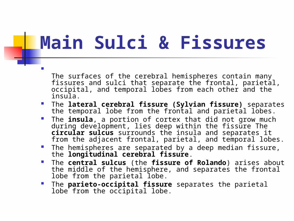
Main Sulci & Fissures
The surfaces of the cerebral hemispheres contain many fissures and sulci that separate the frontal, parietal, occipital, and temporal lobes from each other and the insula.
The lateral cerebral fissure (Sylvian fissure) separates the temporal lobe from the frontal and parietal lobes.
The insula, a portion of cortex that did not grow much during development, lies deep within the fissure The circular sulcus surrounds the insula and separates it from the adjacent frontal, parietal, and temporal lobes.
The hemispheres are separated by a deep median fissure, the longitudinal cerebral fissure.
The central sulcus (the fissure of Rolando) arises about the middle of the hemisphere, and separates the frontal lobe from the parietal lobe.
The parieto-occipital fissure separates the parietal lobe from the occipital lobe.


Corpus Callosum The corpus callosum connects the two
hemispheres It is a large bundle of myelinated and
nonmyelinated fibers, that crosses the longitudinal cerebral fissure and interconnects the hemispheres.
The corpus callosum serves to integrate the activity of the two hemispheres and permits them to communicate with each other.
Most parts of the cerebral cortex are connected with their counterparts in the opposite hemisphere by axons that run in the corpus callosum.


White Matter
The white matter of the adult cerebral hemisphere contains myelinated nerve fibers of many sizes as well as neuroglia.
Transverse (commissural) fibresinterconnect the two cerebral hemispheres (mainly the corpus callosum)
Projection fibres connect the cerebral cortex with lower portions of the brain or the spinal cord.

White matter continued
Association fibres connect the various portions of a cerebral hemisphere and permit the cortex to function as a coordinated whole.


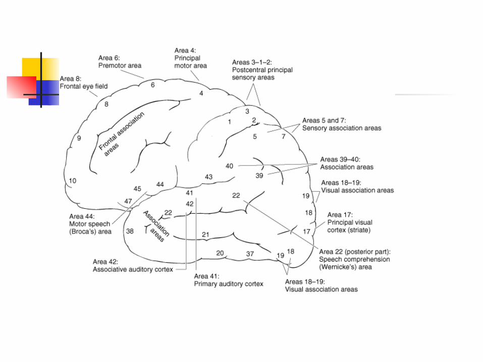
Areas of the cerebrum
Brodmann numbers to identify functions- down to individual sulci
Question localisation now that we know more about connectionism and we have amore dynamic view of the brain works

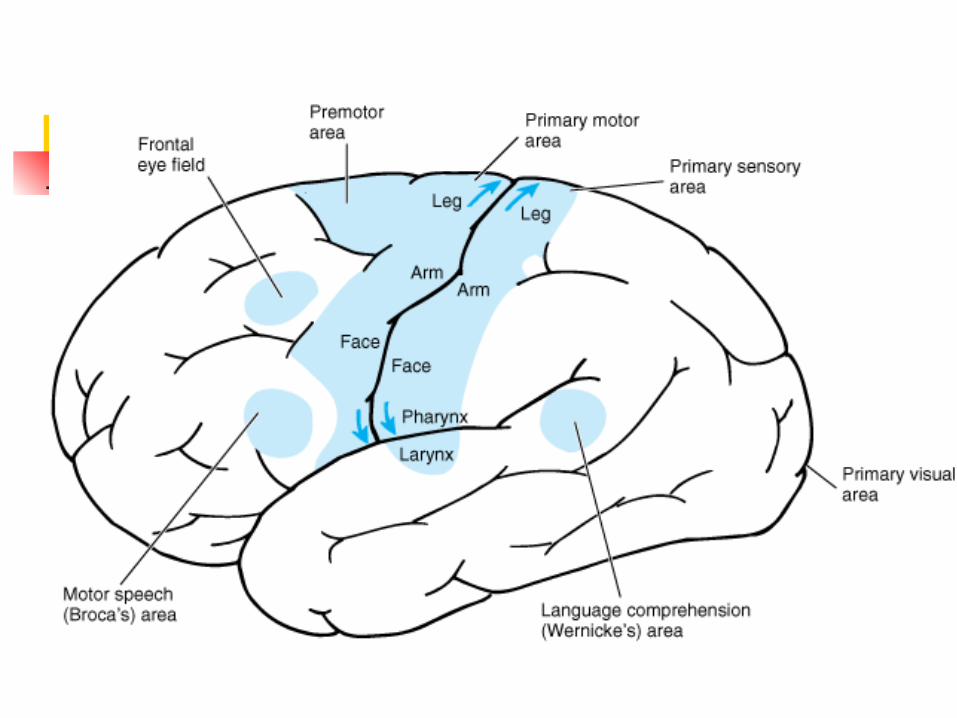

Primary Motor Cortex
The primary motor projection cortex is located on the anterior wall of the central sulcus.
These cells control voluntary movements of skeletal muscle on the opposite side of the body.

Homunculus
Map of motor control Reflects the body Sizes indicate the amount of
‘brain’ needed for various functions
Note vast area for the face- why?


Primary Sensory Cortex The primary sensory projection cortex for
sensory information received from the skin, mucosa, and other tissues of the body and face is located in the postcentral gyrus and is called the somatesthetic area,
This area receives fibers that convey touch and proprioceptive (muscle, joint, and tendon) sensations from the opposite side of the body.
A relatively wide portion of the adjacent frontal and parietal lobes can be considered a secondary sensory cortex because this area also receives sensory stimuli.
The cortical taste area is located close to the facial sensory area.


How do areas compare on the homunculous?

Primary Visual Cortex The primary visual receptive cortex is located in
the occipital lobe. In primates, an extensive posterior portion of the
occipital pole is concerned primarily with high-resolution macular vision;
the more anterior parts are concerned with peripheral vision.
The visual cortex in the right occipital lobe receives impulses from the right half of each retina,
The left visual cortex receives impulses from the left half of each retina. The upper portion of area 17 represents the upper half of each retina, and the lower portion represents the lower half.

Primary auditory cortex The primary auditory receptive area is located
in the superior temporal gyrus toward the lateral cerebral fissure.
The auditory cortex on each side receives the auditory radiation from the cochlea of both ears, and there is point-to-point projection of the cochlea on the acoustic area.
Wernicke's area (in the posterior third of the superior temporal gyrus in the dominant (usually left) hemisphere, is involved in high-order auditory discrimination and speech comprehension.

Basal ganglia The term basal ganglia are masses of gray
matter deep within the cerebral hemispheres. The term is debatable because these masses
are nuclei rather than ganglia Anatomically, the basal ganglia include the
caudate nucleus, the putamen, and the globus pallidus.Together they are called the corpus straitum
Functionally, the basal ganglia and their interconnections and neurotransmitters form the extrapyramidal system.

Extrapyramidal system
Influences motor instructions sent to the periphery
Has a role in stabilising the large and complicated systems that control movement
Helps to direct action and interpret sensory information


About the hemispheres
Why have they evolved? Why do we need them? Why are they so big?


![C brain anatomy jan 2007.ppt [Read-Only] - swostroke.caswostroke.ca/wp-content/uploads/2015/12/C-brain-anatomy-jan-2007.pdf · 7 Brain structures • Cerebral hemispheres – Frontal](https://img.pdfslide.us/doc/110x75/5e030e52d9e2ea2f20418d24/c-brain-anatomy-jan-2007ppt-read-only-7-brain-structures-a-cerebral-hemispheres.jpg)