Embed Size (px)
Citation preview
58 T H E B R I T I S H J O U R N A L O F S U R G E R Y
BRACHIAL PLEXUS ANESTHESIA- AN AID TO THE CASUALTY SURGEON
DESCRIPTION AND RESULTS OF 252 CASES
BY MILES FOX AND DEREK H. BUNTING FROM ANCOATS HOSPITAL, MANCHESTER
THE purpose of this report is to stress the usefulness of brachial block anaesthesia in a general hospital with a very busy Casualty Department. The tech- nique is not difficult to learn and is a safe and satis- factory method in the hands of relatively junior surgical staff by whom it was performed, and who later carried out the necessary operative procedures. It allows patients to be treated promptly, comfort- ably, and with a minimum of risk as out-patients, and, in addition, eases the strain on the anaesthetic staff.
This paper presents the results of 252 patients treated by regional block anaesthesia of the brachial plexus in Ancoats Hospital, Manchester, in the past three years.
METHOD The method employed for anaesthetizing the
brachial plexus is that described by Patrick (I940), a modification of Kulenkampff and Persky’s (1928)
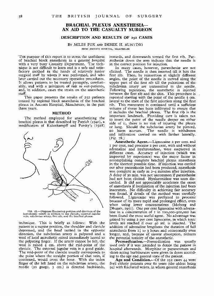
FIG. @.-Diagram illustrating position and direction of the hypodermic needle in relation to the clavicle, external jugular vein, subclavian artery, first rib, and the brachial plexus.
technique. This is briefly as follows: With the patient in a supine position, the shoulder and clavicle depressed, and the head turned in the opposite direction, the subclavian artery is palpated and a weal of local anresthetic raised immediately lateral to the palpating finger. If the artery cannot be felt, the weal is raised 4 cm. above the mid-point of the clavicle. The external jugular vein is a good guide. The mid-point of the clavicle usually corresponds to the point where the straight portion of that vein, if continued, would cross the bone. With the index finger of the left hand on the subclavian artery, the needle (20 gauge, 5 cm.) is directed backwards,
inwards, and downwards toward the first rib. Par- nsthesiae down the arm indicate that the needle is in the correct position for injection.
In many cases, however, paraesthesiae are not elicited. The needle is then inserted till it hits the first rib. Then, by reinsertion at slightly different angles, the point of the needle is moved along the upper part of the first rib till the pulsations of the subclavian artery are transmitted to the needle. Following aspiration, the anaesthetic is injected between the first rib and the skin. This procedure is repeated starting with the point of the needle 4 cm. lateral to the start of the first injection along the first rib. This maneuvre is continued until a sufficient volume of tissue has been infiltrated to ensure that it includes the brachial plexus. The first rib is the important landmark. Providing care is taken not to insert the point of the needle deeper on either side of it, there is no risk of injuring the pleura or lung. Should the subclavian artery be pierced, no harm accrues. The needle is withdrawn and infiltration carried on with farther laterally. (Fig. 78.)
Anaesthetic Agent.-Lignocaine 2 per cent and I per cent, and procaine 2 per cent, with and without adrenaline and hyaluronidase, were employed in different cases. Accuracy of injection (which was improved by experience) was the major factor in accomplishing complete brachial plexus anaesthesia in the shortest possible time. If injection was carried out after paraesthesiae had been produced, anaesthesia was complete as early as 2-4 minutes after injection. A delay of 30 min. was not uncommon if paraesthesiae had not been elicited. Hyaluronidase was soon dis- carded. It did not appreciably accelerate the onset of anzesthesia if localization of the injection had been inaccurate. No difficulty in achieving fair accuracy was found, if details of the method were carefully followed. Lignocaine was preferred to procaine because of its more rapid and prolonged effect, even when using lower concentrations (Moberg and Dhunkr, 1951). One per cent lignocaine with adrena- line in a concentration of I in ~oo,ooo-~oo,ooo has been found the most useful agent. No advantage was gained by using 2 per cent lignocaine, in which toxic levels are reached if over 40 ml. is injected. The addition of adrenaline lengthens the duration of full anaesthesia from I$ to 3 hours and occasionally even longer, and, because of slower absorption, reduces the potential toxicity of the anaesthetic.
Premeditation.-Fremedication was usually used only if it was intended to detain the patient in hospital afterwards. Morphine or papaveretum and short-acting barbiturates were given in doses accord- ing to the age and general state of the patient.
Age and Condition.-Of the 252 cases 44 were frail elderly patients over 60 years (the oldest being 94) with fractured wrists, in whom general anaesthesia
B R A C H I A L P L E X U S A N E S T H E S I A 59
carried out by the resident surgical staff would have involved significant risk. The youngest was 13 years. Although brachial plexus anaesthesia could have been used in younger patients, co-operation in children is often poor and general anaesthesia was preferred.
Five patients had concurrent head injuries with some concussion; in these, regional anaesthesia was used to enable the injuries of the upper extremity to be dealt with at once. Many came in late at night, having eaten or drunk recently. It was possible to proceed with the operation forthwith, without having to wait some time before a general anaesthetic could be administered.
Type of 0peration.-
Fractures of wrist (manipulation) 75 Fractures of forearm (closed reduction) Fractures of forearm (open reduction) 3 Fractures of olecranon (open reduction) 5 Reduction of dislocated elbow 4 Reductions of dislocated shoulder and fractures of
humeral neck 7 Soft-tissue injuries of forearm, hand, and fingers
(including tendon and nerve suture), and bone
Cold operations (median nerve decompression, operations for Dupuytren’s contracture, excision of ganglia, etc.)
Number
I 0
injuries of fingers I19 pemoyal of foreign bodies I1
18 - Total 252
Complications.- I . Puncture of the subclavian artery was not
uncommon. No harmful effects resulted. The needle was withdrawn and injection continued with the point of the needle placed more laterally. On one occasion following injection of about 5 ml. of 2 per cent lignocaine, sudden unconsciousness ensued, which passed off within several minutes. I t was found that injection had been made directly into a blood-vessel, probably a vein.
2. Damage to the plexus, manifesting itself as brachial plexus neuritis or permanent paralysis, did not result in any of the cases described. Care was taken to use a fairly thin needle (20 gauge) to avoid risk of trauma.
3. Pneumothorax occurred in I case, the exploring needle having been inserted past the first rib. The patient became distressed, dyspnoeic, and slightly cyanosed within a matter of minutes. X-ray exami- nation showed a partial right pneumothorax with mediastinal shift to the opposite side. She had bronchitis and emphysema. An emphysematous bulla had probably been ruptured.
4. Horner’s syndrome was not infrequent. It did not inconvenience the patient and disappeared in a short time.
5. Toxic symptoms from the anaesthetic did not occur in any of the patients. When 2 per cent lignocaine was used, as it was at the beginning of this series, no more than 40 ml. was ever injected.
Failure Rate.-Failure to anaesthetize the plexus, so that the intended operation had to be carried out with the aid of other methods of anaes- thesia, occurred in 7 cases. Greater difficulty was experienced, and the failure rate was higher in patients with short and full necks. In the asthenic the cords of the brachial plexus lie so superficial as to be felt digitally.
One of the patients developed a pneumothorax, as described above. Quite often, a slight degree of motor power, never enough to interfere with the operation, remained in the forearm and hand.
DISCUSSION Brachial block anaesthesia is an invaluable help
to the emergency surgeon. He can also spare his anaesthetic colleagues, enabling them to attend to other commitments. The need for this is not apparent in well-staffed teaching hospitals, but in the peri- phery, where a duty anaesthetist may not be readily available, it is a great boon. The method is not difficult to learn and could be acquired by every surgeon. In Ancoats Hospital, which is situated in the midst of a dense industrial area of Manchester, seeing about 22,000 new patients in the Casualty Department each year, the casualty officers are in- structed, so that they and the resident surgical officers are able to deal expeditiously with any patient in whom the method is indicated. The patients remain ambulatory and can be sent home afterwards. Those arriving with a full stomach present no difficulty. Risks of general anaesthesia in infirm and aged patients are avoided. In reduction of fractures and dislocations, radiological control can be performed without submitting the patient to long or repeated periods of general anaesthesia. Shock is alleviated by removal of painful stimuli, and traumatic reflex spasm is avoided or abolished.
I t takes a few minutes only to infiltrate the brachial plexus. Practically all conditions of the upper extremity which necessitate operative or manipulative intervention lend themselves to this form of anaesthesia. If one is working high in the arm or axilla, the intercostobrachial nerve must be blocked.
The technique, as described by Patrick (1940), was found to be reliable and safe in the hands of junior surgical staff. If paraesthesiae were obtained, one injection usually produced satisfactory anaesthesia. If paraesthesiae were not produced after five or six attempts, a block of tissue containing the brachial plexus was infiltrated. Excessive trauma was thus avoided and the anzsthesia produced was satisfactory. The discomfort produced by paraesthesiae is small and of momentary duration. I t is a sign that injection can be carried out and satisfactory anaesthesia expected within a short period. No evidence of damage to the brachial plexus (temporary or per- manent) was found in the 252 cases described. The same conclusions were reached in other larger series (Kulenkampff and Persky, 1928; Bonica, Moore, and Orlov, 1949). The single-injection technique described by Lookman (1958), requiring more practice at accurate localization and producing a higher rate of incomplete anzsthesia, was not tried.
There were 7 failures. After performing over 10 successful injections, none of the residents had a failure. Some sensation of touch and pressure and a little movement quite often remained. This did not interfere with the operation and was therefore not recorded. By varying the percentages of anaesthetic solution injected, it is possible to abolish sensation and yet leave sufficient motor power to help the surgeon identify more easily the proximal ends of
60 T H E B R I T I S H J O U R N A L O F S U R G E R Y
divided tendons (Macintosh and Mushin, 1947). This refinement was not attempted, none of the residents having extensive experience of this form of anaesthesia.
Various agents for anesthetizing the brachial plexus were tried. One per cent lignocaine with adrenaline (I in ~oo ,oo~ too ,ooo) was found to be most effective. No toxic symptoms were recorded. On one occasion about 5 ml. of 2 per cent lignocaine was inadvertently injected into a vein with sudden, but temporary, unconsciousness of the patient.
One case of pneumothorax occurred. The rapid onset and marked distress of the patient made it probable that an emphysematous bulla had been ruptured. Recovery was complete in several days. Bonica and Moore (1950) reported 10 pneumo- thoraces in a series of 1512 blocks. Macintosh and Mushin (1947) had 3 cases in a series of 400 blocks. There was no mortality. Macintosh and Mushin suggest that minor degrees of pneumothorax may be missed as they can occur without obvious symp- toms. Partial collapse of the lung may be a sequel to temporary phrenic nerve paralysis which occasion- ally occurs. It was not noticed in any of the cases described. Horner’s syndrome was seen several times, disappearing with the end of anaesthesia. It did not inconvenience the patient.
SUMMARY Two hundred and fifty-two cases of brachial
block anaesthesia are described. The results are dis- cussed.
We believe that brachial plexus block is a useful, convenient, and safe method of anaesthesia for surgery of the upper extremity and an invaluable aid to the Casualty Surgeon.
We are grateful to Mr. C. H. Cullen and Mr. A. Glass for their kind advice and help in preparing this paper.
REFERENCES BONICA, J. J., and MOORE, D. C. (I950), Anas. and
Analges., 29, 241. - - - _ and ORLOV, M. (1949), Amer. J. Surg., 78,
45. LOOKMAN, A. A. (1999, Anaesthesia, 13, 5. MACINTOSH, R. R., and MUSHIN, W. W. (I947), Local
Analgesia-Brachial Plexus. Oxford : Blackwell Scientific Publications.
MOBERG, E., and DHUNER, K. G. ( I~sI ) , J . BoneJt Surg., 33A, 884.
KULENKAMPFF, D., and PERSKY, M. A. (1928), Ann. Surg., 87, 883.
PATRICK, J. (1940), Brit. 3. Surg., 27, 734.
INTERSTITIAL PRESENTING AS SPIGELIAN HERNIA A REVIEW OF THE LITERATURE AND CASE REPORT
BY B. ALTMAN WESTMINSTER HOSPITAL, LONDON, S.W.1
Interstitial Hernia.-Interstitial hernia, which was first described by Bartholin in 1661, is the commonest type of interparietal hernia. An inter- stitial hernia is one in which the hernial sac passes between the layers of the abdominal wall.
Classification.-The most useful classification is that of Fuld (1921), who discusses these herniae under the following headings :-
I . Properitoneal: The sac passes between the peritoneum and the transversalis fascia.
t. Inguino-interstitial: The sac passes between the transversalis fascia and transversus abdominis, between transversus abdominis and obliquus internus, between obliquus internus and obliquus externus, or rarely between the fibres of obliquus internus.
3. Inguino-superficial : The sac passes through the external inguinal ring to lie superficially in the anterior abdominal wall or thigh.
These herniae originate in the region of the internal inguinal ring and may be associated with an inguinal hernia. Moynihan (1900) thought that this association was an essential part of the condition and in fact defined an interstitial hernia as being bilocular, one loculus being an inguinal sac and the other the interparietal sac. Lower and Hicken (1931), how- ever, found many which were monolocular with no evidence of an associated inguinal hernia.
Incidence.-The relative incidence of these types is given by Lower and Hicken (1931)~ in a study of a large series of cases, as properitoneal 20 per cent,
interstitial 60 per cent, and inguino-superficial 20 per cent.
Types I and z occur three times and type 3 fourteen times more often in men than women. The usual age-groups in which they occur are 30-40 years in men and 50-60 years in women.
AetioZogy.-The factors involved may be dis- cussed under the headings of mechanical and con- genital.
I. Mechanical : The most commonly recorded abnormality is a maldescended testicle causing an obstruction to the ‘normal descent’ of an inguinal hernia. Macready in 1893 found an abnormality of the testis in 73.4 per cent and maldescent in 67.1 per cent. Similar figures have been produced by many others.
Weak areas in the first and third parts of the inguinal canal, narrowing of the inguinal rings, the wearing of a truss, and hydrocele of the canal or cord have also been held to be contributory factors.
2. Congenital: Small peritoneal pouches close to the obliterated hypogastric arteries were found in 22 per cent of necropsies on fetuses by Moynihan (1900)~ and these have been considered to be the starting-point of properitoneal sacs.
In a survey of the subject and review of the literature Barling (1956) concludes that all cases of interparietal hernia should be considered congenital in origin, since mechanical factors, when they do occur, are secondary to a congenital defect. This