Embed Size (px)
Citation preview
BPS 594Pharmacogenomics and Molecular Pharmacology
Genes and GeneticsGenes and Genetics
Debra A. Tonetti, Ph.D.COP 453
Human Molecular Genetics, Strachan & Read, 3rd EditionChapter 1
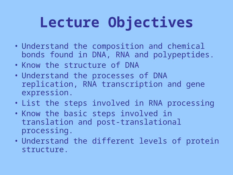
Lecture Objectives
• Understand the composition and chemical bonds found in DNA, RNA and polypeptides.
• Know the structure of DNA• Understand the processes of DNA replication,
RNA transcription and gene expression.• List the steps involved in RNA processing• Know the basic steps involved in translation and
post-translational processing.• Understand the different levels of protein
structure.
Molecular Genetics
• Primarily concerned with the interaction between the information molecules (DNA and RNA) and how this information is translated into proteins.
• In eukaryotes, DNA molecules are found in the chromosomes of the nucleus, mitochondria and also chloroplasts of plant cells.
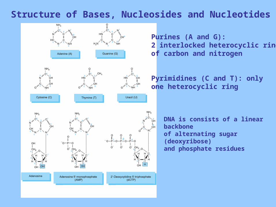
Structure of Bases, Nucleosides and Nucleotides
Purines (A and G): 2 interlocked heterocyclic ringsof carbon and nitrogen
Pyrimidines (C and T): onlyone heterocyclic ring
DNA is consists of a linear backbone of alternating sugar (deoxyribose)and phosphate residues
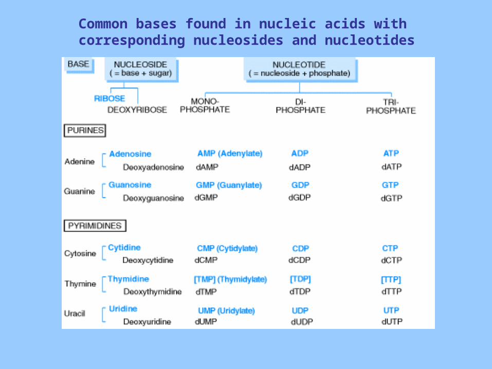
Common bases found in nucleic acids with corresponding nucleosides and nucleotides
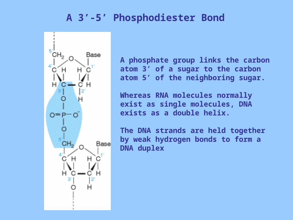
A 3’-5’ Phosphodiester Bond
A phosphate group links the carbon atom 3’ of a sugar to the carbon atom 5’ of the neighboring sugar.
Whereas RNA molecules normally exist as single molecules, DNA exists as a double helix.
The DNA strands are held together by weak hydrogen bonds to form a DNA duplex
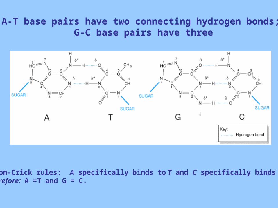
A-T base pairs have two connecting hydrogen bonds; G-C base pairs have three
Watson-Crick rules: A specifically binds to T and C specifically binds to G therefore: A =T and G = C.
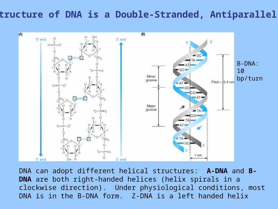
The Structure of DNA is a Double-Stranded, Antiparallel Helix
DNA can adopt different helical structures: A-DNA and B-DNA are both right-handed helices (helix spirals in a clockwise direction). Under physiological conditions, most DNA is in the B-DNA form. Z-DNA is a left handed helix
B-DNA: 10 bp/turn
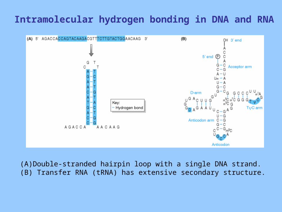
Intramolecular hydrogen bonding in DNA and RNA
(A) Double-stranded hairpin loop with a single DNA strand. (B) Transfer RNA (tRNA) has extensive secondary structure.
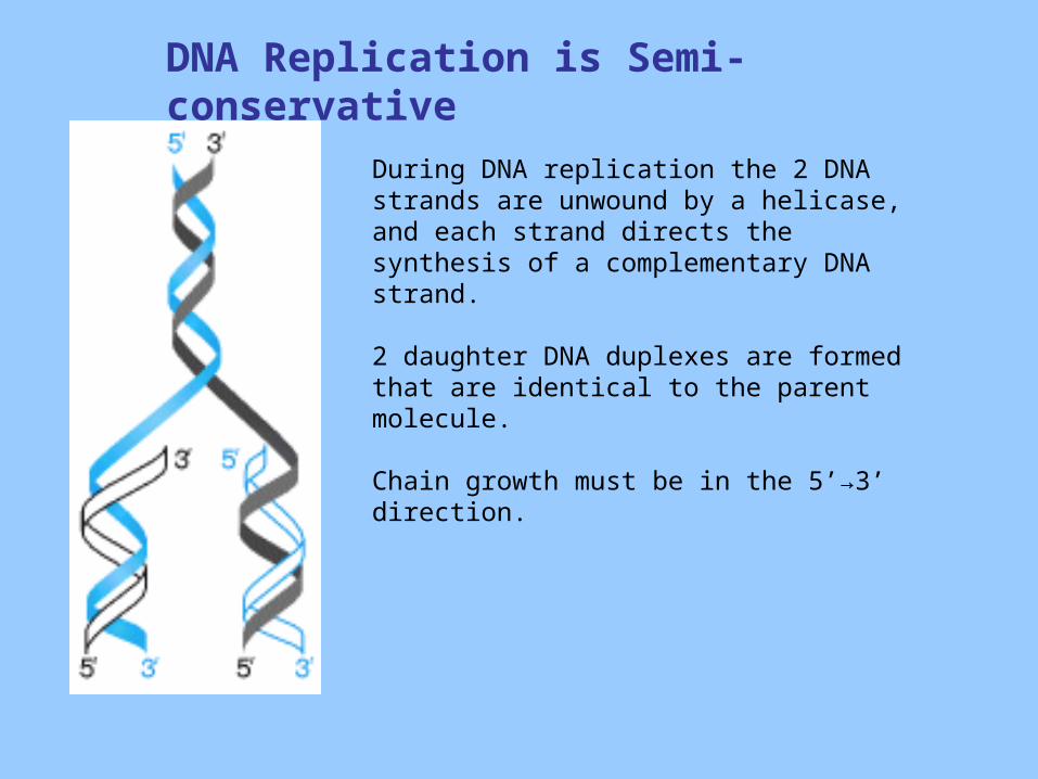
DNA Replication is Semi-conservative
During DNA replication the 2 DNA strands are unwound by a helicase, and each strand directs the synthesis of a complementary DNA strand.
2 daughter DNA duplexes are formed that are identical to the parent molecule.
Chain growth must be in the 5’→3’ direction.
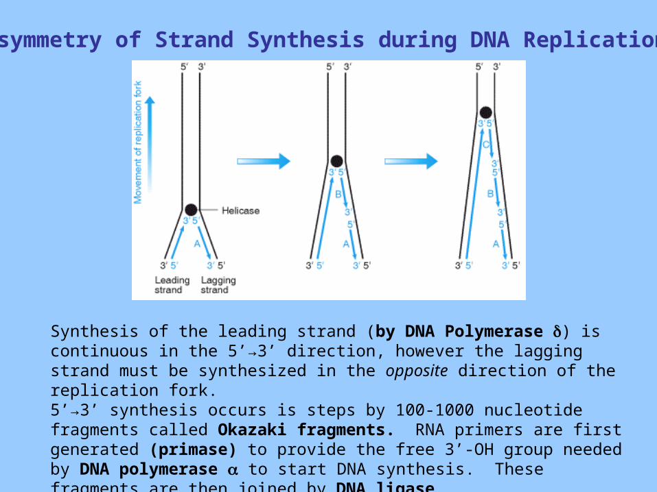
Asymmetry of Strand Synthesis during DNA Replication
Synthesis of the leading strand (by DNA Polymerase ) is continuous in the 5’→3’ direction, however the lagging strand must be synthesized in the opposite direction of the replication fork.5’→3’ synthesis occurs is steps by 100-1000 nucleotide fragments called Okazaki fragments. RNA primers are first generated (primase) to provide the free 3’-OH group needed by DNA polymerase to start DNA synthesis. These fragments are then joined by DNA ligase
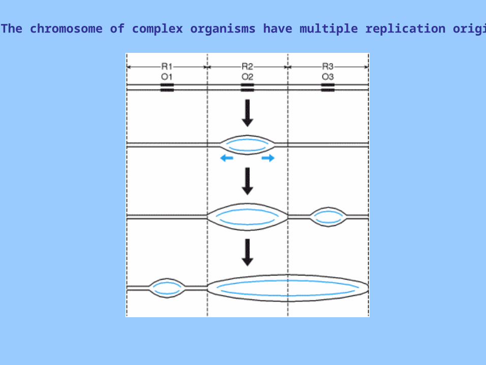
The chromosome of complex organisms have multiple replication origins
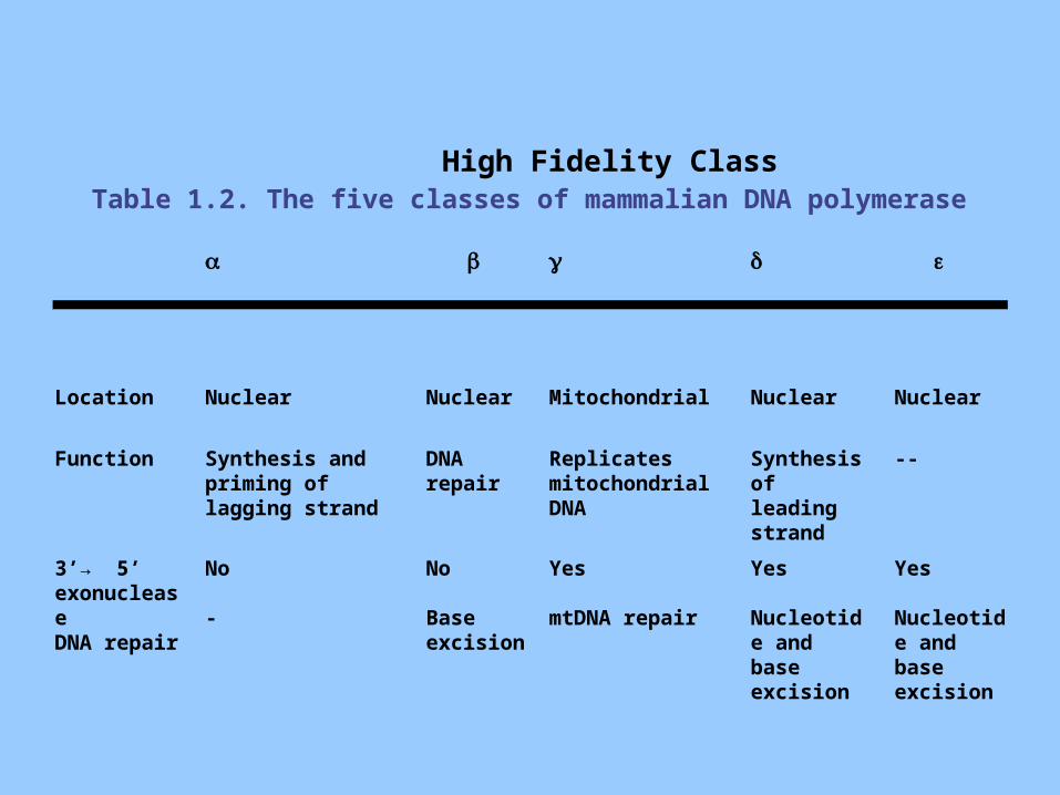
Table 1.2. The five classes of mammalian DNA polymerase
High Fidelity Class
Location Nuclear Nuclear Mitochondrial Nuclear Nuclear
Function Synthesis and priming of lagging strand
DNA repair
Replicates mitochondrial DNA
Synthesis of leading strand
--
3’→ 5’ exonucleaseDNA repair
No
-
No
Base excision
Yes
mtDNA repair
Yes
Nucleotide and base excision
Yes
Nucleotide and base excision
Major Classes of Proteins used in the DNA Replication Machinery
• Topoisomerases: unwind DNA by breaking a single DNA strand. Tension from the supercoil is released.
• Helicases: Unwind the double strand.• DNA polymerases:
– DNA-directed DNA polymerases (some with DNA repair function)
– RNA-directed DNA polymerases (reverse transcriptases) • Telomerase – ends of linear chromosome
• Primases: attach small RNA primer to provide 3’-OH group for DNA polymerase. Is degraded by ribonuclease.
• Ligase: catalyzes the formation of a phosphodiester bond between adjacent 3’OH and 5’-phosphate groups.
• Single-stranded binding proteins: Maintains the stability of the replication fork, prevents single-stranded DNA degradation.
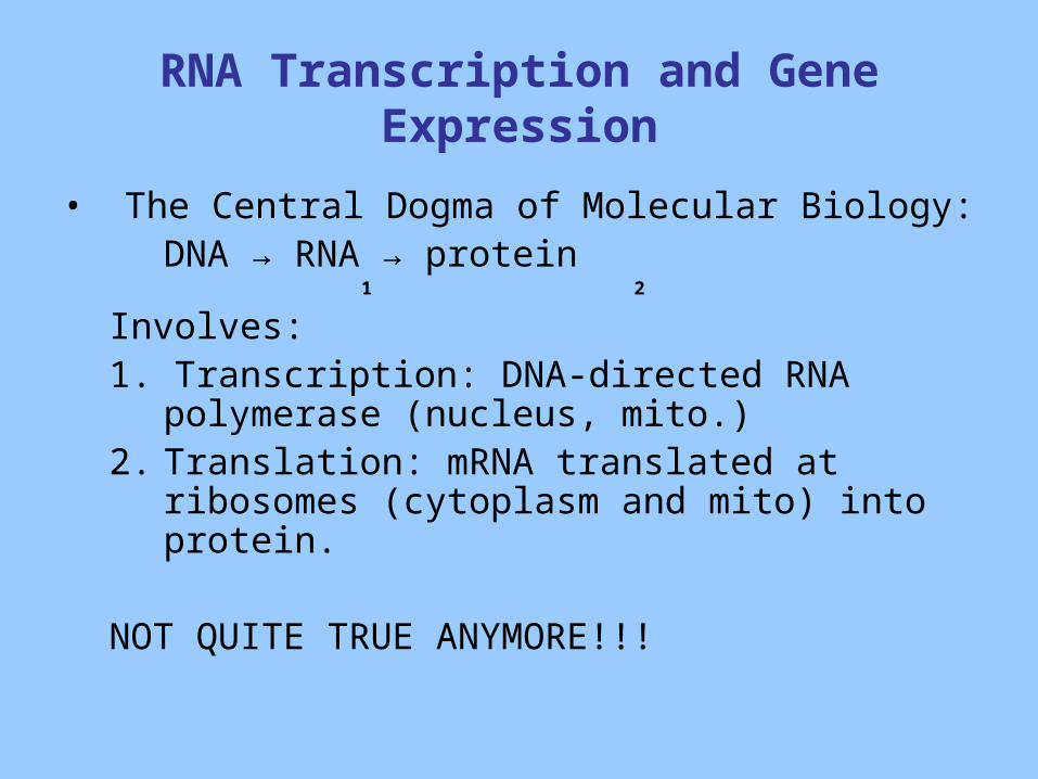
RNA Transcription and Gene Expression
• The Central Dogma of Molecular Biology:DNA → RNA → protein 1 2
Involves:1. Transcription: DNA-directed RNA polymerase
(nucleus, mito.)2. Translation: mRNA translated at ribosomes
(cytoplasm and mito) into protein.
NOT QUITE TRUE ANYMORE!!!
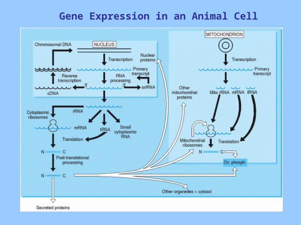
Gene Expression in an Animal Cell
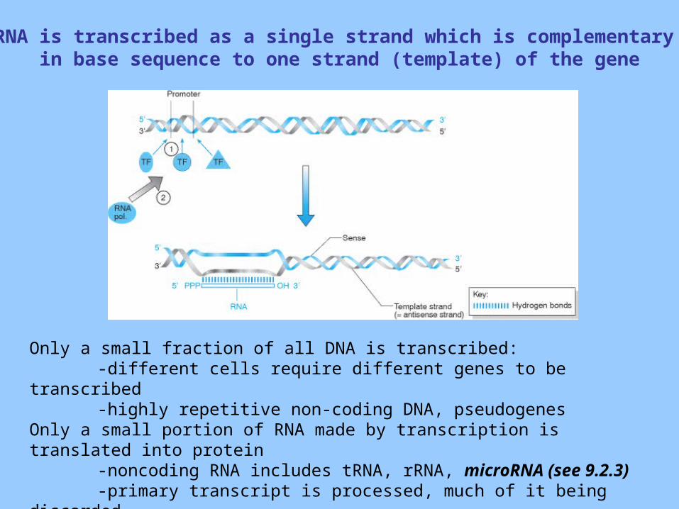
RNA is transcribed as a single strand which is complementary in base sequence to one strand (template) of the gene
Only a small fraction of all DNA is transcribed:-different cells require different genes to be transcribed-highly repetitive non-coding DNA, pseudogenes
Only a small portion of RNA made by transcription is translated into protein-noncoding RNA includes tRNA, rRNA, microRNA (see 9.2.3)-primary transcript is processed, much of it being discarded-only the central part of the mature RNA is translated – sections on each
end remain untranslated.
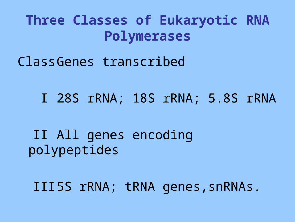
Three Classes of Eukaryotic RNA Polymerases
Class Genes transcribed
I 28S rRNA; 18S rRNA; 5.8S rRNA
II All genes encoding polypeptides
III 5S rRNA; tRNA genes,snRNAs.
Trans-acting Transcription Factors and Cis-acting regulating elements are required for Gene Expression
• Short sequence elements in the vicinity of the gene (cis) are recognized by transcription factors (trans) to guide and recruit RNA polymerase.
• These sequences are often clustered upstream of the coding sequence of the gene and collectively define the promoter region.
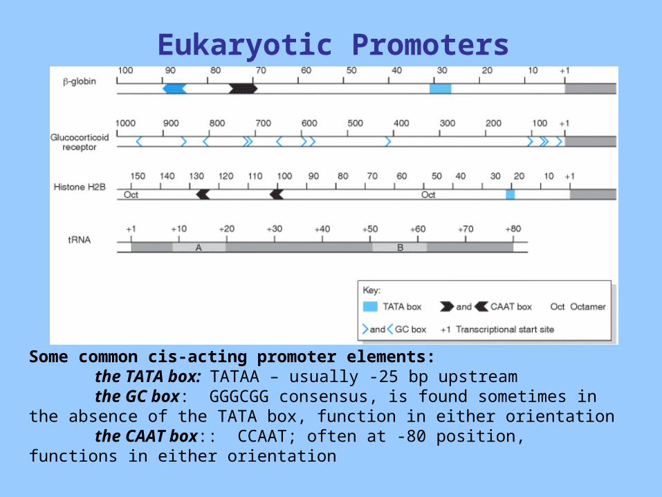
Eukaryotic Promoters
Some common cis-acting promoter elements:the TATA box: TATAA – usually -25 bp upstreamthe GC box: GGGCGG consensus, is found sometimes in the absence
of the TATA box, function in either orientationthe CAAT box:: CCAAT; often at -80 position, functions in either
orientation

Additional Specific Recognition Elements (often tissue specific)
• Enhancers: located at a variable distances from the transcriptional start site; orientation-independent; enhance transcriptional activation
TRE (TPA response element) GTGAGT(A/C)A
transcription factor: AP-1 family (Jun/Fos)
• Silencers: similar to enhancers but inhibit transcriptional activity of specific genes
Tissue-Specific Gene Expression
• The DNA content of every cell is identicalWhat makes the different cell types unique??• Only a portion of genes are expressed in any
one cell type.
How is this achieved??• Transcriptionally inactive or active chromatin
-determined by chromatin conformation: condensed or open
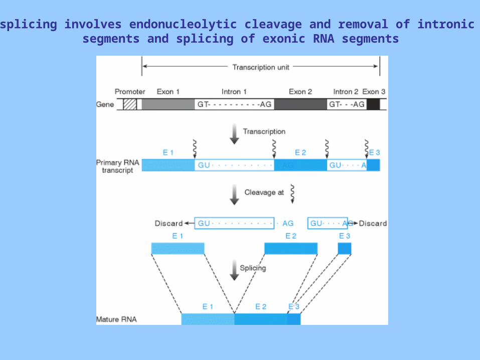
RNA splicing involves endonucleolytic cleavage and removal of intronic RNA segments and splicing of exonic RNA segments
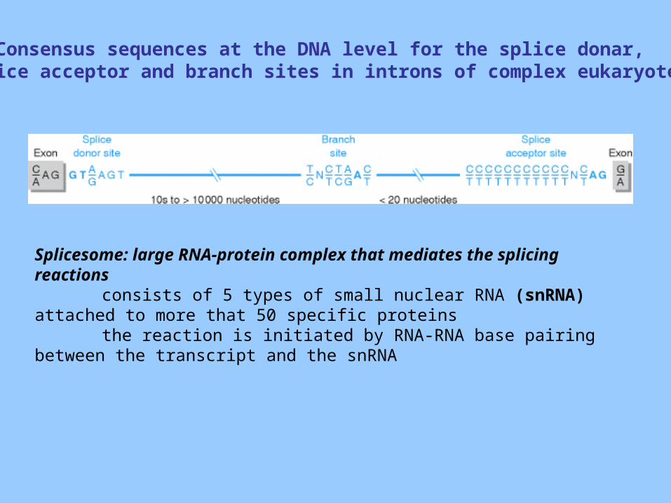
Consensus sequences at the DNA level for the splice donar, splice acceptor and branch sites in introns of complex eukaryotes
Splicesome: large RNA-protein complex that mediates the splicing reactions
consists of 5 types of small nuclear RNA (snRNA) attached to more that 50 specific proteins
the reaction is initiated by RNA-RNA base pairing between the transcript and the snRNA
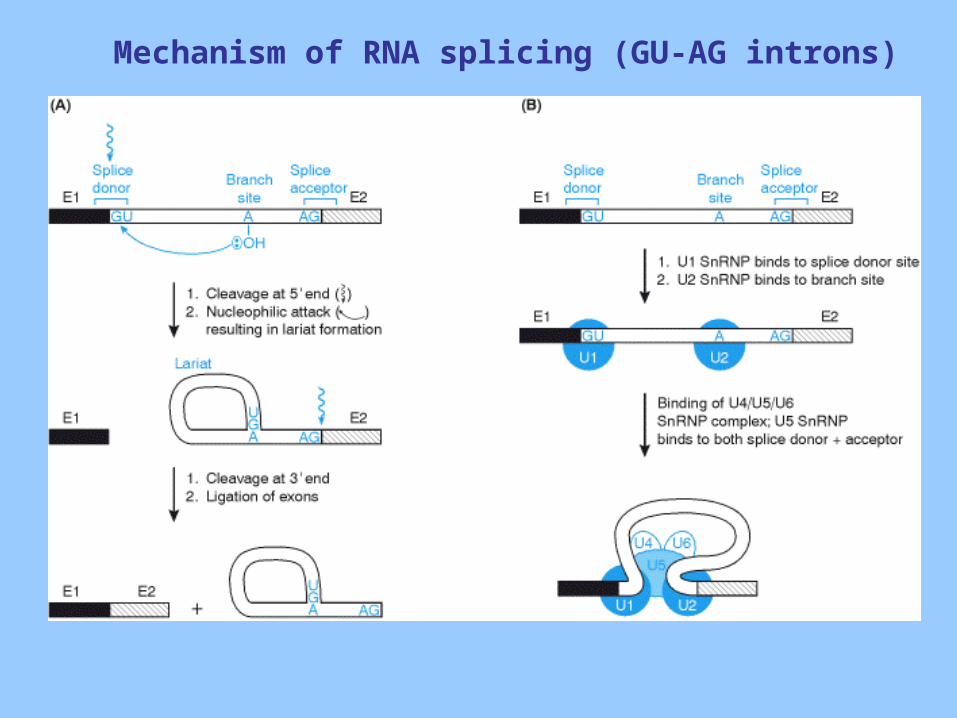
Mechanism of RNA splicing (GU-AG introns)
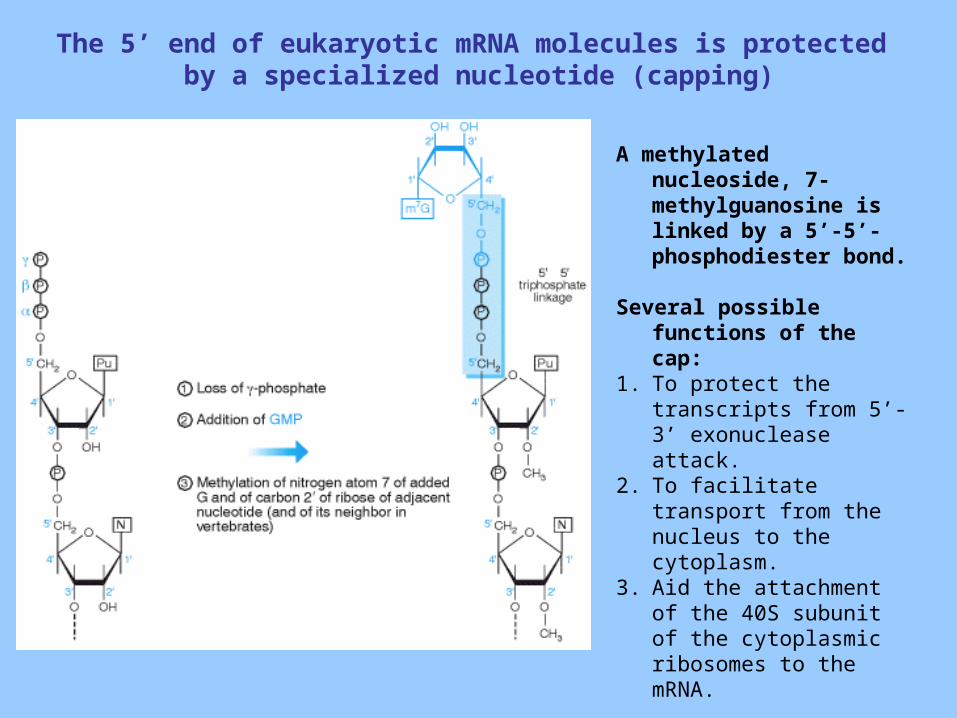
The 5’ end of eukaryotic mRNA molecules is protected by a specialized nucleotide (capping)
A methylated nucleoside, 7-methylguanosine is linked by a 5’-5’-phosphodiester bond.
Several possible functions of the cap:
1. To protect the transcripts from 5’-3’ exonuclease attack.
2. To facilitate transport from the nucleus to the cytoplasm.
3. Aid the attachment of the 40S subunit of the cytoplasmic ribosomes to the mRNA.
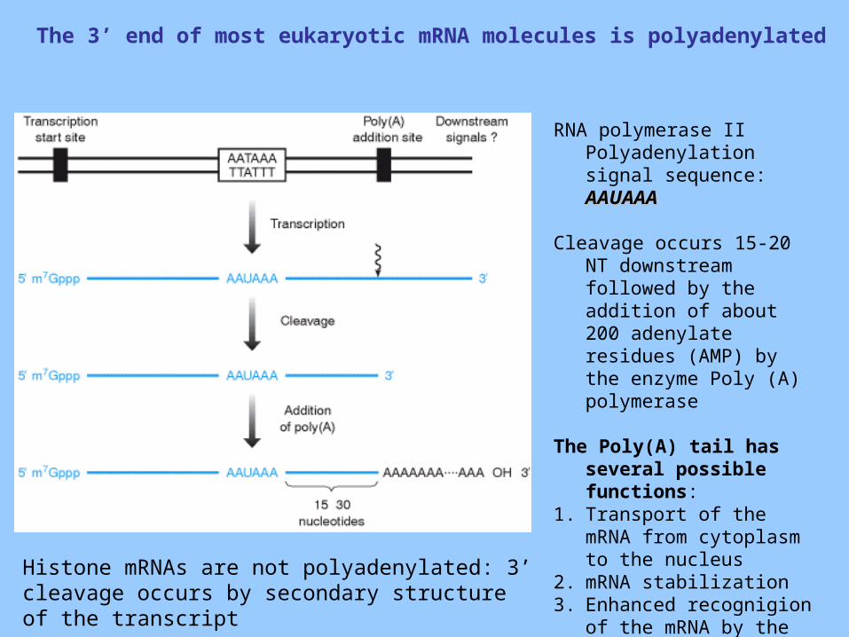
The 3’ end of most eukaryotic mRNA molecules is polyadenylated
RNA polymerase II Polyadenylation signal sequence: AAUAAA AAUAAA
Cleavage occurs 15-20 NT downstream followed by the addition of about 200 adenylate residues (AMP) by the enzyme Poly (A) polymerase
The Poly(A) tail has several possible functions:
1. Transport of the mRNA from cytoplasm to the nucleus
2. mRNA stabilization 3. Enhanced recognigion of
the mRNA by the ribosomal machinery.Histone mRNAs are not polyadenylated: 3’ cleavage
occurs by secondary structure of the transcript
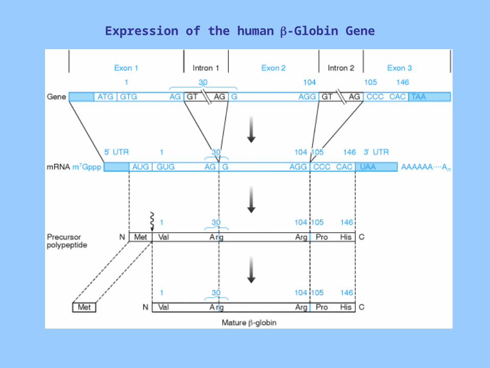
Expression of the human-Globin Gene
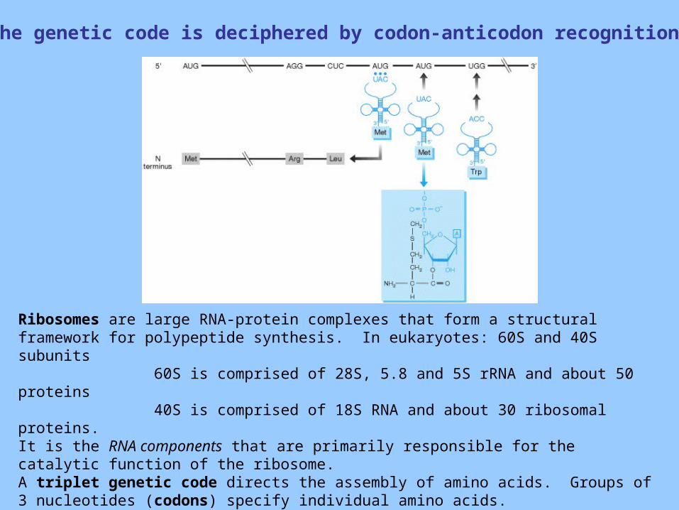
The genetic code is deciphered by codon-anticodon recognition
Ribosomes are large RNA-protein complexes that form a structural framework for polypeptide synthesis. In eukaryotes: 60S and 40S subunits
60S is comprised of 28S, 5.8 and 5S rRNA and about 50 proteins40S is comprised of 18S RNA and about 30 ribosomal proteins.
It is the RNA components that are primarily responsible for the catalytic function of the ribosome.A triplet genetic code directs the assembly of amino acids. Groups of 3 nucleotides (codons) specify individual amino acids.
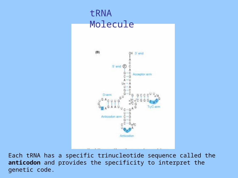
Each tRNA has a specific trinucleotide sequence called the anticodon and provides the specificity to interpret the genetic code.
tRNA Molecule
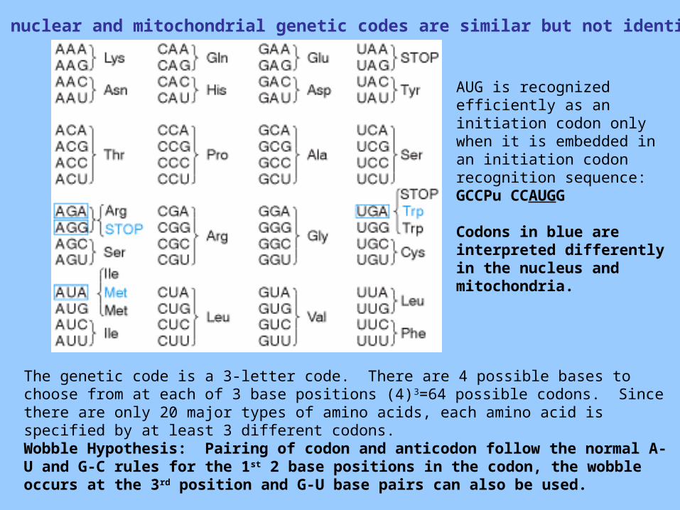
The nuclear and mitochondrial genetic codes are similar but not identical
The genetic code is a 3-letter code. There are 4 possible bases to choose from at each of 3 base positions (4)3=64 possible codons. Since there are only 20 major types of amino acids, each amino acid is specified by at least 3 different codons.Wobble Hypothesis: Pairing of codon and anticodon follow the normal A-U and G-C rules for the 1st 2 base positions in the codon, the wobble occurs at the 3rd position and G-U base pairs can also be used.
AUG is recognized efficiently as an initiation codon only when it is embedded in an initiation codon recognition sequence:GCCPu CCAUGG
Codons in blue are interpreted differently in the nucleus and mitochondria.
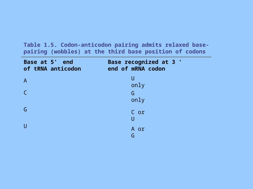
Table 1.5. Codon-anticodon pairing admits relaxed base-pairing (wobbles) at the third base position of codons
Base at 5’ end of tRNA anticodon
Base recognized at 3 ‘
end of mRNA codon
A U only
C G only
G C or U
U A or G
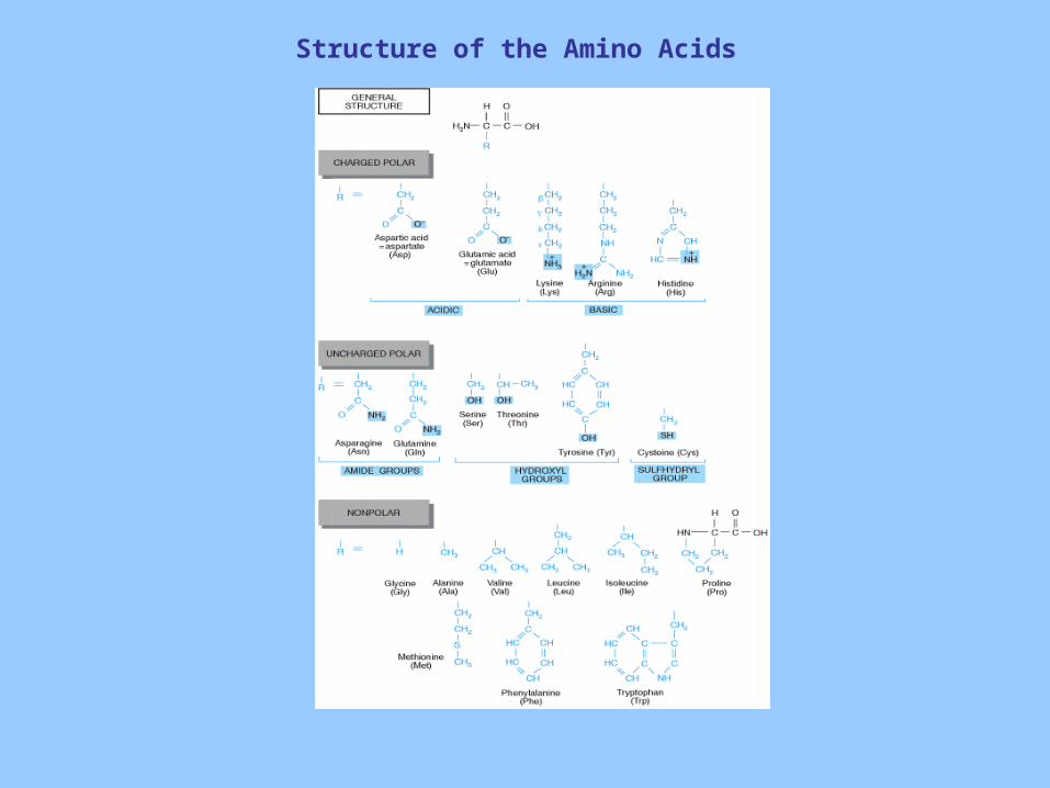
Structure of the Amino Acids
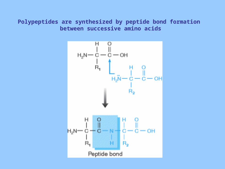
Polypeptides are synthesized by peptide bond formation between successive amino acids
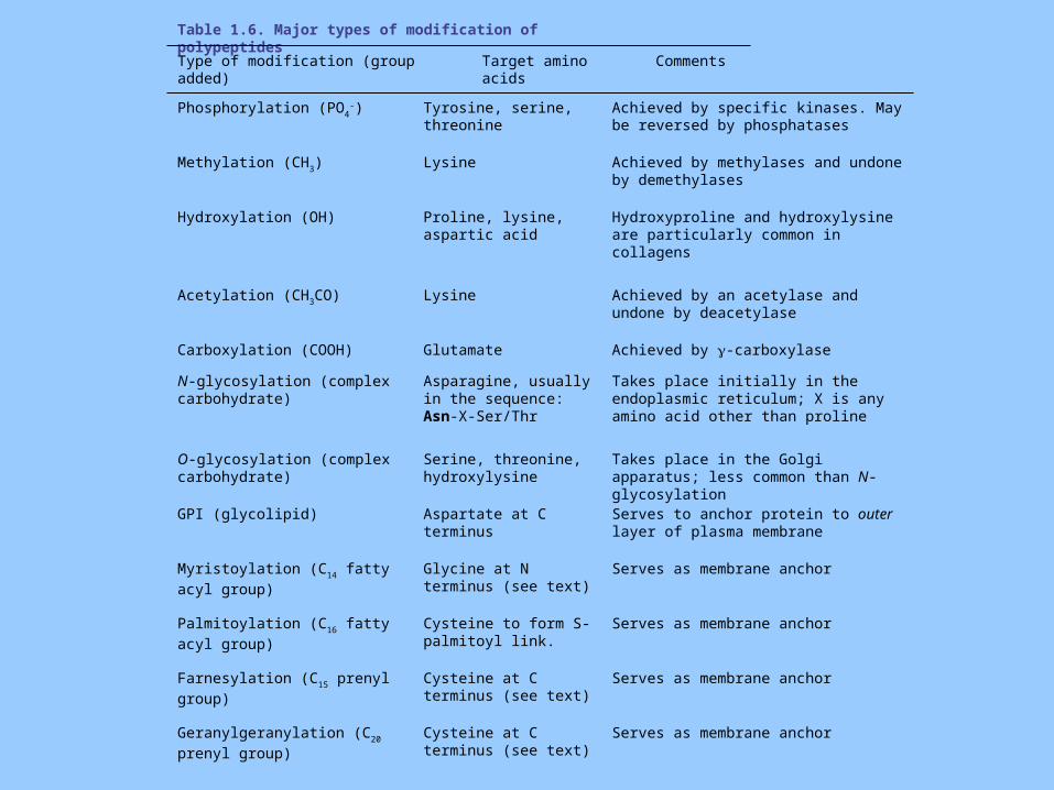
Table 1.6. Major types of modification of polypeptides
Type of modification (group added) Target amino acids Comments
Phosphorylation (PO4-) Tyrosine, serine,
threonineAchieved by specific kinases. May be reversed by phosphatases
Methylation (CH3) Lysine Achieved by methylases and undone by demethylases
Hydroxylation (OH) Proline, lysine, aspartic acid
Hydroxyproline and hydroxylysine are particularly common in collagens
Acetylation (CH3CO) Lysine Achieved by an acetylase and undone by deacetylase
Carboxylation (COOH) Glutamate Achieved by -carboxylase
N-glycosylation (complex carbohydrate)
Asparagine, usually in the sequence: Asn-X-Ser/Thr
Takes place initially in the endoplasmic reticulum; X is any amino acid other than proline
O-glycosylation (complex carbohydrate)
Serine, threonine, hydroxylysine
Takes place in the Golgi apparatus; less common than N-glycosylation
GPI (glycolipid) Aspartate at C terminus Serves to anchor protein to outer layer of plasma membrane
Myristoylation (C14 fatty acyl
group)
Glycine at N terminus (see text)
Serves as membrane anchor
Palmitoylation (C16 fatty acyl
group)
Cysteine to form S-palmitoyl link.
Serves as membrane anchor
Farnesylation (C15 prenyl group) Cysteine at C terminus (see text)
Serves as membrane anchor
Geranylgeranylation (C20 prenyl
group)
Cysteine at C terminus (see text)
Serves as membrane anchor
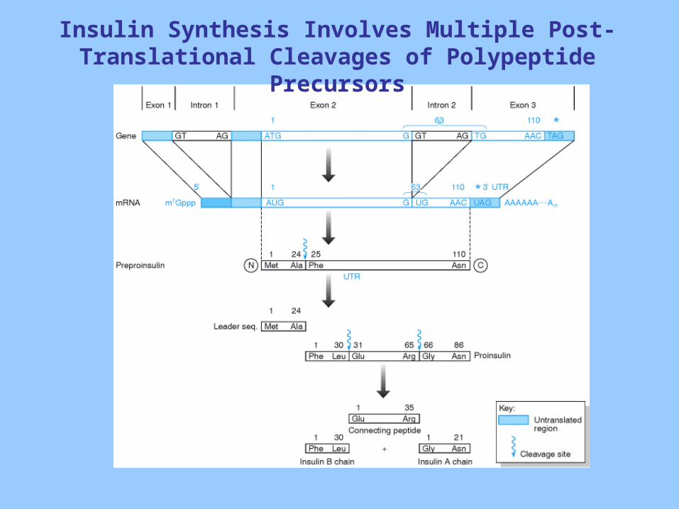
Insulin Synthesis Involves Multiple Post-Translational Cleavages of Polypeptide Precursors
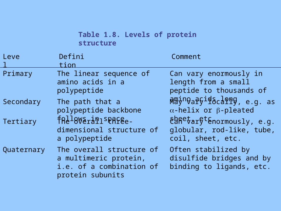
Table 1.8. Levels of protein structure
Level Definition Comment
Primary The linear sequence of amino acids in a polypeptide
Can vary enormously in length from a small peptide to thousands of amino acids long
Secondary The path that a polypeptide backbone follows in space
May vary locally, e.g. as -helix or -pleated sheet, etc.
Tertiary The overall three-dimensional structure of a polypeptide
Can vary enormously, e.g. globular, rod-like, tube, coil, sheet, etc.
Quaternary The overall structure of a multimeric protein, i.e. of a combination of protein subunits
Often stabilized by disulfide bridges and by binding to ligands, etc.
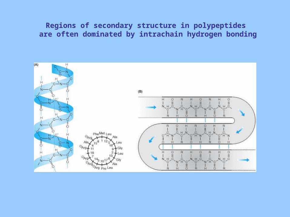
Regions of secondary structure in polypeptides are often dominated by intrachain hydrogen bonding
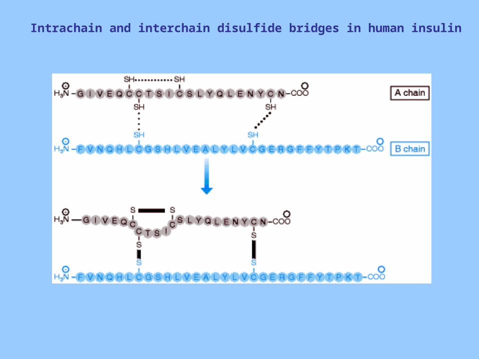
Intrachain and interchain disulfide bridges in human insulin
Chromosome structure and Function
Molecular Biology of the Cell
Chapter 2
Lecture Objectives
• Understand the structure and function of chromosomes.
• Know the two types of cell division, mitosis and meiosis and be able to identify similarities and differences of these processes.
• Learn the nomenclature of chromosomal abnormalities and understand the functional consequences.
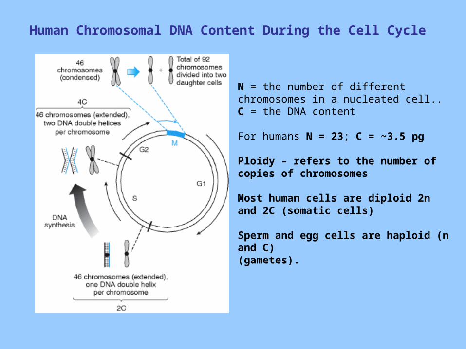
Human Chromosomal DNA Content During the Cell Cycle
N = the number of different chromosomes in a nucleated cell..C = the DNA content
For humans N = 23; C = ~3.5 pg
Ploidy – refers to the number of copies of chromosomes
Most human cells are diploid 2n and 2C (somatic cells)
Sperm and egg cells are haploid (n and C)(gametes).
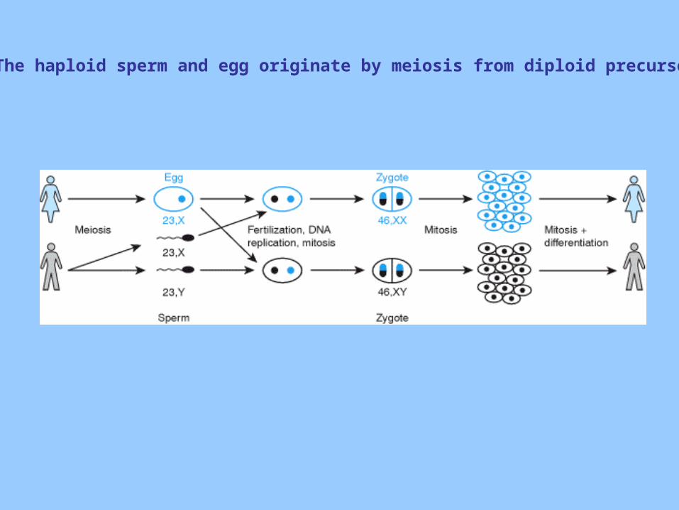
The haploid sperm and egg originate by meiosis from diploid precursors
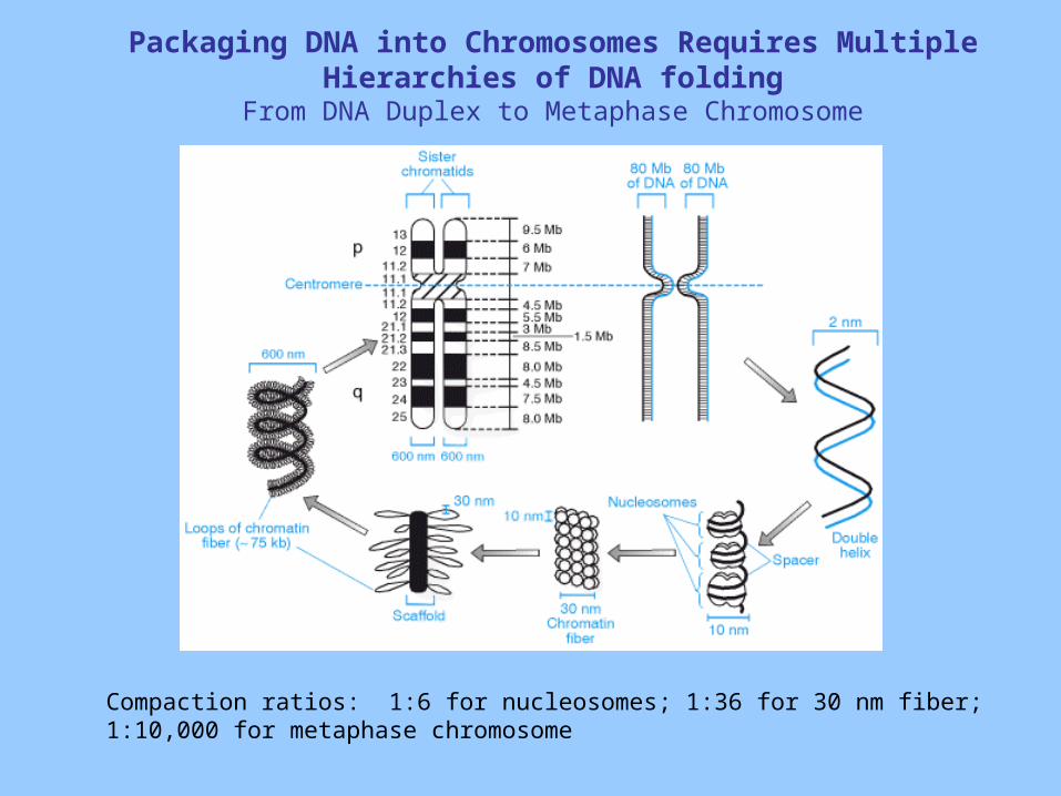
Packaging DNA into Chromosomes Requires Multiple Hierarchies of DNA folding
From DNA Duplex to Metaphase Chromosome
Compaction ratios: 1:6 for nucleosomes; 1:36 for 30 nm fiber; 1:10,000 for metaphase chromosome
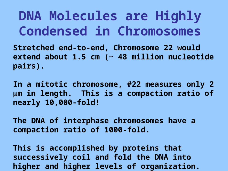
DNA Molecules are Highly Condensed in Chromosomes
Stretched end-to-end, Chromosome 22 would extend about 1.5 cm (~ 48 million nucleotide pairs).
In a mitotic chromosome, #22 measures only 2 m in length. This is a compaction ratio of nearly 10,000-fold!
The DNA of interphase chromosomes have a compaction ratio of 1000-fold.
This is accomplished by proteins that successively coil and fold the DNA into higher and higher levels of organization.
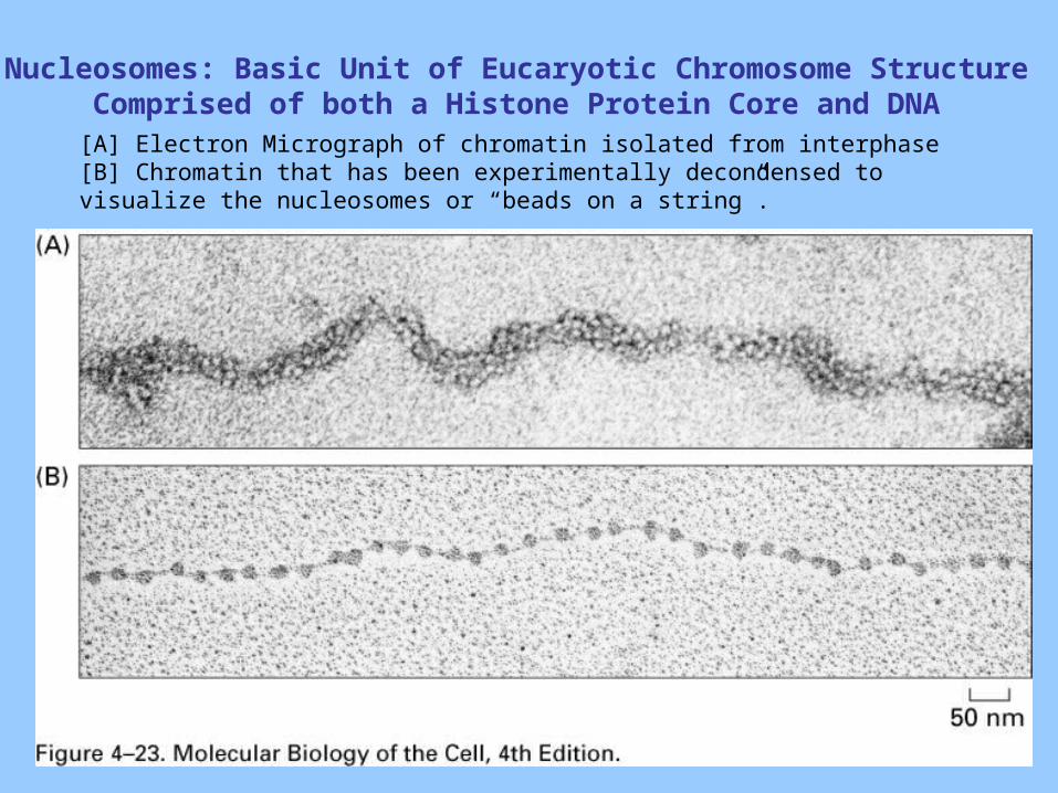
Nucleosomes: Basic Unit of Eucaryotic Chromosome StructureComprised of both a Histone Protein Core and DNA
[A] Electron Micrograph of chromatin isolated from interphase [B] Chromatin that has been experimentally decondensed to visualize the nucleosomes or “beads on a string”.
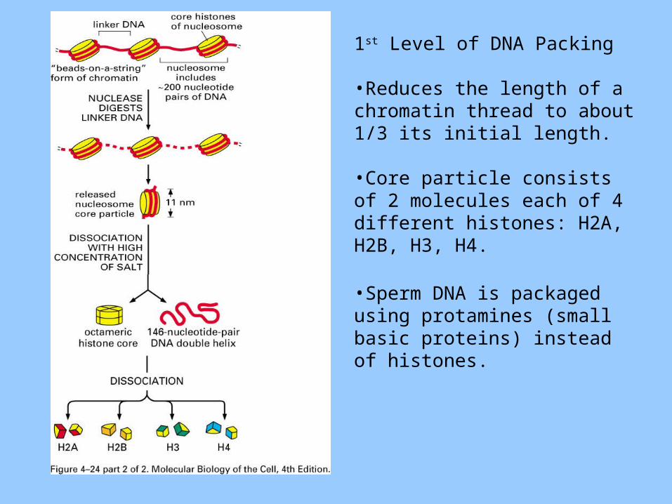
1st Level of DNA Packing
•Reduces the length of a chromatin thread to about 1/3 its initial length.
•Core particle consists of 2 molecules each of 4 different histones: H2A, H2B, H3, H4.
•Sperm DNA is packaged using protamines (small basic proteins) instead of histones.
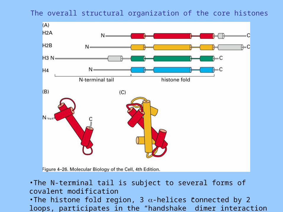
The overall structural organization of the core histones
•The N-terminal tail is subject to several forms of covalent modification•The histone fold region, 3 -helices connected by 2 loops, participates in the “handshake” dimer interaction
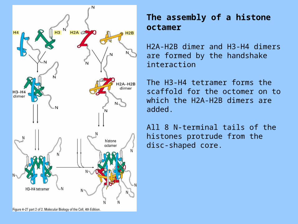
The assembly of a histone octamer
H2A-H2B dimer and H3-H4 dimers are formed by the handshake interaction
The H3-H4 tetramer forms the scaffold for the octomer on to which the H2A-H2B dimers are added.
All 8 N-terminal tails of the histones protrude from the disc-shaped core.
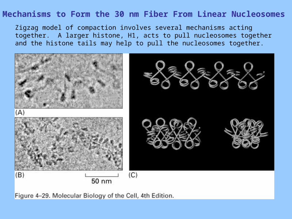
Mechanisms to Form the 30 nm Fiber From Linear Nucleosomes
Zigzag model of compaction involves several mechanisms acting together. A larger histone, H1, acts to pull nucleosomes together and the histone tails may help to pull the nucleosomes together.
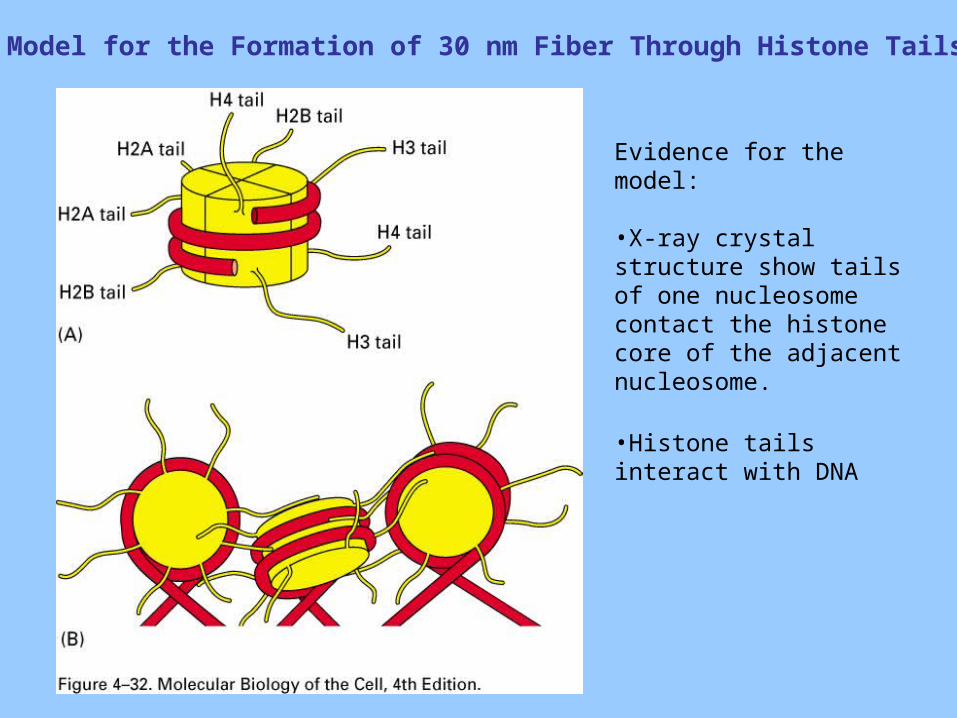
Model for the Formation of 30 nm Fiber Through Histone Tails
Evidence for the model:
•X-ray crystal structure show tails of one nucleosome contact the histone core of the adjacent nucleosome.
•Histone tails interact with DNA
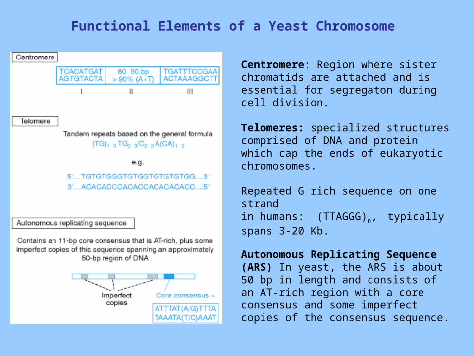
Functional Elements of a Yeast Chromosome
Centromere: Region where sister chromatids are attached and is essential for segregaton during cell division.
Telomeres: specialized structures comprised of DNA and protein which cap the ends of eukaryotic chromosomes.
Repeated G rich sequence on one strand in humans: (TTAGGG)n, typically spans 3-20 Kb.
Autonomous Replicating Sequence (ARS) In yeast, the ARS is about 50 bp in length and consists of an AT-rich region with a core consensus and some imperfect copies of the consensus sequence.
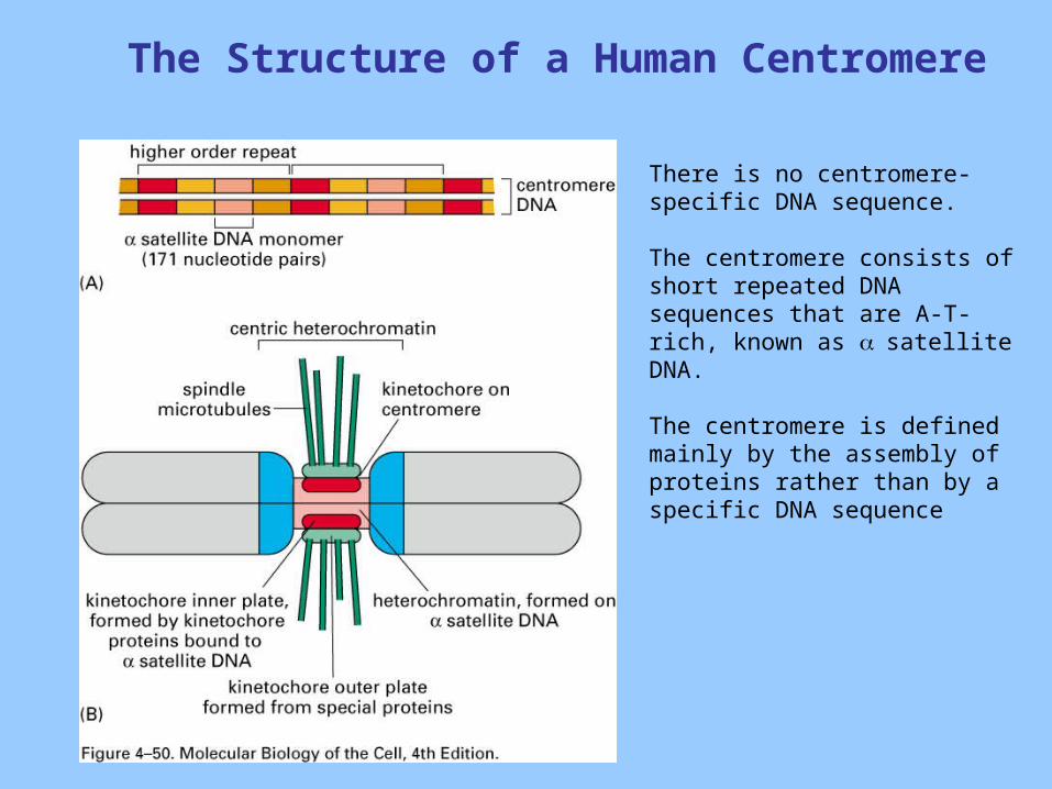
The Structure of a Human Centromere
There is no centromere-specific DNA sequence.
The centromere consists of short repeated DNA sequences that are A-T-rich, known as satellite DNA.
The centromere is defined mainly by the assembly of proteins rather than by a specific DNA sequence
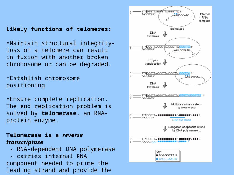
Likely functions of telomeres:
•Maintain structural integrity-loss of a telomere can result in fusion with another broken chromosome or can be degraded.
•Establish chromosome positioning
•Ensure complete replication. The end replication problem is solved by telomerase, an RNA-protein enzyme.
Telomerase is a reverse transcriptase - RNA-dependent DNA polymerase - carries internal RNA component needed to prime the leading strand and provide the template for the lagging strand.
Heterochromatin is Highly Organized and Usually Resistant to Gene Expression
Two types of chromatin exist in interphase nuclei of many higher eucaryotic cells:
Euchromatin is less condensed and associated with genes that are expressed.
Heterochromatin is highly condensed and usually does not contain genes. However genes that are packaged into heterochromatin are resistant to expression. Approximately 10% of the genome is packaged into heterochromatin.
Heterochromatin is responsible for the proper functioning of telomeres and centromeres.
Heterochromatin is dynamic, it can spread and retract and it is tends to be inherited from a cell to its progeny.
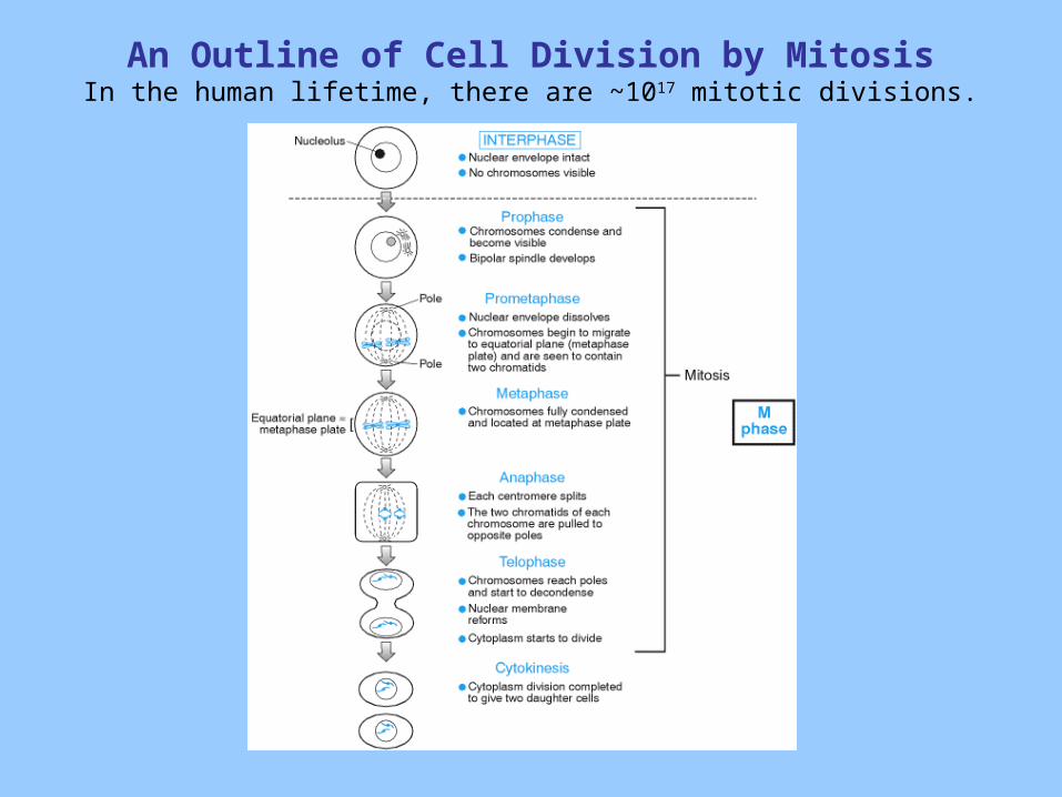
An Outline of Cell Division by MitosisIn the human lifetime, there are ~1017 mitotic divisions.
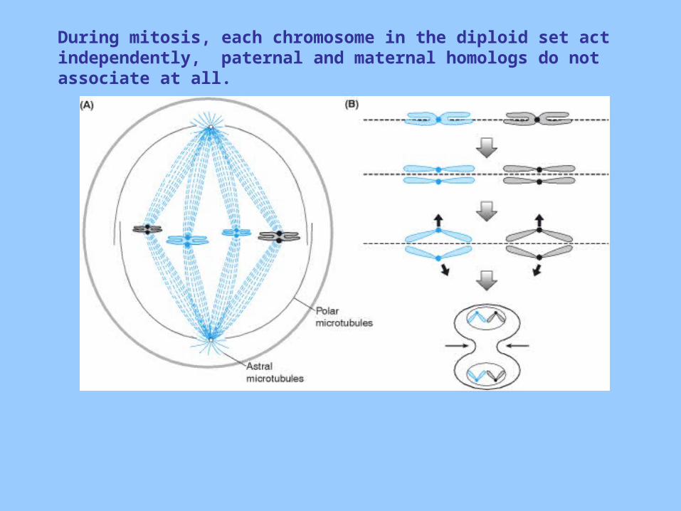
During mitosis, each chromosome in the diploid set act independently, paternal and maternal homologs do not associate at all.
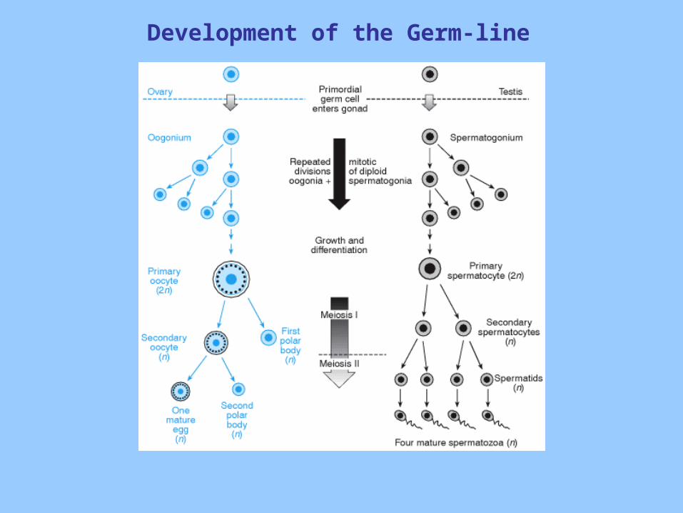
Development of the Germ-line
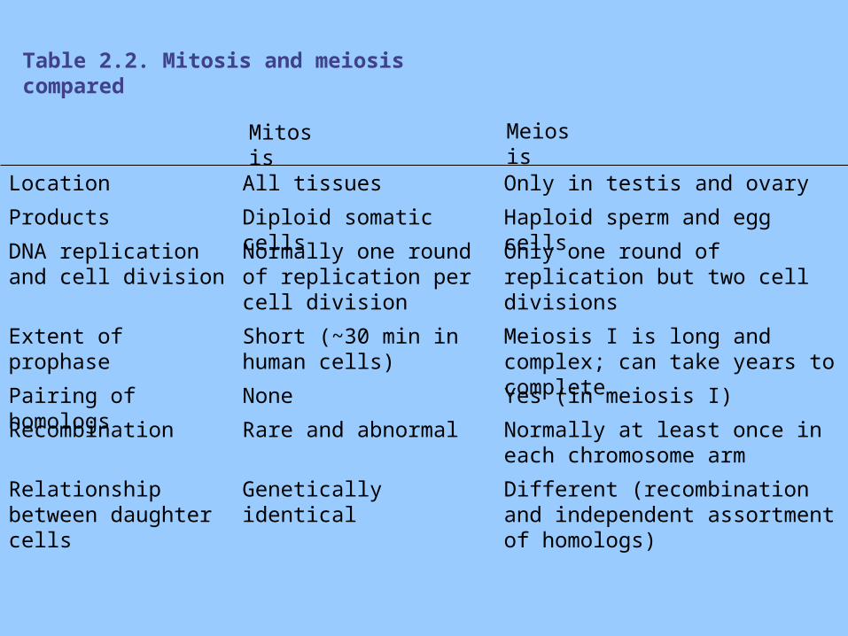
Table 2.2. Mitosis and meiosis compared
Mitosis Meiosis
Location All tissues Only in testis and ovary
Products Diploid somatic cells Haploid sperm and egg cells
DNA replication and cell division
Normally one round of replication per cell division
Only one round of replication but two cell divisions
Extent of prophase Short (~30 min in human cells)
Meiosis I is long and complex; can take years to complete
Pairing of homologs None Yes (in meiosis I)
Recombination Rare and abnormal Normally at least once in each chromosome arm
Relationship between daughter cells
Genetically identical Different (recombination and independent assortment of homologs)
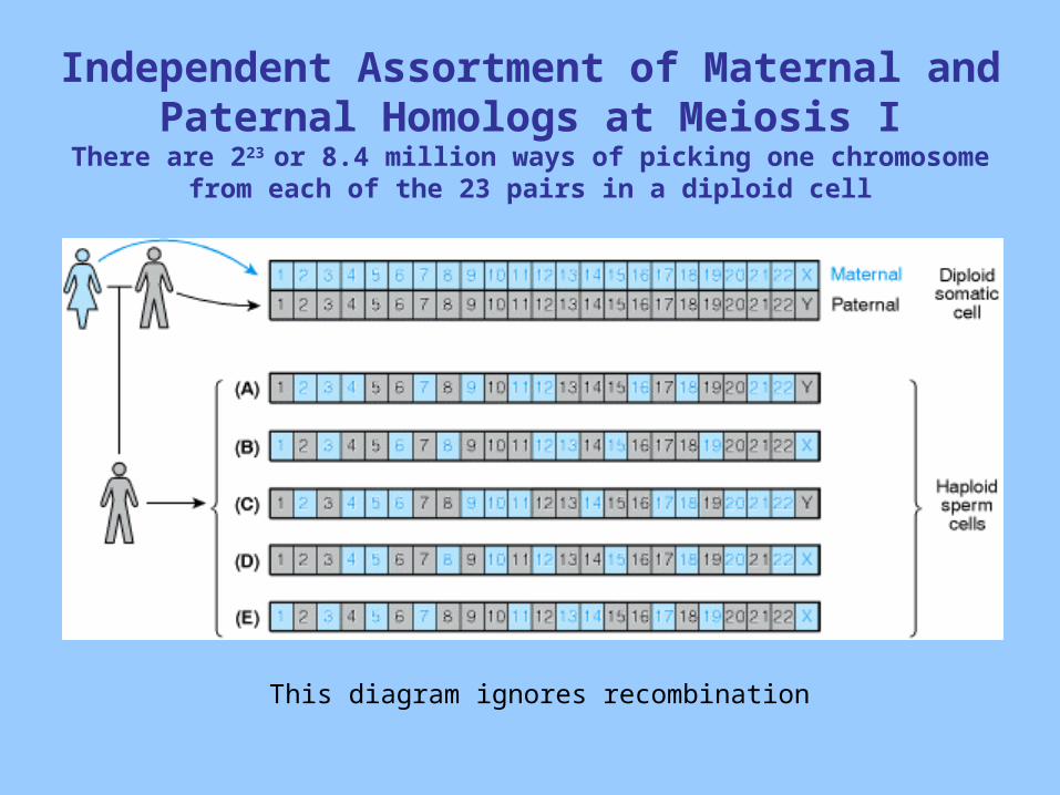
Independent Assortment of Maternal and Paternal Homologs at Meiosis I
There are 223 or 8.4 million ways of picking one chromosome from each of the 23 pairs in a diploid cell
This diagram ignores recombination
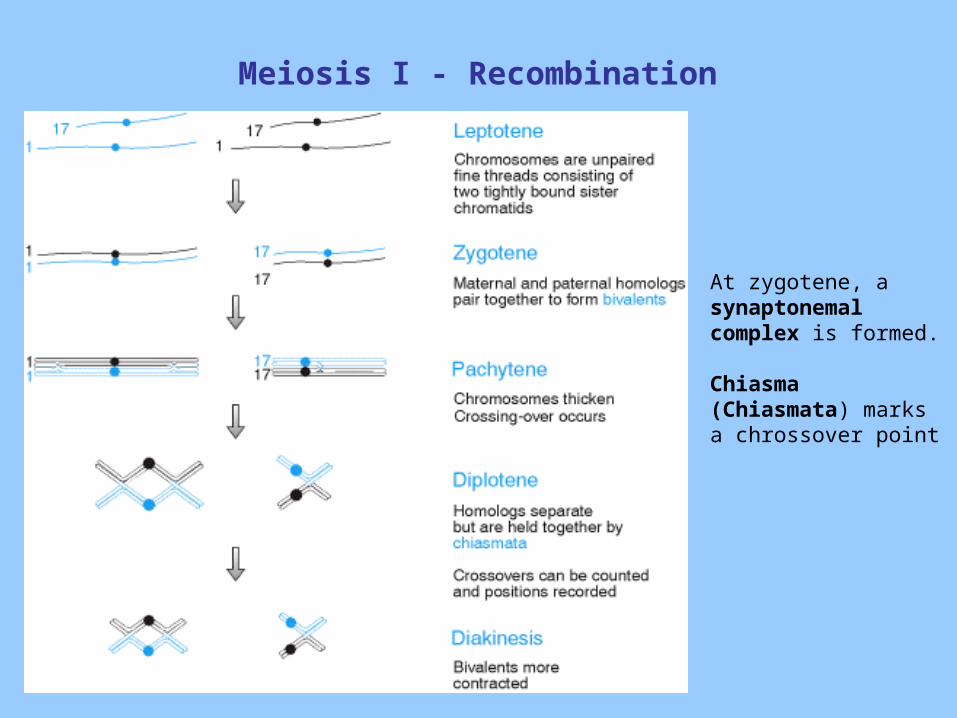
Meiosis I - Recombination
At zygotene, a synaptonemal complex is formed.
Chiasma (Chiasmata) marks a chrossover point
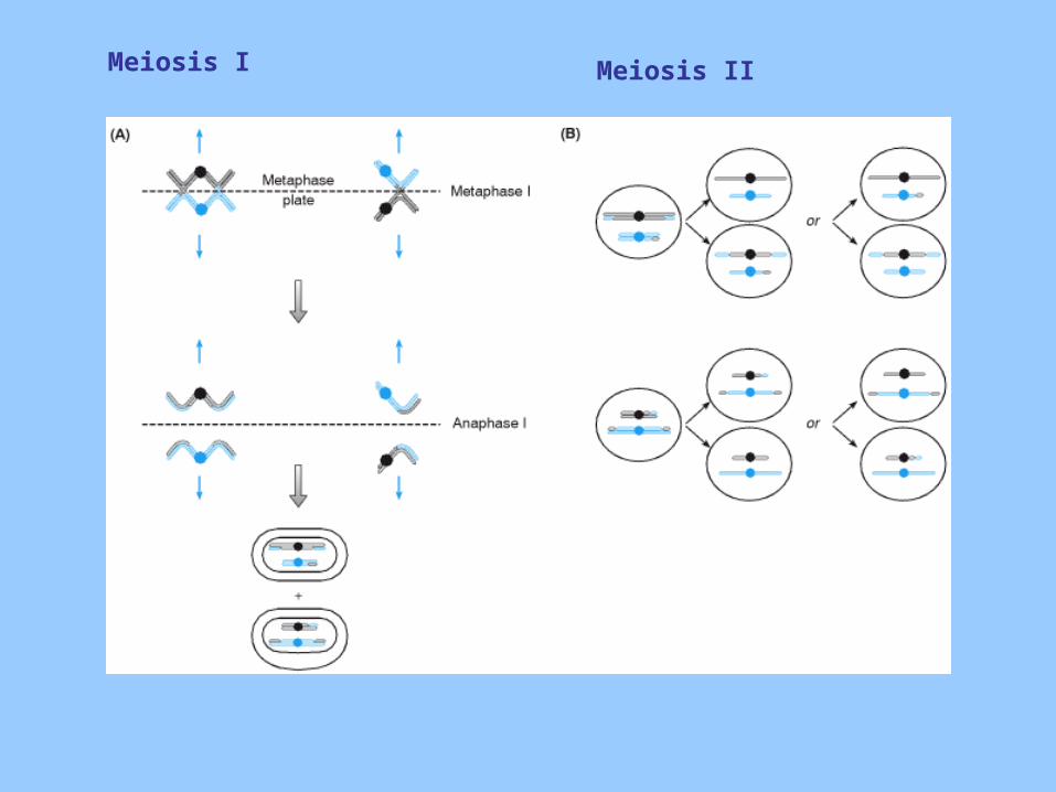
Meiosis I Meiosis II
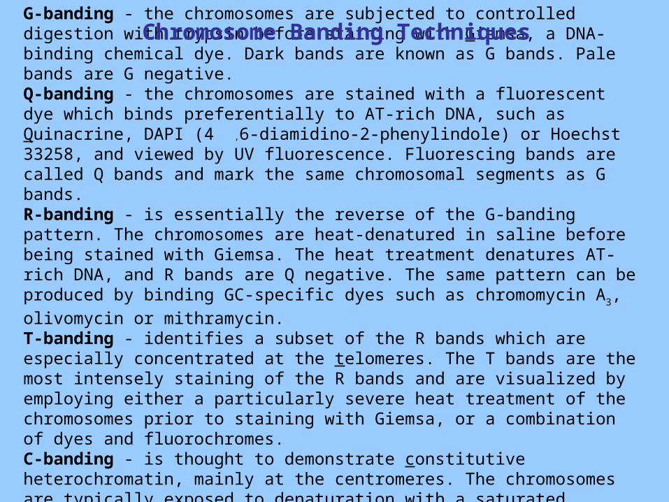
G-banding - the chromosomes are subjected to controlled digestion with trypsin before staining with Giemsa, a DNA-binding chemical dye. Dark bands are known as G bands. Pale bands are G negative.Q-banding - the chromosomes are stained with a fluorescent dye which binds preferentially to AT-rich DNA, such as Quinacrine, DAPI (4 ,6-diamidino-2-phenylindole) or Hoechst 33258, and viewed by UV fluorescence. Fluorescing bands are called Q bands and mark the same chromosomal segments as G bands.R-banding - is essentially the reverse of the G-banding pattern. The chromosomes are heat-denatured in saline before being stained with Giemsa. The heat treatment denatures AT-rich DNA, and R bands are Q negative. The same pattern can be produced by binding GC-specific dyes such as chromomycin A3, olivomycin or
mithramycin.T-banding - identifies a subset of the R bands which are especially concentrated at the telomeres. The T bands are the most intensely staining of the R bands and are visualized by employing either a particularly severe heat treatment of the chromosomes prior to staining with Giemsa, or a combination of dyes and fluorochromes.C-banding - is thought to demonstrate constitutive heterochromatin, mainly at the centromeres. The chromosomes are typically exposed to denaturation with a saturated solution of barium hydroxide, prior to Giemsa staining.
Chromosome Banding Techniques
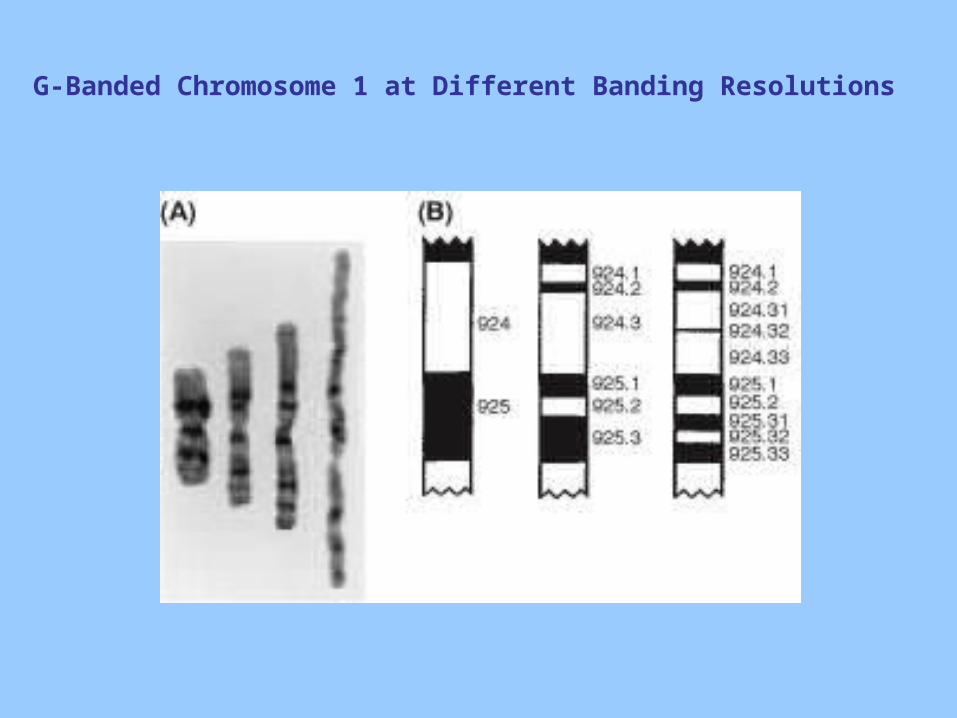
G-Banded Chromosome 1 at Different Banding Resolutions
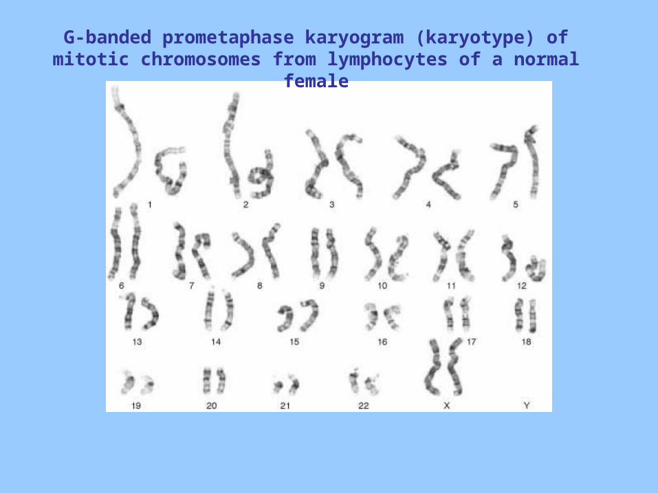
G-banded prometaphase karyogram (karyotype) of mitotic chromosomes from lymphocytes of a normal female
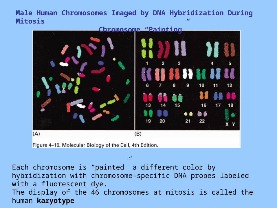
Male Human Chromosomes Imaged by DNA Hybridization During MitosisChromosome “Painting”
Each chromosome is “painted” a different color by hybridization with chromosome-specific DNA probes labeled with a fluorescent dye.The display of the 46 chromosomes at mitosis is called the human karyotype
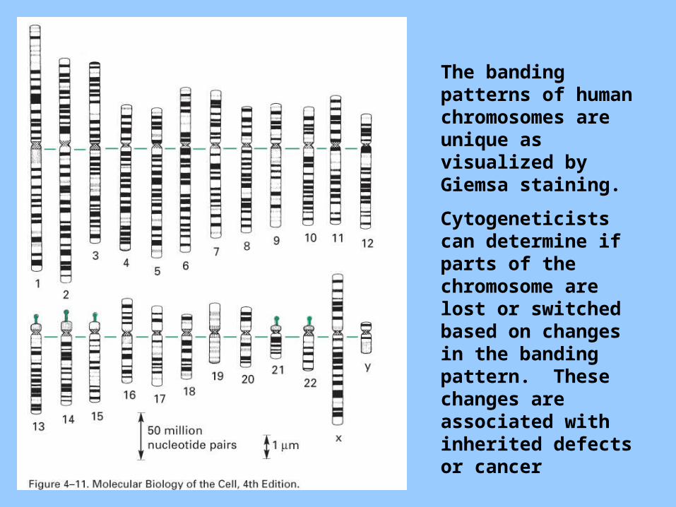
The banding patterns of human chromosomes are unique as visualized by Giemsa staining.
Cytogeneticists can determine if parts of the chromosome are lost or switched based on changes in the banding pattern. These changes are associated with inherited defects or cancer
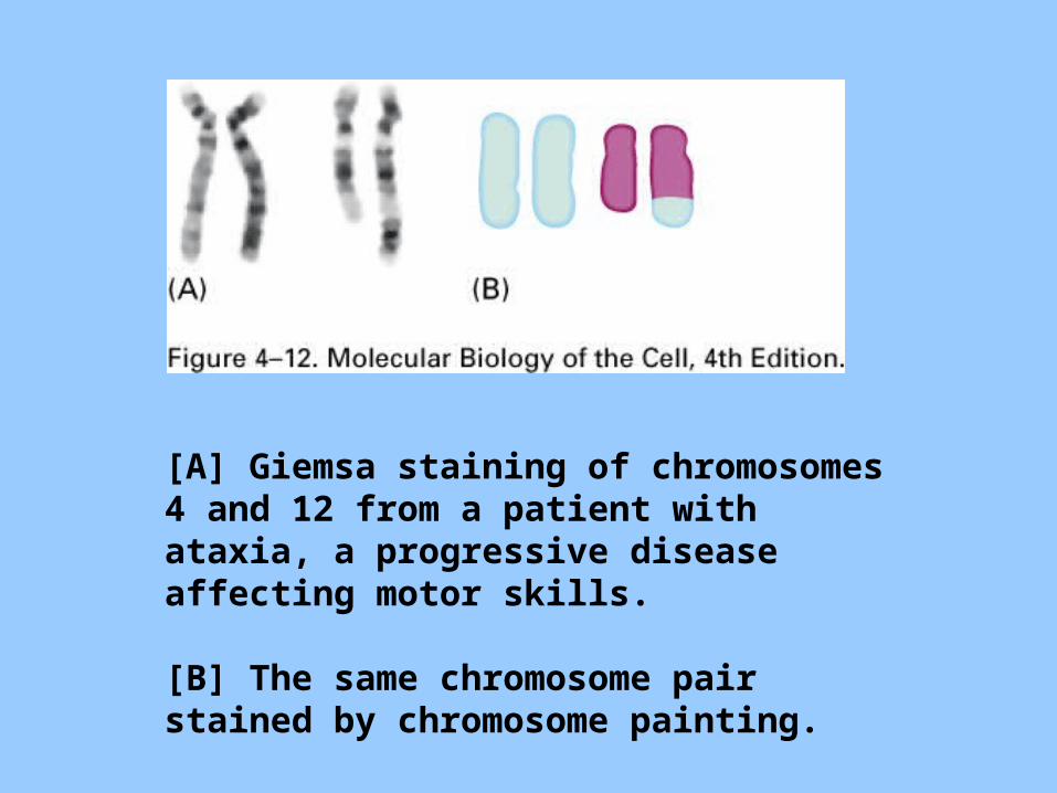
[A] Giemsa staining of chromosomes 4 and 12 from a patient with ataxia, a progressive disease affecting motor skills.
[B] The same chromosome pair stained by chromosome painting.
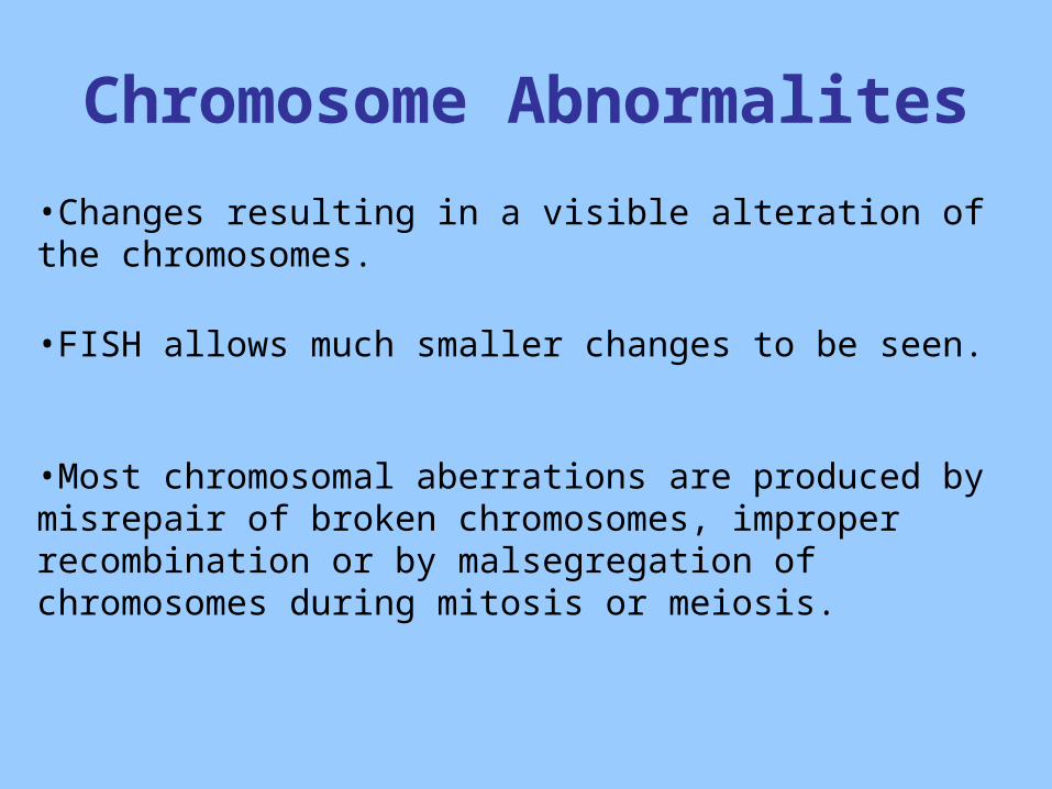
Chromosome Abnormalites
•Changes resulting in a visible alteration of the chromosomes.
•FISH allows much smaller changes to be seen.
•Most chromosomal aberrations are produced by misrepair of broken chromosomes, improper recombination or by malsegregation of chromosomes during mitosis or meiosis.
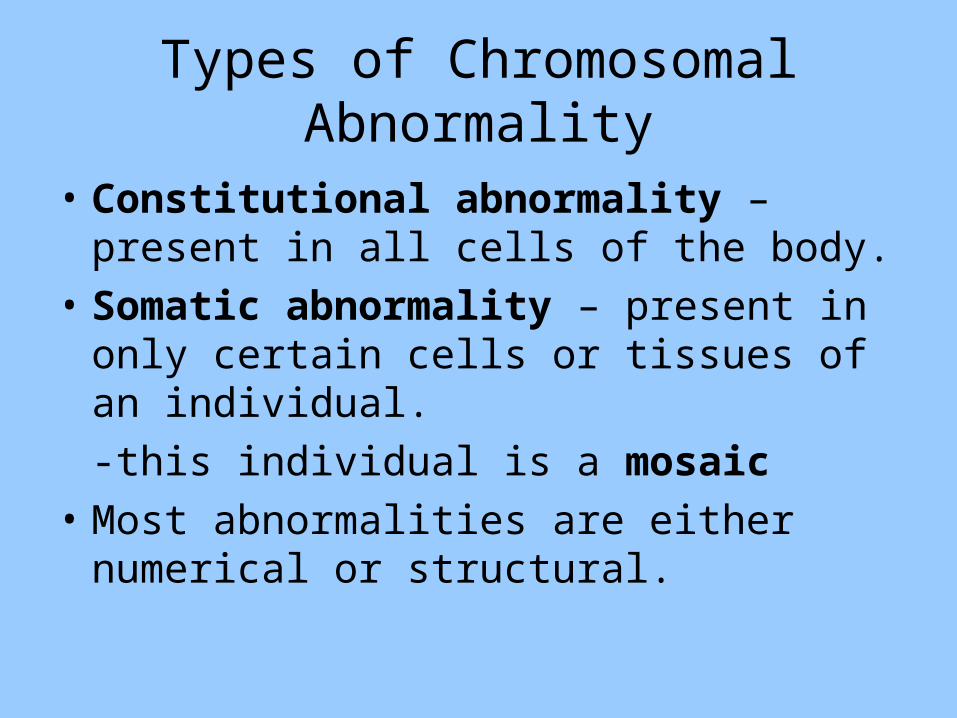
Types of Chromosomal Abnormality
• Constitutional abnormality – present in all cells of the body.
• Somatic abnormality – present in only certain cells or tissues of an individual.
-this individual is a mosaic
• Most abnormalities are either numerical or structural.
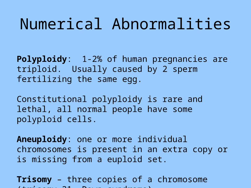
Numerical Abnormalities
Polyploidy: 1-2% of human pregnancies are triploid. Usually caused by 2 sperm fertilizing the same egg.
Constitutional polyploidy is rare and lethal, all normal people have some polyploid cells.
Aneuploidy: one or more individual chromosomes is present in an extra copy or is missing from a euploid set.
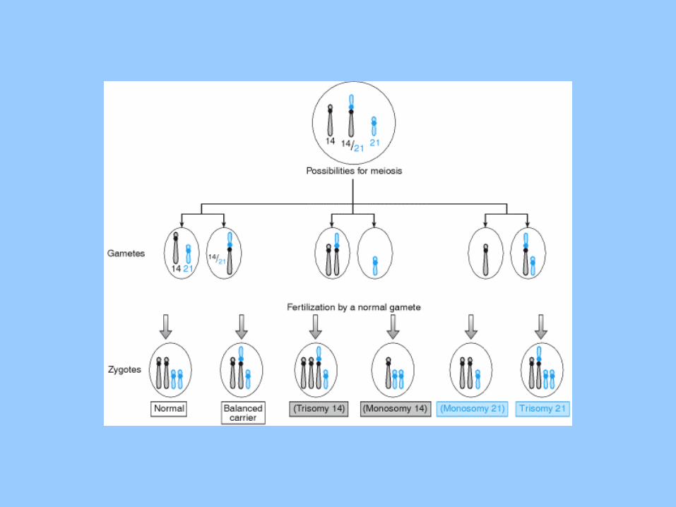
Trisomy – three copies of a chromosome (trisomy 21, Down syndrome).
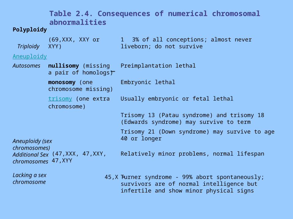
Table 2.4. Consequences of numerical chromosomal abnormalities
Turner syndrome - 99% abort spontaneously; survivors are of normal intelligence but infertile and show minor physical signs
45,X =
Relatively minor problems, normal lifespan(47,XXX, 47,XXY, 47,XYY
Aneuploidy (sex chromosomes)Additional Sex chromosomes
Lacking a sex chromosome
Trisomy 21 (Down syndrome) may survive to age 40 or longer
Trisomy 13 (Patau syndrome) and trisomy 18 (Edwards syndrome) may survive to term
Usually embryonic or fetal lethaltrisomy (one extra chromosome)
Embryonic lethalmonosomy (one chromosome missing)
Preimplantation lethalnullisomy (missing a pair of homologs)
Autosomes
Aneuploidy
1 3% of all conceptions; almost never liveborn; do not survive
(69,XXX, XXY or XYY) Triploidy
Polyploidy
Structural Chromosomal Abnormalities
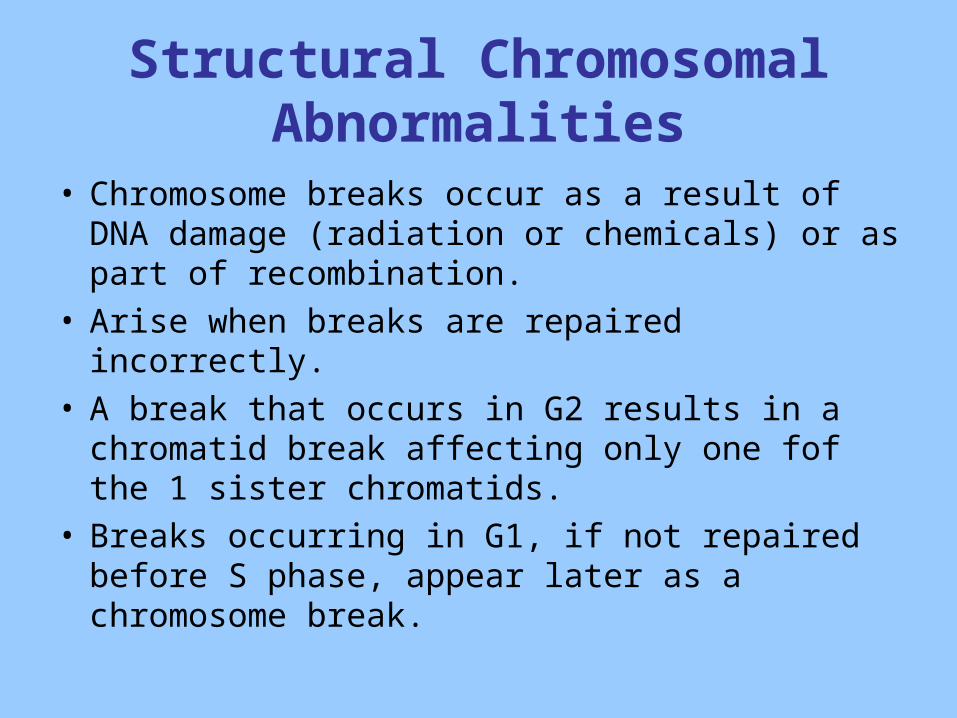
• Chromosome breaks occur as a result of DNA damage (radiation or chemicals) or as part of recombination.
• Arise when breaks are repaired incorrectly.• A break that occurs in G2 results in a chromatid
break affecting only one fof the 1 sister chromatids.
• Breaks occurring in G1, if not repaired before S phase, appear later as a chromosome break.
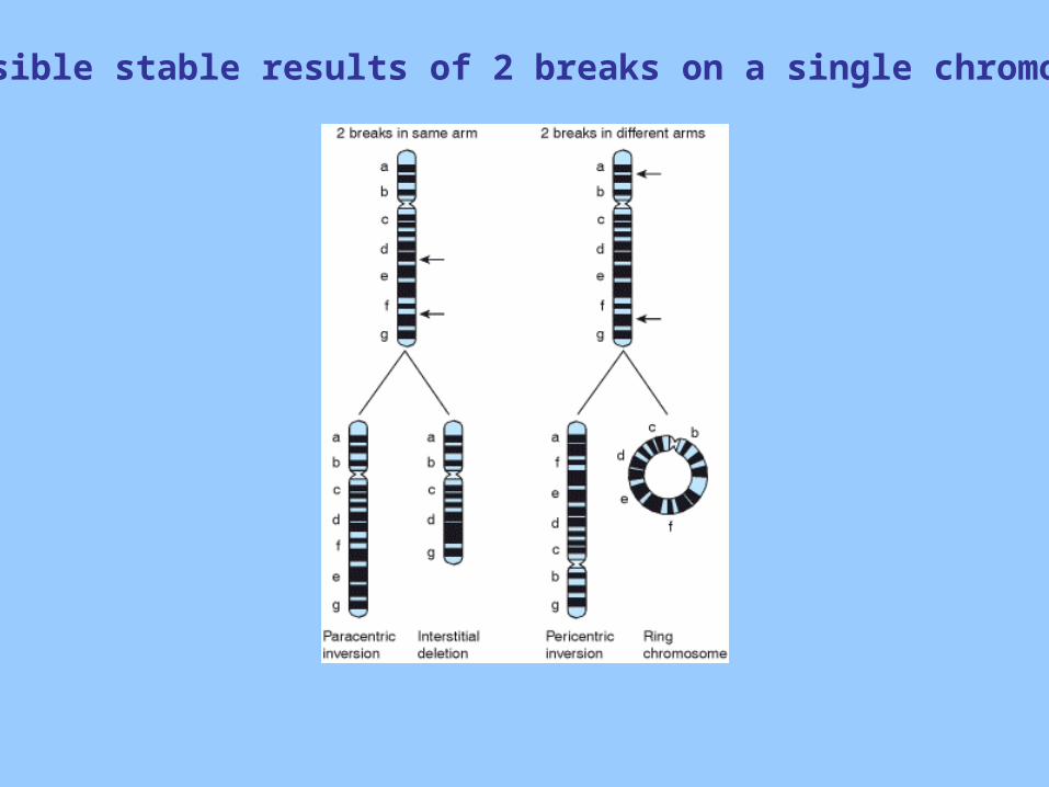
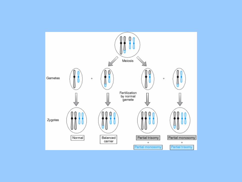
Possible stable results of 2 breaks on a single chromosome
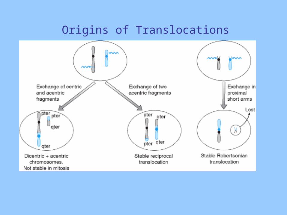
Origins of Translocations