Embed Size (px)
Citation preview

Book of abstracts
Foto: Fotograf: C.F. Møller/Adam Mørk

Welcome Welcome to the Copenhagen Symposium on Separation Sciences 2018 (CSSS 2018)
On behalf of the scientific and organizing committees, it is a great pleasure to welcome you to the 2nd instalment of the Copenhagen Symposium on Separation Sciences.
We are looking forward to two exciting days with 17 invited speakers from all over Europe, who will share with us the latest developments and trends in separation sciences, including sample preparation and detection techniques. It is a great opportunity to get inspired and to network with colleagues from academia and industry. We are also very happy to have several instrument manufacturers and suppliers of analytical equipment and consumables at the sympo-sium as well. Common lunches, coffee breaks and not least an extended poster session provide further opportunities for discussions and exchange. Finally, we hope that many of you will also join us for the symposium dinner on Tuesday evening.
We are grateful to our exhibitors and sponsors without whom this event may not have been possible: Waters, Thermo Fisher, Agilent, BioLab, Phenomenex, MSCi, Mikrolab Aarhus,The Separation Sciences Foundation (SSF), and the NordicPOP network funded by NordForsk.
We wish you a fruitful symposium, and above all, a good time in Copenhagen!
Jörg P. Kutter (symposium chair)Steen Honoré Hansen (honorary chair)
Inga BjørnsdottirBente GammelgaardStig Pedersen-BjergaardCarsten Boye Knudsen

Conference Officials
Conference Chairs
Jörg P. Kutter University of Copenhagen DenmarkSteen Honoré Hansen University of Copenhagen Denmark(Honorary Chair)
Scientific Committee
Jörg P. Kutter University of Copenhagen DenmarkBente Gammelgaard University of Copenhagen DenmarkInga Bjørnsdottir Novo Nordisk A/S DenmarkCarsten Boye Knudsen Zealand Pharma DenmarkStig Pedersen-Bjergaard University of Copenhagen Denmark and University of Oslo Norway
Organizing Committee
Jörg P. Kutter University of Copenhagen DenmarkInga Bjørnsdottir Novo Nordisk A/S DenmarkCarsten Boye Knudsen Zealand Pharma DenmarkBente Gammelgaard University of Copenhagen DenmarkStig Pedersen-Bjergaard University of Copenhagen Denmark and University of Oslo NorwayNarmin Amin University of Copenhagen DenmarkAnders Vejen Schubert University of Copenhagen DenmarkBente Gade PharmaDanmark Denmark

Program at a glance
Foto: Fotograf: C.F. Møller/Adam Mørk

Program – Tuesday, June 26, 2018
8:30 – 8:45 Registration8:45 – 9:00 Welcome/Opening remarks
The long-term perspective…
Session Chair: Jörg P. Kutter, University of Copenhagen
9:00 – 9:30 Michael Lämmerhofer, University of Tübingen, Germany Separation Science towards the year 2030 – what are the major directionsandhowcanthepharmaceuticalindustrybenefitfromthese?
Recent progress in microfluidic and immunoaffinity techniques
Session Chair: Jörg P. Kutter, University of Copenhagen
9:30 – 10:00 Detlev Belder, University of Leipzig, Germany Hyphenated chip-based HPLC 10:00 – 10:30 Trine Grønhaug Halvorsen, University of Oslo, Norway Analysisofproteinsfrombiologicalmatricesusingaffinitybased sample clean-up and LC-MS/MS 10:30 – 11:00 Coffee break
Recent progress in liquid-phase separation – chromatography and electrophoresis I
Session Chair: Nickolaj J. Petersen, University of Copenhagen
11:00 – 11:30 Gert Desmet, Free University of Brussels, Belgium Recentprogressinthedevelopmentofperfectlyorderedseparationmedia
11:30 – 12:00 Peter Myers, University of Liverpool, UK Whydowestillusesilicaasastationaryphase?
12:00 – 12:30 Alberto Cavazzini, University of Ferrara, Italy Advancedchiralstationaryphasesforhighefficientultrafastchiral separationsbyliquidchromatography
12:30 – 13:30 Lunch
13:30 – 15:00 Poster session (with refreshments)

Recent progress in liquid-phase separation – chromatography and electrophoresis II
Session Chair: Bente Gammelgaard, University of Copenhagen
15:00 – 15:30 Nikoline Juul Nielsen, University of Copenhagen, Denmark Pulsedelutionin2Dreversedphaseliquidchromatography
15:30 – 16:00 Frantisek Foret, Academy of Sciences of the Czech Republic, Czech Republic Theroadfrommicrotomacroinpreparativeisotachophoresis
16:00 – 16:30 Coffee break
16:30 – 17:00 Myriam Taverna, Univeristy of Paris-Sud, France Fromelectrokineticpreconcentrationtohighresolutionseparation:anovel capillary electrophoresis approach to determine amyloid peptides directly fromcerebrospinalfluids
In Memoriam
Session Chair: Jörg P. Kutter, University of Copenhagen
17:00 – 17:30 In Memoriam Professor Steen Honoré Hansen Per Helboe, Per Helboe Consultancy and University of Copenhagen
19:30 Symposium dinner
Program – Wednesday, June 27, 2018
8:45 – 9:00 Housekeeping/Announcements
Forefront applications of mass spectrometry
Session Chair: Stig Pedersen-Bjergaard, University of Oslo and University of Copenhagen
9:00 – 9:30 Kevin Pagel, Free University of Berlin, Germany Ionmobilityspectrometryasseparationtechniqueinglycananalysis
9:30 – 10:00 Petur Weihe Dalsgaard, University of Copenhagen, Denmark Ionmobilityasanaddeddimensionfortoxicologyscreening 10:00 – 10:30 Christian Janfelt, University of Copenhagen, Denmark MassspectrometrybyMALDIandDESIforimagingofdrugsand metabolitesinbiologicalsamples
10:30 – 11:00 Coffeebreak

ADME and pharmacometabolomics
Session Chair: Carsen Boye Knudsen, Zealand Pharma
11:00 – 11:30 Deirdre Cabooter, Catholic University of Leuven, Belgium Zebrafishasasmall-animalmodelforADMEstudies: analyticalchallenges
11:30 – 12:00 Isabelle Kohler, Leiden Academic Center for Drug Research, The Netherlands Clinical metabolomics and pharmacometabolomics to enable personalized medicine
12:00 – 13:00 Lunch
Forefront biopharmaceutical characterization
Session Chair: Inga Bjørnsdottir, Novo Nordisk AS
13:00 – 13:30 Henrik Jensen, University of Copenhagen, Denmark FlowInducedDispersionAnalysis(FIDA)quantifiesproteins,protein-ligand interactionsandimmuneresponsesundernativeconditions 13:30 – 14:00 Elena Dominguez Vega, Leiden University Medical Center, The Netherlands CapillaryelectrophoresiscombinedwithMSorSPRdetectionforthe assessmentofproteinheterogeneity,conformationandaffinity 14:00 – 14:30 Davy Guillarme, University of Geneva, Switzerland Modernanalyticaltoolsforthecharacterizationofmonoclonalantibody andantibody-drugconjugates
14:30 Farewell Symposium adjourns

Invited talks

Separation sciences towards the year 2030 – what are the major
directions and how can the pharmaceutical industry benefit from
these?
Michael Lämmerhofer1 1University of Tübingen, Germany
Separation sciences have seen dramatical developments in the last two decades. New technological advances have pushed some techniques close to the physical limits. The quest for
advanced separations and analytical characterizations are growing in pharmaceutical industries.
New therapeutic concepts and more complex drugs impose new challenges on analytical procedures and separation sciences. In addition, regulation authorities ask for new analytical
specifications of well established products. Separations in particular by HPLC are supporting
pharmaceutical industry in all stages of development and production of pharmaceutical products. It comprises support of early developments in drug discovery, in vitro and in vivo pharmacokinetic
testing, synthesis and process development, process control, impurity profiling, final product release, dissolution and content uniformity testing. Upcoming drug products such as peptide and
protein therapeutics, gene therapeutics and nanomedicinces are subject to principally the same
testings, but may be more challenging and may need more than one assay. Automation-facilitated workflows allows workers with little training in separation science to use existing often generic methods efficiently. For final release testing and quality control, a robust well-developed method
that can be readily transferred all over the world may be of prime importance. On the other hand, in the early phase of development in modern drug discovery flexible and rapid problem solving is
of paramount importance. There is an effort to streamline method development in order to
quickly adjust the method to the needs. The advent of UHPLC with sub-2µm fully porous particle and core-shell columns, complementary stationary phase selectivities, fast parallel column
screening approaches, the use of SFC certainly support the needs in this area. On the other hand,
advanced concepts such as well-ordered pillar array columns, multi-dimensional separations and their hyphenation with information-rich multiple and or multi-stage detection systems, ion-
mobility separations as an additional dimension bring advantages in fields where the separation of
highly complex mixtures in order to gain a comprehensive picture of a pharmaceutical product is in the focus. Overall, separation science will remain a key enabling technology in pharmaceutical
industry, yet advances are needed to cope with new challenges.
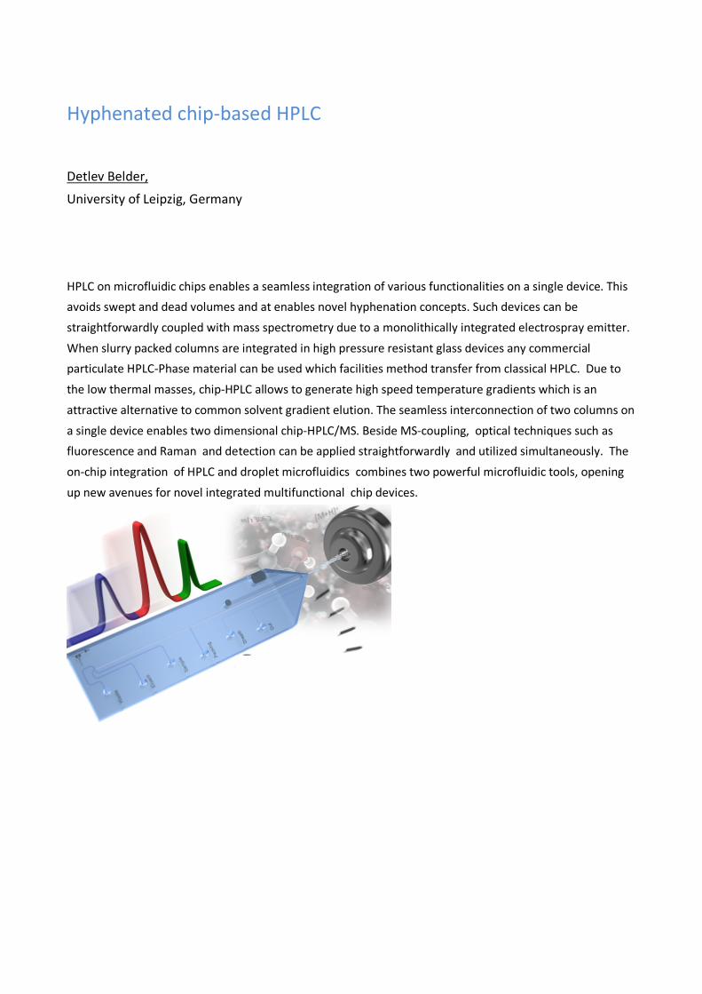
Hyphenated chip-based HPLC
Detlev Belder,
University of Leipzig, Germany
HPLC on microfluidic chips enables a seamless integration of various functionalities on a single device. This avoids swept and dead volumes and at enables novel hyphenation concepts. Such devices can be
straightforwardly coupled with mass spectrometry due to a monolithically integrated electrospray emitter.
When slurry packed columns are integrated in high pressure resistant glass devices any commercial particulate HPLC-Phase material can be used which facilities method transfer from classical HPLC. Due to
the low thermal masses, chip-HPLC allows to generate high speed temperature gradients which is an
attractive alternative to common solvent gradient elution. The seamless interconnection of two columns on
a single device enables two dimensional chip-HPLC/MS. Beside MS-coupling, optical techniques such as fluorescence and Raman and detection can be applied straightforwardly and utilized simultaneously. The
on-chip integration of HPLC and droplet microfluidics combines two powerful microfluidic tools, opening
up new avenues for novel integrated multifunctional chip devices.

Analysis of proteins from biological matrices using affinity based
sample clean-up and LC-MS/MS
Trine Grønhaug Halvorsen, Maren C. S. Levernæs, Cecilia Rossetti, Leon Reubsaet
School of Pharmacy, University of Oslo, Norway
Targeted protein determination using LC-MS/MS after a typical bottom-up pretreatment results in
high specificity and the possibility to differentiate between different (iso)forms in one single step.
This offers a more reliable alternative to traditional immuno-metric assays.
Determination of low abundance proteins (i.e biomarkers) in biological matrices by LC-MS/MS
requires efficient sample clean-up. This can be achieved with affinity extractions based on conventional antibodies as well as synthetic antibodies. Immunoaffinity sample clean-up using monoclonal antibodies targeting the protein of interest has been demonstrated to enable
biomarker determination in the low picomolar range1,2 (protein extractions). In recent years affinity based sample clean-up techniques targeting a proteotypic peptide both using conventional
antibodies (peptide extractions/epitope fishing) and synthetic antibodies (molecularly imprinted
polymers) have been introduced3,4. The aim of the present paper is to give an overview of affinity based sample clean-up techniques targeting proteins and/or their proteotypic peptides and their use in determination of low
abundance proteins in biological matrices using LC-MS/MS. The differences and similarities between the techniques will be highlighted through examples.
In conclusion, each approach has its own distinct advantages and limitations which are important
to be aware of: The main advantage of protein affinity enrichment being the possibility to get
isoform information of the whole protein using a single antibody, while the main advantage of
peptide enrichment is a better clean-up and a more appropriate use of stable isotope labelled
peptide standards for quantitation.
References: (1) Torsetnes, S. B.; Nordlund, M. S.; Paus, E.; Halvorsen, T. G.; Reubsaet, L. J. Proteome Res. 2013, 12, 412. (2) Ackermann, B. L.; Berna, M. J. Expert Review of Proteomics 2007, 4, 175. (3) Anderson, N. L.; Anderson, N. G.; Haines, L. R.; Hardie, D. B.; Olafson, R. W.; Pearson, T. W. J. Proteome Res. 2004,
3, 235. (4) Rossetti, C.; Levernæs, M. C. S.; Reubsaet, L.; Halvorsen, T. G. Journal of Chromatography A 2016, 1471, 19.

Recent Progress in the Development of Perfectly Ordered
Separation Media
Gert Desmet
Vrije Universiteit Brussel, Belgium The present contribution aims at illustrating and demonstrating how micro-machining technology can
boost High Performance Liquid Chromatography (HPLC). Currently, HPLC is routinely used in nearly every
chemical analysis lab. Despite its high degree of maturity, the technique still does not deliver the required
separation power needed to unravel the complex samples encountered in the state-of-the-art research in biology and drug development (e.g., proteomics and metabolomics), or in contemporary food and
environmental analysis, etc…..
One of the reasons for the performance limitations of packed bed HPLC columns is that they are packed
randomly. This randomness forces the liquid to follow different paths with different path lengths, which in turn broadens the individual sample component bands. To solve this packing disorder problem, we have
used advanced photolithographic etching techniques such as the Bosch-process to produce perfectly
ordered porous support columns with optimized hydrodynamic shape and optimized external porosity.
Using this approach, we have been able to realize sub-micrometer plate heights using radially elongated diamond-shaped pillars that are up to 15 times wider than their axial dimension (5 µm). The use of such a
high-aspect ratio pillars allowed for a 5-fold reduction of the minimal plate height compared to beds filled
with pillars with a similar inter-pillar distance (2.5 µm) but with an aspect ratio around unity (cylinders, diamonds). This increase in performance can be largely attributed to a decrease of the B-term band
broadening, which is about a factor of about 25 smaller in the large-aspect ratio columns compared to the
cylindrical pillar columns. In addition, the columns also generate only a minimal C-term band broadening,
as the space formed between the high-aspect ratio pillars is very uniform and basically resembles the through-pore space one would have in a parallel array of flat plates. The concept also enables a drastic
reduction of the footprint of pillar array columns, allowing to fabricate columns offering very high efficiency
on the surface of a single silicon wafer.
Why Do We Still Use Silica as a Stationary Phase?
Peter Myers
University of Liverpool, Department of Chemistry, Liverpool, UK
Silica was used in the first commercial LC columns dating back to the 1970’s and today silica is still the most widely used support for HPLC and UPLC. So, after nearly 50 years why are we still using silica?
Is it that good there has been and still isn’t a need for a replacement?
In this lecture I will describe the problems we had with silica in the 1980’s and how very little progress has been made to date. In the 1980’s we had 5micron spherical totally porous particles. Today we have 1.5micron particles. Is this a real development?
I will describe how bonded phase chemistries have had to develop to help conceal silica problems. In the 1980’the most popular phase was octadecyl. Today the most popular phase is still octadecyl. Is it that good?
Compare this to the computer revolution. In the 1980’s we had Apple, Commodore, Atari, BBC Micro TRS-80, ZX81, ZX Spectrum, Commodore 128, and the Amstrad. Today we have i-pads, computers surfaces and smartphones all of which have hundreds or even thousands of times more processing power than the computers of the 1980’s. Has chromatography moved on that fast?
But the main focus of the lecture will be to look into the future and offer some new alternatives to silica and also offer some new separation methods that do not rely on the classical theory of adsorption or partitioning and certainly do not rely on packed silica columns.

New generation chiral stationary phases for high efficient
ultrafast chiral separations by liquid chromatography
Alberto Cavazzini1, Martina Catani1, Omar H. Ismail2, and Francesco Gasparrini2 1University of Ferrara, Italy, 2Sapienza University of Rome, Italy
This contribution retraces the revolution that has characterized the field of chiral separations by liquid chromatography in the last few years thanks to the design and preparation of new chiral particles suitable for high efficient, ultrafast enantioseparations.
Examples of ultrafast chiral separations have been achieved by using different chiral selectors anchored to silica particles of different geometries and properties. Not only chiral selectors traditionally considered “fast” (e.g., Pirkle type Whelk-O1 CSPs), but also the “slow” ones (such as macrocyclic antibiotics-based CSPs) have been successfully employed to this scope. Latest generation chiral particles include both sub-2 µm fully porous ones (FPPs) and second-generation superficially porous particles (SPPs) of diameter as small as 2 µm. The combination of very short columns (1 cm or even 5mm) and high flow rates has allowed to perform the separation of enantiomers in seconds or even in fractions of seconds in normal- and reversed-phase (NP and RP) chromatography, Hydrophilic interaction chromatography (HILIC) and supercritical-fluid chromatography (SFC).
Current knowledge about ultrafast chiral separations through liquid chromatography will be revised, with particular reference to the fundamentals of mass transfer through chiral particles
and to some aspects that should be further developed for the advancement of the field. Particular
emphasis will be given to SFC, where the potential of new generation chiral stationary phases (CSPs) is enormous.

Pulsed Elution and Active Modulation in 2D reversed phase LC:
Increasing Flexibility Nikoline J. Nielsen1, Simon S Jakobsen1,2, Sylvain Verdier3, Claude Mallet4, Jan H Christensen1
1Analytical Chemistry Group, Department of Plant and Environmental Sciences, Faculty of Science,
University of Copenhagen, Thorvaldsensvej 40, DK-1871 Frederiksberg, Denmark 2Currently employed at Danish Emergency Management Agency, Datavej 16, DK-3460 Birkerød, Denmark 3Haldor Topsøe A/S, Haldor Topsøes Allé 1, DK-2800 Kongens Lyngby, Denmark 4Waters Corporation, 34 Maple Street, Milford, Massachusetts 01757, United States.
The interest in online comprehensive two-dimensional liquid chromatography (LC×LC) have increased as it offers ways to improve the performance of separations in terms of peak capacity and selectivity compared to one-dimensional liquid chromatography. However, the price for greater resolving power in LC×LC is a more complex system, where parameterization of the first dimension constrains the second dimension and vice versa. In this study a way to release these constrains is presented.
The developed method addresses several of the challenges encountered in LC×LC. This includes in particular: loss of peak capacity due to undersampling of the first dimension, limited peak capacity in the second dimension due to short second dimension analysis time and limitations in selection of column dimensions and flow rates.
The method is based on a strategy where the sample is eluted of the first dimension column by pulses of increasing eluotropic strength and width. In between the pulses the first dimension is kept in a no-elution state by applying a flow of mobile phase with weak eluotropic strength. The eluate from the first dimension is diluted with a make-up flow of water before it is trapped on trap columns in the modulator and re-injected onto the second column. It is demonstrated that the LC system is capable of delivering the required pulses of strong solvent and by tuning the length and eluotropic strength of these pulses, analytes with retention factors in water (kw) above 150 can be manipulated to elute in 3-4 pulses. In between the pulses the first dimension can be kept in a no-elution state for up to 10 minutes without changes in which pulse and the number of pulses analytes with kw above 350 elute in. For analytes with kw equal to 150 minor changes in the pulse position of elution was observed, when the no-elution time was increased from 1 to 10 min. The initial experiments were done using 27 nitrogen containing aromatic compounds and UV-detection, and the method applied to alkaline fractions of vacuum gas oils with positive electrospray ionization and time-of-flight detection. Peak capacities of 4018 and 4610 (corresponding to a peak production rate of 7.4 peaks/min and 4.5 peaks/min) was obtained for a 540 min and 1040 min analysis, respectively. The pulsed elution approach combined with a refocussing step between the two dimensions offers great potential with respect to increasing the flexibility of LC×LC.
The road from micro to macro in preparative isotachophoresis.
Frantisek Foret, Vladimira Datinska, Ivona Voracova
Czech Academy of Sciences, Institute of Analytical Chemistry, Brno, Czech Republic
Many clinical and diagnostic applications require high quality nucleic acids for downstream
analytical methods such as quantitative PCR, microarrays, and/or next-generation sequencing. A
complex matrix, such as blood and other bodily fluids, samples from the scene of crime, fossil
samples, etc., very often compromise isolation and purification of nucleic acids.
The most common nucleic acids isolation techniques are based on extraction with inherent
limitations, with regard to quantitative results. In recent years, there is an increasing interest in sorbent free alternatives. Here, we report on two instrumental systems for processing of large sample volumes by discontinuous electrophoresis with theoretically unlimited concentration
factor. In the first system, we used capillary instrument with large bore (0.8 mm ID) fluoropolymer capillary, conductivity detector and fraction collection valve. Sample volumes up 150 �l could be
injected, focused and collected. In order to achieve a high recovery and enrichment, factors including electric current, sample amount and matrix were investigated experimentally as well as by computer simulation.
The second, laboratory constructed, system was designed in a flat arrangement where sample zones migrated towards a fraction collection well. This allowed focusing of 15 ml sample volumes
in a 110 mm device in less than 1 hour. Position of the migrating zone was monitored by laser-
induced fluorescence. While a discontinuous electrolyte system was used, the selected geometry did not lead to a typical isotachophoretic migration when operated at constant current, constant
voltage or constant power modes. These experimental findings were confirmed by theoretical
descriptions derived for each operation mode. Samples dissolved in the terminating electrolyte or
in saline solutions were processed in both systems. The DNA content in the collected fractions
were further analyzed by fluorescence spectrometry and chip capillary electrophoresis.
In conclusion, we have developed two simple, preparative methods for DNA concentration and
purification. The first method, based on a capillary ITP, can process sample volumes up to 150 µl. The second method, based on the flat channel design, allows processing of 15 ml sample volumes.
This is by far the largest described focusing capacity. In the discontinuous electrolyte system the

sample enrichment factor is limited only by the system geometry and even higher loading
capacities are possible if needed.
From electrokinetic preconcentration to high resolution separation: a novel capillary electrophoresis approach to quantify amyloid peptides directly from cerebrospinal fluids Myriam Taverna1, Cédric Crosnier de Lassichère1, Thanh Duc Mai1, Markus Otto2 1 Institut Galien Paris Sud, UMR 8612, Protein and Nanotechnology in Analytical Science (PNAS), CNRS, Univ. Paris-Sud, Univ. Paris-Saclay, Châtenay-Malabry, France 2 University of Ulm, Department of Neurology, Ulm, Germany
The Amyloid Beta (Aβ) peptide 1-42 is an established Cerebrospinal Fluid (CSF) biomarker for the
diagnosis of Alzheimer’s disease (AD) as its level is lowered in the CSF of AD patients(1). Nevertheless, its quantification is not sufficient for a reliable molecular diagnosis of AD. To
discriminate further AD from other neurodegenerative diseases, combinations of various Aβ
isoforms have been proposed (2). Immunoassays are widely used to measure Aβ 1-42 levels. While antibodies specific for Aβ 1-42 and Aβ 1-40 are readily available, this is not the case for other Aβ
peptides found in biological fluids. In addition, unsatisfactory inter-laboratory reproducibility of
these ELISA is a major issue. Our aim is to explore the potential of Capillary Electrophoresis (CE) to propose a new approach of AD diagnosis overcoming current limitations.
The first challenge has been to develop high resolution separation of Aβ peptide family having
close-related structures, differing sometimes only one or two amino acids. CE and microchip capillary electrophoresis were employed for this purpose with encouraging achievements(3,4). CE-
based techniques can provide rapid and highly-efficient separations, however they suffer from poor sensitivities. We thus investigated two strategies to overcome this issue: fluorescent labelling of peptides and on-line preconcentration of the analytes prior to CE operation. Both allowed to
increase the detection sensitivity to some extend (LOQ down to 10 nM)(5, 6). Very recently, we developed a novel electrokinetic preconcentration approach, combining fluorescent labelling and
a new concept of multiple electrokinetic preconcentrations(7). For the first time, reliable
quantification of Aβ 1-42, Aβ 1-40 and Aβ 1-38 down to sub nM in CSF was made possible without recourse to immunoassays or immunoprecipitations. Sensitivity enhancement factors up to 170
with LOQ better than 0.05 nM could be achieved with this antibody free’ approach. Excellent
agreement between our results and the gold standard ELISA was demonstrated for measurements of Aβ 1-42 in CSF, opening the route to new possibilities in AD diagnosis.
References: (1) Querfurth et al., N Engl J Med 2010, 362, 329-344; (2) Lewczuk P et al. JAD 2015, 43, 183-191 (3) Verpillot et al. J chromatogr. A 2008, 1214, 157-164. (4) Mesbah K.et al. Analyst 2014, 139, 6547-6555. (5) Oukacine et al., Anal Chem. 2014, 86, 3317-3322 (6) Mai TD et al. J Chromatogr A. 2016 Jul 1;1453:116-23. (7) Crosnier de Lassichère C et al. Anal Chem. 2018 ;90(4):2555-2563
Ion mobility spectrometry as separation technique in
glycan analysis
Johanna Hofmann,1,2 Heung Sik Hahm,3 Hannes Hinneburg,3 Weston B. Struwe,4
Daniel Kolarich,3 Peter H. Seeberger,1,3 Kevin Pagel1,2 1Freie Universitaet Berlin, Germany, 2 Fritz Haber Institute of the Max Planck Society, Germany, 3Max Planck Institute for Colloids and Interfaces, Potsdam, 4University of Oxford, UK
Currently, the vast majority of glycans are characterized using mass spectrometry-based
techniques (MS). Measuring the molecular weight of a sugar, however, immediately poses a fundamental problem: entire classes of monosaccharide building blocks exhibit an identical atomic
composition and, consequently, an identical mass. Therefore, glycan MS data can be highly
ambiguous and often it is not possible to clearly assign a particular structure. A promising approach to overcome this limitation is to implement an additional gas-phase separation step
using ion mobility-mass spectrometry (IM MS). Here, ions travel through a gas-filled cell aided by
an electric field and are separated according to their collision cross section (CCS). Here, we demonstrate the potential of IM-MS to be used as a tool for the separation and
identification of isomeric glycans and glycopeptides. First, six synthetic oligosaccharide isomers
that differ with respect to their composition, connectivity, or configuration were analyzed. Our data reveal that linkage- and stereoisomers, which are difficult to distinguish using established
techniques, can be separated and unambiguously identified on basis of their CCS. When mixed,
even minor isomeric components with concentrations as low as 0.1% are still clearly detectable.1
Second, we extended our investigations to glycopeptides. Our data show that glycopeptides,
which merely differ in the regiochemistry of the attached glycan can be distinguished using
fragmentation and subsequent IM-MS analysis.2 Further studies revealed that a similar approach
can also be used to identify typical features in larger glycans.
References
1. J. Hofmann, H. S. Hahm, P. H. Seeberger, K. Pagel, Nature 2015, 256, 241-244. 2. H. Hinneburg, J. Hofmann, W.B. Struwe, A. Thader, F. Altmann, D. Varón Silva, P.H. Seeberger, K. Pagel, D. Kolarich, Chem. Commun. 2016, 52, 4381-4384.
Ion mobility as an added dimension for toxicology screening
Petur W. Dalsgaard, Christian B. Mollerup, and Kristian Linnet
Section of Forensic Chemistry, University of Copenhagen, Denmark
Several forensic laboratories have started to use UHPLC-HR-MS for drug screening because of its
advantages compared to GC-MS, HPLC-DAD and immunoassays. But compared to GC-MS, these UHPLC-HR-
MS methods lack the access to large libraries, inter laboratory reproducible spectra, and standardized gradients. Most laboratories using UHPLC-HR-MS build in-house libraries or get libraries with up to 2500
compounds from the manufacturer of the instruments. HR-MS instruments can in theory screen for an
unlimited number of compounds, but in practice it gives a lot of false positive findings and is very time
consuming regarding to data analysis. We set out to develop a practical UPLC-IMS-QTOF-MS drug screening by processing HDMSE acquired data
from forensic blood samples against a >4000 compounds library (Targeted and Suspect Screening). Also we
set out to find a practical way to identify unknown peaks (Non-Targeted Screening).

Mass spectrometry by MALDI and DESI for imaging of drugs and
metabolites in biological samples
Christian Janfelt
Department of Pharmacy, University of Copenhagen, Copenhagen, Denmark
Mass spectrometry imaging (MSI) relies on the recording of mass spectra in every point on the surface of a
sample, from which images can be generated of every detected compound. The images are extremely
specific and information rich thanks to the use of mass spectrometry. No prior labeling by fluorescence or radioactivity is needed; drugs and metabolites are easily distinguished and can be imaged relative to
endogenous compounds, e.g. lipids, which may serve as biomarkers of different tissue types for histological
classification.
The presentation will provide an introduction to MSI by DESI (Desorption Electrospray Ionization) and MALDI (Matrix Assisted Laser Desorption Ionization) along with examples from the work of our group, and
the possibilities for quantitative MSI will be discussed.
The examples include a study of the inflammatory response to brain ischemia demonstrating the use of
biomarkers for diagnostic purposes, the use of whole-body imaging of drugs and metabolites in mice for comparison of administration route, as well as examples of the use of MSI in drug delivery studies involving
human and porcine skin, rat intestine and porcine buccal mucosa. Finally, some examples will be shown of
MSI in plant science, a field with an unparalleled number of metabolites and an increasing use of MSI.
Zebrafish as a small-animal model for ADME studies: analytical
challenges Deirdre Cabooter, Stanislav Kislyuk, Wannes Van den Bosch, Erwin Adams, Peter de Witte, Patrick
Augustijns
University of Leuven (KU Leuven), Department of Pharmaceutical and Pharmacological Sciences,
Herestraat 49, 3000 Leuven, Belgium
The present study explores the potential of 10-day old zebrafish (Danio rerio) as a predictive model
for ADME studies. For this purpose, an analytical method to measure the whole-body uptake of pharmaceuticals in zebrafish using state-of-the-art equipment is developed. 10-day old zebrafish
are incubated with pharmaceuticals displaying a variety in physicochemical properties via the route
of immersion at the maximum tolerated concentration, after which the zebrafish are homogenized and extracted using a powerful batch sonicator. Samples are then analyzed using ultra-high
performance liquid chromatography (UHPLC) on a reversed-phase 2.1 mm I.D. column, coupled to
a state-of-the-art Waters Xevo TQ-S mass spectrometer. Recovery, matrix effects, linearity and sensitivity are investigated for all compounds. It is demonstrated that the lower limits of
quantification of the analytical method are so good, that a single zebrafish can be used to study the
whole-body uptake of a particular drug. A clear correlation between lipophilicity and absorption of the drugs is observed in zebrafish using this methodology.
Subsequently, a similar methodology is used to study the uptake of pharmaceuticals in the brain of zebrafish and hence explore the potential of zebrafish as a predictive blood-brain-barrier model. For
this purpose, a brain extraction procedure allowing to isolate the intact brain from the head of zebrafish larvae is developed. This brain extraction procedure is established for a zebrafish strain exhibiting red fluorescence of the brain, allowing to control the integrity of the extracted parts. To
improve the sensitivity of the analysis further, the diameter of the analytical column is reduced to 300 µm I.D and quantitative experiments are carried out on pooled samples of six zebrafish (n=6). The selective semi-permeable nature of the blood-brain-barrier is demonstrated by measuring the
uptake in the brain and trunk separately and the obtained results are discussed with regards to brain-to-plasma ratios typically obtained for more traditional murine models.

Clinical metabolomics and pharmacometabolomics to enable
personalized medicine
Isabelle Kohler1, Amy Harms1, Shazhad Ahmad2, Cornelia van Duijn2, and Thomas Hankemeier1
1Leiden Academic Center for Drug Research, Division of Systems Biomedicine and Pharmacology,
Analytical Biosciences and Metabolomics, Leiden, The Netherlands; 2Erasmus Medical Center,
Department of Epidemiology, Rotterdam, The Netherlands
Clinical metabolomics, i.e., the comprehensive measurement of metabolite intermediates and end-
products present in patient-derived samples has gained much attention in the last decade to support the
discovery of new biomarkers, aiming for improved diseased diagnosis and prognosis, better understanding of underlying pathophysiological mechanisms, and individualization of therapies. The latter represents an
essential challenge in drug development and discovery, where the “one-size-fits-all” paradigm is considered
no longer valid and has seen a significant shift towards personalized health care and tailored medical
treatments. Pharmacometabolomics, which involves the determination of individual metabolic states including influences from genetics, environment and gut microbiome, is expected to play a crucial role in
personalized medicine, by providing comprehensive metabolite signatures upon drug exposure, increased
probability of successful selection of drug candidates, better understanding in biomolecular mechanisms, as
well as adequate patients subclassifications for inclusion in clinical trials. Combining (pharmaco)metabolomics with other omics strategies, such as genomics and transcriptomics,
will also allow for a better understanding of the biomolecular mechanisms of diseases, for instance
neurodegenerative disorders such as Alzheimer’s disease, which still remains poorly understood despite
the growing number of affected people.

Flow Induced Dispersion Analysis (FIDA) quantifies proteins,
protein-ligand interactions and immune responses under native
conditions
Morten E. Pedersen1, Sarah I. Gad1, Nicklas N. Poulsen2, Brian Sørensen1, Jesper Østergaard1,2, Nickolaj J. Petersen2, Niels H. H. Heegaard3,† and Henrik Jensen1,2
1FIDA-Tech Aps, Universitetsparken 2 (C/O University of Copenhagen), 2100 Copenhagen, Denmark, 2 Department of Pharmacy, University of Copenhagen, Universitetsparken 2, 2100
Copenhagen, Denmark; 3 Department of Autoimmunology & Biomarkers, Statens Serum Institut, Artillerivej 5, 2300 Copenhagen, Denmark
Aim: In this work we demonstrate the application of FIDA (Flow Induced Dispersion Analysis) for quantification of proteins and protein –ligand interactions under native conditions using nano-microliter samples amounts.
FIDA is a novel capillary-based technology for assessing protein concentration in complex solutions (e.g. plasma samples) and for measuring in-solution binding under native conditions. FIDA is based on quantifying Taylor dispersion in a pressure driven flow of a ligand (indicator molecule) interacting with the protein of interest (e.g. an antibody-based drug). The indicator appears small (i.e. it has a high apparent diffusivity) when it is not bound to the antibody, but upon binding it will appear larger (i.e. it has a lower apparent diffusivity). The change in apparent diffusivity/size forms the basis for an accurate measure of protein concentration and interaction.1,2
In this presentation, FIDA is demonstrated for assessing in-solution protein concentration, quantification of an antibody-based drug compound (sub-nanomolar sensitivity), and for detection of immune responses in patients. The detection of immune responses constitutes a key element in relation to immunogenicity testing of protein-based drugs and for diagnosis of autoimmune diseases. Therefore, FIDA is demonstrated for rapid (minutes) measurement of autoantibodies against dsDNA (a diagnostic marker for Systemic Lupus Erythematosus, SLE) in patient samples3.
Conclusions: FIDA may be used as an alternative to ELISA and Surface Plasmon Resonance (SPR) based methodologies for protein quantification and for measuring protein interactions such as immunogenicity of biopharmaceuticals under native conditions using nano- to microliter sample amounts. FIDA is performed on a fully automated platform resembling those used for LC and CE (accepting vials and 96 well plates).
[1] N. N. Poulsen, et al, Analyst, 2015, 140, 4365-4369. [2] Henrik Jensen and Jesper Østergaard;Analyte Quantification using Flow Induced Dispersion Analysis, EP2542885B1, US9310359,US20130059313, WO2011107406A1. National Phase. [3] N. N. Poulsen, et al, Anal. Chem., 2016,88, 9056-9061.
CE combined with MS or SPR for the assessment of protein
heterogeneity, conformation and affinity
Elena Domínguez-Vega1, Rob Haselberg2, Manfred Wuhrer1, Govert W. Somsen2 1Leiden University Medical Center, The Netherlands, 2Vrije Universiteit Amsterdam, The
Netherlands
Over the last decades, capillary electrophoresis (CE) has demonstrated to be an excellent technique for the
analysis of intact proteins and their proteforms. CE has the intrinsic capacity to produce narrow peaks for macromolecules and the selectivity to separate closely-related protein variants and isoforms. Moreover, it
can provide proteoform resolution under near-physiological conditions, while maintaining protein affinity
and conformational integrity. Changes in protein three-dimensional structure – e.g. as a result of modification or unfolding – can be reflected in the protein electrophoretic mobility. Thanks to recent
technological developments in MS-interfacing, combination of CE with native MS has been made possible,
offering further information on aggregation, complexation and folding states. Moreover, when combined
with affinity-specific detectors such as surface plasmon resonance (SPR) binding to target biomolecules can be selectively monitored.
In this lecture methods will be presented for the coupling of (native) CE with MS and SPR for the
assessment of the heterogeneity, conformation and affinity of proteins in complex samples. The
performance of the CE-MS and CE-SPR systems will be demonstrated by instructive examples. Assessment of protein heterogeneity of various proteins of clinical and pharmaceutical relevance using different CE-MS
approaches will be shown. CE-MS also showed good potential for revealing unfolding intermediates and
conformers of amyloidogenic and pharmaceutical proteins. A microfluidic CE-SPR flow cell was developed
and has demonstrated to allow selective binding assessment of mixture components, as illustrated by affinity profiling of heterogeneous enzymes and antibodies to immobilized inhibitors and antigens.

Modern analytical tools for the characterization of monoclonal
antibody and antibody-drug conjugates
Davy Guillarme1, Valentina D’Atri, Alexandre Goyon, Balazs Bobaly, Szabolcs Fekete 1 School of Pharmaceutical Sciences, University of Geneva, University of Lausanne, CMU, 1 rue Michel Servet, 1211 Geneva 4, Switzerland
The characterization of therapeutic monoclonal antibodies (mAbs) and antibody-drug conjugates
(ADCs), is a tremendous challenge to state-of-the-art analytical technologies. Indeed, subtle changes
in these large (> 150 kDa) molecules can have profound effects on efficacy and pharmacokinetic
properties, thus it is important to have the ability to rapidly and accurately assess changes in the
distribution of different isoforms (e.g., glycosylation, oxidation, deamidation, lysine truncation…) of
such biomolecules.
Today, the most widely used analytical approaches for therapeutic protein characterization are
liquid chromatography (LC) and mass spectrometry (MS), probably due to the remarkable
developments of these strategies in the past few years, enabling a new level of performance.
The aim of this presentation will be to review the possibilities and trends of LC and LC-MS using
different modes of chromatography for the characterization of biopharmaceuticals at the protein
level. The historical chromatographic modes, namely size exclusion chromatography (SEC), ion
exchange chromatography (IEX) and hydrophobic interaction chromatography (HIC) have been
successfully combined in our laboratory with MS using a 2D setup. On the other hand, reversed
phase liquid chromatography (RPLC) and hydrophilic interaction chromatography (HILIC) were
directly combined to MS, and HILIC was found particularly useful to assess glycosylation at the
protein level of analysis.

Poster presentations

Thiol-ene microchips for efficient and diversified online
enzymatic treatment of proteins during an HDX-MS workflow
Gerard Comamala1; Rasmus R. Svejdal1; Vibe S. Nielsen1; Jörg P. Kutter1; Kasper D. Rand1 1University of Copenhagen, Copenhagen, Denmark
HDX-MS has become a routine technique to study the conformation of proteins, however certain
protein systems including large complexes, membrane proteins or glycoproteins present a
challenge to the traditional HDX-MS workflow. Enzymatic sample treatment using alternative
proteases or glycosidases has shown potential to improve HDX-MS analysis of such proteins,
however, the use of such rare enzymes is costly and requires additional manual sample handling.
Here we describe the successful immobilization of Pepsin, Rhizopus-Pepsin, Nepenthesin II and
PNGase A onto monolith-functionalized microchips and demonstrate their utility in an online HDX-
MS workflow. The microchips enable efficient and reproducible sample treatment during HDX-MS
and provide advantages over traditional packed columns in terms of fabrication, customization,
efficient and localized enzyme immobilization and low internal volume.
Microchips were interfaced via a custom-designed 3D-printed interface to a conventional HDX-MS
setup to compare performance at different flowrates. Pepsin microchips yielded 99.3% sequence
coverage (SQ) when tested at a sample residence-time (SRT) of 3s, which corresponds to a typical
SRT for on-column digestion in conventional HDX-MS workflows. The microchip format could thus
yield online digestion at comparable efficiency to traditional packed columns. At an SRT of 0.8s,
Pepsin microchips yielded 80% SQ whereas Nepenthesin-II microchips retained 99.3% SQ, showing
the superiority of Nepenthesin-II in on-chip digestion. Differences in peptide diversity from on-chip
digestions at 3s SRT using Pepsin, Rhizopus-Pepsin and Nepenthesin-II microchips confirmed
successful immobilization and native specificity of the enzymes on the microchip-format.
After obtaining proof of concept of on-chip deglycosylation with PNGase A in a standard linear
channel chip, a microchip with an S-shaped channel with 3-fold the length was designed and
tested. A SRT of 2min yielded a deglycosylated peptide signal similar to that yielded by an
overnight in-solution deglycosylation with PNGase A (S/N=1000), while SRTs of 24s and 12s yielded
peptide signals with S/N=120 and S/N=15 respectively. The microchip also deglycosylated
glycopeptides from an on-column digested mAb when placed downstream of a pepsin column
(S/N=50, RT=24s). No significant differences were found between 4 consecutive runs for the
PNGase A- or Pepsin-immobilized microchips, indicating reproducibility and robustness for
continuous use over multiple-sample runs. Microchips retained 70-100% of their activity when
tested under the same conditions after storage at 4°C for prolonged.
Poster 1

A set of analytical tools for the screening of anti hIAPP aggregation compounds: toward new treatments of type 2
diabetes
Corentin Berardet1,2, Julia Kaffy2, Frédéric Halgand3, Guillaume Van der Rest3, Sandrine Ongeri2, Myriam Taverna1
1Institut Galien Paris Sud, UMR 8612Univ. Paris-Sud, CNRS, Université Paris-Saclay, 92296, Châtenay-Malabry, France 2BioCIS, UMR 8076, Univ. Paris-Sud, CNRS, Université Paris-Saclay, 92296, Châtenay Malabry, France 3Laboratoire de Chimie Physique, UMR 8000, Univ. Paris-Sud, CNRS, Université Paris-Saclay, 91400, Orsay, France
Human Islet Amyloid Polypeptide (hIAPP) is a 37 amino-acids peptide co-secreted with insulin
by pancreatic β-cells and involved in glucose homeostasis. Its aggregation leads to β-cells degeneration and is believed to promote Type II diabetes by preventing insulin secretion and proper utilization. It has been shown that intermediary formed oligomers are the key species
responsible for the toxicity towards β-cells. However, no reliable techniques exist to identify and separate these early formed species. For that reason, we have developed a set of analytical tools allowing the evaluation of new synthesized drug candidates toward their
inhibitory activity of this oligomerization process. A CE method allowing the separation of three different species of hIAPP has been developed.
Separation is fast and compatible with a real time kinetic monitoring. Peaks identification was
performed with complementary techniques like filtrations on membranes or mass
spectrometry experiments. The targeted oligomers could be this way identified on the CE
profile. In addition to this oligomerization monitoring, a specifically designed inhibitor was
tested for its activity against hIAPP aggregation. This inhibitor was designed as a mimic of a
hexapeptide adopting a helical conformation targeting the monomeric hIAPP. We showed by
IMS-MS that the inhibitor is complexing specifically IAPP monomers preventing somehow the
aggregation. No complex with small oligomers were detected. Moreover, the CE experiments
revealed that the addition of this compound to hIAPP dramatically reduces the oligomer production and preserves the monomeric species.
Poster 2

Determination of tramadol and its metabolites in hair after a
single-dose intake using UHPLC-MS/MS Linda Tuong Vy Le Dang1, Sys Stybe Johansen1, Marie Katrine Klose Nielsen1, Pernilla Haage2,3,
Fredrik Kugelberg2,3 and Robert Kronstrand2,3 1Section of Forensic Chemistry, Department of Forensic Medicine, Faculty of Health Sciences,
University of Copenhagen, Copenhagen, Denmark, 2National Board of Forensic Medicine,
Department of Forensic Genetics and Forensic Toxicology, Linköping, Sweden, 3Division of Drug
Research, Linköping University, Linköping, Sweden
Hair analysis is a useful tool in forensic investigations, such as drug-facilitated sexual assaults,
when drug exposure is no longer detectable in blood and urine samples. The aim of this study is to
validate an applicable ultra-high-performance liquid chromatography-tandem mass spectrometry (UHPLC-MS/MS) method, and to provide reference concentrations of tramadol and its metabolites in hair after a single intake, as litterature on single doses of tramadol in hair are scarce.
Tramadol and its metabolites were extracted from washed hair (10 mg), by overnight incubation (18 h) in an extraction media. Parameters examined in the method validation included linearity, lower limit of quantification, precision and accuracy (bias), matrix effects , selectivity, carry-over
and extraction recovery. The linearity ranged from 0.0005-1.0 ng/mg for tramadol and O-desmethyltramadol, and from 0.001-2.0 ng/mg for N-desmethyltramadol. RSD was observed to be lower than 20%, and the bias within 79-108%. The bias was also examined using quality control
samples (n=9), and RSD and bias were within ±20%. Extraction recovery was evaluated to >50% for
all three analytes. For the proof of concept, samples from two subjects were analyzed. The subjects were given
respectively 50 and 100 mg of tramadol. Hair samples were collected at the day of the ingestion of
tramadol, and 14, 30, 60 and 120 days after ingestion. The highest analyte concentrations were found in the first segment (0-0. 5 cm), from the samples taken 14 days upon ingestion. For the
subject who was given 100 mg of tramadol, a peak concenctration of 0.49 ng/mg of tramadol was
found. The metabolite to drug concentration was 0.51 for N-desmethyltramdol to tramadol, and 0.10 for O-desmethyltramadol to tramadol. For the subject who was given 50 mg of tramadol, the
peak concentration was 0.19 ng/mg, and the metabolite to drug concentration was 0.12 for N-
desmethyltramdol to tramadol, and 0.29 for O-desmethyltramadol to tramadol. Tramadol and its
metabolites were found in all relevant segments, from the subject given a 50 mg dose. A different
pattern was observed for the subject given 100 mg, as the analyte findings were found in earlier segments than the expected ones.
Poster 3

A quantitative analysis of colonic mucosal oxylipins and endocannabinoids in Ulcerative Colitis patients and cytokines
gene expression
Joseph Diab 1, Rania Al-Mahdi 2, Sandra Gouveia-Figueira 3, Terkel Hansen 1, Einar Jensen 1, Rasmus
Goll 2, Thomas Moritz 3, Jon Florholmen 2, and Guro Forsdahl 1 1 Natural Products and Medicinal Chemistry Research Group, Department of Pharmacy
Faculty of Health Sciences, University of Tromsø The Arctic University of Norway 2 Research Group of Gastroenterology and Nutrition, Department of Clinical Medicine
Faculty of Health Sciences, University of Tromsø The Arctic University of Norway 3 Swedish Metabolomics Center, Swedish University of Agricultural Sciences, Umeå, Sweden
Inflammatory Bowel Disease (IBD) is a chronic inflammatory disorder affecting up to 0.5% of the general population in the western world. IBD’s major forms, Ulcerative Colitis (UC) and Crohn’s
Disease (CD), are characterized by a dysregulated mucosal immune response triggered by the intestinal flora. The onset of IBD symptoms appears to be caused by an imbalance between pro- and anti-inflammatory molecules. The omega 3 and omega 6 polyunsaturated fatty acids (ω-3 and ω-6
PUFAs) and their bioactive lipids derivatives, known as oxylipins and endocannabinoids (eCBs), are involved in prompting and resolving the inflammatory response. Therefore, quantifying these bioactive lipids in the colon mucosa is needed to capture the inflammatory signatures in IBD.
We have quantified 36 oxylipin and 11 eCB metabolites simultaneously in colon biopsies taken from three different groups, namely, treatment naive UC patients in the debut of the disease, deep remission UC patients, and healthy subjects. We have further analyzed the cytokines profile in colon
biopsies from the same patients to evaluate a potential link between the lipids profile and the inflammatory events mediated by pro- and anti-inflammatory cytokines.
To our knowledge, this is the first study of such a large number of oxylipins and eCBs in the colon
mucosa of treatment-naive and deep remission UC patients. Our data revealed higher levels of ω-6 oxylipins and lower levels of ω-3 eCBs in inflamed colon mucosa. These finding provide further
evidence of the altered balance between pro and anti-inflammatory lipid mediators in IBD and
suggest potential targets for intervention.
Das, U. N. (2016). "Inflammatory bowel disease as a disorder of an imbalance between pro- and anti-inflammatory molecules and deficiency of resolution bioactive lipids." Lipids Health Dis 15: 11-18. Kaplan, G. G. (2015). "The global burden of IBD: from 2015 to 2025." Nat Rev Gastroenterol Hepatol 12(12): 720-727.
Poster 4

Investigating morphine permeation and metabolization in the
novel ex vivo insect brain model of the grasshopper Schistocerca
gregaria
Olivia Dzwonkowski, Nickolaj J. Petersen, and Claus Cornett
Department of Pharmacy, University of Copenhagen, Denmark
The novel ex vivo insect brain model of the desert locust Schistocerca gregaria enables
permeation studies of drug compounds through a blood brain barrier like membrane, the neural
lamella (1, 2, 3) . After a drug compound migrates through the neural lamella into the brain, it is
also converted by metabolizing enzymes. The current locust model may have several advantages for the use in preclinical studies. There are less ethical restrictions, low cost for the purchase and maintenance of the animals and more
interestingly, they have shown metabolite formation similar to humans and therefore can give indication of how new drug candidates may be metabolized before starting preclinical/ clinical testing on mammalian species (4).
Here, we investigated the permeation profile of morphine through the grasshopper brain barrier and evaluated the scope and extent of its metabolization. Using the ex vivo insect brain model, grasshoppers were decapitated and the brains were dissected leaving the neural lamella intact.
The brains were incubated in a solution of morphine of different concentrations. After given time points, the brains were transferred into an insect buffer solution and washed two times, and the
neural lamella was removed. The brains were homogenized in precipitation solvent, and further
prepared for instrumental analysis. Samples were analysed by UHPLC coupled to high resolution
accurate mass spectrometry. Quantification of morphine and estimation of metabolite
concentrations were obtained using full scan data acquisition mode.
The full scan mode allowed searching for metabolites using a novel mass deviation filter (MDF)
technique, which enables high-resolution mass spectrometers to be utilized for detecting both
predicted and unexpected drug metabolites based on narrow, well-defined mass defect ranges for these metabolites (5). The observed morphine metabolites were further identified by
fragmentation experiments where m/z values of metabolite candidates were included for targeted
mass spectrometry data acquisition mode. We found four metabolites; two of them also appear in
human.
Poster 5

References
1. Andersson O, Hansen SH, Hellman K, Olsen LR, Andersson G, Badolo L, et al. Thegrasshopper: a novel model for assessing vertebrate brain uptake. J Pharmacol Exp Ther[Internet]. 2013;346(2):211–8. Available from:http://www.ncbi.nlm.nih.gov/pubmed/23671124
2. Hellman K, Aadal Nielsen P, Ek F, Olsson R. An ex Vivo Model for Evaluating Blood–BrainBarrier Permeability, Efflux, and Drug Metabolism. ACS Chem Neurosci [Internet].2016;acschemneuro.6b00024. Available from:http://pubs.acs.org/doi/abs/10.1021/acschemneuro.6b00024
3. Andersson O, Badisco L, Hansen AH, Hansen SH, Hellman K, Nielsen PA, et al.Characterization of a novel brain barrier ex vivo insect-based P-glycoprotein screeningmodel. Pharmacol Res Perspect [Internet]. 2014 Aug 9 [cited 2014 Sep 24];2(4):n/a-n/a.Available from: http://doi.wiley.com/10.1002/prp2.50
4. Olsen LR, Gabel-Jensen C, Nielsen PA, Hansen SH, Badolo L. Identification of a FunctionalHomologue to the Mammalian CYP3A4 in Locusts. Drug Metab Dispos [Internet].2014;(July):1153–62. Available from: http://www.ncbi.nlm.nih.gov/pubmed/24778367
5. Zhang H, Zhang D, Ray K, Zhu M. Mass defect filter technique and its applications to drugmetabolite identification by high-resolution mass spectrometry. J Mass Spectrom [Internet].2009;44(7):999–1016. Available from: http://doi.wiley.com/10.1002/jms.1610

Performance and robustness of nano-electromembrane extraction performed in micro-chip format Frederik André Hansen1, Drago Sticker1, Jörg Kutter1, Stig Pedersen-Bjergaard1,2 and Nickolaj J
Petersen1 1Department of Pharmacy, University of Copenhagen, Denmark. 2School of Pharmacy, University of Oslo, Norway.
Electromembrane extraction (EME) is a sample preparation technique that utilizes an electric field
to transport analyte ions across a supported liquid membrane into a clean acceptor solution.
Generally considered a microextraction technique, EME often have µL acceptor volumes. Recently,
we proposed the concept of “nano-electromembrane extraction” with acceptor volumes of just 8
nL, enabling the unique combination of soft extractions (recoveries < 1 %) and high enrichment
factors for sample volumes in the microliter range [1]. Robustness and precision of fabrication was, however, an issue, and the device was not suitable for commercialization or mass production.
In the present work, we have transferred nano-EME to a microfluidic chip format for the first time. The chip was composed of a thiol-ene polymer, and integrated a porous polypropylene membrane
needed for EME in such a way, that only a very small area of the membrane (600 µm × 100µm) was in contact with the acceptor solution of 6 nL. Even so, the chip could be fabricated with excellent repeatability and was easily mass-produced.
Extractions were performed from a 70 µL sample reservoir into a stagnant 6 nL acceptor solution. Following extraction, the acceptor solution was analyzed with LC-MS, connected to the chip via a
10-port switch valve. The setup allowed fast semi-automatic extractions and analyses. During 5minutes of extraction, 60-fold enrichment was achieved while only extracting 0.4 % from the 70 µL
sample reservoir, thus soft extraction conditions. At an extraction voltage of 15 V, the current was
less than 40 nA, which enabled stable extractions for more than 60 minutes in duration, withenrichment factors up to 400. Finally, the quantitative reliability and compatibility with biological
matrices such as urine, plasma and whole blood was evaluated.
References
1. Payán et al. Nano-electromembrane extraction. Anal. Chim. Acta. 2013. 785: 60-66.
Poster 6

Development of an LC-ICP-MS-IDA method for quantitation of a
model peptide and its selenium containing analogue in biological
matrices Freja Grønbæk-Thorsen, Stefan Stürup, Bente Gammelgaard, Laura Hyrup Møller
Department of Pharmacy, University of Copenhagen, Denmark
Peptides are attractive as drug compounds as they usually offer high selectivity, high efficacy and
low toxicity. However, different challenging aspects, as formulation and delivery of peptides, are
important to consider in the development. Analytical methods with adequate selectivity and
sensitivity are required, and for small peptides a kind of labeling may be nessecary. Recently,
selenium (Se) was suggested as a promising elemental label allowing for easy and sensitive quantification in biological matrices using Se-based inductively coupled plasma mass spectrometry
(ICP-MS). The Se-label may be introduced by exchange of a sulfur (S) containing amino acid with a Se-containing analogue [1]. Furthermore, new ICP-MS/MS technology allows for quantification of unmodified S-containing peptides by measurement of S [2].
The aim of this study was to develop a method for quantification of the cell penetrating peptide penetratin (Pen) and its selenium labelled analogue (PenMSe) in human plasma by S- and Se-based LC-ICP-MS/MS and isotope dilution analysis (IDA).
A sample preparation for removal of plasma proteins, but retaining Pen/PenMSe prior to LC-ICP-MS/MS analysis, was developed using protein precipitation. Furthermore, the study included a separation of Pen/PenMSe from human plasma by reversed phase UHPLC. The detection was
performed by ICP-MS/MS and for quantification of Se and S, a post-column isotope dilution
analysis was applied.
Validation of the Se-based and S-based method was performed in both human plasma and
aqueous solution. The Se-based quantification showed high selectivity and sensitivity and low
detection and quantification levels were observed. The S-based quantification was possible as
well. A comparison of Se- and S-based detection will be addressed, including benefits and
drawbacks of Se-labelling regarding analytical figures of merit and peptide stability.
[1] L.H. Møller, J.S. Bahnsen, H.M. Nielsen, J. Østergaard, S. Stürup, B. Gammelgaard, Selenium asan alternative peptide label – comparison to fluorophore-labelled penetratin, European Journal ofPharmaceutical Sciences, 67 (2015) 76-84.[2] L.H. Møller, A. Macherius, T.H. Hansen, H.M. Nielsen, C. Cornett, J. Ostergaard, S. Sturup, B.Gammelgaard, Quantification of pharmaceutical peptides in human plasma by LC-ICP-MS sulfurdetection, J. Anal. At. Spectrom., 31 (2016) 1877-1884.
Poster 7

High speed, high sensitivity and highly reproducible and accurate
label free quantification using the PASEF method on a TIMS QTOF
Heiner Koch1, Markus Lubeck1, Romano Hebeler1, Peter Milland2, Paul Shan3, Markus Lubeck1 and
Scarlet Koch1
Scarlet Koch, Bruker Daltonik GmbH, Germany 1 Bruker Daltonik GmbH, Germany, 2 Bruker Daltonics, Scandinavia, 3Bioinformatics Solutions Inc.,
Canada,
Mass spectrometry (MS)-based proteomics has become a powerful technology for the identification and
quantification of thousands of proteins. The depth and quality of quantification is dependant on four different parameters: chromatographic performance, performance of the instrument, optimized
acquisition methods and powerful data analysis software. We recently introduced the Parallel
Accumulation Serial Fragmentation (PASEF) method (Meier et al., JPR 2015) on TIMS QTOF instrument,
delivering high robustness, speed and sensitivity, which are very important when performing shotgun proteomics experiments. Here we show highly reproducible and accurate quantification using the this
instrument with the PASEF acquisition method and the software packages of PEAKS and MaxQuant which
have optimized several parameters of algorithm for the processing of 4-dimensional PASEF data.
To investigate the quantitative performance of the PASEF method for shotgun proteomics, we first
analyzed 200 ng of a complex peptide mixture derived from a mammalian cell line, separated using the
recently release high performance Ion Opticks UHPLC nano column (Melbourne, Australia). Using a 90 min gradient and optimized PASEF parameters using a cycle time of 1.1s (including 1 100ms MS TIMS scan and
10 PASEF scans containing on average 12 MS/MS scans per 100ms PASEF scan), more than 5300 proteins
families identified in each run with a good reproducibility. Comparing label free intensities between
technical replicates shows excellent reproducibility with a R2 = 0.98 using either a 90 min gradient or a 30 min gradient and a low coefficient of variation. Accurate quantification of different expressed proteins
remains challenging over a multiple orders of dynamic range. To evaluate of the accuracy of our system, we
performed a spike-in experiment containing E.coli digest in different ratios (1:2, 1:5 and 1:10) into HeLa
digest. 100 ng of sample material was injected and each concentration was masureed in triplicate. All three expected concentration ratios could be determined with high accuracy.
Poster 8

Investigations of enantiomers of azole antifungal
drugs and their potential toxicity using microfluidic approaches
Nan Lu1, Julia L. Kallberg1, Andreas Kretschmann1, Drago Sticker1, Jörg P. Kutter1
1University of Copenhagen, Denmark
Abstract: Recent studies indicate that pharmaceuticals may disrupt the hormonal system in
humans, and are likely to contribute to the increasing incidences of endocrine related diseases,
such as infertility, obesity, and some types of cancers(1). Enantiomeric separation is an important
prerequisite for the investigation of enantiospecific toxicity as for example the endocrine
disrupting (ED) potency of individual drug enantiomers. To facilitate this, fast, reliable, and cost-
effective screening methods detecting the enantiospecific endocrine disrupting potential of chiral
drugs, for example azole antifungal drugs, are needed. Miniaturized labs (so-called lab-on-a-chip
devices) offer the unique possibility to combine different analytical and toxicological tests on a
micro-scale, and reduce the duration of tests to minutes or seconds. In the current study, we
present an approach towards inexpensive free-flow electrophoresis(2) chips fabricated by a double
molding technique using thiol-ene polymers. Azole antifungal enantiomers were separated in a
continuous fashion within such a setup, allowing the individual enantiomers to be collected in
sufficient quantities to make them available for further down-stream functionalities, such as
gradient generators, and, not least, on-chip cell cultures to test the ED potenital of these drug
entantiomers.
Keywords: lab-on-a-chip; enantiomer separation; azole antifungal drugs
References:
(1) Bergman, Å., et al., State of the science of endocrine disrupting chemicals 2012, I.-O.P.f.t.S.M.o.
Chemicals, Editor. 2013, World Health Organization, United Nations Environment Programme:
http://www.who.int/ceh/publications/endocrine/en/.
(2) S. Köhler, C. Benz, H. Becker, E. Beckert, V. Beushausen, D. Belder, Micro free-flow
electrophoresis with injection molded chips, RSC Advances, 2012, 2, 520–525.
Poster 9

Comparison of validation results from blood and brain data for 30
pharmaceuticals by a fully automated SPE and UHPLC-MS/MS
method
Michael Nedahl, Marie Katrine Klose Nielsen, Sys Stybe Johansen, Kristian Linnet
Department of Forensic Medicine, Faculty of Health and Medical Sciences, University of
Copenhagen
Introduction
In routine postmortem forensic toxicology, brain tissue has become a valuable supplementary
matrix to blood. The quantification of drugs in different matrices in autopsy cases is essential to
determine if drug intoxication was involved in the cause of death. A validated method capable of quantifying multiple drugs in both brain and blood is therefore needed. This method concerned 30 basic drugs, primarily pharmaceuticals and their metabolites.
Aim To validate a multi-target method for the quantification of 30 basic drugs in blood and brain and compare the obtained results.
Method The fully automated solid-phase extraction (SPE) and ultra-high-performance liquid chromatography–tandem mass spectrometry (UHPLC-MS/MS) method was validated in blood and
brain. Stable isotope labeled internal standards were employed for each compound. Results
The method was successfully validated in blood and brain with concentrations ranging from below
therapeutic to toxic levels. The average suppression of signal was markedly less in brain than in
blood (13% vs 27%), while the average extraction recovery was similar (67% vs 64%). Statistical analysis of calibration curves prepared in blood and brain, respectively, showed no significant
difference. The average accuracy in brain was equal to or better than blood for 62% of the
compounds. The average precision in brain was improved or equal to blood for 95% of the compounds. The internal standards successfully corrected all differences between the matrices.
Discussion/conclusion
By employing stable isotope labeled internal standards for each compound, it is possible to
quantify compounds in the brain using blood calibrators in routine postmortem cases. Individual
compounds showed markedly improved results in brain when compared to blood. These results emphasize the fact that brain tissue is a usefulness matrix in postmortem forrensic cases.
Poster 10

Seamless Coupling of HPLC and Droplet Microfluidics on a Single
Glass Chip
Andrea J. Peretzki1, Renata F. Gerhardt1 and Detlev Belder1 1University of Leipzig, Germany
We present a microfluidic approach, which combines chip HPLC and droplet microfluidics on one
single glass device. Analyte bands eluting from a packed HPLC column on chip are
compartimentalized by a downstream droplet generator into numerous discrete nanoliter sized
droplets in a segmented oil carrier phase. This effectively prevents post column diffusive peak
dispersion and allows the long-time conservation of chromatographic separations. This technical marriage of chip-HPLC and droplet microfluidics now allows to join the potential of both functionalities on novel integrated lab-on-a-chip devices. The combination of high performance
separations and directed steering of liquid samples at picoliter scale opens up the possibility to integrate further downstream processes. We could recently demonstrate a first proof of concept using fluorescence detection[1].
[1] R. F. Gerhardt, A. J. Peretzki, S. K. Piendl, D. Belder, Anal. Chem., 2017, 89, 13030–13037.
Poster 11

In Depth Analytical Comparison of Infliximab and Biosimilars Phenomenex
Monoclonal antibodies (mAbs) encompass a rapidly growing therapeutic market. While new
antibodies are continually being discovered, expiring patents of the earliest antibodies have
prompted generics, or biosimilars, to emerge. Because mAbs are such large proteins, creating an
exact replica is nearly impossible. While the amino acid sequence remains largely the same, post
translational modifications(PTMs), like glycosylation, will vary depending on the cell line and
manufacturing processes used. Because of these variations, it is necessary to fully characterize
these new biosimilars. Here, we show an in-depth characterization of an antibody therapeutic
mAb that recently came off patent, Infliximab, and other biosimilars, including Renflexis
Spoiler alert, Renflexis very different than Remicade, especially glycosylation.
Poster 12
A stability-indicating UPLC method for simultaneous determination of bupivacaine and degradation products
Amalie Møller Sørensen and Karen Marie Dollerup Holm The Capital Region Hospital Pharmacy, Denmark
The Capital Region Hospital Pharmacy manufactures over 60 different registered products containing varies combinations and concentrations of 27 well-known salts and drugs ranging from anesthetics, antidotes to heart medication in different formulations. These products have for over 20 years saved the Danish health care system millions in commercial drug costs. Since initial registration, demands to product-control with focus on degradation products have changed and this calls for more advanced analytics than earlier. To insure compliance with the most recent guidelines especially chromatographic separation principals have become the golden standard. Additional to including the active product ingredient (API) and its degradation products, method development aims to include preservatives in the same method, making several older and manually challenging analytical methods obsolete during routine analysis. This paper presents the development and validation of a simple stability-indicating UPLC method for bupivacaine hydrochloride in a solution for injection. The potential carcinogenic compound, 2,6-dimethylaniline, is a possible degradation product of bupivacaine. Because of the potential carcinogenic effect of 2,6-dimethylaniline, the limit of content is very low, and a specific method for determination is needed. Development of a reverse phase C18 UPLC-method allows detection and quantification of a great number of components in short time by application of a gradient of phosphate buffer and acetonitrile. Successful separation of bupivacaine from 2,6-dimethylaniline and five other impurities listed in the European Pharmacopeia was achieved within 6 minutes. The developed method was validated and linearity (r = 1,0000), accuracy, and precision were found to be acceptable within the concentration ranges of 0,25 – 650 µg·mL–1 for bupivacaine and 0,05 – 0,4 µg·mL–1 for 2,6-dimethylaniline. The limits of quantitation were found to be lower than the maximum accepted limits for impurities according to the ICH guidelines; 0,05 µg·mL–1 for bupivacaine; and 0,005 µg·mL–1 for 2,6-dimethylaniline. The method is recently approved by the Danish Medicines Agency for investigation and control of the stability of bupivacaine hydrochloride in solutions for injection and to monitor potential formation of the potential carcinogenic degradation product. A color reaction method as well as a UV-VIS method were made obsolete.
Poster 13

Controlled study of single dose intake of zopiclone in hair using a
validated UHPLC-MS/MS analytical method Stine L Hansen1, Sys S Johansen1, Marie K K Nielsen1, Gunnel Nilsson2, and Robert Kronstrand2. 1Section of Forensic Chemistry, Department of Forensic Medicine, Faculty of Health and Medical
Sciences, University of Copenhagen, Frederik V's Vej 11, 2100 Copenhagen, Denmark, 2Department of Forensic Genetics and Forensic Toxicology, Swedish National Board of Forensic
Medicine, Linköping, Sweden.
Analysis in hair is useful for identifying past drug exposure. Segmental hair analysis may
differentiate a single exposure from chronic use, which can be helpful in drug-facilitated sexual
assaults when drug exposure is no longer detectable in blood and urine samples.
The aim of this study was to develop and validate an analytical method for quantification of zopiclone and metabolites, N-desmethyl zopiclone and N-oxide zopiclone, in human hair and to find reference values after single dose intake of zopiclone.
Eight healthy volunteers ingested a single dose of 10 mg zopiclone. Hair samples were collected prior to, and 14, 30, 60 and 120 days after intake. Zopiclone and the metabolites were extracted from 10 mg of washed hair by incubating in an extraction media and measured by ultra-high
performance liquid chromatography-tandem mass spectrometry (UHPLC-MS/MS). The method was linear in the range 0.0005-1.0 ng/mg for all three analytes with limits of quantifications of 0.0005 ng/mg for zopiclone and N-oxide zopiclone and 0.001 ng/mg for N-
desmethyl zopiclone. For zopiclone, precision and accuracy were determined at five concentration
levels to 1.6-9.9% and 98.9-100.3% (n=9), respectively. Intermediate precision of quality controls in hair were less than 13% (n=16) for two levels of zopiclone and less than 8% (n=11) for two levels
of zopiclone N-desmethyl.
Studies of the volunteers showed zopiclone and N-desmethyl zopiclone in all relevant segments and in some neighboring segments, except in one volunteer where the analytes were not detected
in the last two hair samples. The peak values for zopiclone and N-desmethyl zopiclone were 0.015-
0.59 ng/mg (median=0.068 ng/mg) and 0.033-0.26 ng/mg (median=0.072 ng/mg), respectively. Only traces of N-oxide zopiclone were found in four volunteers. The ratio of N-desmethyl
zopiclone/zopiclone varied from 0.19 to 2.2 (median=0.90) and therefore N-desmethyl zopiclone
showed to be as good a marker as zopiclone to provide information concerning zopiclone intake.
The validation of the method was accepted at all measured parameters. Zopiclone and N-
desmethyl zopiclone could be detected in all relevant hair segments of all sampling times except for one volunteer.
Poster 14

Studying the longitudinal diffusion coefficient in liquid
chromatography Huiying Song1, Devin Makey2, Donatela Sadriaj1, Monika Dittmann3, Gert Desmet4, Dwight Stoll2,
Deirdre Cabooter1 1University of Leuven, Belgium, 2Gustavus Adolphus College, United States, 3Agilent Technologies,
Germany, 4Vrije Universiteit Brussel, Belgium
The longitudinal (b-term) diffusion coefficient is the single remaining source of axial band
broadening in the absence of a flow. An exact knowledge of the b-term is essential when studying
mass transfer mechanisms in high performance liquid chromatography (HPLC)[1,2]. In this study, a
general framework for an accurate assessment of the b-term coefficient was developed. For this
purpose, three methodologies that are commonly used for the determination of longitudinal diffusion were first thoroughly studied and compared: peak parking, fitting of experimentally obtained plate height curves and the dynamic method. These methods were assessed based on their
mutual agreement, intra- and inter-day variability and measurement/analysis time. It was observed that data obtained by the curve fitting and peak parking method were highly reproducible (RSD < 1%) and in excellent agreement (deviation less than 1%), while the accuracy of the dynamic method
was compromised by the baseline variation inherent to the long analysis times [3].
With a reliable protocol to measure the b-term in place (peak parking with additional curve fitting
data to verify the accuracy of the obtained results), the effect of the test molecule, mobile phase composition, (thickness of the) stationary phase (layer) and zone retention factor on the resulting b-term coefficient was subsequently studied. In this way, a first attempt is made to rationalize the
parameters influencing the b-term in packed particle columns.
[1] H. Song, G. Desmet, D. Cabooter, Evaluation of the kinetic performance differences betweenhydrophilic-interaction liquid chromatography and reversed-phase liquid chromatography underconditions of identical packing structure, Anal. Chem. 87 (2015) 12331–12339.[2] F. Gritti, G. Guiochon, Comparison between the intra-particle diffusivity in the hydrophilicinteraction chromatography and reversed phase liquid chromatography modes. Impact on thecolumn efficiency, J. Chromatogr. A1297 (2013) 85–95.[3] H. Song, D. Sadriaj, G. Desmet, D. Cabooter. Methodologies to determine b-term coefficientsrevisited. J. Chromatogr. A 1532 (2018) 124-135.
Poster 15

Assessing Performance and Method Transfer of Monoclonal
Antibody and Peptide Bioseparation Methods Using a Novel
Biocompatible UHPLC System
Lena Stensrud1, Brooke M. Koshel2, Ximo Zhang2, Robert Birdsall2, Corey Reed2, Stephan Koza2,
Zhimin Li2, and Paula Hong2 1Waters A/S, Baldersbuen 46, 2640 Hedehusene, Denmark, 2Waters Corporation, 34 Maple Street,
Milford, MA USA
Many of the top-selling pharmaceuticals currently on the market and in the pipeline are biologics. Because biologics are more complex than small molecules, analytical methods for analysis and regulatory requirements tend to be less straightforward. To date, many of the methods used in
development and quality control laboratories are HPLC-based. While this may be sufficient in some cases, there are noted advantages of updating legacy systems and methods with more modern instrumentation. Regulators also recognize and support the notion for improving process
performance through lifecycle management in an effort to enhance product quality and patient safety.
The product lifecycle includes development and manufacturing activities as well as technology
transfers. When adopting new technology, it is of critical importance that instrumentation be robust and easily deployed. In this work, a new-to-market biocompatible UHPLC platform will be
used to demonstrate the benefits of laboratory modernization in support of lifecycle
management. Ion exchange, size exclusion, and peptide mapping are among the methods used to demonstrate equivalency across HPLC, UHPLC, and UPLC platforms. By updating from an HPLC
platform, better resolution, shorter run time, and greater peak capacity can be achieved. As
analytical assays are transferred to various in-house laboratories and contract organizations, it is imperative that results are consistent among sites. Method transfer across multiple instrument
platforms is also demonstrated and assessed using retention time and peak area percent. By
modernizing laboratory instrumentation, legacy methods can be successfully reproduced or
updated to take advantage of new column technologies and lower dispersive systems for various
improvements in performance.
Poster 16
Assessing the Impact of Increased Pre-Column System Volume on
Peak Shape for High Organic Diluent Samples Using U(H)PLC
Lena Stensrud1, Christopher Henry2, Paul Rainville3, Margaret Maziarz3, Pat McConville3, and Paula
Hong3 1Waters A/S, Baldersbuen 46, 2640 Hedehusene, Denmark, 2Waters Corporation, Stamford Ave,
Wilmslow SK9 4AX, UK, 3Waters Corporation, 34 Maple Street, Milford, MA USA
Ideally when running a chromatographic method, sample diluent should be as close to method
starting conditions as possible. This is done in order to minimise the possibility of band spreading
and peak distortion which can lead to poor peak symmetry, peak splitting or unusable data. In practice this is not always possible as sample solubility often dictates the amount of organic
content needed to ensure complete dissolution. With older, higher dispersion volume LC systems, this phenomenon is less problematic due to sufficient sample/solvent mixing which mitigated peak problems brought about by solvent effects. However for modern lower dispersion U(H)PLC
systems, high organic diluents can be problematic when injected in larger volumes and can result in poor peak symmetry or splitting.
To understand this phenomenon and investigate means for a simple solution to overcome this issue, five USP methods were selected (i.e. acetaminophen, itraconazole, ketoconazole, loratidine and bicalutamide) which require samplediluent organic levels ranging between 67- 100% organic.
Within this body of work, all methods were conducted on the Waters ACQUITY Arc system (system
dispersion ~25µl) and the Waters ACQUITY UPLC H-Class (~7µl) with structured and iterative
modifications to increase volume to assess the impact of additional volume on peak symmetry
problems brought on as a result of high organic diluents.
Poster 17
A simple sheathless CE-MS interface for MS analysis of intact
proteins at native conditions
Tam T. T. N. Nguyen, Nickolaj J. Petersen and Kasper D. Rand
Department of Pharmacy, University of Copenhagen, Denmark
CE-MS has shown considerable potential in protein research and pharmaceutical development,
however, technical challenges of hyphenation of CE with MS have limited its use. Here, we have
developed a simple and sensitive sheathless CE-MS interface based on the novel concept of
forming a sub-micrometer fracture directly in the capillary. The simple interface design allowed the
generation of a stable ESI spray capable of ionization at low nanoliter flow-rates (45-90 nL/min) for
high sensitivity MS analysis of challenging samples like those containing proteins and peptides. The
merit of the CE-MS interface for native analysis was demonstrated by analysis of Ubiquitin in both
native and denatured conditions. Evidently, the mass spectra of the native Ubiquitin sample
showed a single charge-state distribution of relatively low charge (unfolded form) while the mass
spectra of the denatured Ubiquitin comprised two charge state distributions of both low and high
charge (mixture of folded and unfolded forms). Moreover, the interface was successfully applied
for analysis of sample containing Amyloid Beta in a native buffer by employing a direct CE infusing
and Ion Mobility Mass Spectrometry (IMS). IMS spectra of monomer, dimer and trimer of Amyloid
Beta were achieved. Our findings underscore a considerable potential of the current CE-MS setup
for native MS applications to probe the conformational properties of proteins. For instance, in one
such implementation, the CE-MS setup could allow CE separation of mixtures of proteins or
protein complexes at native conditions followed by analysis of the conformation of each CE-
separated protein component by conventional MS or gas-phase techniques like ion mobility (IMS)
and gas-phase HDX-MS.
Poster 18

Standardized pH-Gradient Ion Exchange Chromatography for the Separation of Monoclonal Antibody Charge Variants
Robert van Ling1, Alexander Schwahn2, Shanhua Lin3, Ken Cook4, Mauro de Pra5, Rowan Moore4 and Kyle D’Silva4
1Thermo Fisher Scientific, Breda, The Netherlands 2 Thermo Fisher Scientific, Reinach, Switzerland
3 Thermo Fisher Scientific, Sunnyvale CA, USA 4Thermo Fisher Scientific, Hemel Hempstead, UK 5Thermo Fisher Scientific, Germering, Germany
Monoclonal antibodies (mAbs) are prone to modifications such as sialylation, deamidation or C-terminal lysine
truncation. Traditionally, salt gradient cation exchange chromatography has successfully been used for the
assessment of the mAb charge variant profile. However, significant efforts are often required to tailor salt
gradient methods to individual mAbs and generally long run times are needed to achieve the desired resolution.
In the fast-paced drug development environment, standardized, rapid and robust platform methods are
desirable, accommodating the majority of mAbs analyzed.
Here, we present the charge variant profile of top-selling mAbs, analyzed by strong cation exchange with a linear
pH gradient method utilizing next generation UHPLC technology. The pH gradient method serves as a platform
method for the mAb charge variant analysis, covering a pH range from 5.6 – 10.2, allowing to determine the pI
value of the charge variants when combined with an on-line pH monitor. Bevacizumab, Cetuxiamab, Infliximab,
and Trastuzumab were analyzed on a small particle ion exchange column using a full pH gradient of 10 min.
Separations of multiple charge variants was achieved for all mAbs analyzed. Additional resolution improvements
and a significant shortening of the analysis time was achieved by optimizing the utilized pH range for each mAb
in conjunction with the application of an elevated flow rate to further decrease the applied gradient slope. This
easy and fast method optimization approach allowed the registration of the charge variant profile for each mAb
within 5 min while retaining the high-resolution separation normally only associated with longer gradient runs.
Poster 19

Sponsors

Travel guide To and from the airport
TaxiTaxis can pickup passengers at terminal 1 and 3 and will get you to downtown Copenhagen in about 20 minutes depending on traffic. Taxis are usually waiting outside each terminal. It will cost approximately DKK 250-300. Dan Taxi: +45 70252525 TaxiNord: +45 48484848
Train The train station is located by terminal 3. You can take a free shuttle bus from terminal 1 to terminal 3, which will take 5 minutes.
According to date and time of your arrival you can coordinate your individual transportation via www.rejseplanen.dk (the image below shows transportation from the airport to DGI Byen on June 26 at 08:00).
The trains run every 10 minutes during the day and will get you to Copenhagen Central Station in about 13 minutes. During the night the trains run 1-3 times an hour.
Citypass tickets can be bought at the ticket machines in terminal 3 just above the railway, and at the DSB ticket sales counter, also located in terminal 3. Please note that the machines only accept coins and credit cards, not notes.

Metro
The metro is located right above terminal 3.
All the trains go in the same direction from the airport (M2 to Vanløse Station), so you do not have to worry about getting on the wrong train.
The trains run with 4-6 minutes intervals during the day and evening. During the night the train runs every 15-20 minutes. It will take you 13 minutes to Nørreport Station (hub in city centre) from the airport.
Tickets can be bought at the metro station and at the DSB ticket sales counter in terminal 3. Please note that the machines only accept coins and credit cards, not notes.
BusBus 5C will take you directly to Copenhagen Central Station, City Hall Square, Nørreport and other stations. It takes about 30-35 minutes from the airport to the Central Station.
The bus runs every 10 minutes at day. The bus runs all night as well, but not as often.
Tickets can be bought at the ticket machines in terminal 3, or you can buy a ticket on the bus. Please note that the bus drivers only accept coins.
Emergency
Numbers for police, ambulance and fire services: who to ca/I and what to say in an emergency ...
Fire, Police, Ambulance - Tel: 112The European SOS number 112 can be dialled to reach emergency services - medical, fire and police - from anywhere in Europe. This Pan-European emergency number 112 can be called from any telepho ne (landline, pay phone or mobile cellular phone). Calls are free. It can be used for any life-threatening situation, including:
• Serious medical problems (accident, unconscious person, severe injuries, chest pain, seizure) • Any type of fire (house, car)• Life-threatening situations (crimes)
Copenhagen & Hovedstaden Emergency Medical NumberThe out-of-hours emergency medical service number (lægevagten) in Copenhagen and the Hovedsta den region is 1813. It operates between 16:00 and 08:00 on weekdays, and throughout the day on weekends and public holidays. Callers will speak to a doctor, and may be referred to an emergency consultation. Callers should have their health cards to hand.
CSSS2018 related urgent casesJorg P. Kutter: +45 2671 0519

Map
![VWR HPLC Troubleshooting.ppt - University College Dublin HPLC Troubleshooting.pdf · Microsoft PowerPoint - VWR HPLC Troubleshooting.ppt [Compatibility Mode] Author: john.sloane Created](https://img.pdfslide.us/doc/110x75/5f07d80f7e708231d41f08c2/vwr-hplc-university-college-dublin-hplc-troubleshootingpdf-microsoft-powerpoint.jpg)