Embed Size (px)
Citation preview
Bones
• Organs that contain several types of tissues• Dominated by bone connective tissue
• Contain nervous tissue and blood tissue
• Contain cartilage in articular cartilages
• Contain epithelial tissue lining blood vessels
Function of Bones
• Support – provides hard framework
• Movement – skeletal muscles use bones as levers
• Protection of underlying organs
• Mineral storage – reservoir for important minerals
• Blood-cell formation – bone contains red marrow
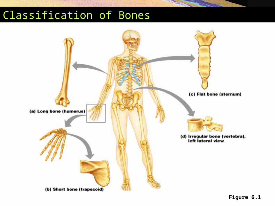
Classification of Bones
Figure 6.1
Classification of Bones
• Long bones – longer than wide – a shaft plus ends
• Short bones – roughly cube-shaped
• Flat bones – thin and flattened, usually curved
• Irregular bones – various shapes, do not fit into other categories
Bone Surface Markings
Depressions or Openings
• Foramen – opening
• Fissure – narrow slits between bones
• Fossa – shallow depression
• Sulcus – groove
• Meatus – tubelike passageway or canal
Bone Surface Markings
Processes or outgrowths (joints or attachments)
• Condyle – large round protuberance
• Facet – smooth flat articular surface (see vertebrae)
• Trochanter – very large projection
• Tuberosity – large rounded roughened projection
Gross Anatomy of Bones
• Compact bone – dense outer layer of bone
• Spongy bone – internal network of bone
Structure of a Typical Long Bone
• Diaphysis – “shaft” of a bone
• Epiphysis – ends of a bone
• Blood vessels – well vascularized
• Medullary cavity – hollow cavity – filled with marrow
• Membranes – periosteum, Sharpey’s fibers, and endosteum

Structure of a Long Bone
Figure 6.3a-c
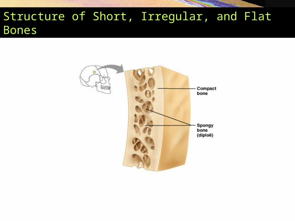
Structure of Short, Irregular, and Flat Bones
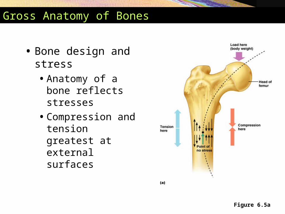
Figure 6.5a
Gross Anatomy of Bones
• Bone design and stress• Anatomy of a bone
reflects stresses
• Compression and tension greatest at external surfaces
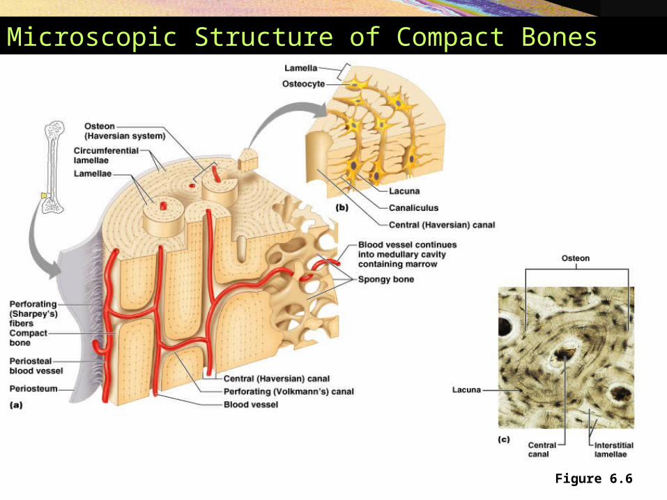
Microscopic Structure of Compact Bones
Figure 6.6
Chemical Composition of Bone
• 35% organic components• Composed of cells, fibers, and organic substances
• Collagen – abundant
• 65% inorganic mineral salts• Primarily calcium phosphate
• Resists compression
Bone Development
• Ossification (osteogenesis) – bone-tissue formation• Membrane bones – formed directly from
mesenchyme (bones of the skull and clavicles)• Intramembranous ossification
• Majority of the bones – develop initially from hyaline cartilage• Endochondral ossification
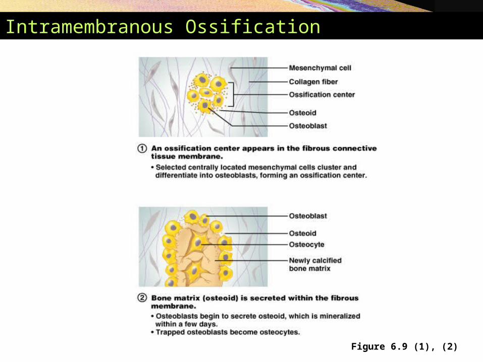
Intramembranous Ossification
Figure 6.9 (1), (2)
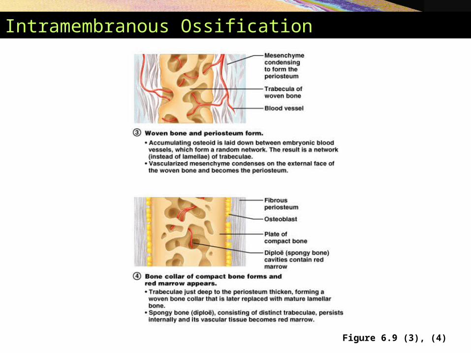
Intramembranous Ossification
Figure 6.9 (3), (4)
Endochondral Ossification
• All bones except some bones of the skull and clavicles
• Bones are modeled in hyaline cartilage
• Begins forming late in 2nd month of human development
• Continues forming until early adulthood
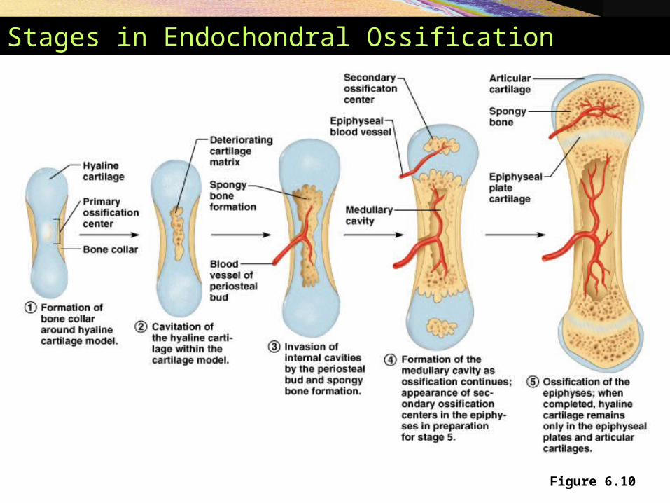
Stages in Endochondral Ossification
Figure 6.10
Anatomy of Epiphyseal Growth Areas
• In epiphyseal plates of growing bones• Cartilage is organized for quick, efficient growth
• Cartilage cells form tall stacks
• Chondroblasts at the top of stacks divide quickly• Pushes the epiphysis away from the diaphysis
• Lengthens entire long bone
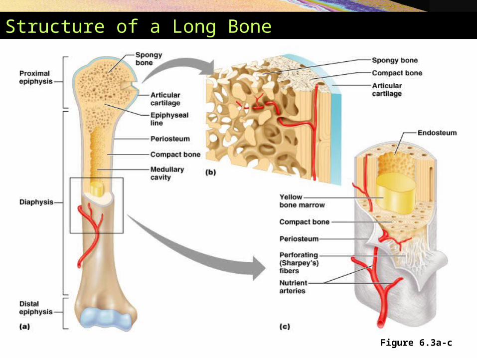
Structure of a Long Bone
Figure 6.3a-c
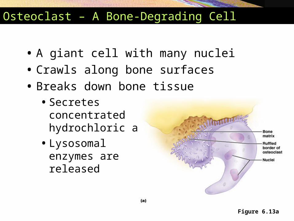
Osteoclast – A Bone-Degrading Cell
• A giant cell with many nuclei
• Crawls along bone surfaces
• Breaks down bone tissue• Secretes
concentrated hydrochloric acid
• Lysosomal enzymes are released
Figure 6.13a
Anatomy of Epiphyseal Growth Areas
• Older chondrocytes signal surrounding matrix to calcify
• Older chondrocytes then die and disintegrate• Leaves long trabeculae (spicules) of calcified
cartilage on diaphysis side• Trabeculae are partly eroded by osteoclasts• Osteoblasts then cover trabeculae with bone
tissue• Trabeculae finally eaten away from their tips by
osteoclasts
Postnatal Growth of Endochondral Bones
• During childhood and adolescence• Bones lengthen entirely by growth of the
epiphyseal plates
• Cartilage is replaced with bone tissue as quickly as it grows
• Epiphyseal plate maintains constant thickness
• Whole bone lengthens
Postnatal Growth of Endochondral Bones
• As adolescence draws to an end• Chondroblasts divide less often
• Epiphyseal plates become thinner• Cartilage stops growing
• Replaced by bone tissue
• Long bones stop lengthening when diaphysis and epiphysis fuse
Postnatal Growth of Endochondral Bones
• Growing bones widen as they lengthen• Osteoblasts – add bone tissue to the external
surface of the diaphysis
• Osteoclasts – remove bone from the internal surface of the diaphysis
• Appositional growth – growth of a bone by addition of bone tissue to its surface
Hormonal Regulation of Bone Growth
• Growth hormone – produced by the pituitary gland• Stimulates epiphyseal plates
• Thyroid hormone – ensures that the skeleton retains proper proportions
• Sex hormones• Promote bone growth
• Later induces closure of epiphyseal plates
Bone Remodeling
• Bone deposit and removal• Occurs at periosteal and endosteal surfaces
• Bone remodeling • Bone deposition – accomplished by osteoblasts
• Bone reabsorption – accomplished by osteoclasts
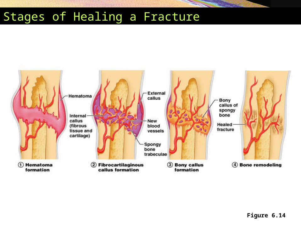
Stages of Healing a Fracture
Figure 6.14
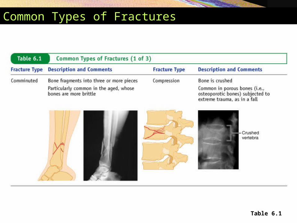
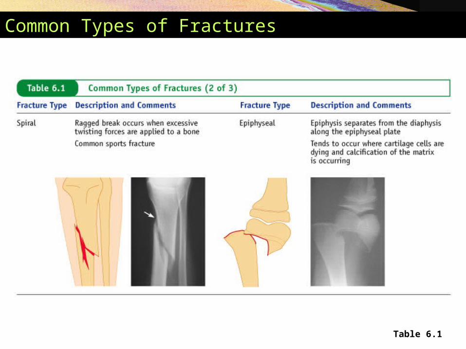
Common Types of Fractures
Table 6.1
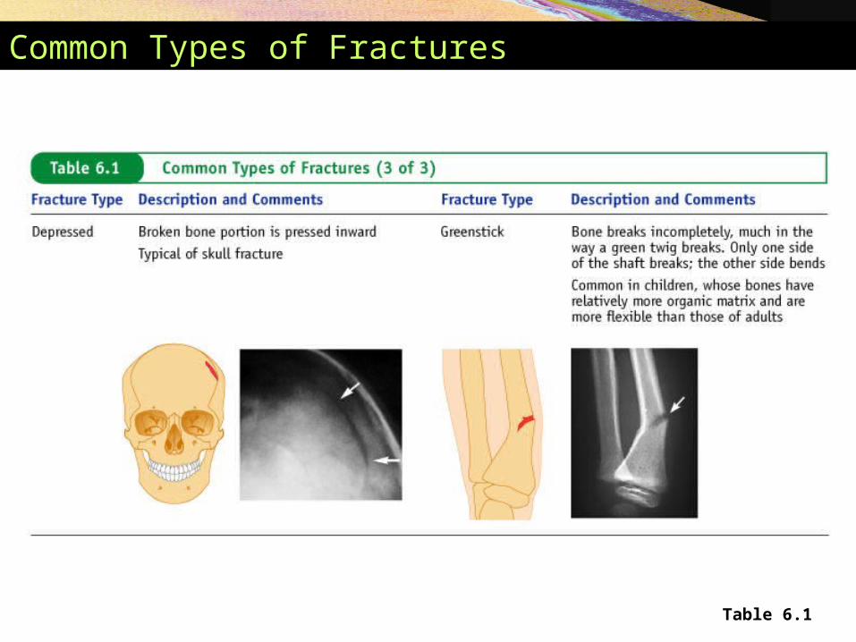
Common Types of Fractures
Table 6.1
Common Types of Fractures
Table 6.1
Disorders of Bones
• Osteoporosis – characterized by low bone mass• Bone reabsorption outpaces bone deposition
• Occurs most of in women after menopause
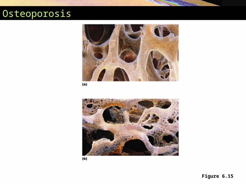
Osteoporosis
Figure 6.15
Disorders of Bones
• Osteomalacia – occurs in adults – bones are inadequately mineralized
• Rickets – occurs in children – analogous to osteomalacia
• Paget's disease – characterized by excessive rate of bone deposition
• Osteosarcoma – a form of bone cancer
The Skeleton Throughout Life
• Cartilage grows quickly in youth
• Skeleton shows fewer chondrocytes in the elderly
• Bones are a timetable• Mesoderm – gives rise to embryonic mesenchyme
cells
• Mesenchyme – produces membranes and cartilage
• Membranes and cartilage ossify
The Skeleton Throughout Life
• Skeleton grows until the age of 18–21 years
• In children and adolescents• Bone formation exceeds rate of bone reabsorption
• In young adults • Bone formation and bone reabsorption are in balance
• In old age reabsorption predominates
• Bone mass declines with age