Embed Size (px)
Citation preview
1
PowerPoint® Lecture Slides prepared by Leslie Hendon University of Alabama, Birmingham
C H A P T E R
Copyright © 2011 Pearson Education, Inc.
Part 1
6 Bones and Skeletal Tissues
Copyright © 2011 Pearson Education, Inc.
The Skeletal System consists of bones, cartilages, and joints. Review of Cartilage • Location and basic structure
• Found throughout adult body • Ear and epiglottis • Articular cartilages and costal cartilage • Larynx, trachea, and nose • Intervertebral discs, pubic symphysis, and articular discs
• Is surrounded by perichondrium (dense irreg. C.T.)-- functions as a girdle (resists outward pressure) and in growth & repair
• Consists primarily of water • Resilient tissue—it springs back to original shape
Copyright © 2011 Pearson Education, Inc.
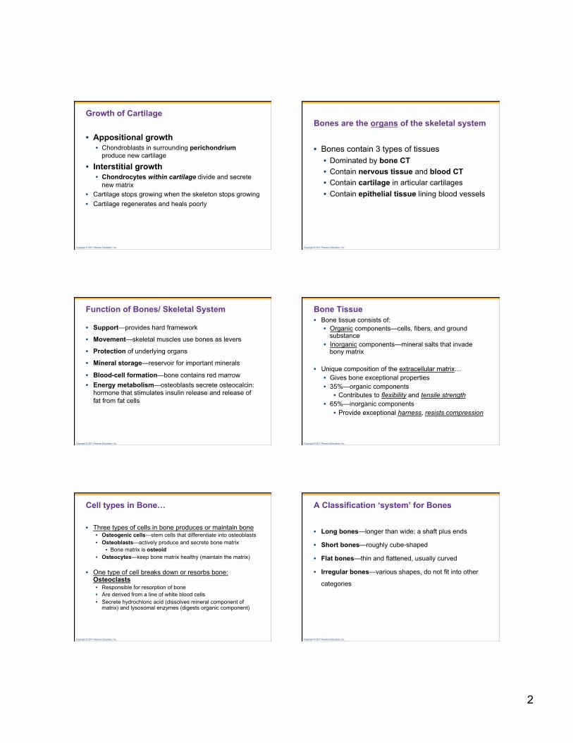
Types of Cartilage (review)
• All cartilages share some similarities • Cell type is the chondrocyte • Chondrocytes are located within lacunae • Matrix contains • Fibers • Jellylike ground substance
Copyright © 2011 Pearson Education, Inc.
Gelatinous ground substance
Chondrocyte in a lacuna Elastic fibers
Lacuna
Matrix Chondrocyte in a lacuna
Perichondrium
Chondrocyte in a lacuna
Collagen fibers
(a) Hyaline cartilage (180×) (b) Elastic cartilage (470×)
(c) Fibrocartilage (285×)
Microscopic appearance of cartilage
Figure 6.2
Copyright © 2011 Pearson Education, Inc.
Types of Cartilage (review)
• Hyaline cartilage (glassy) • Most abundant cartilage • Provides support through flexibility & resilience
• Elastic cartilage—contains many elastic fibers • Able to tolerate repeated bending
• Fibrocartilage—resists strong compression and strong tension • A intermediate between hyaline and dense regular
C.T.
Copyright © 2011 Pearson Education, Inc.
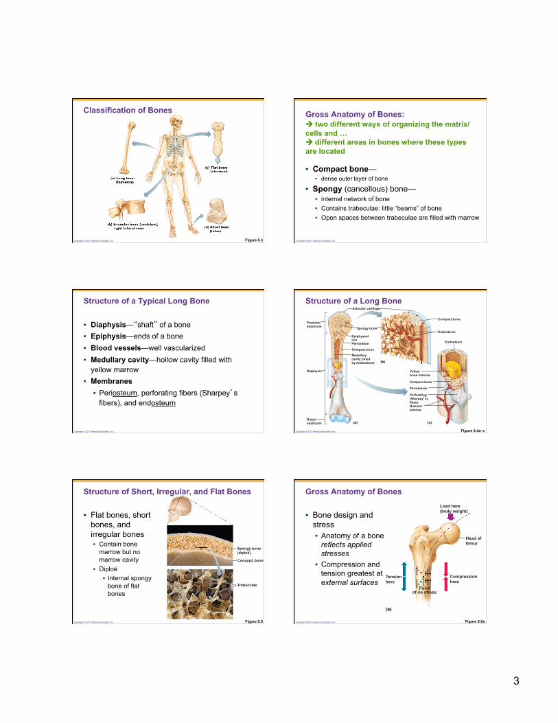
Hyaline cartilages Elastic cartilages Fibrocartilages
Cartilages
Cartilage in external ear Cartilages in
nose Articular cartilage of a joint
Costal cartilage Cartilage in intervertebral disc
Pubic symphysis
Articular cartilage of a joint
Meniscus (padlike cartilage in knee joint)
Cartilage: Where to find different types of cartilage Epiglottis
Larynx
Trachea
Cricoid cartilage
Lung
Respiratory tube cartilages in neck and thorax
Thyroid cartilage
2
Copyright © 2011 Pearson Education, Inc.
Growth of Cartilage
• Appositional growth • Chondroblasts in surrounding perichondrium
produce new cartilage
• Interstitial growth • Chondrocytes within cartilage divide and secrete
new matrix • Cartilage stops growing when the skeleton stops growing • Cartilage regenerates and heals poorly
Copyright © 2011 Pearson Education, Inc.
Bones are the organs of the skeletal system
• Bones contain 3 types of tissues • Dominated by bone CT • Contain nervous tissue and blood CT • Contain cartilage in articular cartilages • Contain epithelial tissue lining blood vessels
Copyright © 2011 Pearson Education, Inc.
Function of Bones/ Skeletal System
• Support—provides hard framework
• Movement—skeletal muscles use bones as levers
• Protection of underlying organs
• Mineral storage—reservoir for important minerals
• Blood-cell formation—bone contains red marrow • Energy metabolism—osteoblasts secrete osteocalcin:
hormone that stimulates insulin release and release of fat from fat cells
Copyright © 2011 Pearson Education, Inc.
Bone Tissue • Bone tissue consists of:
• Organic components—cells, fibers, and ground substance
• Inorganic components—mineral salts that invade bony matrix
• Unique composition of the extracellular matrix… • Gives bone exceptional properties • 35%—organic components
• Contributes to flexibility and tensile strength • 65%—inorganic components
• Provide exceptional harness, resists compression
Copyright © 2011 Pearson Education, Inc.
Cell types in Bone…
• Three types of cells in bone produces or maintain bone • Osteogenic cells—stem cells that differentiate into osteoblasts • Osteoblasts—actively produce and secrete bone matrix
• Bone matrix is osteoid • Osteocytes—keep bone matrix healthy (maintain the matrix)
• One type of cell breaks down or resorbs bone: Osteoclasts • Responsible for resorption of bone • Are derived from a line of white blood cells • Secrete hydrochloric acid (dissolves mineral component of
matrix) and lysosomal enzymes (digests organic component)
Copyright © 2011 Pearson Education, Inc.
A Classification ‘system’ for Bones
• Long bones—longer than wide; a shaft plus ends
• Short bones—roughly cube-shaped
• Flat bones—thin and flattened, usually curved
• Irregular bones—various shapes, do not fit into other
categories
3
Copyright © 2011 Pearson Education, Inc.
Classification of Bones
Figure 6.3 Copyright © 2011 Pearson Education, Inc.
Gross Anatomy of Bones: two different ways of organizing the matrix/cells and … different areas in bones where these types are located
• Compact bone— • dense outer layer of bone
• Spongy (cancellous) bone— • internal network of bone • Contains trabeculae: little “beams” of bone • Open spaces between trabeculae are filled with marrow
Copyright © 2011 Pearson Education, Inc.
Structure of a Typical Long Bone
• Diaphysis—“shaft” of a bone • Epiphysis—ends of a bone • Blood vessels—well vascularized • Medullary cavity—hollow cavity filled with
yellow marrow • Membranes • Periosteum, perforating fibers (Sharpey’s
fibers), and endosteum
Copyright © 2011 Pearson Education, Inc.
Structure of a Long Bone
Figure 6.4a–c
Proximal epiphysis
(b)
(c) (a)
Yellow bone marrow
Endosteum Epiphyseal line
Articular cartilage
Periosteum
Spongy bone
Compact bone Medullary cavity (lined by endosteum)
Compact bone
Compact bone Periosteum Perforating (Sharpey’s) fibers Nutrient arteries
Diaphysis
Distal epiphysis
Endosteum
Copyright © 2011 Pearson Education, Inc.
Compact bone
Trabeculae
Spongy bone (diploë)
Figure 6.5
Structure of Short, Irregular, and Flat Bones
• Flat bones, short bones, and irregular bones • Contain bone
marrow but no marrow cavity
• Diploë • Internal spongy
bone of flat bones
Copyright © 2011 Pearson Education, Inc.
Load here (body weight)
Head of femur
Compression here
Point of no stress
Tension here
(a)
Gross Anatomy of Bones
• Bone design and stress • Anatomy of a bone
reflects applied stresses
• Compression and tension greatest at external surfaces
Figure 6.6a
4
Copyright © 2011 Pearson Education, Inc.
Figure 6.6b Bone anatomy and bending stress.
Trabeculae of spongy bone
Load here
Compression lines Tension lines
Copyright © 2011 Pearson Education, Inc.
Bone Markings
• Superficial surfaces of bones reflect stresses on them
• There are three broad categories of bone markings: • Projections for muscle attachment • Surfaces that form joints • Depressions and openings
Copyright © 2011 Pearson Education, Inc.
Bone Markings
Table 6.1 Copyright © 2011 Pearson Education, Inc. Table 6.1 (2 of 3)
Copyright © 2011 Pearson Education, Inc. Table 6.1 (3 of 3)
Copyright © 2011 Pearson Education, Inc.
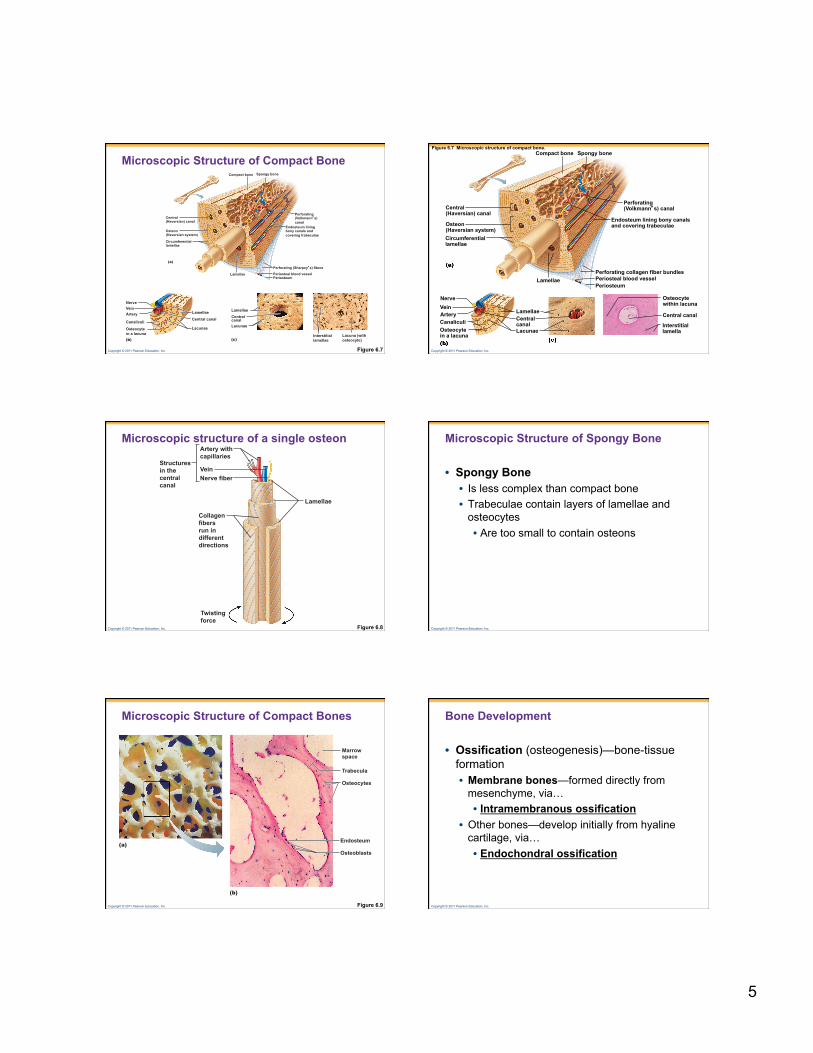
Microscopic Structure of Compact Bone • Compact Bone
• Contains passage ways for blood vessels, lymph vessels, and nerves
• Matrix is organized in Osteons (Haversian canals) —long cylindrical structures • Function in support • Structurally—resembles rings of a tree in cross-section • Osteons contain:
• Lamellae • Central canal • Perforating canals • Canaliculi
5
Copyright © 2011 Pearson Education, Inc. Figure 6.7
(a)
Compact bone
Endosteum lining bony canals and covering trabeculae
Perforating (Volkmann’s) canal
Perforating (Sharpey’s) fibers
Periosteal blood vessel Periosteum
Lamellae
Circumferential lamellae
Osteon (Haversian system)
Central (Haversian) canal
Spongy bone
(c) Interstitial lamellae
Lacunae
Lamellae Central canal
Lacuna (with osteocyte) (b)
Nerve Vein Artery
Canaliculi
Osteocyte in a lacuna
Lacunae
Lamellae Central canal
Microscopic Structure of Compact Bone
Copyright © 2011 Pearson Education, Inc.
Figure 6.7 Microscopic structure of compact bone.
Central (Haversian) canal
Osteon (Haversian system) Circumferential lamellae
Compact bone Spongy bone
Perforating (Volkmann’s) canal
Endosteum lining bony canals and covering trabeculae
Lamellae Perforating collagen fiber bundles Periosteal blood vessel Periosteum
Nerve Vein Artery Canaliculi Osteocyte in a lacuna
Lamellae Central canal Lacunae
Osteocyte within lacuna
Central canal
Interstitial lamella
Copyright © 2011 Pearson Education, Inc.
Structures in the central canal
Artery with capillaries
Vein Nerve fiber
Lamellae
Collagen fibers run in different directions
Twisting force
Microscopic structure of a single osteon
Figure 6.8 Copyright © 2011 Pearson Education, Inc.
Microscopic Structure of Spongy Bone
• Spongy Bone • Is less complex than compact bone • Trabeculae contain layers of lamellae and
osteocytes • Are too small to contain osteons
Copyright © 2011 Pearson Education, Inc.
Trabecula
Osteocytes
Endosteum
Marrow space
(b)
(a) Osteoblasts
Microscopic Structure of Compact Bones
Figure 6.9 Copyright © 2011 Pearson Education, Inc.
Bone Development
• Ossification (osteogenesis)—bone-tissue formation • Membrane bones—formed directly from
mesenchyme, via… • Intramembranous ossification
• Other bones—develop initially from hyaline cartilage, via… • Endochondral ossification
6
Copyright © 2011 Pearson Education, Inc.
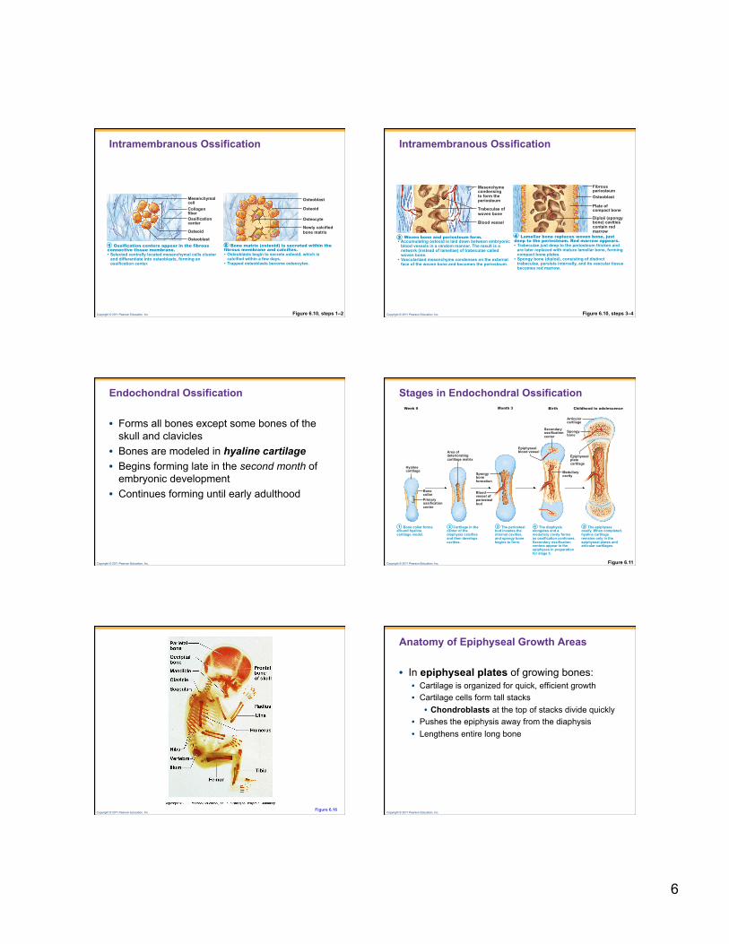
Intramembranous Ossification
Figure 6.10, steps 1–2
Mesenchymal cell Collagen fiber Ossification center Osteoid Osteoblast
Osteoid Osteocyte Newly calcified bone matrix
Osteoblast
Ossification centers appear in the fibrous connective tissue membrane. • Selected centrally located mesenchymal cells cluster and differentiate into osteoblasts, forming an ossification center.
Bone matrix (osteoid) is secreted within the fibrous membrane and calcifies. • Osteoblasts begin to secrete osteoid, which is calcified within a few days.
• Trapped osteoblasts become osteocytes.
1 2
Copyright © 2011 Pearson Education, Inc.
Intramembranous Ossification
Figure 6.10, steps 3–4
Mesenchyme condensing to form the periosteum
Blood vessel
Trabeculae of woven bone
Fibrous periosteum Osteoblast Plate of compact bone Diploë (spongy bone) cavities contain red marrow
Woven bone and periosteum form. • Accumulating osteoid is laid down between embryonic blood vessels in a random manner. The result is a network (instead of lamellae) of trabeculae called woven bone.
• Vascularized mesenchyme condenses on the external face of the woven bone and becomes the periosteum.
Lamellar bone replaces woven bone, just deep to the periosteum. Red marrow appears. • Trabeculae just deep to the periosteum thicken and are later replaced with mature lamellar bone, forming compact bone plates.
• Spongy bone (diploë), consisting of distinct trabeculae, persists internally, and its vascular tissue becomes red marrow.
3 4
Copyright © 2011 Pearson Education, Inc.
Endochondral Ossification
• Forms all bones except some bones of the skull and clavicles
• Bones are modeled in hyaline cartilage • Begins forming late in the second month of
embryonic development • Continues forming until early adulthood
Copyright © 2011 Pearson Education, Inc.
Hyaline cartilage
Area of deteriorating cartilage matrix
Epiphyseal blood vessel
Spongy bone formation
Epiphyseal plate cartilage
Secondary ossification center
Blood vessel of periosteal bud
Medullary cavity
Articular cartilage
Childhood to adolescence Birth Week 9 Month 3
Spongy bone
Bone collar Primary ossification center
Bone collar forms around hyaline cartilage model.
Cartilage in the center of the diaphysis calcifies and then develops cavities.
The periosteal bud invades the internal cavities, and spongy bone begins to form.
The diaphysis elongates and a medullary cavity forms as ossification continues. Secondary ossification centers appear in the epiphyses in preparation for stage 5.
The epiphyses ossify. When completed, hyaline cartilage remains only in the epiphyseal plates and articular cartilages.
1 2 3 4 5
Stages in Endochondral Ossification
Figure 6.11
Copyright © 2011 Pearson Education, Inc. Figure 6.16
Copyright © 2011 Pearson Education, Inc.
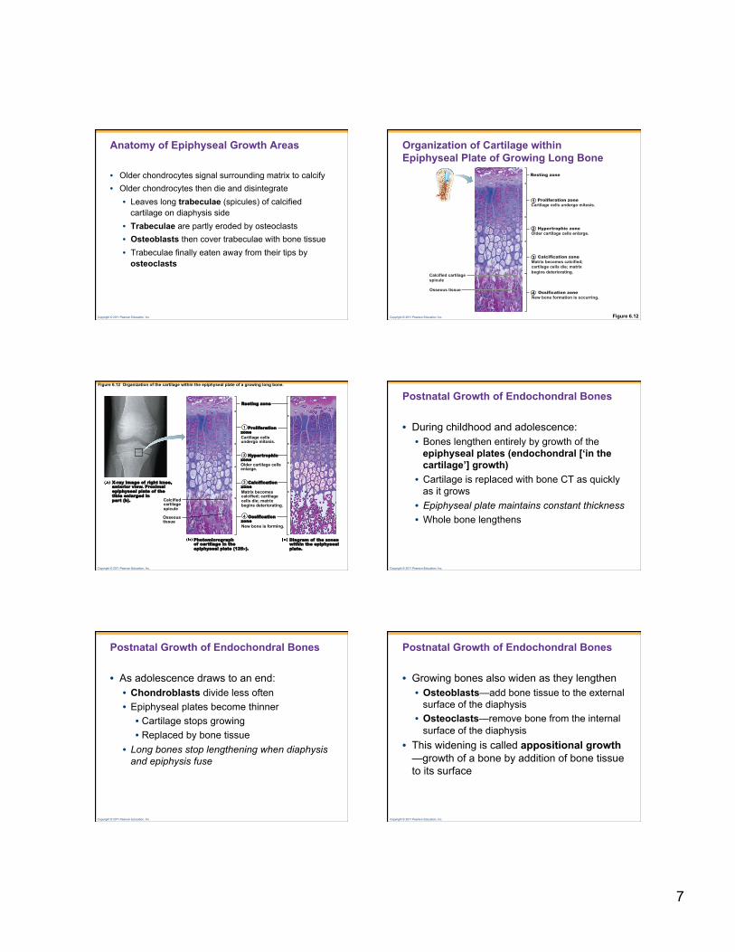
Anatomy of Epiphyseal Growth Areas
• In epiphyseal plates of growing bones: • Cartilage is organized for quick, efficient growth • Cartilage cells form tall stacks
• Chondroblasts at the top of stacks divide quickly • Pushes the epiphysis away from the diaphysis • Lengthens entire long bone
7
Copyright © 2011 Pearson Education, Inc.
Anatomy of Epiphyseal Growth Areas
• Older chondrocytes signal surrounding matrix to calcify • Older chondrocytes then die and disintegrate
• Leaves long trabeculae (spicules) of calcified cartilage on diaphysis side
• Trabeculae are partly eroded by osteoclasts • Osteoblasts then cover trabeculae with bone tissue • Trabeculae finally eaten away from their tips by
osteoclasts
Copyright © 2011 Pearson Education, Inc. Figure 6.12
Organization of Cartilage within Epiphyseal Plate of Growing Long Bone
Calcified cartilage spicule
Osseous tissue
Resting zone
Proliferation zone Cartilage cells undergo mitosis.
Hypertrophic zone Older cartilage cells enlarge.
Ossification zone New bone formation is occurring.
Calcification zone Matrix becomes calcified; cartilage cells die; matrix begins deteriorating.
1
2
3
4
Copyright © 2011 Pearson Education, Inc.
Figure 6.12 Organization of the cartilage within the epiphyseal plate of a growing long bone.
Calcified cartilage spicule
Osseous tissue
Cartilage cells undergo mitosis.
Older cartilage cells enlarge.
Matrix becomes calcified; cartilage cells die; matrix begins deteriorating.
New bone is forming.
Resting zone
Proliferation zone
Hypertrophic zone
Calcification zone
Ossification zone
Diagram of the zones within the epiphyseal plate.
Photomicrograph of cartilage in the epiphyseal plate (125×).
X-ray image of right knee, anterior view. Proximal epiphyseal plate of the tibia enlarged in part (b).
1
2
3
4
Copyright © 2011 Pearson Education, Inc.
Postnatal Growth of Endochondral Bones
• During childhood and adolescence: • Bones lengthen entirely by growth of the
epiphyseal plates (endochondral [‘in the cartilage’] growth)
• Cartilage is replaced with bone CT as quickly as it grows
• Epiphyseal plate maintains constant thickness • Whole bone lengthens
Copyright © 2011 Pearson Education, Inc.
Postnatal Growth of Endochondral Bones
• As adolescence draws to an end: • Chondroblasts divide less often • Epiphyseal plates become thinner • Cartilage stops growing • Replaced by bone tissue
• Long bones stop lengthening when diaphysis and epiphysis fuse
Copyright © 2011 Pearson Education, Inc.
Postnatal Growth of Endochondral Bones
• Growing bones also widen as they lengthen • Osteoblasts—add bone tissue to the external
surface of the diaphysis • Osteoclasts—remove bone from the internal
surface of the diaphysis • This widening is called appositional growth
—growth of a bone by addition of bone tissue to its surface
8
Copyright © 2011 Pearson Education, Inc.
Bone Growth influenced by hormones (also nutrition)
• Growth hormone—produced by the pituitary gland • Stimulates epiphyseal plates
• Thyroid hormone—ensures that the skeleton retains proper proportions
• Sex hormones (estrogen and testosterone)
• Promote bone growth
• Later induces closure of epiphyseal plates
Copyright © 2011 Pearson Education, Inc.
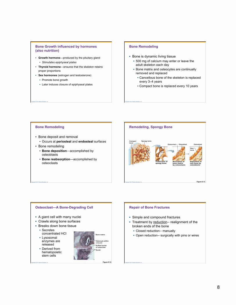
Bone Remodeling
• Bone is dynamic living tissue • 500 mg of calcium may enter or leave the
adult skeleton each day • Bone matrix and osteocytes are continually
removed and replaced • Cancellous bone of the skeleton is replaced
every 3–4 years • Compact bone is replaced every 10 years
Copyright © 2011 Pearson Education, Inc.
Bone Remodeling
• Bone deposit and removal • Occurs at periosteal and endosteal surfaces
• Bone remodeling • Bone deposition—accomplished by
osteoblasts • Bone reabsorption—accomplished by
osteoclasts
Copyright © 2011 Pearson Education, Inc.
Remodeling, Spongy Bone
Figure 6.13
Compact bone
Spongy bone
Trabeculae of spongy bone
Resorption of bone matrix by osteoclasts
Deposition of new bone by osteoblasts
New bone
Osteoblast Osteoblast Osteoclast
Copyright © 2011 Pearson Education, Inc.
Osteoclast—A Bone-Degrading Cell
• A giant cell with many nuclei • Crawls along bone surfaces • Breaks down bone tissue • Secretes
concentrated HCl • Lysosomal
enzymes are released
• Derived from hematopoietic stem cells
Figure 6.14
Osteocyte within a lacuna
Bone matrix
Ruffled border of osteoclast Nuclei
Copyright © 2011 Pearson Education, Inc.
Repair of Bone Fractures
• Simple and compound fractures • Treatment by reduction-- realignment of the
broken ends of the bone • Closed reduction-- manually • Open reduction-- surgically with pins or wires
9
Copyright © 2011 Pearson Education, Inc.
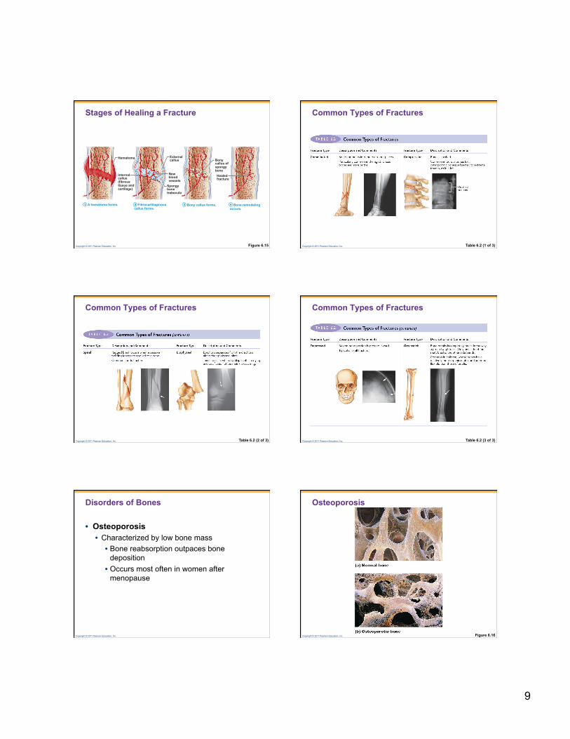
Stages of Healing a Fracture
Figure 6.15
Hematoma External callus
New blood vessels
Spongy bone trabecula
Internal callus (fibrous tissue and cartilage)
A hematoma forms. Fibrocartilaginous callus forms.
Bony callus forms.
Bony callus of spongy bone Healed fracture
Bone remodeling occurs.
1 2 3 4
Copyright © 2011 Pearson Education, Inc.
Common Types of Fractures
Table 6.2 (1 of 3)
Copyright © 2011 Pearson Education, Inc.
Common Types of Fractures
Table 6.2 (2 of 3) Copyright © 2011 Pearson Education, Inc.
Common Types of Fractures
Table 6.2 (3 of 3)
Copyright © 2011 Pearson Education, Inc.
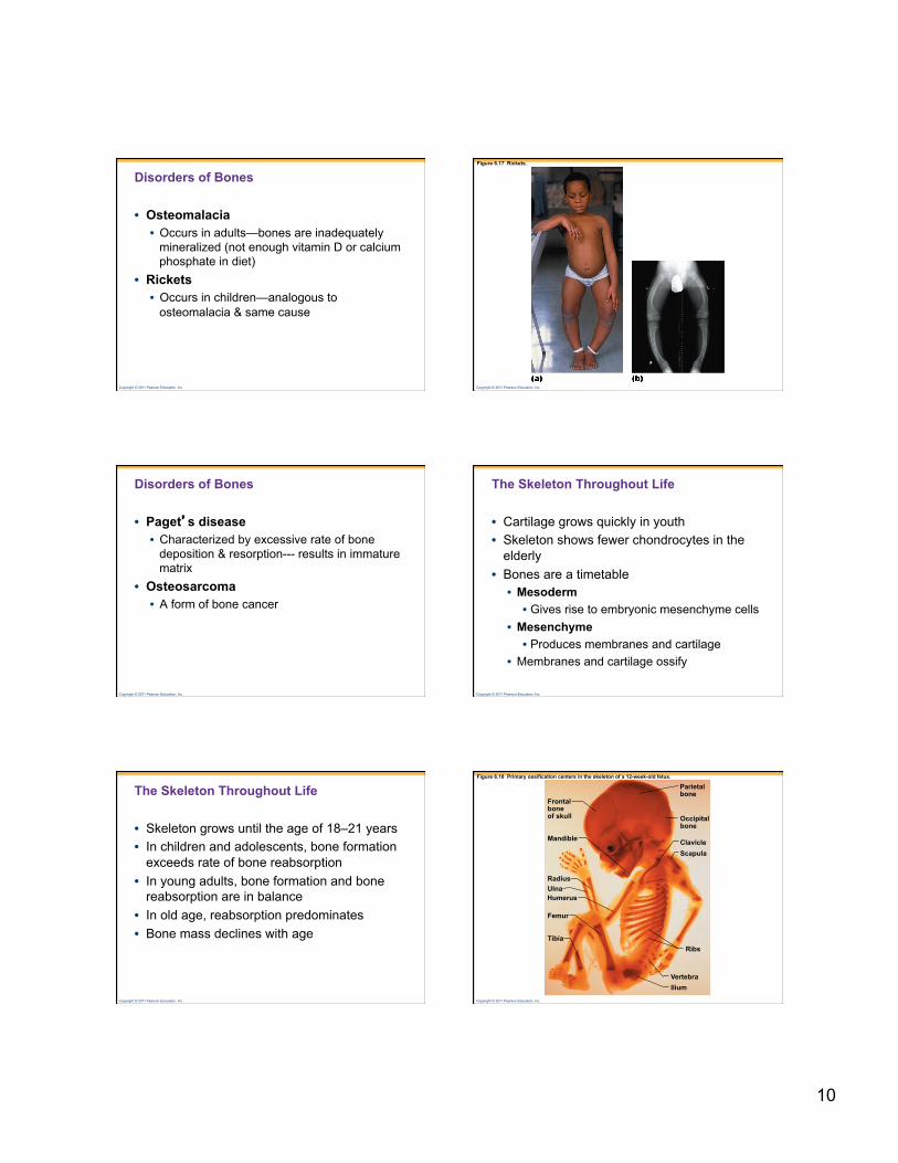
Disorders of Bones
• Osteoporosis • Characterized by low bone mass • Bone reabsorption outpaces bone
deposition • Occurs most often in women after
menopause
Copyright © 2011 Pearson Education, Inc.
Osteoporosis
Figure 6.16
10
Copyright © 2011 Pearson Education, Inc.
Disorders of Bones
• Osteomalacia • Occurs in adults—bones are inadequately
mineralized (not enough vitamin D or calcium phosphate in diet)
• Rickets • Occurs in children—analogous to
osteomalacia & same cause
Copyright © 2011 Pearson Education, Inc.
Figure 6.17 Rickets.
Copyright © 2011 Pearson Education, Inc.
Disorders of Bones
• Paget’s disease • Characterized by excessive rate of bone
deposition & resorption--- results in immature matrix
• Osteosarcoma • A form of bone cancer
Copyright © 2011 Pearson Education, Inc.
The Skeleton Throughout Life
• Cartilage grows quickly in youth • Skeleton shows fewer chondrocytes in the
elderly • Bones are a timetable • Mesoderm • Gives rise to embryonic mesenchyme cells
• Mesenchyme • Produces membranes and cartilage
• Membranes and cartilage ossify
Copyright © 2011 Pearson Education, Inc.
The Skeleton Throughout Life
• Skeleton grows until the age of 18–21 years • In children and adolescents, bone formation
exceeds rate of bone reabsorption • In young adults, bone formation and bone
reabsorption are in balance • In old age, reabsorption predominates • Bone mass declines with age
Copyright © 2011 Pearson Education, Inc.
Figure 6.18 Primary ossification centers in the skeleton of a 12-week-old fetus.
Frontal bone of skull
Mandible
Radius Ulna Humerus
Femur
Tibia
Ilium Vertebra
Ribs
Scapula Clavicle
Occipital bone
Parietal bone


![Bones and Skeletal Tissues - Department Faculty …faculty.elac.edu/LEOT/doc/2013/sp/Anat-MariebA6-9[LectNotes1].pdf · SKELETAL SYSTEM •1) Components –Cartilages –Bones –Tendons](https://img.pdfslide.us/doc/110x75/5b8258647f8b9a2b678e5f78/bones-and-skeletal-tissues-department-faculty-lectnotes1pdf-skeletal-system.jpg)