Embed Size (px)
Citation preview

Bone Grafting theMandible
Patrick J. Louis, DDS, MD
KEYWORDS
� Bone grafting � Mandible � Dentate � Edentulous
Multiple bone grafting techniques have been used to reconstruct the partially dentateand edentulous mandible.1–6 Many of these techniques have been successful. In thisarticle, the various bone grafting techniques to reconstruct mandibular defects arereviewed. Other issues discussed include whether autogenous bone is necessaryfor reconstruction of the mandibular ridge and the importance of membranes.
BLOCK ONLAY GRAFTS
Block onlay grafts have been used extensively for reconstruction of mandibular alve-olar defects.7–11 Autogenous and nonautogenous options are available for vertical andhorizontal deficiencies of the mandible. Autogenous bone grafts have the advantageof transferring osteogenic cells to the recipient site.12–15 Autogenous donor sites forblock grafts include the calvarium, mandible, zygoma, and ilium. Intramembranousbone (calvarium, mandible, and zygoma) has the advantage of decreased rates ofresorption compared with endochondral bone (ilium), due to its dense cortical struc-ture and microarchitecture.16–20
When a small amount of bone is needed, local grafts harvested from the mandibularsymphysis or ramus have been used extensively (Fig. 1).10,21–24 These sources havethe advantage of being convenient due to their proximity to the reconstruction siteand low risk of morbidity.25 The disadvantage of the mandible as a bone source isthe limited amount of bone available and the fact that mandibular harvesting cansometimes interfere with the planned reconstruction if too close to the reconstructionsite (Table 1).26 The most common postoperative morbidity associated with chin andramus grafts has been reported as temporary paresthesia. For the chin it ranges from10% to 50%, and for the ramus, it ranges from 0% to 5%.21,27 The zygomaticomax-illary region also can be used as a harvest site.28 The advantage of this site is thatwhen performing grafts in the maxilla, simple extension of the incision is needed.The procedure is associated with a low risk of morbidity. The disadvantage is the
This article was previously published in the May 2011 issue of Oral and Maxillofacial SurgeryClinics of North America.Oral and Maxillofacial Surgery, School of Dentistry, University of Alabama at Birmingham, 19197th Avenue South, SDB 419, Birmingham, AL 35294, USAE-mail address: [email protected]
Dent Clin N Am 55 (2011) 673–695doi:10.1016/j.cden.2011.07.006 dental.theclinics.com0011-8532/11/$ – see front matter � 2011 Elsevier Inc. All rights reserved.
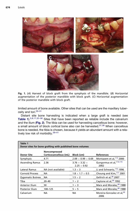
Fig. 1. (A) Harvest of block graft from the symphysis of the mandible. (B) Horizontalaugmentation of the posterior mandible with block graft. (C) Horizontal augmentationof the posterior mandible with block graft.
Louis674
limited amount of bone available. Other sites that can be used are the maxillary tuber-osity and tori.29–31
Distant site bone harvesting is indicated when a large graft is needed (seeTable 1).5,8,17,32–36 Sites that have been reported as reliable include the calvariumand the ilium (Fig. 2). The tibia can be used for harvesting cancellous bone; however,a small amount of block cortical bone also can be harvested.37,38 When cancellousbone is needed, the tibia is chosen, because it yields an abundant amount with a rela-tively low risk of morbidity.39–41
Table 1Donor sites for bone grafting with published bone volumes
Donor SiteNoncompressedCorticocancellous (mL) Block (cm) References
Symphysis 4.71 2.09 � 0.99 � 0.69 Montazem et al,113 2000
Ascending Ramus 2.36 3.76 � 3.32 �2.25 � 0.92
Gungormus et al,114,115
2002
Lateral Ramus NA (not available) 1.3 � 3 Li and Schwartz,116 1996
Cornoid Process NA 1.8 � 1.7 � 0.5 Choung and Kim,117 2001
Zygomatic Butress NA 1.5 � 2 Gellrich et al,28 2007
Tiba 20–40 1 � 2 Caytone et al,39 1992
Anterior Ilium 50 1 � 3 Marx and Morales,49 1988
Posterior Ilium 100–125 5 � 5 Marx and Morales,49 1988
Calvarium NA NA Moreira-Gonzalez et al,43
2006

Fig. 2. (A) Clinical photograph of a patient who had avulsion of one of the mandibular inci-sors secondary to a motor vehicle accident. (B) Scalp incision for access to the calvarium. (C)Outline and harvest of block graft from the outer table of the right parietal bone for recon-struction. (D) Block graft for augmentation of the anterior mandible. (E) Particulate autog-enous bone graft has been placed over the block graft and will be covered witha membrane. (F) Postoperative view of bone graft site with excellent bone contour. Notethe fixation screw is visible through the mucosa. (G) Implant placement after exposure ofthe augmented site. (H) Restored mandibular incisor immediately after final restoration.Note the excellent ridge contour. (I) Restored mandibular incisor several months after finalrestoration placed.
Bone Grafting the Mandible 675
The types of grafts harvested from the calvarium and the ilium are entirely different.Calvarial grafts are usually harvested in strips from the parietal bone. The averagethickness of the parietal bone is 7.45 mm.42,43 A split thickness harvest technique isused, which can yield graft thicknesses of approximately 3 mm. The main advantageof calvarial grafts is their dense cortical structure that resists resorption.16–18 Theamount of available cortical graft is abundant. However, this procedure usuallyrequires general anesthesia in a hospital or ambulatory care setting, but there area few reports of office based techniques.44,45 The incidence of complication is low,

Fig. 2. (continued)
Louis676
with the most common reported complication being seroma/hematoma.46 Largeramounts of bone can be harvested from the ilium, but these grafts typically havea thin cortical layer and a thick cancellous portion.35,47 The main advantages of thesegrafts are the large volume of bone that can be harvested and carved into variousshapes.48,49 The main disadvantage is the morbidity associated with bone graftharvest. The reported complication incidence is higher than with other donor sites.Reported complications include gait disturbance, paresthesia, hematoma/seroma,and fracture.49–53
Horizontal Segmental Defects
These cases have adequate alveolar bone height but inadequate width. The deficiencyis most commonly on the facial surface of the mandible. The planning of these casesshould include physical and radiographic examination. The site and the plannedimplant diameter dictate the amount of bone required. In the mandibular incisor,region a 3 mm diameter implant is usually indicated. The amount of bone needed is1 mm to 1.5 mm of bone on both the facial and lingual aspect of the implant. Thus,in the incisor region, the minimum amount of bone needed is 4 mm to 5 mm. In thecanine and premolar region, the usual implant diameter is approximately 4 mm, andthe width of bone needed is 6 mm to 7 mm. In the molar region, the approximatesize of the implant needed is usually 5 mm, and the minimum width required is 7mm to 8 mm.When grafting these various sites, the thickness of the block graft shouldbe slightly larger than the final planned width.For most segmental deficiencies in the mandible, autogenous grafts can be har-
vested from the symphysis or ramus. The planned recipient site is usually exposedthrough a crestal incision. Depending on location of the deficiency, the donor sitemay be accessed through extension of this incision. Once the site is exposed, thedefect can be measured and the size of the bone graft determined.
Bone Grafting the Mandible 677
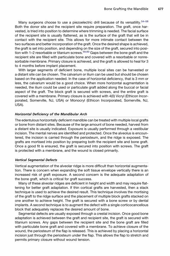
Many surgeons choose to use a piezoelectric drill because of its versatility.54–58
Both the donor site and the recipient site require preparation. The graft, once har-vested, is tried into position to determine where trimming is needed. The facial surfaceof the recipient site is usually flattened, as is the surface of the graft that will be incontact with the recipient site. This allows for more intimate contact between thetwo surfaces and better incorporation of the graft. Once the desired shape is achieved,the graft is set into position, and depending on the size of the graft, secured into posi-tion with 1–2 resorbable or titanium screws.59,60 Gaps between the bone graft and therecipient site are filled with particulate bone and covered with a resorbable or nonre-sorbable membrane. Primary closure is achieved, and the graft is allowed to heal for 3to 4 months before implant placement.With larger segments of deficient bone, multiple local sites can be harvested or
a distant site can be chosen. The calvarium or ilium can be used but should be chosenbased on the application needed. In the case of horizontal deficiency, that is 3 mm orless, the calvarium would be a good choice. When more horizontal augmentation isneeded, the ilium could be used or particulate graft added along the buccal or facialaspect of the graft. The block graft is secured with screws, and the entire graft iscovered with a membrane. Primary closure is achieved with 4(0) Vicryl (Ethicon Incor-porated, Somerville, NJ, USA) or Monocryl (Ethicon Incorporated, Somerville, NJ,USA).
Horizontal Deficiency of the Mandibular Arch
The edentulous horizontally deficient mandible can be treated with multiple local graftsor bone from distant sites. Because of the large amount of bone needed, harvest froma distant site is usually indicated. Exposure is usually performed through a vestibularincision. The mental nerves are identified and protected. Once the alveolus is encoun-tered, the incision is carried through the periosteum, and the ridge is exposed. Thegrafts are mortised into position by preparing both the recipient site and bone graft.Once a good fit is ensured, the graft is secured into position with screws. The graftis protected with a membrane, and the wound is closed primarily.
Vertical Segmental Defects
Vertical augmentation of the alveolar ridge is more difficult than horizontal augmenta-tion. There is concern when expanding the soft tissue envelope vertically there is anincreased risk of graft exposure. A second concern is the adequate adaptation ofthe bone graft, which is critical for graft success.Many of these alveolar ridges are deficient in height and width and may require flat-
tening for better graft adaptation. If thin cortical grafts are harvested, then a stacktechnique is used to achieve the desired result. This technique involves the mortisingof the graft to the ridge surface and the placement of multiple block grafts stacked onone another to achieve height. The graft is secured with a bone screw or by dentalimplants. A second technique is to augment the defect with a single corticocancellousblock that adequately replaces the desired amount of bone.Segmental defects are usually exposed through a crestal incision. Once good bone
adaptation is achieved between the graft and recipient site, the graft is secured withtitanium screws. Any gaps between the recipient site and the bone graft are filledwith particulate bone graft and covered with a membrane. To achieve closure of thewound, the periosteum of the flap is released. This is achieved by placing a horizontalincision just through the periosteum under the flap. This allows the flap to stretch andpermits primary closure without wound tension.
Louis678
Vertical Deficiency of the Mandibular Arch
This generally requires a large amount of autogenous bone from the calvarium or theilium. As discussed earlier, reconstruction can be performed using a stacked calvarialgraft that is shaped and mortised to the desired height (Fig. 3). Iliac crest grafts do notrequire stacking; instead the ridge is exposed through a vestibular incision, the mentalnerves identified, the block secured with titanium screws, and gaps filled with cancel-lous particulate, then covered with a membrane before performing a 2-layer closure.
Allogeneic Block Grafts
Allogeneic block grafts have not had extensive use in implant dentistry.61–63 Asystemic review of the literature by Waasdorp identified 35 papers, with only ninepublications meeting inclusion criteria (2 case reports, 6 case series, and 1 prospec-tive, multicenter, consecutive case series).64 There were no randomized controlledclinical trials identified. Observational studies generally report graft incorporation ratesof 90% or greater and implant survival of 99% to 100%. The shortcomings of thesestudies is that most reports involved selected defects in anterior regions and had shortterm follow-ups of less than 3 years. The reviewers concluded that case-based reportsdocument the potential for allogeneic block grafts to support alveolar ridge
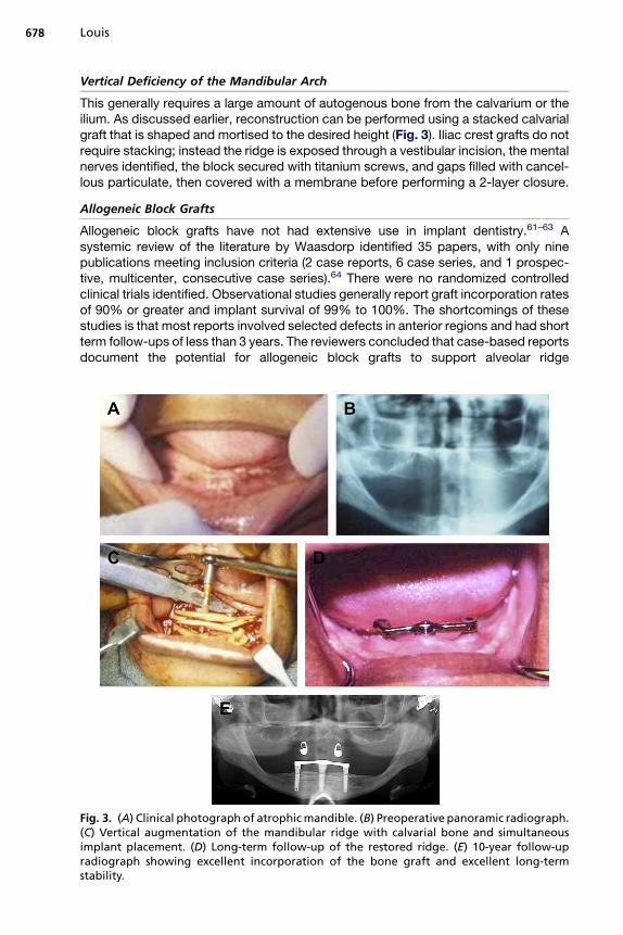
Fig. 3. (A) Clinical photograph of atrophicmandible. (B) Preoperative panoramic radiograph.(C) Vertical augmentation of the mandibular ridge with calvarial bone and simultaneousimplant placement. (D) Long-term follow-up of the restored ridge. (E) 10-year follow-upradiograph showing excellent incorporation of the bone graft and excellent long-termstability.

Bone Grafting the Mandible 679
augmentation and implant placement, but there is insufficient evidence to establishtreatment efficacy relative to graft incorporation, alveolar ridge augmentation, andlong-term dental implant survival.Allogeneic block grafts can be substituted for autogenous grafts to avoid donor site
morbidity, but the risk of exposure is higher, especially with vertical augmentation.61
BARRIER MEMBRANES
Barrier membranes have been studied extensively in the dental and maxillofacial liter-ature.27,65–73 Membranes have been shown to protect both block and particulateonlay grafts.74 The surgeon has the choice of resorbable versus nonresorbable andrigid versus nonrigid membranes when performing grafting procedures. Studiessuggest that there is less resorption of block and particulate grafts when a membraneremains in place throughout the healing phase of the graft.74,75 Significant resorptioncan occur for block grafts left unprotected, but not always.76 Vertical augmentation isparticularly problematic with both block and particulate grafts, with resorption ratesthat have been reported higher than for horizontal augmentation.76 Cordaro reportedresorption rates of 23.5% for lateral augmentation and 42% for vertical augmentationwhen using block grafts without a membrane.76 Particulate onlay grafts can undergoeven more resorption.75
Block grafts have structural integrity and thus maintain space and resist resorp-tion.17,18 Particulate grafts lack structural integrity and can be displaced.75 With hori-zontal augmentation, where the vertical height is adequate and only width is lacking,particulate graft material can be used and protected with a membrane. The choice ofparticulate graft material becomes important if a nonrigid membrane is used. Theparticulate graft must have some strength to resist deformation by the soft tissueenvelope.75,77 In this case, it is preferable to use a mineralized bone product suchas autogenous bone, mineralized allogeneic bone, or anorganic bovine bone. If a rigidmembrane is used, then the choice of material becomes less crucial, as a rigidmembrane will maintain space and protect the graft.78–80
When vertical augmentation is indicated, block or particulate graft can be chosen. Ifa block graft is chosen, it has the advantage of structural integrity to maintain space;however, because of high resorption potential, membrane protection is war-ranted.74,76 A nonrigid, resorbable or nonresorbable membrane can be used.When a particulate graft is selected for vertical augmentation, a rigid membrane
must be used to protect the graft. The technique of vertical augmentation with partic-ulate grafts protected by a rigid membrane has been reported to be successful.78,79,81
These techniques have been associated with a high rate of exposure that can nega-tively affect the graft. Exposure rates are as high as 50%, particularly when largevertical augmentations are performed.78 One of the techniques that has beendescribed to minimize graft exposure is the use of PRP (platelet-rich plasma gel)placed over the rigid membrane before wound closure.82
PARTICULATE GRAFTS
Particulate onlay grafts have been used extensively for reconstruction of mandibularalveolar defects. Autogenous and nonautogenous options are available. The sourceof autogenous particulate is the same as has been discussed for block harvest (seeTable 1). For small amounts of bone, local sites can be used, including the symphysis,ramus, and maxillary tuberosity. This is usually harvested with a bone shaver. Whengreater volumes of bone are needed, then distant sites are employed. Particulate

Louis680
grafts have the advantage of rapid vascularization, but they must be protected bya membrane to reduce the risk of resorption.Allogeneic and xenogenic particulate grafts have been growing in popularity. These
grafts are readily available and abundant. They can be used in combination withautogenous bone or alone. Recent studies have shown success with these graft mate-rials for vertical ridge augmentation when protected with a rigid membrane.82,83
Segmental Horizontal Defects
The reconstruction of the partially edentulous mandible with particulate graft materialcan be performed with various bone graft materials. The type of graft material neededwill depend on the size of the defect and type of membrane used. With horizontalaugmentation, where the vertical height is adequate, and only width is lacking, partic-ulate graft material can be used and protected with a membrane (Fig. 4). Whena mineralized bone graft is used, a nonrigid membrane is suggested. In this technique,the graft is placed through a crestal incision. The defect is grafted, then protected withmembrane. If a rigid membrane is used, it must be fixed with screws. If a nonrigidmembrane is used, the edges of the membrane must be tucked and secured under-neath the flap.
Fig. 4. (A) Preoperative radiograph of a patient missing tooth #30. This site has adequateheight but not width. (B) Preoperative photograph. (C) Harvest of bone during a rightmaxillary tuberosity reduction. (D, E) Horizontal augmentation of the right posteriormandible during implant placement. (F) Final restoration in place, area #30.

Bone Grafting the Mandible 681
Demineralized bone graft materials tend to be soft and easily compressed by thesoft tissue envelope. If only demineralized product is used to augment a horizontaldefect, a rigid membrane should be used to protect the graft. This membrane mustbe secured with screws to the underlying bone to prevent micromotion. To closethe wound, a periosteal releasing incision may be required. The particulate graftmust have some strength to resist deformation. In this case, it is preferable to usemineralized product such as autogenous bone, mineralized allogeneic bone, or anor-ganic bovine bone as the graft of choice. However, if a rigid membrane is used, thenthe choice of material becomes less crucial.The technique is done through a vestibular incision to expose the edentulous space.
The incision is extended to near the junction of the attached mucosa. Mobilization ofthe facial, crestal, and lingual aspect of the soft tissues is usually performed. Thedissection around the cervical portion of the teeth is performed in a tunneling fashionsubperiosteally to mobilize the gingival cuff. As an alternative to a vestibular incision,a crestal incision can be used in conjunction with releasing incision at least 2 teeth oneither side of the defect. Once the ridge is exposed, a titanium mesh membrane canbe trimmed and contoured to fit into position as a crib. This is then filled with bone graftand secured with at least 2 screws on the facial aspect. When a crestal incision is usedfor exposure, the periosteum must be released along the facial vestibule to facilitatea tension-free closure of the wound. This is usually not necessary when a vestibularincision is used. Closure is performed in layers with 3.0 Vicyrl suture material placeddeep to the mucosa. The mucosa is closed with 4.0 Vicyrl or Monocryl suture.
Segmental Vertical Defects
When using a particulate bone graft for vertical ridge augmentation, a rigid membranemust be used to prevent deformation and resorption of the graft. The reconstruction isperformed in similar fashion as described in the section Segmental Horizontal Defects(Fig. 5).
Horizontal Deficiency of the Mandibular Arch
To reconstruct horizontal deficiency of the mandibular alveolus with particulate graft,the principles of graft protection during the healing phase are important. The ridge isexposed through a vestibular incision. After identification of the mental nerves, thedissection is carried toward the ridge. The periosteum is incised, and a subperiostealdissection is performed to expose the ridge. The particulate graft is placed and thencovered with a membrane. The choice of particulate graft material becomes importantif a nonrigid membrane is used. The particulate graft must have some strength to resistdeformation.75,77 In this case, it is preferable to use mineralized bone as the graft ofchoice. If a rigid membrane is used, the choice of material becomes less crucial, asa rigid membrane will maintain space and protect the graft. Following membraneplacement, the wound is closed primarily.78–80
Vertical Deficiency of the Mandibular Arch
When augmenting the highly atrophic edentulous mandible vertically, an intraoral orextraoral approach can be used. The intraoral approach is more straightforward. Inthis technique, an incision is made in the depth of the mandibular vestibule just ante-rior to the retromolar pad extending anteriorly across the midline. After identifying andprotecting the mental nerves, a subperiosteal dissection is achieved. In the extremelyatrophic mandible, the inferior alveolar nerve may be exposed along the crest due todehiscence of the canal. In this case, a preferred technique is to start anteriorly andidentify the mental foramina and then dissect posteriorly. With this technique, the

Fig. 5. (A) Preoperative panoramic radiograph showing inadequate bone height of the leftposterior mandible. (B) Preoperative view of the narrow posterior mandibular ridge. (C)Preoperative cast. (D) Exposure of the posterior mandible through a vestibular incision.(E) Try in of the titanium mesh. (F) Mineralized and demineralized bone mixture. (G)Augmentation of the posterior mandibular ridge with allogeneic bone and titaniummesh. (H) Soft tissue flap draped over the mesh. (I) Posterior alveolar ridge 1 week postop-eratively. (J) Posterior alveolar ridge 4 months postoperatively. (K) Surgical exposure of theridge. (L) After removal of the mesh there is excellent bone height and width. (M) Surgicalguide in place. (N) Panoramic radiograph 4 months postoperatively. (O) Cone-beamcomputed tomography (CT) scan showing a cross-sectional view of the posterior ridge at4 months. Note the excellent bone height and width. (P) Mirror view of the alveolar ridgewith implants in place. (Q) Postoperative radiograph with implants in place. (R) Final resto-ration in place.
Louis682
dissection is carried onto the lingual surface of the mandible in a subperiostealfashion. The lingual extent of the dissection anteriorly is to the genial tubercles. Thegenioglossus and geniohyoid should not be detached.Once the mandible is exposed, the preferred technique for vertical augmentation is
the use of preformed titanium mesh. A right hemi-tray and left hemi-tray are chosenbased on the desired level of augmentation. This is determined by preoperative plan-ning with study models and radiographs. The mesh is contoured and trimmed to fit.Relief over the mental foramina is necessary to avoid injury to the nerve. It is desirableto extend the mesh to the posterior aspect of the mandible in the region of the retro-molar pad, as the crest of the ridge begins to turn superiorly to form the ramus. This isdone to avoid any sharp areas at the end of the mesh that may later become exposed.In patients with larger mandibles, two trays may not be long enough to extend poste-riorly to the retromolar region while still covering the anterior aspect of the mandible. In

Fig. 5. (continued)
Bone Grafting the Mandible 683
these cases, a third piece of titanium mesh must be contoured and overlapped ontothe anterior aspects of both hemi-trays in order to protect the graft that will be placedin this location.Themesh is filled with the bone graft and secured into position with at least 2 screws
on the facial surface of each hemi-tray and 1 screw on the lingual, usually in the ante-rior region just above the genial tubercles. If an additional midline mesh is needed tocover the graft, it can be secured on the facial and lingual of the ridge. PRP gel can beplaced over the mesh before closure of the incision to minimize exposure. Becausethe incision is made in the vestibule, the soft tissue is thick and should be closed intwo layers.An extraoral approach also can be used when augmenting the mandible (Figs. 6
and 7). This approach is more difficult than an intraoral approach because it involvestransposition of the inferior alveolar nerves. This incision is made under sterile condi-tions along the submental crease. The incision must be extended along the full lengthof the submental crease in order to achieve adequate exposure. This dissection iscarried sharply down to the inferior border of the mandible where a subperiostealdissection is performed to expose the facial and superior aspect of the mandible.From this approach, the mental nerves can be easily identified. When there is a dehis-cence of the inferior alveolar nerve, the neurovascular bundle is mobilized and trans-posed posteriorly. In cases where there is no dehiscence, the inferior alveolar nervetransposition is performed in order to achieve adequate mobilization of the mentalnerve and provide room for the bone graft and titanium mesh trays. Once the inferioralveolar nerve is transposed, usually to the second molar region, the titanium meshtray can be contoured and trimmed.The posterior extent of the inferior aspect of the tray should be placed just anterior to
the exit of the inferior alveolar neurovascular bundle. The superior aspect of the traycan extend posteriorly to the retromolar region. The mucosa must be fully mobilizedin the retromolar pad region and on the lingual aspect of the mandible to preventpinching or tearing of the mucosa when the titanium mesh is inserted. Once themesh has been properly contoured and fitted, it is filled with the particulate bone graftand secured as described previously. PRP is placed and the wound closed.
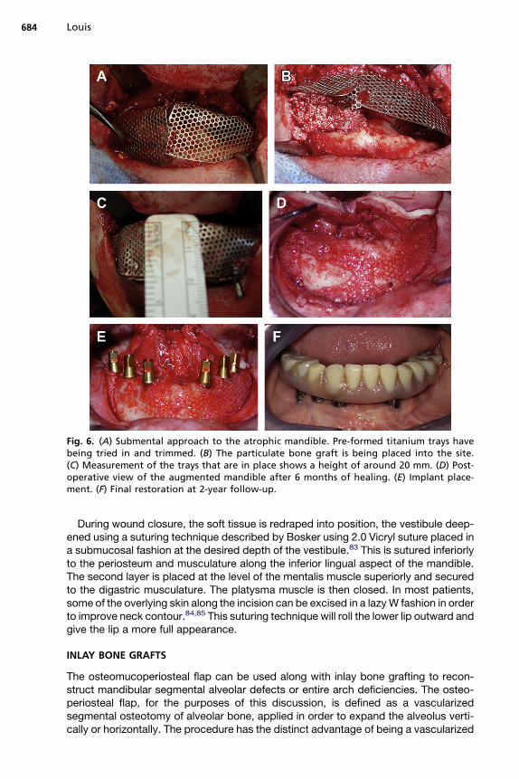
Fig. 6. (A) Submental approach to the atrophic mandible. Pre-formed titanium trays havebeing tried in and trimmed. (B) The particulate bone graft is being placed into the site.(C) Measurement of the trays that are in place shows a height of around 20 mm. (D) Post-operative view of the augmented mandible after 6 months of healing. (E) Implant place-ment. (F) Final restoration at 2-year follow-up.
Louis684
During wound closure, the soft tissue is redraped into position, the vestibule deep-ened using a suturing technique described by Bosker using 2.0 Vicryl suture placed ina submucosal fashion at the desired depth of the vestibule.83 This is sutured inferiorlyto the periosteum and musculature along the inferior lingual aspect of the mandible.The second layer is placed at the level of the mentalis muscle superiorly and securedto the digastric musculature. The platysma muscle is then closed. In most patients,some of the overlying skin along the incision can be excised in a lazyW fashion in orderto improve neck contour.84,85 This suturing technique will roll the lower lip outward andgive the lip a more full appearance.
INLAY BONE GRAFTS
The osteomucoperiosteal flap can be used along with inlay bone grafting to recon-struct mandibular segmental alveolar defects or entire arch deficiencies. The osteo-periosteal flap, for the purposes of this discussion, is defined as a vascularizedsegmental osteotomy of alveolar bone, applied in order to expand the alveolus verti-cally or horizontally. The procedure has the distinct advantage of being a vascularized
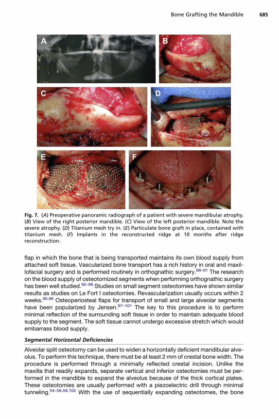
Fig. 7. (A) Preoperative panoramic radiograph of a patient with severe mandibular atrophy.(B) View of the right posterior mandible. (C) View of the left posterior mandible. Note thesevere atrophy. (D) Titanium mesh try in. (E) Particulate bone graft in place, contained withtitanium mesh. (F) Implants in the reconstructed ridge at 10 months after ridgereconstruction.
Bone Grafting the Mandible 685
flap in which the bone that is being transported maintains its own blood supply fromattached soft tissue. Vascularized bone transport has a rich history in oral and maxil-lofacial surgery and is performed routinely in orthognathic surgery.86–91 The researchon the blood supply of osteotomized segments when performing orthognathic surgeryhas been well studied.92–96 Studies on small segment osteotomies have shown similarresults as studies on Le Fort I osteotomies. Revascularization usually occurs within 2weeks.95,96 Osteoperiosteal flaps for transport of small and large alveolar segmentshave been popularized by Jensen.97–101 The key to this procedure is to performminimal reflection of the surrounding soft tissue in order to maintain adequate bloodsupply to the segment. The soft tissue cannot undergo excessive stretch which wouldembarrass blood supply.
Segmental Horizontal Deficiencies
Alveolar split osteotomy can be used to widen a horizontally deficient mandibular alve-olus. To perform this technique, there must be at least 2 mm of crestal bone width. Theprocedure is performed through a minimally reflected crestal incision. Unlike themaxilla that readily expands, separate vertical and inferior osteotomies must be per-formed in the mandible to expand the alveolus because of the thick cortical plates.These osteotomies are usually performed with a piezoelectric drill through minimaltunneling.54–56,58,102 With the use of sequentially expanding osteotomes, the bone
Louis686
can be forced buccally. Interposition (inlay) bone grafts with either banked or autoge-nous bone can be placed to keep the segments separated. If the segment is unstable,then a small miniplate can be placed for stabilization. Closure can be performed withor without a resorbable membrane.
Segmental Vertical Defects
Segmental vertical osteotomies are performed through a vestibular incision. Areleasing incision is not needed. Soft tissue reflection is only enough to perform theosteotomy (Fig. 8). The osteotomy has both vertical and horizontal components.The horizontal component of the osteotomy is placed so that the strength of the basalbone is not significantly compromised and the transport segment is large enough for
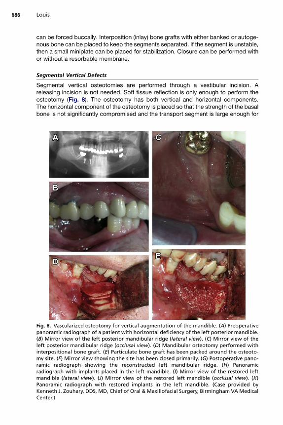
Fig. 8. Vascularized osteotomy for vertical augmentation of the mandible. (A) Preoperativepanoramic radiograph of a patient with horizontal deficiency of the left posterior mandible.(B) Mirror view of the left posterior mandibular ridge (lateral view). (C) Mirror view of theleft posterior mandibular ridge (occlusal view). (D) Mandibular osteotomy performed withinterpositional bone graft. (E) Particulate bone graft has been packed around the osteoto-my site. (F) Mirror view showing the site has been closed primarily. (G) Postoperative pano-ramic radiograph showing the reconstructed left mandibular ridge. (H) Panoramicradiograph with implants placed in the left mandible. (I) Mirror view of the restored leftmandible (lateral view). (J) Mirror view of the restored left mandible (occlusal view). (K)Panoramic radiograph with restored implants in the left mandible. (Case provided byKenneth J. Zouhary, DDS, MD, Chief of Oral & Maxillofacial Surgery, Birmingham VAMedicalCenter.)

Fig. 8. (continued)
Bone Grafting the Mandible 687
stabilization if needed. The vertical arms of the osteotomy connect the outer aspect ofthe horizontal osteotomy to the crest of the alveolus. There are usually two verticalarms, placed on either side of the planned alveolar segment that will be transported.The vertical arms should slightly converge toward the base of the transport segment.Once the bony cuts are made, the segment is moved to the desired level. Block orparticulate inlay graft is used to hold the segment in the desired position. If particulategraft is used without a miniplate for stabilization, a mineralized bone graft material ispreferable. If additional stability is needed, a miniplate can be placed. Since the inci-sion is made in the vestibule, a single layered tension free closure is usually easilyachieved.
Vertical Deficiency of the Mandibular Arch
Total mandibular osteotomy for correction of vertical deficiency of the mandibleshould be performed with caution. In cases of mandibular atrophy, an osteotomymay weaken the mandible and predispose it to fracture. Although absolute minimumheight that must be present before considering this technique has not established,there must be enough bone to safely perform the osteotomy. The procedure is per-formed through a vestibular incision. Once the mental nerves have been identifiedand protected, the dissection is carried to the level of the planned osteotomy. Minimal
Louis688
periosteal reflection is performed. The planned osteotomy is performed with a piezo-electric drill, staying above the inferior alveolar nerve posterior to the mental foramen.Once the osteotomy is complete, the alveolar segment is moved superiorly to thedesired height. The segment is stabilized with miniplates and interpositional bonegraft. A tension-free closure can be performed with resorbable suture (eg, Vicryl orMonocryl).
POSTOPERATIVE CARE
Postoperative care for bone grafting treatment includes use of antibiotics such asamoxicillin or clindamycin for 7 to 10 days. Patients are placed on a chlorhexadinemouth rinse for at least the first 2 to 4 weeks. If a wound dehiscence develops, thepatient is maintained on chlorhexadine mouth rinse until the second-stage surgery.If exposure of hardware occurs, appropriate cleaning should also include use ofa soft toothbrush to remove any plaque or debris and irrigation. Follow-up visits areusually at 2 weeks, 1 month, and 3 months postoperatively. Temporization variesprimarily based on if the patient is partially dentate. Most patients with some teethcan undergo immediate temporization using a partially or completely tooth-born pros-thesis. Edentulous patients could undergo immediate temporization by relining anexisting denture. However, to avoid pressure on the wound, in patients where hard-ware has been place, a period of rest is usually indicated to minimize the risk of wounddehiscence. The temporization could be delayed 2 to 4 weeks. In patients with atro-phic mandibles that have undergone complete or segmental osteotomies, a strict non-chew diet for 6 to 8 weeks is recommended to minimize the risk of fracture. The
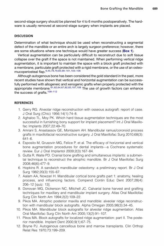
Box 1
Recommended options for defect reconstruction
I. Horizontal segmental defects
Onlay block graft with rigid membrane
Onlay particulate graft with membrane
Vascularized osteotomy with mineralized or block inlay graft
II. Horizontal deficiency of the entire arch
Onlay block graft with membrane
Onlay particulate graft with rigid membrane
Onlay particulate mineralized graft with membrane
Vascularized osteotomy with mineralized or block inlay graft
III. Vertical segmental defects
Onlay block graft with membrane
Onlay particulate graft with rigid membrane
Vascularized osteotomy with mineralized or block inlay graft
Vascularized osteotomy with inlay graft and rigid stabilization
IV. Vertical deficiency of the entire arch
Onlay block graft with membrane
Onlay particulate graft with rigid membrane
Vascularized osteotomy with inlay graft and rigid stabilization
Bone Grafting the Mandible 689
second-stage surgery should be planned for 4 to 6 months postoperatively. The hard-ware is usually removed at second-stage surgery when implants are placed.
DISCUSSION
Determination of what technique should be used when reconstructing a segmentaldefect of the mandible or an entire arch is largely surgeon preference; however, thereare some situations where one technique would have greater success (Box 1).Vertical augmentation can be particularly difficult to reconstruct due to soft tissue
collapse over the graft if the space is not maintained. When performing vertical ridgeaugmentation, it is important to maintain the space with a block graft protected witha membrane, particulate graft protected with a rigid membrane, or the use of an osteo-mucoperiosteal flap.9,45,78,79,83,99–101,103–106
Although autogenous bone has been considered the gold standard in the past, morerecent studies have shown that vertical and horizontal augmentation can be success-fully performed with allogeneic and xenogenic grafts when properly protected with theappropriate membrane.61,62,64,67,82,83,107,108 The use of growth factors can enhancethe success of grafts.109–112
REFERENCES
1. Gerry RG. Alveolar ridge reconstruction with osseous autograft: report of case.J Oral Surg (Chic) 1956;14(1):74–8.
2. Aghaloo TL, Moy PK. Which hard tissue augmentation techniques are the mostsuccessful in furnishing bony support for implant placement? Int J Oral Maxillo-fac Implants 2007;22:49–70.
3. Amrani S, Anastassov GE, Montazem AH. Mandibular ramus/coronoid processgrafts in maxillofacial reconstructive surgery. J Oral Maxillofac Surg 2010;68(3):641–6.
4. Esposito M, Grusovin MG, Felice P, et al. The efficacy of horizontal and verticalbone augmentation procedures for dental implants—a Cochrane systematicreview. Eur J Oral Implantol 2009;2(3):167–84.
5. Gutta R, Waite PD. Cranial bone grafting and simultaneous implants: a submen-tal technique to reconstruct the atrophic mandible. Br J Oral Maxillofac Surg2008;46(6):477–9.
6. Hopkins R. A sandwich mandibular osteotomy: a preliminary report. Br J OralSurg 1982;20(3):155–67.
7. Aalam AA, Nowzari H. Mandibular cortical bone grafts part 1: anatomy, healingprocess, and influencing factors. Compend Contin Educ Dent 2007;28(4):206–12 [quiz: 13].
8. Donovan MG, Dickerson NC, Mitchell JC. Calvarial bone harvest and graftingtechniques for maxillary and mandibular implant surgery. Atlas Oral MaxillofacSurg Clin North Am 1994;2(2):109–22.
9. Pikos MA. Atrophic posterior maxilla and mandible: alveolar ridge reconstruc-tion with mandibular block autografts. Alpha Omegan 2005;98(3):34–45.
10. Pikos MA. Mandibular block autografts for alveolar ridge augmentation. AtlasOral Maxillofac Surg Clin North Am 2005;13(2):91–107.
11. Pikos MA. Block autografts for localized ridge augmentation: part II. The poste-rior mandible. Implant Dent 2000;9(1):67–75.
12. Boyne PJ. Autogenous cancellous bone and marrow transplants. Clin OrthopRelat Res 1970;73:199–209.

Louis690
13. Cushing M. Autogenous red marrow grafts: their potential for induction of osteo-genesis. J Periodontol 1969;40(8):492–7.
14. Dragoo MR, Sullivan HC. A clinical and histological evaluation of autogenousiliac bone grafts in humans. I. Wound healing 2 to 8 months. J Periodontol1973;44(10):599–613.
15. Rivault AF, Toto PD, Levy S, et al. Autogenous bone grafts: osseous coagulumand osseous retrograde procedures in primates. J Periodontol 1971;42(12):787–96.
16. Buchman SR, Ozaki W. The ultrastructure and resorptive pattern of cancellousonlay bone grafts in the craniofacial skeleton. Ann Plast Surg 1999;43(1):49–56.
17. Ozaki W, Buchman SR. Volume maintenance of onlay bone grafts in the cranio-facial skeleton: microarchitecture versus embryologic origin. Plast ReconstrSurg 1998;102(2):291–9.
18. Ozaki W, Buchman SR, Goldstein SA, et al. A comparative analysis of the micro-architecture of cortical membranous and cortical endochondral onlay bonegrafts in the craniofacial skeleton. Plast Reconstr Surg 1999;104(1):139–47.
19. Zins JE, Whitaker LA. Membranous versus endochondral bone: implications forcraniofacial reconstruction. Plast Reconstr Surg 1983;72(6):778–85.
20. Zins JE, Whitaker LA. Membranous vs endochondral bone autografts: implica-tions for craniofacial reconstruction. Surg Forum 1979;30:521–3.
21. Clavero J, Lundgren S. Ramus or chin grafts for maxillary sinus inlay and localonlay augmentation: comparison of donor site morbidity and complications. ClinImplant Dent Relat Res 2003;5(3):154–60.
22. Felice P, Iezzi G, Lizio G, et al. Reconstruction of atrophied posterior mandiblewith inlay technique and mandibular ramus block graft for implant prostheticrehabilitation. J Oral Maxillofac Surg 2009;67(2):372–80.
23. Hoppenreijs TJ, Nijdam ES, Freihofer HP. The chin as a donor site in earlysecondary osteoplasty: a retrospective clinical and radiological evaluation.J Craniomaxillofac Surg 1992;20(3):119–24.
24. Precious DS, Smith WP. The use of mandibular symphyseal bone in maxillofacialsurgery. Br J Oral Maxillofac Surg 1992;30(3):148–52.
25. Booij A, Raghoebar GM, Jansma J, et al. Morbidity of chin bone transplantsused for reconstructing alveolar defects in cleft patients. Cleft Palate CraniofacJ 2005;42(5):533–8.
26. Park HD, Min CK, Kwak HH, et al. Topography of the outer mandibular symphy-seal region with reference to the autogenous bone graft. Int J Oral MaxillofacSurg 2004;33(8):781–5.
27. Chiapasco M, Abati S, Romeo E, et al. Clinical outcome of autogenous boneblocks or guided bone regeneration with e-PTFE membranes for the reconstruc-tion of narrow edentulous ridges. Clin Oral Implants Res 1999;10(4):278–88.
28. Gellrich NC, Held U, Schoen R, et al. Alveolar zygomatic buttress: a new donorsite for limited preimplant augmentation procedures. J Oral Maxillofac Surg2007;65(2):275–80.
29. Drew H, Zweig B. Use of a buccal exostosis autograft for alveolar ridge augmen-tation: an aid to implant placement. J N J Dent Assoc 2007;78(3):40–2.
30. Tolstunov L. Maxillary tuberosity block bone graft: innovative technique andcase report. J Oral Maxillofac Surg 2009;67(8):1723–9.
31. Moraes Junior EF, Damante CA, Araujo SR. Torus palatinus: a graft option foralveolar ridge reconstruction. Int J Periodontics Restorative Dent 2010;30(3):283–9.
Bone Grafting the Mandible 691
32. Diaz-Romeral-Bautista M, Manchon-Miralles A, Asenjo-Cabezon J, et al. Autog-enous calvarium bone grafting as a treatment for severe bone resorption in theupper maxilla: a case report. Med Oral Patol Oral Cir Bucal 2010;15(2):e361–5.
33. Gutta R, Waite PD. Outcomes of calvarial bone grafting for alveolar ridge recon-struction. Int J Oral Maxillofac Implants 2009;24(1):131–6.
34. Cranin AN, Demirdjan E, DiGregorio R. A comparison of allogeneic and autog-enous iliac monocortical grafts to augment the deficient alveolar ridge ina canine model. I. Clinical study. J Oral Implantol 2003;29(3):124–31.
35. Crespi R, Vinci R, Cappare P, et al. Calvarial versus iliac crest for autologousbone graft material for a sinus lift procedure: a histomorphometric study. Int JOral Maxillofac Implants 2007;22(4):527–32.
36. FeliceP,Marchetti C, IezziG,et al. Vertical ridgeaugmentationof theatrophicposte-rior mandible with interpositional bloc grafts: bone from the iliac crest vs. bovineanorganic bone. Clinical and histological results up to one year after loading froma randomized–controlled clinical trial. ClinOral Implants Res 2009;20(12):1386–93.
37. Bogdan S, Nemeth Z, Huszar T, et al. The proximal tibia. A possible donor site inpreprosthetic surgery. Fogorv Sz 2008;101(2):58–63.
38. Bogdan S, Nemeth Z, Huszar T, et al. Comparison of postoperative complica-tions following bone harvesting from two different donor sites for autologousbone replacement (hip bone and proximal epiphysis of the tibia). Orv Hetil2009;150(7):305–11.
39. Catone GA, Reimer BL, McNeir D, et al. Tibial autogenous cancellous bone asan alternative donor site in maxillofacial surgery: a preliminary report. J OralMaxillofac Surg 1992;50(12):1258–63.
40. Marchena JM, Block MS, Stover JD. Tibial bone harvesting under intravenoussedation: morbidity and patient experiences. J Oral Maxillofac Surg 2002;60(10):1151–4.
41. O’Keeffe RM Jr, Riemer BL, Butterfield SL. Harvesting of autogenous cancellousbone graft from the proximal tibial metaphysis. A review of 230 cases. J OrthopTrauma 1991;5(4):469–74.
42. Pensler J, McCarthy JG. The calvarial donor site: an anatomic study incadavers. Plast Reconstr Surg 1985;75(5):648–51.
43. Moreira-Gonzalez A, Papay FE, Zins JE. Calvarial thickness and its relation tocranial bone harvest. Plast Reconstr Surg 2006;117(6):1964–71.
44. Al-Sebaei MO, Papageorge MB, Woo T. Technique for in-office cranial bone har-vesting. J Oral Maxillofac Surg 2004;62:120–2.
45. Salvato G, Agliardi E. Calvarial bone grafts in severe maxillary atrophy: prepros-thetic surgery with sedation. Implant Dent 2007;16(4):356–61.
46. Iturriaga MT, Ruiz CC. Maxillary sinus reconstruction with calvarium bone graftsand endosseous implants. J Oral Maxillofac Surg 2004;62(3):344–7.
47. Kao SY, Yeung TC, Chou IC, et al. Reconstruction of the severely resorbed atro-phic edentulous ridge of the maxilla and mandible for implant rehabilitation:report of a case. J Oral Implantol 2002;28(3):128–32.
48. Marx RE. Bone harvest from the posterior ilium. Atlas Oral Maxillofac Surg ClinNorth Am 2005;13(2):109–18.
49. Marx RE, Morales MJ. Morbidity from bone harvest in major jaw reconstruction:a randomized trial comparing the lateral anterior and posterior approaches tothe ilium. J Oral Maxillofac Surg 1988;46(3):196–203.
50. Beirne OR. Comparison of complications after bone removal from lateral andmedial plates of the anterior ilium for mandibular augmentation. Int J Oral Max-illofac Surg 1986;15(3):269–72.

Louis692
51. Falkensammer N, Kirmeier R, Arnetzl C, et al. Modified iliac bone harvesting—morbidity and patients’ experience. J Oral Maxillofac Surg 2009;67(8):1700–5.
52. Nkenke E, Weisbach V, Winckler E, et al. Morbidity of harvesting of bone graftsfrom the iliac crest for preprosthetic augmentation procedures: a prospectivestudy. Int J Oral Maxillofac Surg 2004;33(2):157–63.
53. Schaaf H, Lendeckel S, Howaldt HP, et al. Donor site morbidity after bone har-vesting from the anterior iliac crest. Oral Surg Oral Med Oral Pathol Oral RadiolEndod 2010;109(1):52–8.
54. Dibart S, Sebaoun JD, Surmenian J. Piezocision: a minimally invasive, periodon-tally accelerated orthodontic tooth movement procedure. Compend ContinEduc Dent 2009;30(6):342–4, 346, 348–50.
55. Sohn DS, Lee JS, An KM, et al. Piezoelectric internal sinus elevation (PISE) tech-nique: a newmethod for internal sinus elevation. Implant Dent 2009;18(6):458–63.
56. Lee HJ, Ahn MR, Sohn DS. Piezoelectric distraction osteogenesis in the atrophicmaxillary anterior area: a case report. Implant Dent 2007;16(3):227–34.
57. Moon JW, Choi BJ, Lee WH, et al. Reconstruction of atrophic anterior mandibleusing piezoelectric sandwich osteotomy: a case report. Implant Dent 2009;18(3):195–202.
58. Sohn DS, Ahn MR, Lee WH, et al. Piezoelectric osteotomy for intraoral harvest-ing of bone blocks. Int J Periodontics Restorative Dent 2007;27(2):127–31.
59. Quereshy FA, Dhaliwal HS, El SA, et al. Resorbable screw fixation for cortical on-lay bone grafting: a pilot study with preliminary results. J Oral Maxillofac Surg2010;68(10):2497–502.
60. Burger BW. Use of ultrasound-activated resorbable poly-D-L-lactide pins (Son-icPins) and foil panels (Resorb-X) for horizontal bone augmentation of the maxil-lary and mandibular alveolar ridges. J Oral Maxillofac Surg 2010;68(7):1656–61.
61. Barone A, Varanini P, Orlando B, et al. Deep-frozen allogeneic onlay bone graftsfor reconstruction of atrophic maxillary alveolar ridges: a preliminary study.J Oral Maxillofac Surg 2009;67(6):1300–6.
62. Dori S, Peleg M, Barnea E. Alveolar ridge augmentation with hip corticocancel-lous allogenic block graft prior to implant placement. Refuat Hapeh Vehashi-nayim 2008;25(3):28–38, 54.
63. Petrungaro PS, Amar S. Localized ridge augmentation with allogenic blockgrafts prior to implant placement: case reports and histologic evaluations.Implant Dent 2005;14(2):139–48.
64. Waasdorp J, Reynolds MA. Allogeneic bone onlay grafts for alveolar ridgeaugmentation: a systematic review. Int J Oral Maxillofac Implants 2010;25(3):525–31.
65. Askari A, Amato F, Abitbol T. The regenerative potential of ePTFE membranesand freeze-dried bone allografts—two case reports. Periodontal Clin Investig1994;16(2):20–4.
66. Becker W, Hujoel P, Becker BE. Effect of barrier membranes and autologousbone grafts on ridge width preservation around implants. Clin Implant Dent Re-lat Res 2002;4(3):143–9.
67. Beitlitum I, Artzi Z, Nemcovsky CE. Clinical evaluation of particulate allogeneicwith and without autogenous bone grafts and resorbable collagen membranesfor bone augmentation of atrophic alveolar ridges. Clin Oral Implants Res 2010;21(11):1242–50.
68. Chen CC, Wang HL, Smith F, et al. Evaluation of a collagen membrane with andwithout bone grafts in treating periodontal intrabony defects. J Periodontol 1995;66(10):838–47.
Bone Grafting the Mandible 693
69. Chiapasco M, Zaniboni M. Clinical outcomes of GBR procedures to correct peri-implant dehiscences and fenestrations: a systematic review. Clin Oral ImplantsRes 2009;20(Suppl 4):113–23.
70. Fugazzotto PA. GBR using bovine bone matrix and resorbable and nonresorb-able membranes. Part 1: histologic results. Int J Periodontics Restorative Dent2003;23(4):361–9.
71. Gielkens PF, Bos RR, Raghoebar GM, et al. Is there evidence that barriermembranes prevent bone resorption in autologous bone grafts during the heal-ing period? A systematic review. Int J Oral Maxillofac Implants 2007;22(3):390–8.
72. Gielkens PF, Schortinghuis J, de Jong JR, et al. The influence of barriermembranes on autologous bone grafts. J Dent Res 2008;87(11):1048–52.
73. Hammerle CH, Jung RE, Yaman D, et al. Ridge augmentation by applying bio-resorbable membranes and deproteinized bovine bone mineral: a report oftwelve consecutive cases. Clin Oral Implants Res 2008;19(1):19–25.
74. Rasmusson L, Meredith N, Kahnberg KE, et al. Effects of barrier membranes onbone resorption and implant stability in onlay bone grafts. An experimentalstudy. Clin Oral Implants Res 1999;10(4):267–77.
75. Stavropoulos F, Dahlin C, Ruskin JD, et al. A comparative study of barriermembranes as graft protectors in the treatment of localized bone defects. Anexperimental study in a canine model. Clin Oral Implants Res 2004;15(4):435–42.
76. Cordaro L, Amade DS, Cordaro M. Clinical results of alveolar ridge augmenta-tion with mandibular block bone grafts in partially edentulous patients prior toimplant placement. Clin Oral Implants Res 2002;13(1):103–11.
77. Gutta R, Baker RA, Bartolucci AA, et al. Barrier membranes used for ridgeaugmentation: is there an optimal pore size? J Oral Maxillofac Surg 2009;67(6):1218–25.
78. Louis PJ, Gutta R, Said-Al-Naief N, et al. Reconstruction of the maxilla andmandible with particulate bone graft and titanium mesh for implant placement.J Oral Maxillofac Surg 2008;66(2):235–45.
79. von Arx T, Hardt N, Wallkamm B. The TIME technique: a new method for local-ized alveolar ridge augmentation prior to placement of dental implants. Int JOral Maxillofac Implants 1996;11(3):387–94.
80. von Arx T, Hardt N, Wallkamm B. The TIME technic. Local osteoplasty withmicro-titanium mesh (TIME) for alveolar ridge augmentation. Schweiz Mon-atsschr Zahnmed 1995;105(5):650–63.
81. von Arx T, Kurt B. Implant placement and simultaneous periimplant bone graft-ing using a microtitanium mesh for graft stabilization. Int J Periodontics Restor-ative Dent 1998;18(2):117–27.
82. Torres J, Tamimi F, Alkhraisat MH, et al. Platelet-rich plasma may prevent tita-nium–mesh exposure in alveolar ridge augmentation with anorganic bovinebone. J Clin Periodontol 2010;37(10):943–51.
83. Artzi Z, Dayan D, Alpern Y, et al. Vertical ridge augmentation using xenogenicmaterial supported by a configured titanium mesh: clinicohistopathologic andhistochemical study. Int J Oral Maxillofac Implants 2003;18(3):440–6.
84. Bosker H, van Dijk L. The transmandibular implant: a 12-year follow-up study.J Oral Maxillofac Surg 1989;47(5):442–50.
85. Bosker H, Wardle ML. Muscular reconstruction to improve the deterioration offacial appearance and speech caused by mandibular atrophy: technique andcase reports. Br J Oral Maxillofac Surg 1999;37(4):277–84.
Louis694
86. Byrne RP, Hinds EC. The ramus “C” osteotomy with body sagittal split. J OralSurg 1974;32(4):259–63.
87. Dupont C, Ciaburro TH, Prevost Y. Simplifying the Le Fort I type of maxillary os-teotomy. Plast Reconstr Surg 1974;54(2):142–7.
88. Freihofer HP Jr. The lip profile after correction of retromaxillism in cleft and non-cleft patients. J Maxillofac Surg 1976;4(3):136–41.
89. Steinhauser EW. Advancement of the mandible by sagittal ramus split andsuprahyoid myotomy. J Oral Surg 1973;31(7):516–21.
90. Steinhauser EW. Variations of Le Fort II osteotomies for correction of midfacialdeformities. J Maxillofac Surg 1980;8(4):258–65.
91. Tessier P. Treatment of facial dysmorphias in craniofacial dysostosis, Crouzon’sand Apert’s diseases. Total osteotomy and sagittal displacement of the facialmassive. Faciostenosis, sequelae of Lefort 3 fracture. Dtsch Zahn Mund Kiefer-heilkd Zentralbl Gesamte 1971;57(9):302–20.
92. Bell WH. Revascularization and bone healing after anterior maxillary osteotomy:a study using adult rhesus monkeys. J Oral Surg 1969;27(4):249–55.
93. Bell WH, Fonseca RJ, Kenneky JW, et al. Bone healing and revascularizationafter total maxillary osteotomy. J Oral Surg 1975;33(4):253–60.
94. Bell WH, Levy BM. Revascularization and bone healing after maxillary corticot-omies. J Oral Surg 1972;30(9):640–8.
95. Bell WH, Schendel SA, Finn RA. Revascularization after surgical repositioning ofone-tooth dento-osseous segments. J Oral Surg 1978;36(10):757–65.
96. Quejada JG, Kawamura H, Finn RA, et al. Wound healing associatedwith segmental total maxillary osteotomy. J Oral Maxillofac Surg 1986;44(5):366–77.
97. Jensen OT. Alveolar segmental sandwich osteotomies for posterior edentulousmandibular sites for dental implants. J Oral Maxillofac Surg 2006;64(3):471–5.
98. Jensen OT. Distraction osteogenesis and its use with dental implants. Dent Im-plantol Update 1999;10(5):33–6.
99. Jensen OT, Bell W, Cottam J. Osteoperiosteal flaps and local osteotomies foralveolar reconstruction. Oral Maxillofac Surg Clin North Am 2010;22(3):331–46, vi.
100. Jensen OT, Kuhlke L, Bedard JF, et al. Alveolar segmental sandwich osteotomyfor anterior maxillary vertical augmentation prior to implant placement. J OralMaxillofac Surg 2006;64(2):290–6.
101. Jensen OT, Mogyoros R, Owen Z, et al. Island osteoperiosteal flap for alveolarbone reconstruction. J Oral Maxillofac Surg 2010;68(3):539–46.
102. Peivandi A, Bugnet R, Debize E, et al. Piezoelectric osteotomy: applications inperiodontal and implant surgery. Rev Stomatol Chir Maxillofac 2007;108(5):431–40.
103. Maestre-Ferrin L, Boronat-Lopez A, Penarrocha-Diago M. Augmentation proce-dures for deficient edentulous ridges, using onlay autologous grafts: an update.Med Oral Patol Oral Cir Bucal 2009;14(8):e402–7.
104. Gongloff RK, Cole M, Whitlow W, et al. Titanium mesh and particulate cancellousbone and marrow grafts to augment the maxillary alveolar ridge. Int J Oral Max-illofac Surg 1986;15(3):263–8.
105. Roccuzzo M, Ramieri G, Spada MC, et al. Vertical alveolar ridge augmentationby means of a titanium mesh and autogenous bone grafts. Clin Oral ImplantsRes 2004;15(1):73–81.
106. Aparicio C, Jensen OT. Alveolar ridge widening by distraction osteogenesis:a case report. Pract Proced Aesthet Dent 2001;13(8):663–8 [quiz: 70].
Bone Grafting the Mandible 695
107. Fontana F, Santoro F, Maiorana C, et al. Clinical and histologic evaluation of allo-geneic bone matrix versus autogenous bone chips associated with titanium-reinforced e-PTFE membrane for vertical ridge augmentation: a prospectivepilot study. Int J Oral Maxillofac Implants 2008;23(6):1003–12.
108. Pieri F, Corinaldesi G, Fini M, et al. Alveolar ridge augmentation with titaniummesh and a combination of autogenous bone and anorganic bovine bone:a 2-year prospective study. J Periodontol 2008;79(11):2093–103.
109. Jung RE, Thoma DS, Hammerle CH. Assessment of the potential of growthfactors for localized alveolar ridge augmentation: a systematic review. J Clin Pe-riodontol 2008;35(Suppl 8):255–81.
110. Herford AS, Boyne PJ, Williams RP. Clinical applications of rhBMP-2 in maxillo-facial surgery. J Calif Dent Assoc 2007;35(5):335–41.
111. Zhang H, Zhou X, Wang X. Mandibular ridge augmentation by sandwich osteot-omy and BMP-HA implantation. Zhonghua Kou Qiang Yi Xue Za Zhi 1997;32(1):37–9.
112. Jung RE, Windisch SI, Eggenschwiler AM, et al. A randomized–controlled clin-ical trial evaluating clinical and radiological outcomes after 3 and 5 years ofdental implants placed in bone regenerated by means of GBR techniqueswith or without the addition of BMP-2. Clin Oral Implants Res 2009;20(7):660–6.
113. Montazem A, Valauri DV, St-Hilaire H, et al. The mandibular symphysis asa donor site in maxillofacial bone grafting: a quantitative anatomic study.J Oral Maxillofac Surg 2000;58(12):1368–71.
114. Gungormus M, Yilmaz AB, Ertas U, et al. Evaluation of the mandible as an alter-native autogenous bone source for oral and maxillofacial reconstruction. J IntMed Res 2002;30(3):260–4.
115. Gungormus M, Yavuz MS. The ascending ramus of the mandible as a donor sitein maxillofacial bone grafting. J Oral Maxillofac Surg 2002;60(11):1316–8.
116. Li KK, Schwartz HC. Mandibular body bone in facial plastic and reconstructivesurgery. Laryngoscope 1996;106(4):504–6.
117. Choung PH, Kim SG. The coronoid process for paranasal augmentation in thecorrection of midfacial concavity. Oral Surg Oral Med Oral Pathol Oral RadiolEndod 2001;91(1):28–33.