Embed Size (px)
Citation preview
COPROPHILOUS FUNGAL COMMUNITY OF WILDRABBIT IN A PARK OF A HOSPITAL (CHILE):
A TAXONOMIC APPROACH
(Comunidades fúngicas coprófilas de conejos silvestres en un parque de un Hospital (Chile): un enfoque taxonómico)
Eduardo Piontelli, L, Rodrigo Cruz, C & M. Alicia Toro .S.M.Universidad de Valparaíso, Escuela de Medicina
Cátedra de micología, Casilla 92 V Valparaíso, Chile. e-mail <eduardo.piontelli@ uv.cl >
Key words: Coprophilous microfungi,wild rabbit, hospital zone, Chile.Palabras clave: Microhongos coprófilos, conejos silvestres, zona de hospital, Chile
RESUMEN
Durante los años 2005-2006 se efectuó un estudiode las comunidades fúngicas coprófilos en excementos deconejos silvestres en un parque de un hospital regional(V Región, Chile), colectándose 21 muestras en 7 mesesen 2 períodos estacionales (fríos y cálidos). Un total de60 especies y 44 géneros fueron detectados en el períodode muestreo, 46 especies en los períodos cálidos y 39 enlos fríos. La distribución de los grandes grupos fue:Zygomycota(11,6 %), Ascomycota (50 %), géneros mitos-póricos asociados (36,8 %) y Basidiomycota (1,6 %).Los géneros de ascomycetes con mayor frecuencia de es-pecies fueron: Chaetomium (5), Saccobolus y Sordaria(3) Podospora, Schizothecium y Sporormiella (2).Lasespecies constantes y dominantes fueron: Chaetomiumcuniculorum, Ch. murorum, Coprinus spp., Iodophanuscarneus, Pilaira anomala, Schizothecium tetrasporum,Sordaria humana, Sporormiella teretispora y Stilbellafimetaria. Bajo un enfoque taxonómico orientado haciala mayoría de los hongos meiosporicos (Ascomycota), sedestaca la presencia de nuevos registros para Chile:Fusariella hughesii, Neurospora caulospora, Pleuro-ascus nicholsonii, Rhytidospora bispora, Saccobolusglobuliferellus, entre otros.
INTRODUCTION
Coprophilous fungi are usually associated withherbivore dung which may serve as a growth factor formany kinds of microorganisms which as a whole (thattogether) play an important role in the ecosystem, reciclingthe nutrients contained in animal faeces. Fungal dung
Boletín Micológico Vol. 21 : 1 - 17 2006
ABSTRACT
During year 2005-through 2006 a study on copro-philous fungal communities present in wild rabbit dungwas carried out in the park of a regional hospital (VRegion, Chile), 21 samples in seven months under twoseasonable periods (cold and warm) being collected.Sixty species and 44 genera as a total were recorded inthe sampling period, 46 species in warm periods and 39in the cold ones. Major groups were arranged as follows:Zygomycota (11,6 %), Ascomycota (50 %), associatedmitosporic genera (36,8 %) and Basidiomycota (1,6 %).Ascomycetes genera having the highest occurrence were:Chaetomium (5), Saccobolus and Sordaria (3) Podos-pora, Schizothecium and Sporormiella (2). Constant anddominant species were : Chaetomium cuniculorum, Ch.murorum, Coprinus spp., Iodophanus carneus, Pilairaanomala, Schizothecium tetrasporum, Sordaria huma-na, Sporormiella teretispora and Stilbella fimetaria.Under a taxonomic approach focused on the majority ofmeiosporic fungi (Ascomycota), the presence of newrecords of fungi for Chile is emphasized: Fusariellahughesii, Neurospora caulospora, Pleuroascusnicholsonii, Rhytidospora bispora, Saccobolusglobuliferellus, among others.
inhabitant is a diverse and interesting special groupmember including all classes of the Kingdom fungi rangingfrom the Zygomycota, Ascomycota (and their relatives)to Basidiomycota (Furuya & Udagawa, 1972;Wicklow,1981; Valdoserra & Guarro, 1992; Richardson, 2001). Theiruniversal distribution is influenced by the presence ofherbivores in an area, types of vegetation, kind of dung,climatic conditions and latitudinal environmental gra-
1
Coprophilous fungal community of wild rabbit in a park of a hospital - E. Piontelli et al.
dients. However many of these coprophilous fungi havea wide ecological range and low preferences for particularherbivore dung substrate (Lunqvist 1972; Richardson,1972; Wicklow, 1975; Tisdall & Oades, 1982; Caretta et al.,1994).
Coprophilous fungi are popular subjects for study,but little work has been done in Chile in their particulargeographic zone, in which diversity and distribution ofrare species and undescribed fungi, new for science, areapparently common (Spegazzini, 1921; Lazo, 1979;Undagawa,1980; Piontelli et al., 1981-1997; Muroi &Undagawa, 1984; Valdoserra & Guarro, 1988, 1994).
Some of these authors focused on special taxono-mic groups from many habitats and type of dung, howeverscarce data are available from places where dung of wildrabbits are highly affected by human activity such ahospital zone.
The aim of this work was to obtain data about therichness in species of coprophilous fungi (especiallyAscomycota and related taxa) in dung of these Lago-morphs, through a seven- month study in an anthropo-genic soil in a park of a hospital, and some comments onthe taxonomic and phylogenetic literature contributionrelated to the most detected meiosporic fungi.
MATERIALS AND METHODS
a) Basic characteristics of place. The Peñablanca Hospital(V Región, Chile, 33º S.Lat. and 72º 30’Long. W, at 30 kmfrom Valparaiso city), is enclosed in a 20 hectare park. Thevegetation consists of several grasses (mainly species ofAvena, Lolium, Cynodon, Hordeum, Bromus and Poa) anda wooded area having mostly species of: Pinus, Acacia,Eucalyptus, Populus, Cupressus, Jubaea and Peumus. Thecity (Villa Alemana) has about 100.000 inhabitants with atypical template warm climate, with winter rainfall (360mma year) and a prolonged dry station (7-8 month).b) Samples and identification. A total of 21 samples werecollected in sterile plastic bags from 3 different sites in a200 m2 area, since May 2005 through April 2006 (May,June, July, August, January, March and April), 3 sampleseach month. In the laboratory the samples were placed(duplicate) in one layer in 6 sterile Petri dishes (10 cm) withtwo sheets of sterile moist paper towels on their bases.Before incubation plates were kept at -20º C for 24 hr toavoid the presence of acari. Each plate was then incubatedat 19-25º C room temperature under natural day and nightregimes for 40 days. Substrate moisture was maintainedby the periodic addition of sterile distilled water (as neces-sary). Cultures were examined under a dissectingmicroscope for fungal fructification at 5,10,20,30,40-dayintervals.
Developed fungi were transferred to glass slides
and stained with lactophenol cotton blue and sealed withnail-varnish before their observation at low and highmagnification. Some microfungi were further cultivated onspecial identification media according to specific mono-graphies. Species occurring more than once over the 6Petri dishes were counted as one occurrence (onlypresence - absence). To get an approximate quantitativeestimate on the occurrence frequency of a taxa in eachsample, the former was marked with one or several signs(x): low presence(x), medium (xx) and high presence (xxx).Most morphometric data on Ascomycota were recordeddirectly on samples collected from the coprophiloussubstrate by using water or lactophenol preparations. Foridentification of fungi, the following literature was generallyused: van Brumelen, 1967; Mirza & Cain, 1969; Ellis, 1971-76; Lundqvist, 1972; Bezerra & Kimbrough, 1975; Bell,1983-2005; Seifert et als, 1983; Ellis & Ellis,1988; Domsch et al.,1993 (see further references in text for more details).
RESULTSA total of 60 species and 44 genera collected from
dung of wild rabbits (Oryctolagus cuniculi) were recordedin the sampling period, the latter being distributed underthe following major taxonomic groups: Zygomycota (11,6%), Ascomycota (50 %), associated mitosporic genera(36,8 %) and Basidiomycota (1,6 %) (Table 1). Ascomy-cetes genera having highest frequency of species were:Chaetomium (5) , Saccobolus and Sordaria (3)Podospora, Schizothecium and Sporormiella (2). Amongthe 14 genera present in cold months, Chaetomiumtrigonosporum, Cheilimenia sp., Neurospora calospora(Fig.2,7-2,7a), Podospora appendiculata, Sporormiellateretispora (Fig. 2,10) and Trichodelitschia munkii (Fig.1, 6), were the only genera developed, whereas out of the15 genera present in warm months: Coprotus sexde-cimsporus (Fig.1,3), Lasiobolus intermedius (Fig.2,8),Rhytidospora bispora (Fig.1,2), Saccobolus globu-liferellus (Fig.3,15) y Thelebolus stercoreus (Fig. 3,13),did developed only in that period (Table 1). Among theexisting taxonomic groups, 46 species were detected inwarm climates and 39 in cold months.
Constant and dominant species present among 5to 7 within the sampling months were: Chaetomium cuni-culorum (Fig.3,16 -3, 16a), Ch. murorum, Coprinus spp.,Iodophanus carneus, Pilaira anomala, Schizotheciumtetrasporum (Fig 2,11-2,11a), Sordaria humana (Fig.3,17-3,17a), Sporormiella teretispora and Stilbella fimetaria(Table 1).
Frequent species (among 3 to 5 within the samplingmonths) were mainly ascomycetes: Ascobolus sp. (Fig.1,5),Chaetomium elatum, C. bostrichodes, Gymnoascusreessii (Fig. 1,4), Pleuroascus nicholsonii (Fig. 2,9-2,9a),
2
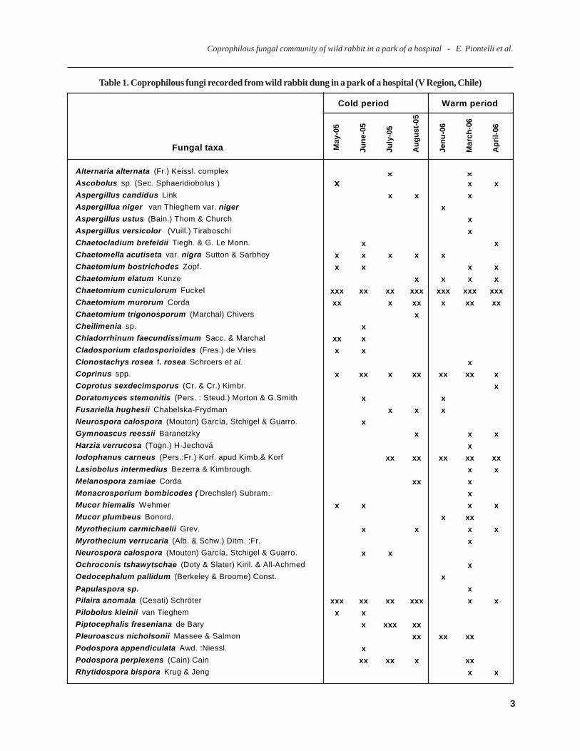
Table 1. Coprophilous fungi recorded from wild rabbit dung in a park of a hospital (V Region, Chile)
Cold period Warm period
Fungal taxa May
-05
June
-05
July
-05
Aug
ust-0
5
Jenu
-06
Mar
ch-0
6
Apr
il-06
Alternaria alternata (Fr.) Keissl. complex x x
Ascobolus sp. (Sec. Sphaeridiobolus ) x x xAspergillus candidus Link x x xAspergillua niger van Thieghem var. niger xAspergillus ustus (Bain.) Thom & Church xAspergillus versicolor (Vuill.) Tiraboschi xChaetocladium brefeldii Tiegh. & G. Le Monn. x xChaetomella acutiseta var. nigra Sutton & Sarbhoy x x x x xChaetomium bostrichodes Zopf. x x x xChaetomium elatum Kunze x x x xChaetomium cuniculorum Fuckel xxx xx xx xxx xxx xxx xxxChaetomium murorum Corda xx x xx x xx xxChaetomium trigonosporum (Marchal) Chivers xCheilimenia sp. xChladorrhinum faecundissimum Sacc. & Marchal xx xCladosporium cladosporioides (Fres.) de Vries x xClonostachys rosea f. rosea Schroers et al. xCoprinus spp. x xx x xx xx xx xCoprotus sexdecimsporus (Cr. & Cr.) Kimbr. xDoratomyces stemonitis (Pers. : Steud.) Morton & G.Smith x xFusariella hughesii Chabelska-Frydman x x xNeurospora calospora (Mouton) García, Stchigel & Guarro. xGymnoascus reessii Baranetzky x x xHarzia verrucosa (Togn.) H-Jechová xIodophanus carneus (Pers.:Fr.) Korf. apud Kimb.& Korf xx xx xx xx xxLasiobolus intermedius Bezerra & Kimbrough. x xMelanospora zamiae Corda xx xMonacrosporium bombicodes ( Drechsler) Subram. xMucor hiemalis Wehmer x x x xMucor plumbeus Bonord. x xxMyrothecium carmichaelii Grev. x x x xMyrothecium verrucaria (Alb. & Schw.) Ditm. :Fr. xNeurospora calospora (Mouton) García, Stchigel & Guarro. x xOchroconis tshawytschae (Doty & Slater) Kiril. & All-Achmed xOedocephalum pallidum (Berkeley & Broome) Const. xPapulaspora sp. xPilaira anomala (Cesati) Schröter xxx xx xx xxx x xPilobolus kleinii van Tieghem x xPiptocephalis freseniana de Bary x xxx xxPleuroascus nicholsonii Massee & Salmon xx xx xxPodospora appendiculata Awd. :Niessl. xPodospora perplexens (Cain) Cain xx xx x xxRhytidospora bispora Krug & Jeng x x
Coprophilous fungal community of wild rabbit in a park of a hospital - E. Piontelli et al.
3
Coprophilous fungal community of wild rabbit in a park of a hospital - E. Piontelli et al.
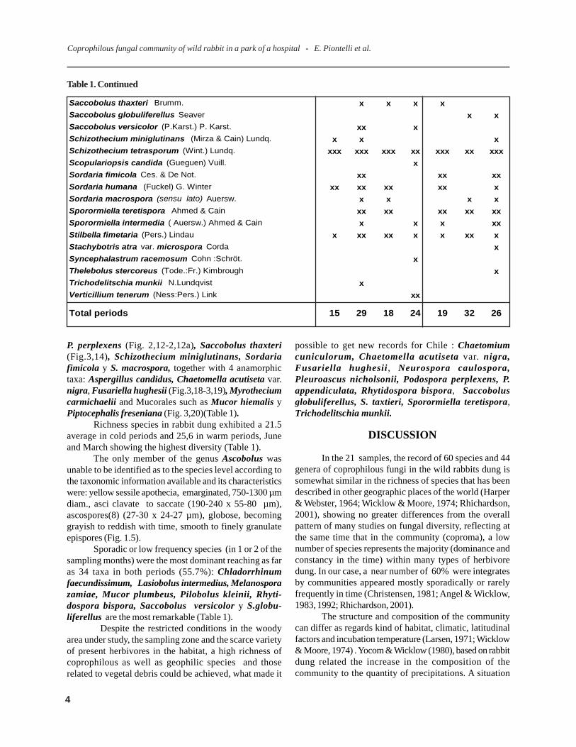
Table 1. Continued
P. perplexens (Fig. 2,12-2,12a), Saccobolus thaxteri(Fig.3,14), Schizothecium miniglutinans, Sordariafimicola y S. macrospora, together with 4 anamorphictaxa: Aspergillus candidus, Chaetomella acutiseta var.nigra, Fusariella hughesii (Fig.3,18-3,19), Myrotheciumcarmichaelii and Mucorales such as Mucor hiemalis yPiptocephalis freseniana (Fig. 3,20)(Table 1).
Richness species in rabbit dung exhibited a 21.5average in cold periods and 25,6 in warm periods, Juneand March showing the highest diversity (Table 1).
The only member of the genus Ascobolus wasunable to be identified as to the species level according tothe taxonomic information available and its characteristicswere: yellow sessile apothecia, emarginated, 750-1300 µmdiam., asci clavate to saccate (190-240 x 55-80 µm),ascospores(8) (27-30 x 24-27 µm), globose, becominggrayish to reddish with time, smooth to finely granulateepispores (Fig. 1.5).
Sporadic or low frequency species (in 1 or 2 of thesampling months) were the most dominant reaching as faras 34 taxa in both periods (55.7%): Chladorrhinumfaecundissimum, Lasiobolus intermedius, Melanosporazamiae, Mucor plumbeus, Pilobolus kleinii, Rhyti-dospora bispora, Saccobolus versicolor y S.globu-liferellus are the most remarkable (Table 1).
Despite the restricted conditions in the woodyarea under study, the sampling zone and the scarce varietyof present herbivores in the habitat, a high richness ofcoprophilous as well as geophilic species and thoserelated to vegetal debris could be achieved, what made it
possible to get new records for Chile : Chaetomiumcuniculorum, Chaetomella acutiseta var. nigra,Fusariella hughesii, Neurospora caulospora,Pleuroascus nicholsonii, Podospora perplexens, P.appendiculata, Rhytidospora bispora, Saccobolusglobuliferellus, S. taxtieri, Sporormiella teretispora,Trichodelitschia munkii.
DISCUSSION
In the 21 samples, the record of 60 species and 44genera of coprophilous fungi in the wild rabbits dung issomewhat similar in the richness of species that has beendescribed in other geographic places of the world (Harper& Webster, 1964; Wicklow & Moore, 1974; Rhichardson,2001), showing no greater differences from the overallpattern of many studies on fungal diversity, reflecting atthe same time that in the community (coproma), a lownumber of species represents the majority (dominance andconstancy in the time) within many types of herbivoredung. In our case, a near number of 60% were integratesby communities appeared mostly sporadically or rarelyfrequently in time (Christensen, 1981; Angel & Wicklow,1983, 1992; Rhichardson, 2001).
The structure and composition of the communitycan differ as regards kind of habitat, climatic, latitudinalfactors and incubation temperature (Larsen, 1971; Wicklow& Moore, 1974) . Yocom & Wicklow (1980), based on rabbitdung related the increase in the composition of thecommunity to the quantity of precipitations. A situation
Saccobolus thaxteri Brumm. x x x xSaccobolus globuliferellus Seaver x xSaccobolus versicolor (P.Karst.) P. Karst. xx xSchizothecium miniglutinans (Mirza & Cain) Lundq. x x xSchizothecium tetrasporum (Wint.) Lundq. xxx xxx xxx xx xxx xx xxxScopulariopsis candida (Gueguen) Vuill. xSordaria fimicola Ces. & De Not. xx xx xxSordaria humana (Fuckel) G. Winter xx xx xx xx xSordaria macrospora (sensu lato) Auersw. x x x xSporormiella teretispora Ahmed & Cain xx xx xx xx xxSporormiella intermedia ( Auersw.) Ahmed & Cain x x x xxStilbella fimetaria (Pers.) Lindau x xx xx x x xx xStachybotris atra var. microspora Corda xSyncephalastrum racemosum Cohn :Schröt. xThelebolus stercoreus (Tode.:Fr.) Kimbrough xTrichodelitschia munkii N.Lundqvist xVerticillium tenerum (Ness:Pers.) Link xx
Total periods 15 29 18 24 19 32 26
4
that in our case did not exhibit major differences in dominantand constant species yet a higher relative quantity in warmmonths than in the cold ones (Table 1), noting that rainperiod appears generally at the end of autumn and in winterseason. However, in warm periods, common morning mistscan supply a suitable environmental humidity in thesampling area. Richness of species seems to be alwaysmore abundant in lagomorphs dung than in ruminants(Richardson, 2001;Wicklow et al., 1980), but this situationis closely related mainly to the number of samples togetherwith the patient and constant observation of the incubatedplates. In our study we did not pretend to make aquantitative assessment of diversity and species richnessbut rather a primary estimate on a substrate that has notbeen seemingly studied in the country.
When considering single taxa by groups, everymember of Zygomycetes fructified within the first week ofobservation, and Pilaria anomala proved to be the mostconstant and dominant species of the group ( Wood &Cooke, 1987), mainly in cold periods. In temperate zones,frequency of Piptocephalidaceae species (Mucoraceae,according to Benny, 2005) is common in rabbit dung, whileChaetocladiaceae species (Mucoraceae, according toBenny 2005) were much less frequent (Richardson,2005),a situation that was in agreement with our study. In thehorse dung, Piontelli et al. (1981) found only similarities inthe presence and frequency of P. kleinii, Ch. brefeldiiand M . hiemalis, whereas Pilaria anomala was absentin that substrate.
Ascomycota species and mitosporic fungi were thebiggest group of species in this substrate and we willfocus our discussion mainly on the former by analyzingthem as to the occurrence of gymnothecial, cleistothecial,perithecial and apothecial types.
The presence of Gymnoascus reessii gymnothecia(Eurotiomycetes, Onygenales, Gymnoascaceae), wasdetected late within 15 to 20 days, and coincides with thecolonization in different kinds of faeces and types of soilsrich in organic matter (together with other Gymnoascales)in many geographic regions (Domsch, et al , 1993; Currah,1985, Delgado et al., 2001). Molecular analyses onGymnoascaceae family correspond undoubtedly to apolyphiletic group included within the Onygenales(Sugiyama et al., 1999-2002; Guarro & Cano, 2002) andtaxonomic approaches designed at present are viewingGymnoascus under a broader concept (Sole et al. 2002),tending to a synonymization of various taxa that had beenconsidered as valid according to Currah’ s approach (1985). This situation while not completely approved nowadays,needs a further sequence analysis on multiple genes datafor every member of this family.
Among taxa forming cleistothecial ascomata wemust take into account the presence of two species rarely
found in the literature and which exhibit an even uncertainposition within the Ascomycota, like Pleuroascusnicholsonii (¿Leotiomycetes, Pseudoeurotiaceae?) andRhytidospora bispora (Sordariomycetes, Helotiales,¿Ceratostomataceae?) Both taxa made their appearancewithin 10-15 day incubation of the substrate.
P.nicholsonii, is seemingly the most knownspecies of the genus and which shows a cosmopolitandistribution, even though its findings are rarely found inthe literature. It has been firstly isolated in England fromGuinea pig dung (1901), in USA from rat woodrat (cf.Malloch & Benny, 1973), from rabbit dung in Spain, ( Bar-rasa & Moreno,1984) and in Australia from an unde-terminate substrate (Bell, 2005). Our isolation does notdiffer morphologically from what has been described inthe literature and it was characterized by having apseudoparenquymatous peridium, blackish and brownascoma together with hyaline spiralate appendages ontheir walls, 8 globose ascospores without germinative pore(2,39-3,5 µm) with a 3.15µm average and with the presenceof filaments which break down as enlarged to subgloboseconidia, sometimes taking the shape of arthroconidia(Fig.2,9a). This species has presented different taxonomichistories with time but they have not been clearly definedyet, from its first inclusion in the Onygenaceae,Amauroascaceae or joint to the Arachnomyces speciesbut they were not closely related to perithecial Eurotiales,but closely to the so-called discomycetes andloculoascomycetes, according to Suh & Blackwell (1999).These authors change the concept of the Pseudoerotiaceaefamily and reduce the number of their members, whileGernandt et al. (2001), grouped the Pseudoeurotiaceae (inincertae sedis), next to the basal Helotiales, at aconsiderable distance from the Eurotiales, a situation thatcoincides with Gibas et al. (2002), who join P. nicholsoniito Leotiomycetes, and far from Arachnomyces species.Sogonov et al.(2005), comment also briefly in thePseudoeurotiaceae the relationship between the closelyrelated species of Connersia rilstonii with P. nicholsonii(both with similar anamorphs).
Rhytidospra bispora, is one of the five species ofthe genus since its description by Jeng & Cain (1977). Itshabitat seems to be strictly coprophilous, but its raredetection in distant geographic zones does not allow toconfirm this situation: it was isolated in Mexico from cowdung by Krug & Jeng (1979), and it occurred likewise inSpain with the same type of dung by Valdoserra et al.(1991) and during this study with rabbit dung in the centralzone of Chile. Our findings in a habitat dispersed by rodentsand not by ruminants may indicate scarse selectivity inthe case of a determinate kind of substrate. It showed nomorphological differences from the two descriptionsappearing in the literature. Superficial ascomata,
Coprophilous fungal community of wild rabbit in a park of a hospital - E. Piontelli et al.
5
Coprophilous fungal community of wild rabbit in a park of a hospital - E. Piontelli et al.
cephalothecoid, non ostiolate globose (250-310 µm diam)yellowish to brown. Unitunicate asci,two spored,subglobose to elliptic 13- 17 x10-12 µm ; pitted ascospore,one celled, elipsoidal with a germ pore at each end.
The taxonomic position of this genus (Pezizomy-cotina, Sordariomycetes, Hypocreales, Ceratostomataceae)has not been clearly determined in the literature andEricsson (2006), considers it as incertae sedis.
Valdoserra & Guarro, (1994), described a newspecies of the genus (R. citriformis) in the coastal zone ofnorthern Chile (Pisagua) in cow dung. Both species arequite similar, with two spored asci, yet they differ in sizeand shape of ascospores; ellipsoid with truncate ends inR. bispora and limoniform in R. citriformis.
Among genera that gave rise to the formation ofperithecioid ascomata within 6 to 10 days, members of theSordariales Order (Pezizomycotina, Sordariomycetes sensustricto), a taxon recently analysed molecularly by Huhndorf& Miller (2004), wherein only genera within Chaetomia-ceae, Lasiosphaeriaceae and Sordariaceae families areretained among the redefined Sordariales. These threefamilies are significant because of the high frequency oftheir members in our research.
The highest presence of species corresponded toChaetomium (Chaetomiaceae), being dominant andfrequent in most of the samples. Its species are isolatedfrom a great variety of substrata: plant remains, queratine,feathers, seeds, dried spices, textiles, paper, cotton fibres,soil and indoor air (C. murorum, C.globosum, C. elatum),yet they seem to prefer sources containing cellulose; thisis why they are frequently found in herbivore dung whichare abundant in these carbonate nutrients as well as witha high nitrogen content. Although members of the taxonare not considered true coprophilous, but they do exhibittheir competitive, defensive (mycotoxic) and metabolicabilities in this substrate.
C.globosum and other species of the genus producesterigmatocystin,chaetoglobosin and chaetomin, amongother toxins. (Sekita et al. 1981). Chaetomium bostrichodesseems to bear effective chemical defenses against fungus-feeding arthropods since sciarid larvae (Lycoriella mali)avoided their ascomata, but this was not the case with theother fungi coexisting in rabbit dung (Wicklow, 1988, inWiklow, 1992). Other species can rarely be agent ofopportunistic mycoses (De Hoog et al, 2000; Sigler &Verweij, 2003).
Among the present species, it is worth to point outChaetomium cuniculorum and Ch. murorum, the formerbeing common in rabbit dung and the latter in diversefaeces (Bell, 1983, 2005), but we have found no referenceson C. cuniculorum in national research papers. In Chile,Piontelli et al. (1981), isolated frequently Ch. murorum
and C.elatum in equine dung, being these the only speciesmatching with those from the rabbit dung in ratherneighboring geographic zones, whereas Piontelli & Grixiolli(1997) in the patagonic zone and Lazo (1979) in the centralzone failed to isolate anyone member of this taxon inbovine and equine dung.
The literature on genus is large and it is condensedin the modern taxonomic treatment by von Arx, Guarro &Figueras (1986), which we employ in our determinations,together with the always useful monography by Ames(1963) and the recent and beautiful illustrated guide byBell (2005). Lee & Hanlin (1999), study the phylogeneticrelationships of Chaetomium and similar genera basedon ribosomal DNA sequences, and Huhndorf & Miller(2004) the molecular system of the Sordariales andespecially the Lasiophaeriaceae family.
Two species of Podospora and two ofSchizothecium were identified among the members ofLasiosphaeriaceae, S.tetrasporum showing overalldominance and constancy in time. Podospora is awidespread genus abundant in species (almost 80, Kirk etal, 2001) and which exhibits a great polymorphism , mainlyin the structure of ascomata wall and ascosporeappendages, having in mind that other taxa in the familybear similar characteristics to Podospora (Arnium,Cercophora, Strattonia, Tripterospora and Zopfiella).The genus is considered as one of the most frequentcolonizers on diverse kinds of herbivore dung besides ofhaving mostly coprophilous species, together with someof them present in plant material, seeds or soil, as it can beseen in various taxonomic papers in the literature(Mirza &Cain, 1969; Lundqvist, 1972; Krug & Khan, 1989; Bell &Mahoney, 1995; Doveri, 1998, Stchigel et al, 2002; Bell,2005, among others ).
Schizothecium, is a genus similar to Podosporaand both of them have been treated in time like synonymsdue to its lack of morphological characters that are notsufficient for its separation (Furuya & Undagawa, 1972;Krug & Khan, 1989; Bell & Mahoney, 1995; Doveri, 1998,Lorenzo & Havrylenko, 2001; Stchigel et al, 2002; Bell,2005, among others). In its complicated history as rega-rds nomenclature, Scizothecium revives thanks toLundqvist‘s investigation (1972), who recognizes 17species which are identified by the following charac-teristics: swollen agglutinated hairs on the ascomata, lackof filiform interascal paraphyses, early stage of septationof ascospore and persistent pedicel.
Most mycologists did not accept separation ofthese two genera (all those included above) as well as Bell& Mahoney (1995) that in their revision of Podosporaspecies with swollen agglutinated parithecial hairs,retained P.aloides, P.conica, P. curvuloides, P. dakotensis,P.miniglutinans, P. tetraspora and P. vesticola in the
6
genus rather than in Schizothecium, as it is Lundqvist ‘sthought (1972).
However, Cai et al. (2005), in a phylogenetic studyof multiple gene sequences, focused mainly in therelationships of Schizothecium and Podospora, and in areevaluation of the taxonomic significance of morpholo-gical character of Schizothecium species having thegeneral characteristics below: perithecia ornamented withswollen agglutinated hair or prominent protruding peridialcell, cylindrical to clavate asci usually without an apicalring, ascospore with a large brown cell, an small hyalinepedicel with or without gelatinous appendages and withphialidic anamorphs. They also observed that every strainunder study is grouped in a strong supported mono-phyletic clade and concluded that they must have theirown generic condition rather than being treated ascongeneric to Podospora. However there is still a certainsuperposition of morphological characters, between thesetwo genera in asci and ascospore what makes separationof both taxa difficult. (Bell & Mahoney,1995). Notwith-standing, the actual phylogenic analysis indicate thatascomatal morphologies in Sordariales are morephylogenetically informative than ascospore characters,host or habitat association (Cai et al, 2005; Miller &Huhndorf, 2005).
Schizothecium species can be considered mostlycoprophilous (with mucilaginous appendages) yet someof them are non-coprophilous (soil, plants or otherhabitats) and they lack these appendages. Cai et al. (2005),based on the literature, added a listing in their paper anda nomenclature of 24 Schizothecium species bearing sevennew combinations thus making valid Lundqvist ‘sinvestigation (1972) and suggested the need to completehis descriptions in future studies. Based on this latelycontribution, we separated two species from Podosporaand included them in Schizothecium (S. tetrasporum andS. miniglutinans). S. tetrasporum (≡ Podosporatetraspora) was not isolated in Santiago and Linares (Lazo,1979), nor in the patagonic zone (except for S. miniglu-tinans that resulted dominant), although it was indeedpresent in equine dung in the V Region (Piontelli et al.1981).These two seemingly cosmopolitan species seem to adaptthemselves quite well to diverse climates. Krug & Khan(1989) state that S. tetrasporum is a species feasible to befound in temperate zones and at a high altitude (Mirza &Cain, 1969; Udagawa & Kobayasi, 1979). BothSchizothecium species were also cited by Lorenzo &Havrylenko (2001) for Argentina.
The two Podospora species (P. appendiculata andP. perpexens), have not been seemingly recorded in Chile.The former did not show any morphological differencefrom descriptions made by Mirza and Cain, 1969 andLundqvist 1972, whereas in the latter, ascospores show
their dark primary cell slightly longer than what it isdescribed by these two authors, yet matching more closelyto Bell ‘s findings ( 2005) in Australia (40-47 x 19-23 um)(x=44,3 x 21,6)
Among Sordariaceae it is noteworthy the case ofSordaria, a quite coprophilous genus, yet some speciesare common too in the soil, seeds and vegetal detritus.Three species were frequent in both sampling periods; S.fimicola, S. humana and S. macrospora (sensu lato), agroup of cosmopolitan species very much related one tothe other and very common in dung of several herbivores(being rabbit dung one of them) under temperate zones(Lundqvist, 1972). S. fimcola did not show any significantmorphometric difference from the literature (Moreau, 1953;Lundqvist, 1972; Guarro & von Arx, 1987; Richardson &Watling, 1997), whereas S. humana, exhibited certainproblems due to the different measures in ascospores,mainly in width since average measures exceeded standardfigures, what left it far from S. fimicola and made it closeto S. lappae (14-15 µm wide). S. humana, is described ina different genus for the fact of lacking unsheathedascospores under the name of Asordaria humana (Guarro& von Arx, 1987), but Asordaria is considered at presenta synonym of Sordaria (Kirk et al. 2001). S. macrospora(sensu lato) showed ascospores with a 31.5x20-21umaverage having some intermediate measures with itsneighboring species S. superba (26,4 x 16,1µm). This iswhy we prefer to keep all analized preparations under thefirst name..
S. macrospora and S .fimicola were firstly isolatedin horse dung (Piontelli et al.,1981) in the country, exceptfor the patagonic zone(Piontelli & Grixolli, 1997).
Another sordariaceae member having scarcepresence was Neurospora calospora (=Gelasinosporacalospora). Neurospora species have been found mostlyin the moist tropics and subtropics as a primary colonizerof trees and shrubs killed by wildfires, yet they also appearin diverse soils and in the bark of many trees expandingsignificantly the known geographic range and habitats ofthe genus. Colonization occurred beneath the bark ofdiverse deciduous and conifer hosts. Ascosporemorphology is the character for the distinction betweenthe genera Neurospora and Gelasinospora, however itwas not an accurate predictor of phylogeneticrelationships as inferred from the sequence data analyzed(Dettman et al. 2001). García et al .(2004), based onultrastructural and 28S rDNA sequence data, included theGelasinospora species in Neurospora and describedthe new genus Pseudogelasinospora to match toGelasinospora amorphoporcata. The similarity of bothgenera has been earlier recognized in the literature(Dowding, 1933; von Arx, 1982) together with anothergenus similar to Gelasinospora (a non-ostiolated
Coprophilous fungal community of wild rabbit in a park of a hospital - E. Piontelli et al.
7
Coprophilous fungal community of wild rabbit in a park of a hospital - E. Piontelli et al.
counterpart) Anixiella Saito (Cain, 1961), which wasrefused by von Arx (1982). In our research work we doaccept the inclusion of Gelasinospora in Neurospora,however in a recent paper, Cai et al (2006), in a phylogeneticinvestigation of Sordariaceae based in multiple genesequences and morphology, agreed with previous studiesthat heterothallic Neurospora are monophyletic eventhough homothallic ones may have a multiple origin,considering there is insufficient evidence to place currentlyaccepted Gelasinospora and Neurospora species withinthe same genus. It seems that history of both genera needsfurther study.
Sporormiaceae (Dothideomycetes, Pleosporales),characterized by fissitunicate asci, dark brown septatespores with germ slit, included several genera verycommon on dung and morphologically very similar tounclear circunscription (Arenal et al, 2004; Doveri, 2004;Nyberg, 2005). Recently Kruys et al. (2005), in aphylogenetic relationship of coprophilous Pleosporalesshowed that Delitschiaceae, Sporormiaceae, Zopfiaceaeand Testudinaceae families form a monophyletic group,yet they must be retained as separate families since theyfail to have a significant support. Sporormiella ,Sporormia and Preussia are considered highly relatedgenera and they are bound to be confused by theirmorphological features. Sporormiella Ell & Everh., oneof the 8 taxa included within the family by Barr (2000), isthe most abundant in species and its members comemostly from a coprophilous habitat, yet some species (cf.S. intermedia) may occur on wood and soils (Ahmed &Cain, 1972; Guarro et al, 1997; Barr, 2000). The genusincluded species with ostiolate ascomata, asci oblong tocylindrical, short stipitate or sessile, while Preussia itscounterpart, presents ascomata non ostiolate (cleisto-thecioid), asci widely clavate and usually stipitate, it wouldinclude mainly species isolated from debris, wood andsoil. Sporormiella, is generally considered a synonym ofPreussia, a situation disapproved by some authors in theliterature (Valdoserra & Guarro, 1992; Guarro et al, 1997;Arenal et al, 2004).
In a phylogenetic study, Nyberg (2005) has provedthat the presence or absence of the ostiole and a type ofsubstrate is not a useful character for delimiting Preussiaand Sporormiella (Guarro et al. ,1997; von Arx & van derAa, 1987), but the characters of the ascus may beimportant for the circunscription of some species of thisgenera, and suggested to continue treating these asseparate because they do not form a monophiletic group.To this respect Bell (2005) has the same opinion. Thegreatest treatment of the genus belongs to Ahmed & Cain(1972) and that of Furuya & Udagawa(1972) is also useful.However separation of certain species becomes quitedifficult because of the overlap between some of these, a
situation occurring either in the coprophilous substratumas well as in the culture (Arenal et al, 2004; Bell, 2005;Nyberg, 2005).
Two members of the genus Sporormiella (S.intermedia and S. teretispora ,were isolated with a highfrequency and constancy in both periods, the latter beingthe most constant as well as dominant. Determination ofthe preparations designed for S. teretispora revealedstable measures on their asci (182-210 x 30-35 µm) andascospores (60-69 x 11,5-13 µm) what induced us to includeit in this species, following Ahmed & Cain (1972) andKhan & Cain (1979) monographie, whereas S. intermediawas identified because its ascospores were always lesserthan 60um long (48-55 x 9,5-10,5 µm). Considering thefeasible changeability of these two species, it is difficultto ascertain absolutely and only through morphologythat they are the result of variants from a single (S.intermedia), since there is a coincidence in their structuressuch as the germinative furrow generally obliquous, thebiseriate arrangement of ascospores in the asci, and theshape and size of asci and paraphyses. Bell (1983) hadsome difficulties in placing his collection within thesetwo species, because of a possible overlap between them,and in a recent work (Bell, 2005), broadly definedS.intermedia (sensu lato) with a larger ascospore range.(50-) 55-77 x (9-) 10-13 (-14).
S. intermedia is one of the most well-knownspecies in many herbivore dung (Ahmed & Cain, 1972;Ellis & Ellis, 1988; Valdoserra & Guarro, 1990; Bell, 1983,2005) and its distribution along several regions of Chilehas firstly been confirmed by Lazo (1979) and Piontelli etal.(1997), whose measurements on ascospores (48,5- 54,4x 8,8-11,9 µm), do fit the original description. Members ofPleosporales, mainly those of the Sporormiaceae familyplay a significant role in the production of secondarymetabolites having diverse functions, such as fitotoxic,antifungal, antibactericide and cholesterol biosynthesisinhibitors (S. minima). Various antifungal compounds(Terezines A-D) and the fungal metabolite Hialophyrone(Wang et al,1995; Huang & Kaneko, 1996; Hatory et al.,2004). have been isolated from S.teretispora.
Another family within Pleosporales which wasrarely represented included only one member ofPhaeotrichaceae as Trichodelitschia munkii, withperithecia neck ornamented with dark non septate bristles,asci bitunicate-fissitunicate with apical rings andascospore uniseriate, dark, bicellular with prominent polargerm pore (see Lundqvist, 1964; Luck-Allen 1970; Barr2000).
Nyberg (2005), in a phylogenetic relationship ofcoprophilous Pleosporales families grouped with highsupport Trichodelitschia with Phaeotricum (Lundqvist,1964; Barr 2000).There is, as it seems no isolation records
8
of this species in Chile.Within Pezizomycetes, the Pezizales Order is an
interesting group of fungi bearing diverse habitat,appearence, nutritional strategies and ecological behavior(saprophytic, parasitic and ectomycorrhizal), which wasrepresented by three families (Pezizaceae, Ascobolaceaeand Ascodesmiaceae ). Considering its frequency in thetwo climatic periods, a single member of Pezizaceae isnoteworthy as Iodophanus carneus, one of the mostcommon discomycetes found in different types of dungwith a worldwide distribution and having Oedocephalumanamorph in some species. (Kimbrough et al. 1969,Kimbrough, 1982). The genus Iodophanus is a segregateof the coprophilous genus Ascophanus and ischaracterized by the pinkish color of its gregariousapothecia, presence of diffuse amyloidy of asci, hialineelipsoidal ascospores and carotenoid pigments inparaphyses and excipular cells. Dominance of thiscoprophilous species has been earlier recorded in diversechilean geographic zones in other coprophilous substrate(Lazo, 1979; Piontelli et al.,1981; Piontelli & Grixolli, 1997).
In a study of multiple nuclear genes, Pezizaceae issupported as a monophyletic group (Hansen et al, (2005),a situation confirmed also by De Hoog et al. (2005) basedon one gene only, however this family seems not to beclosely related with other families thus it would benecessary to restrict possibly Pezizaceae as the uniquetaxon within Pezizales, as well as to carry out further futureconfirmatory studies which allow at the same time to applynew ordinal names for the other families. It seems advisableto maintain still the current taxonomic situation (Kirk et al,2001; Eriksson, 2006) considering families of the orderunder the concept of Pezizales sensu lato.
The Ascobolaceae family(Pezizomycetes,Pezizales) was represented by members of two generaonly, Ascobolus and Saccobolus. The unique species ofAscobolus had a sporadic occurrence in the two periodsand it was not possible to confirm it at a species level asby the available information. Based on the morphologydescribed under results it can be included in theSphaeridiobolus section because of its close similaritiesto the A.brassicae, species yet having much biggerascospores (26-30µm diam). Ascobolus species havingspherical or subspherical ascospores and described byvan Brumelen (1967) are few in number: A. brassicae, A.crosslandii, A subglobosusus, A.reticulatus, however allof them are under dimension ranks and exhibit a differentepispore (Fig. 1,5). A revision of some of the lately literature(van Brumelen,1980; Dissing, 1989; Wang & van Brumelen1997; Gamundi et al, 2004; Dokmetzian, et al, 2004, 2005),failed to find any similarity to our record which it will beexamined later on.
It is worth while to state that based on recent studies
related to the mechanism of forcible ascospore dischargein A. immersusus, 8 ascospores can be discharged as asingle mucilage-embebbed projectile with a 0,3 Mpa or 3atm. pressure, with a glycerol as a mayor osmolyte (Fischer,et al, 2004).
The other most well-known member of theAscobolaceae family was Saccobolus with 3 species, mostof them showing a low frequency, except for S. taxterii.The genus is cosmopolitan and widely dispersed indifferent coprophilous substrate. The most usefulmonography keeps still being that of van Brumelen (1967),however his number of species has increased at presentin the literature (van Brumelen, 1976; Aas, 1978; Dissing,1989; Wang & van Brumelen 1997). The identification ofspecies (26 according to Kirk et al, 2001) conveys somedifficulties in the sense that it is based on various factorswhich are not sometimes easy to evaluate because theyare very alike: the ascomata color with aging process, thecolor of paraphyses, the arrangement of ascospore in theascus (6 group), all of them linked together in a jelliedmass, change in color of the latter with age (generallyreddish wine) as well as and the shape and ornamentationof those. This situation observed in various papers aboutthe species of the genus (for example Bell, 1983, 2005),could be seen in the most common species that weidentified as S. taxterii, a species related to S. verrucis-porus, and S. versicolor (being the latter a very changea-ble species). We discarded the former either for its coarselypunctate ascospores and fine anastomosing fissure, andthe latter for its more o less smooth and fissuredascospores.
The Ascodesmidaceae was rarely represented andLasiobolus intermedius was the only species of the genusfound. This taxa (operculate discomycetes), with hialinethick-walled bristles on sessile minute apothecia had beenplaced previously in the family Theleboleceae (Kimbrough& Korf , 1967). Lasiobolus is another genus which exhibitshigh changeability in bristle morphology and ascosporearrangement (Bezerra & Kimbrough, 1975); according tovan Brumelen (1998), the asci of Lasiobolus and Coprotusagree with the earlier defined Octospora type. Phylo-genetically Lasiobolus and Ascodesmis, which have beensuggested as closely related to Orbicula and Lasiobo-lidium, are a sister lineage to the Pseudombrophila(Landvik et al.,1998; Hansen et al., 2005).
In our isolations, the ellipsoidal, hyalineascospores (many with Bary bubble) measured 17,5 -19,5x 10,50-12,50 µm (with a 18,5 x 11,5µm average), dimensionsthat do agree with the ranks of species L. cuniculi, L.intermedius y L. ciliatus, however considering otherisolations collected from other substrate in the chileancentral zone and the Patagonia (Piontelli & Grixolli, 1997),where practically the same dimensions resulted, we
Coprophilous fungal community of wild rabbit in a park of a hospital - E. Piontelli et al.
9
Coprophilous fungal community of wild rabbit in a park of a hospital - E. Piontelli et al.
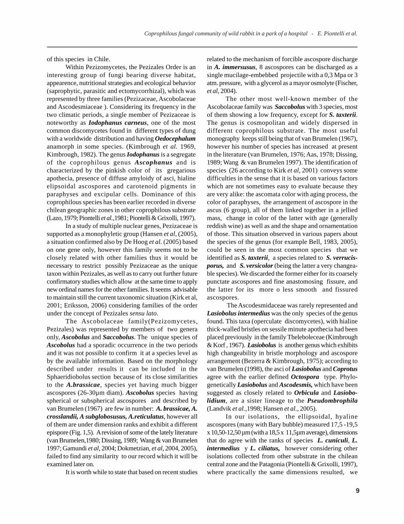
Figure 1.- 1,1-1,2. Rhytidospora bispora, 1,1. Ascoma peridium and ascospores. 1-2. Bispored asci. 1,3. Coprotussexdecimsporus, asci and ascospores. 1,4. Gymnoascus reesii, peridial gymnothecial hyphae with hooked appendagesand ascospore. 1,5. Ascobolus sp. Asci with round ascospores. 1,6 .Trichodelitschia munkii, bitunicate asci an ascosporeswith 2 polar germ pores (Bar =20 µm).
classified the species as, L. intermedius (?). This decisionbased mainly on Bezerra and Kimbrouugh (1975),nomenclature includes it among those species havingascospores lower than 20µm (in our case they kept alwaysnear to their maximum length limit). Moreover anothermorphological features were taken into account which
proved to be seemingly quite changeable such as thearrangement of ascospores in the ascus (uniseriate orbiseriate), length and width of asci (100-200 x 18-25 µm),the non septate pointed setae, slightly ventricose at theirbases (120-270 x 9-15 µm), the filiform paraphyses and thecells of ectal excipulum. We commented with other authors
10
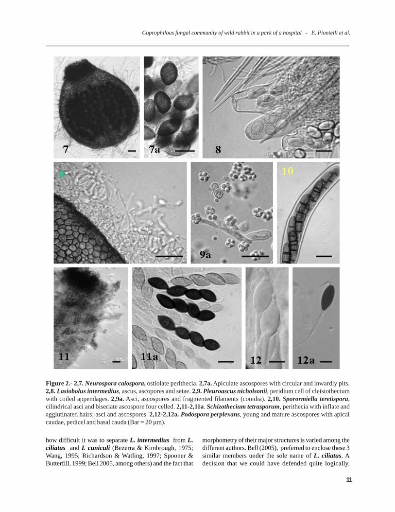
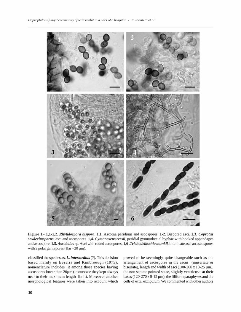
Figure 2.- 2,7. Neurospora calospora, ostiolate perithecia. 2,7a. Apiculate ascospores with circular and inwardly pits.2,8. Lasiobolus intermedius, ascus, ascopores and setae. 2,9. Pleuroascus nicholsonii, peridium cell of cleistotheciumwith coiled appendages. 2,9a. Asci, ascospores and fragmented filaments (conidia). 2,10. Sporormiella teretispora,cilindrical asci and biseriate ascospore four celled. 2,11-2,11a. Schizothecium tetrasporum, perithecia with inflate andagglutinated hairs; asci and ascospores. 2,12-2,12a. Podospora perplexans, young and mature ascospores with apicalcaudae, pedicel and basal cauda (Bar = 20 µm).
how difficult it was to separate L. intermedius from L.ciliatus and L cuniculi (Bezerra & Kimbrough, 1975;Wang, 1995; Richardson & Watling, 1997; Spooner &Butterfill, 1999; Bell 2005, among others) and the fact that
morphometry of their major structures is varied among thedifferent authors. Bell (2005), preferred to enclose these 3similar members under the sole name of L. ciliatus. Adecision that we could have defended quite logically,
Coprophilous fungal community of wild rabbit in a park of a hospital - E. Piontelli et al.
11
Coprophilous fungal community of wild rabbit in a park of a hospital - E. Piontelli et al.
Figure 3.- 3,13. Thelebolus stercoreus, mature ascus and ascospores. 3,14. Saccobolus thaxteri, group of maturespore cluster in asci. 3,15. Saccobolus globuliferellum, asci and spore cluster loosely united. 3,16-3,16a. Chaetomiumcuniculorum, details of perithecium hairs and mature ascospores. 3,17-3,17a. Sordaria humana, mature ascus with 7ascospore (arrow indicate a genetic fusion of 2 acospores); immature ascus tip with apical ring. 3,18-3,19. Fusariellahughesii, cylindrical to narrowly ellipsoid conidia generally 3 septate, and monophialidic conidiogenous cell. 3,20.Piptocephalis freseniana, merosporangia, mitospores and inverted conical head cell (Bar =10 µm).
12
however the small number of ascomata found (only five)together with the information collected from themorphological features analyzed induced us to take thisdecision, that might be considered as well under thebroader term of L.ciliatus group or sensu lato.
Lasiobolus is one of the most frequent disco-mycetes ocurring in ruminant dung (Richardson, 1972),yet a special approach intended for a particular substratumis still missing (Caretta et al, 1998). L. ciliatus, speciallyand L. intermedius, its possible parental species, havebeen isolated from temperate through cold zones, such assouthern soils of the argentinian Patagonia and chileanTierra del Fuego (Lazo, 1979; Piontelli et al, 1981;Valdoserra & Guarro, 1992; Piontelli & Grixolli, 1997;Gamundi et al, 2004).
The two last discomycetes bearing a scarcerepresentation in the substratum were: Coprotus sexde-cimsporus and Thelebolus stercoreus, species that areconsidered by Eriksson (2006) within the Leotiomycetes(Thelebolales, Thelebolaceae), or may be Coprotus withinPyronemataceae, Pezizales (Kirk et al., 2001). It issignificant to remark according to Hambleton & Sigler(2005), that two members of the Myxotrichaceae familyformed a clade with 2 members of the Thelebolaceae(Ascozonus woolhopensis y T. stercoreus ) with a highstatistical support . Based on Currah’s investigation (1994),it is suggested that the Myxotrichaceae might represent adistinct evolutionary line from the members of Onygenales(inoperculate discomycetes) and this distant relationshiphas been confirmed by Sugiyama et al, 1999; Sugiyama &Mikawa, 2001; Hambleton et al.,2005: Jiang & Yao, 2005;Wang et al,. 2006, and others, who relate the taxon withLeotiomycetes (Leotiales and Erysiphales).
Coprotus, is an essentially coprophilousgenus that has a late occurrence in this substratum (20-25days), with light colored small and sessile apothecia freeof setae, eigth to multispored non amyloid asci withhyaline, smooth and ellipsoid ascospores and a conspi-cuous and constant du Bary bubble, were the majorfeatures of the genus. The only species present was C.sexdecimsporus, the morphological characteristics ofwhich agreed with the description of the type species ofthe genus according to Kimbrough et al, (1972). Theseauthors supplied the keys for northamerican species,determined their taxonomic distribution suitable at thatage, together with their similarities to other related or alikegenera such as: Ascophanus, Rhypariobolus, Ascozonusand Coprobia among other. However their operculateasci as well as du Bary bubble in the ascospore allowtheir differentiation from these. Other authors added morespecies to the genus or else described the normal ones indifferent countries (Jeng & Krug, 1977; Aas, 1983; Genéet al., 1993; Wang, 1999; Doveri et al., 2000; Dokmetzian
et al., 2005). C. sexdecimsporus, seems to have nopreference for a particular type of dung (Kimbrough et al,1972) and its presence had been detected earlier in Chile inhorse and cow dung (Lazo, 1979; Piontelli et al. 1981), butthis was also the case also in several places in Argentina,Tierra del Fuego and Antarctic zone (Gamundi et al., 2004;Dokmetzian et al., 2005).
Thelebolus is a morfologically simple and smallcleistohymenial ascomycete with asci containing 8 to over2000 ascospores. Its echology continues being stilluncertain for various species and most of them arepsychrophilic and frequently isolated from the Antarctica(Montemartini et al., 1993; Azmy & Seppelt, 1998; De hooget al., 2005). De Hoog et al.(2005), in a phylogenetic andecological study of Thelebolus in Antarctic zones agreewith the idea of classifying this genus and other relatedsuch as Ascozonus and Caccobius (among others) in aseparate Thelebolales order near the Leotiales in a singlefamily Theleboleceae (Landvik et al., 1998; Kirk et al., 2001),at a certain distance from other discomycetes members ofthe Pezizales. Van Breumelen (1998), reconsiders therelationship within the Thelebolales based on ascusultrastructure, finding at least 3 different types of asci inThelebolus species and maintain the earlier definition ofOctospora type intended for Lasiobolus and Coprotusasci
T. stercoreus, exhibiting 2 to 4 asci together with256 spores (we observed 92 spores in a single plane), isone of the most changeable species within the genus andcapable of bearing an hymenium with a variable number ofasci from 1 to 40 as regards the quantity of ascosporespresent (> number of ascospores < number of asci), asituation which has meant problems for the definition ofspecies in the genus, since the research work performedby Kimbroug & Korf (1967). De Hoog et al. (2005) acceptnowadays only 4 species in the genus, 2 of them new forthe Antarctica while T. stercoreus includes the well-knownT. caninus, T. crustaceus, T. psychrophilous species assynonyms, among others. Its cosmopolitan distributionoccurs essentially in different coprophilous substratecollected from mammals and poultry in cold and temperateregions yet it has never been found in the Antarctica (DeHoog et al., 2005; Gamundi et al., 2004). In Chile,Thelebolus stercoreus has been isolated from cattle andhorse dung in temperate and cold zones (Piontelli et al.,1981; Piontelli & Grixolli, 1997).
We have focused the discussion based on oursecond objective and we will just add a brief comment onsome representative mitosporic fungi related toAscomycota, generally considered only as a small partwithin the dung inhabitants, in a substratum that canbecome a box of surprises as regards change in abundance
Coprophilous fungal community of wild rabbit in a park of a hospital - E. Piontelli et al.
13
Coprophilous fungal community of wild rabbit in a park of a hospital - E. Piontelli et al.
Aas,O. (1983). The genus Coprotus (Pezizales) in Norway. NordicJ. Bot. 3:253-259
Ahmed, S.I. & Cain, R.F.(1972). Revision of the genera Sporormiaand Sporormiella. Can. J, Bot. 50:419-477
Ames, L.M. (1963). A monograph of the Chaetomiaceae. Theunites States Army research and developpmental series.
Angel, K. & Wickow, D.T. (1983). Coprophilous fungal communityin semiarid to mesic grassland. Can. J. Bot. 61:594-602
Arenal, F.; Platas, G.& Pelaez, F. (2004). Variability of sporeleng in some species of genus Preussia (Sporormiella). Mycotaxon89:137-151
Arx, J.A.von. (1982). A key to the species of Gelasinospora.Persoonia 11:443-449
Arx, J.A. von. & Aa, H.A. van der. (1987).Spororminula tenerifegen. et sp. nov. Trans. Br. mycol. Soc 89:117-120
Arx, J.A.von.; Guarro, J. & Figueras, M.J. (1986). TheAscomycete genus Chaetomium. Nova Hedwigia 84:1-161
Azmi, O.R. & Seppelt, R.D. (1998) The broad-scale distributionof microfungi in the Windmill Island region, continental Antarctica.Polar biology 19:92-10
Barr, M.E. (2000). Notes on coprophilous bitunicate ascomycetes.Mycotaxon 76:105-1012
Barrasa, J.M. & Moreno, C. (1984). Hongos copróilos de EspañaIII. Anales Jard. Bot. Madrid . 41:3-17
Bell, A. (1983). Dung Fungi. An Illustrated Guide to CoprophilousFungi in New Zealand. Victoria University Press, Private BagWellington.
Bell, A. (2005). An illustrated guide to the coprophilousAscomycetes of Australia. CBS, Utrecht, The Netherland
Bell, A. & Mahoney, D.P. (1995). Coprophilous fungi i New
REFERENCES
Zealand. I. Podospora species with swollen agglutinated perithecialhairs. Mycologia 87:375-396
Benny, G.L. (2005) Zygomycetes. http:/www.zygomycetes.org/
Bezerra, J.L. & Kimbrough, J.W. (1975). The genus Lasiobolus(Pezizales,Ascomycetes).Can. J. Bot.53:1206-1229
Cai, L,; Jeewon, R & Hyde, K. (2005). Phylogenetic evaluationand taxonomic revision of Schizothecium based on ribosomal FNAand protein code genes. Fungal diversity 19:1-21
Cai, L,; Jeewon, R. & Hyde, K. (2006). Phylogeneticinvestigations of Sordariaceae based eo multiple gene sequence andmorphology, Mycol. Res. 110:137-150
Cain, R.F.(1961). Anixiella and Diplogelasinospora, two generawith clestothecia and pitted ascospores. Can. J. Bot. 39:1667-1677
Caretta, G.; Mangiarotti, A.M. & Piontelli, E. (1994).Coprophilous fungi on horse, goat and sheep dung from Lombardia(Italy). Micologia Italiana 23:11-20
Caretta, G. & Piontelli, E. (1996).Coprophilous fungi fromconfined deers in Pavia (Lombardia, Italy). Boletín Micológico11.41-50
Caretta,G.; Piontelli.E.; Picco.A.M. & Del Frate, G. (1999).Some Filamentous Fungi on Grassland Vegetation from Kenya.Mycopathologia 145:155-169
Caretta, G.; Piontelli, E.; Savino, E. & Bulgheroni, A. (1998).Some coprophilous fungi from Kenya. Mycopathologia 145:125-134
Christensen, M. (1981). Species diversity and dominance in fungalcommunities. In The fungal community: Its organization and rolein the ecosystem (Wicklow, D.T. & Carrol, G.C. eds.), Marcel Dekker,N.York. pp. 201-232
Currah, R.S.(1985). Taxonomy of the Onygenales: Arthroderma-taceae, Gymnoascáceae, Myxotrichaceae and Onygenaceae.
and diversity of this taxa (Seifert et al. 1983), nearly to40% and which matches not only with the kind ofcoprophilous substratum and other physiological orenvironmental variants, but also largely with thecomposition of the vegetation (grasses and leaf litter), thesoil mycota and the kind of antifungal antibiosis in thecoproma community, a situation that we proved as anexample in the case of Stilbella erythrocephala, whichwhen growing, refused the presence of any neighboringmycobiota (Leher et al., 2006). Among the records of themost well known fungal propagules in the litter,Alternaria, Cladosporium and Aspergillus had a poorrepresentation, while some taxa of rare occurrence suchas: Chaetomella acutiseta var. nigra, Clonostachys roseaf. rosea, Fusariella hughesii, Harzia verrucosa,
Ochroconis tshawytschae, among others, could beobserved in herbivore dung (Table 1), taking no accountof those that our observation failed to detect in the lownumber of samples collected (Richardson, 2001recommended to collect 40-50).
From the clinical point of view, diversity of fungalspecies detected in this coprophilous substratumbelonging to the external habitat of a public hospital, isnot seemingly a significant source of dispersion ofpotentially pathogenous fungi for man.
This paper presents a limited range of coprophilousfungi in special habitats affected by anthropogenicactivities and these results are a source of a rich andattractive microfungal community in a selective andbeautiful microcosms of biodiversity.
14
Substrates. Croom Helm, London and Sidney.
Eriksson,O.E.(2006). Outline of Ascomycota. Myconet 12.1-82
Fischer, M.; Cox, J.; Davis, D.J.; Wagner, A.; Taylor, R.;Huerta, A.J.; Money, N.P. (2004). New information on themechanism of forcible ascospore discharge from Ascobolusimmersus. Fungal Genet. Biol. 41.:698-707
Furuya,K. & Udagawa, S-I. (1972). Coprophilous Pyrenomycetesfrom Japan I. J.Gen. Microbiol. 18:433-454
Gamundi, J.I.; Minter, D.W.; Romero, I., A;, Barrera; V. A.;Gaiotti, A.L.; Messuti, M.I.; Stecconi, M. (2004). Checklist ofthe discomycetes (Fungi) od Patagonia, Tierra del Fuego and Adjacentantartic areas. Darwiniana 42:63-164
García, D.; Stchigel, A.M.; Cano, J.; Guarro, J.; Hawksworth,D.L. (2004). A synopsis of re-circumscription of Neurospora (Syn.Gelasinospora) Based on ultrastructural and 28S rDNA sequencesdata. Mycol. Res. 108: 1119-1142
Gené, J.; ElShafie, A.E. & Guiarro, J.(1993). Two newcoprophilous Pezizales from the Sultanate of Oman. Mycotaxon46:275-284
Gernandt,D.S.; Platt,J.L.;Stone, J.K.; Spatafora, J.W.;Holst-Jensen, A.;Hamelin, R.C.; Kohn, L.M. (2001).Phylogenetics of Helotiales and Rhytismatales based on partialsmall subunit nuclear ribosomal DNA sequences.Mycologia:93:915–933.
Gibas, C.F.; Sigler, L.; Summerbell.& Currah, R.S.(2002).Phylogeny of the genus Arachnomyces and its anamorphsand the establishment of Aragnomycetales, a new Eurotiomyceteorder in the Ascomycota. Stud. Mycol. 47:131-139
Guarro, J.; Abdullah, S.K.; Gene, J.& Al-Saadoon, A.H. (1997).A new species of Preussia from submerged plant debris. Mycol Res.101:305-308
Guarro, J. & Cano, J. (2002). Phylogeny of Onygenalean fungiof medical interest. Stud. In Mycol. 47:1-4
Hambleton, S. & Sigler, L. (2005). Meliniomyces, a newanamorph genus for root-associated fungi with phylogeneticaffinities to Rhizoscyphus ericae (=Hymenoscyphus ericae)Leotiomycetes. Studies in Mycology 53: 1-27.
Hambleton, S.; Nickerson, N.L. & Seifert, K.A. (2005).Leohumicola, a new genus of heat-resistant hyphomycetes. Studiesin Mycology 53: 29-52.
Hansen, K.; Lobuglio, K.f. & Pfister, D.H. (2005).Evolutionaryrelaqtionships of the cup-fungus genus Peziza and Pezizaceae inferredfrom multiple nuclear genes: RPB2, beta tubulin, and LSU rDNA.Mol. Phylogenet. Evol. 36:1-23
Harper, J.E. & Webster, J.(1964). An experimental análisis ofthe coprophilous fungal succession.Trans. Br. myco. Soc. 47:511-530
Hatory, H,: Shibata, T.; Tsurumi, Y.;Nakanishi. T.; Katsuoka,M.et al. (2004). FR171456, a novel Cholesterol synthesis inhibitorsproduced by Sporormiella minima Nº 15604. J.Antibit. 57:253-259
Hoog, G.S. de.; Göttlich, E.; Platas, G..; Genilloud, Leotta
Mycoraxon24:1-216
Currah, R.S. (1994). Peridial morphology and evolution in theprototunicate ascomycetes. In: Ascomycete ssystematics: problemsand perpective in the nineties (Hawksworth, D.L. ed.), Plenumpress, N.York pp 281-293
De Hoog, G.S.; Guarro, J.; Gené, J & Figueras, M.J. (2000).Atlas of clinical fungi. 2nd ed. CBS, Nederland, Univ. Rovira i Virgili,Reus , Spain.
Delgado, A.A..; Piñeiro, C.A. & Urdaneta, G.L. (2001). Hon-gos coprófilos del estado de Zulia, Venezuela. Clases: Plectomycetes,yDiscomycetes.División Ascomycota. Rev. Científica FCV-Luz11:297-305
Dettman, J.R.;Harbisnski, F.M. & Taylor, J. (2001), Ascosporemorphology is a poor predictor of the phylogenetic relationshipsof Neurospora and Gelasinospora. Fungal Genetic and Biology34:49-61
Dissing, H. (1989). four new coprophilous species of Ascobolusand Saccobolus from Greenland (Pezizales). Opera Botanica 100:43-50
Dokmetzian, D.A.; Gimenez, M.C.; Cinto, I.E. & Renalli,M.E. (2004). Estudio sistemático y Biológico de las Ascoboláceaede Argentina XIX. Dos nuevas especies de Ascobolus (Ascomycota).Hickenia 3(49):205-211
Dokmetzian, D.A.;Ramos, A.M.;Cinto, I.E.; Suárez, M.E.;Renalli, M.E. (2004). Seis especies del género Coprotus(Pyronemataceae) de Argentina estudiadas en cultivo. Hickenia 3(57):243-252
Dokmetzian.; Renalli, M.E. & Saidman, B.O. (2005). Isozymeanalysisi of twelve species of the genus Ascobolus. Mycotaxon92:295-309
Domsch, K.H.; Gams, W. & Anderson, T-H. (1993). Compen-dium of soil fungi. Vol.I (reprint). IHW-Verlag Eching.
Doveri, F.; Cacialli, G. & Caroti, V. (1998).Indagine preliminaresiu funghi fimicoli dell’isola della Cona con aggiornamento sulgenere Podospora in Italia. Contributo allo studio dei funghi fimicoliXXIII. Pagine di Micologia 9:25-69
Doveri, F.; Cacialli, G. & Caroti, V. (2000). Guide pourl’identification des Pezizales fimicoles d’Italie. Contribution ál’etude des champignon fimicole XXXII. Documents Mycologiques.117-118:3-97
Doveri, F. (2004). Fungi fimicoli Italici: A guide to the recognitionof basidiomycetes and ascomycetes living in fecal material. Ass.Micol. Bresadola, Trento
Dowding, E.S.(1933). Gelasinospora a new genus ofPyrenomycetes wiyh pitted ascospores. Can. J.Res. Sect. C 9:294-305
Ellis, M.B.(1971). Dematiaceous Hyphomycetes. Commonw,Mycol. Inst. Kew.
Ellis, M.B.(1976).More Dematiaceous Hyphomycetes. Commonw.Mycol. Inst. Kew.
Ellis, M. B. & Ellis, J.P. (1988). Microfungi on Miscellaneous
Coprophilous fungal community of wild rabbit in a park of a hospital - E. Piontelli et al.
15
Coprophilous fungal community of wild rabbit in a park of a hospital - E. Piontelli et al.
G. & Brummelen, J. van. (2005). Evolution taxonomy andecology of the genus Thelebolus in Antarctica. – Studies in Mycology51: 33-76.
Huang, L.H. & Taneko, T. (1996). Pyrenomycetes andLoculoascomycetes as source of secondary metabolites. J. Indust.Microbio. Biotechn.17:402-416
Huhndorf, S.M, & Miller, A.N. (2004). Molecular systematic ofthe Sordariales: the order and the family Lasiophaeriaceae redefined.Mycologia 96:368-387
Jeng, R.S. & Krug, J.C. (1977). New species and new records ofcoprophilous Pezizales from Argentina and Venezuela. Can. J. Bot.55:2987-3000
Jiang, Y & Yao,Y-J. (2005). ITS sequences analysis and ascomataldevelopment of Pseudogymnoascus roseus. Mycotaxon 94:55-73
Khan, R.S. & Cain, R.F. (1979). The genera Sporormiella andSporormia in East Africa. Can. J. Bot. 57:1174-1186
Kimbrough, J. W. & Korf, R.P. (1967). A synopsisof the generaand species of the tribe Thrlebolaceae(Pseudoascobolaceae) Am.J. Bot 54:9-23
Kimbrough, J.W.; Luck-Allen, e.R. & Cain, R.F. (1969).Iodophanus, the Pezizaceae segregate of Ascophanus (Pezizales).Amer. J. Bot. 56:1187-1202
Kimbrough, J. W. (1982). The Discomycetes. In an Encyclopediaof Taxonomy and Classification of Living Organisms. McGraw-Hill Book Company, N.Y. 231-242 pp.
Krug, J.C. & Khan, R.S. (1989). New records and new species ofPodospora from East Africa. Can. J. Bot. 67:1174-1182
Landvik, S.; Kristiansen, R. & Schumacher,T. (1998).Phylogenetic andd structural studies in the Thelebolaceae(Ascomycota). Mycoscience 39:49-56
Larsen, K. (1971). Danish endocoprophilous fungi, and theirsequence of occurrence. Bot. Tidsskrift 66:1-32
Lazo; W. (1979) Hongos coprófilos de Chile. Archivos de Biol. yMed. Exper. 12:637 (Resumen).
Lee,S. & Hanlin, R.T. (1999). Phylogenetic relationships ofChaetomium and similar genera based on ribosomal DNA sequences.Mycologia 91:434-442
Leher, N-A.;Meffert, A.;Antelo, L.;Stemer, O.;Anke, H.;Weber, R.W.S. (2006).Antiamoebins, myrocin B and the basis ofantifungal antibiosis in the coprophilous fungus Stilbella erythro-cephala (Syn, S.fimetaria). FEMS Microbiol. Ecol. 55:105-112
Lorenzo.L.E. & Havrylenkom M. (2001). The genera Arniumand Podospora from Argentina. Mycologia 93:1221-1230
Luck-Allen, E.R. (1970). A new Species of Trichodelitschia. NovaHedwigia 19:305-309
Lundqvist,N.(1964). The genus Trichodelitschia in Sweden. Bot.Tidskr. 58:267-272
Lundqvist,N.(1972) Nordic Sordariaceae s. lat. Symbolae BotanicaeUpsalienses 20:1-314
Malloch, D. & Benny, G.L. (1973). California Ascomycetes: fournew species and a new record. Mycologia 65:648-660
Miller, A.N. & Huhndorf, S.M. (2005). Multi-gene phylogeniesindicate ascomal wall morphology is a better predictor ofphylogenetic relationships than ascospore morphology in theSordariales (Ascomycota). Molecular Phylogenetics and Evolution,35:60-75
Mirza, J.H. & Cain,R.f. (1969). Revision of the genus Podospora.Can. J. Bot. 47:1999-2048
Montematini, A. Caretta, G. & Del Frate, G.. (1993). Notes onThelebolus microsporus isoalted in Antarctica. Mycotaxon 48:343-358
Moreau, C.(1953). Les genres Sordaria et Pleurage. IN: Encycl.Mycol. 25:1-330
Nyberg, A.K. (2005). Phylogenetic relationships and speciesrichness of coprophilous Ascomycetes. Umeà Univ, Dep.Ecologyand Environ.Sci., Umeà, Sweden. Doctoral dissertation. pp. 1-26
Piontelli, E.;Toro SantaMaria, M.A. & Caretta, G. (1981).Coprophilous fungi of the horse. Mycopathologia 74:89-105
Piontelli, E. & Grixolli, A. (1997). Microhongos de la PatagoniaChilena: Algunos Ascomycetes coprófilos. Boletín. Micológico12:13-24
Richardson, M.J. (1972). Coprophilous Ascomycetes on differentdung types. Trans. Br. mycol. Soc. 59:37-48
Richardson, M.J. & Watling, R. (1997). Keys to fungy on dung.British Mycological Society.
Richardson, M.J. (2001). Diversity and occurrence of coprophilousfungi. Mycol. Res.105:387-402
Richardson, M.J. (2005).The occurrence and distribution ofPiptocephalis, Syncephalis and Chaetocladium species ondung.Mycol Res.109:1425-1428
Seifert, K.; Kendrick, B. & Murase, G . (1983). A key to Hy-phomycetes on dung. University of Waterloo Biology Series No.27. Ontario, Canada.
Sekita S, Yoshihira K, Natori S, Udagawa S, Muroi T,Sugiyama Y, Kurata H, Umeda, M. (1981). Mycotoxinproduction by Chaetomium spp. and related fungi.Can J Microbiol.27:766-72
Sigler, L. & Verweij, P.E. (2003). Aspergillus, Fusarium, andother opportunistic moniliaceous fungi. In: Manual of clinicalmicrobiology 8th Ed. (Murray et al.,Eds.). American Society forMicrobiology, Washington. Pp. 1726-1760.
Sogonov, M.V.; Schroers, K-J.; Gams, W.; Dijksterhuis, J.;Summerbell, R.C. (2005). The Hyphomycete Teberdiniahygrophila gen. nov., sp. nov. and related anamorphs ofPseudeurotium species. Mycologia,97:695-709
Sole,, M.; Cano, J. ; Pitarch, L.B.; Stshigel, A.M.: Guarro,J. (2002). Molecular phylogeny of Gymnoascus and related genera.Stud. In Mycol. 47:141-152
Spooner, B.M.& Butterfill, G.B. (1999). Coprophilousdiscomycetes from the Azores. Kew Bulletin 54: 541-560
16
Stchigel, A.M.; Calduch, M. & Zaror, L. (2002). A new speciesof Podospora from soil in Chile. Mycologia 94:554-558
Sugiyama, M.; Ohara,, A. & Mikawa, T. (1999). MolecularPhylogeny of onygenalean fungi based on small subunit ribosomalDNA (SSU rDNA) sequences. Mycoscience 40:251-258
Sugiyama, M. & Mikawa, T. (2001). Phylogenetic analysis ofthe non pathogenic genus Spiromastix (Onygenaceae) and relatedonygenalean taxa based on LSU sequences. Mycoscience 42:413-421
Sugiyama, M.; Summerbel, R.C. & Mikawa, T. (2002).Molecular phylogeny of onygenalean fungi based on small subunit(SSU) and large subunit (LSU) ribosomal DNA sequences.Stud inMycol. 47:5-23
Suh, S-O. & Blackwell, M. (1999). Molecular phylogeny ofthe cleistothecial fungi placed in Cephalothecaceae andPseudoerotiaceae. Mycologia 91:836-848
Tisdall, J. M. & Oades, J.M (1982). Organic matter and water-stable aggregates in soils. Journal of Soil Science 33(141-163)
Undagawa, S.& Kobayasi,Y. (1979).Coprophilous fungi ofMexican volcano Popocatepetl. J.Jpn. Bot. 54:161-168
Valldosera M. & Guarro J. 1988. Some coprophilous ascomy-cetes from Chile. - Tran. Br. mycol. Soc. 90:601-605.
Valdoserra, M. & Guarro, J. (1992). Estudios sobre hongoscoprófilos aislados en España.XVII. Recopilación de la bibliografíaexistente y relación de todos los ascomicetos citados. Bol. Soc.Micol. Madrid 17:39-55
van Brumelen, J. (1967). A World monograph of the generaAscobolus and Saccobolus (Ascomycetes, Pezizales). Persoonia,Supplement 1:1-260
van Brumelen, J. (1976). Some new species of Saccobolus,Persoonia. 8:421-430
van Brumelen, J. (1980). Two species of Ascobolus new toBritain.Persoonia 11:87-92
van Brumelen, J. (1998).Reconsideration of relationships withinthe Thelebolaceae based on ascus ultrastructure. Persoonia 16:425-469
Wang, Y-Z. (1999). The coprophilous Discomycetes of TaiwanIII. Bull. Nat. Mus. Natur. Sci. 12:49-74
Wang, Y-Z; Gloer, J.B.: Scott, J.A. & Malloch, D. (1995).Terezines A-D: new aminoacid derived bioactive metabolites fromthe coprophilous fingus Sporormiella teretispora. J. Nat. Prod. 58:93-99
Wang, Y-Z. & Brumelun, J.van. (1997). A new species ofAscobolus from Taiwan. Mycotaxon 65:443-446
Wang, Z.; Binder, M. & Hibbett, D.S. (2005). Life history andsystematics of the aquatic discomycete Mitrula (Helotiales,Ascomycota) based on cultural, morphological, and molecularstudies. American Journal of Botany 92:1565-1574
Wang, Z-Y.; Binder, M.; Schoch, C.L.; Johnston, P.R.;Spatafora, J.W.; Hibbett, D.S. (2006).Evcolution of helotialeanfungi (Leotiomycetes, Pezizomycotina): A nuclear rDNAphylogeny. Mol. Phylogenet. Evol. 41:295-312
Wood, S.N. & Cooke, R.C. (1987). Use of semi-natural resourceunit in experiment studies on coprophilous succession. Trans. Br.mycol. Soc.87:337-339
Wicklow, D.T. (1975). Relationships between coprophilous fungiand fecal substrates in a Colorado grassland. Mycologia 67:63-74
Wicklow, D.T. (1981). The coprophilous fungal community: Amycological system for examinin ecological ideas. In: The fungalcommunity its organization an role in the ecosystem (Wicklow,D.T. & Carrol, G.C.eds.), Marcel Dekker, N.york. pp.47-70
Wicklow, D.T. (1992). The coprophilous fungal community: Anexperimental system.In The fungal community: Its organizationand role in the ecosystem (Carrol, G.C. & Wicklow, D.T. eds.), 2ndedn,Marcel Dekker, N.York. pp. 715-728
Wicklow, D.T. & Moore, V. (1974). Effect of incubationtemperature on the coprophilous fungus succession. Trans. Br. myco.Soc. 62:411-415
Wicklow, D.T.; Angel, K. & Lussenhop, J. (1980). Fungalcommunity expression in lagomorph versus ruminant feces.Mycologia 72:1015-1021
Yocom,D.H. & Wicklow ,D.T. (1980). Community differentia-tions along a dune succession: an eperimental approach withcoprophilous fungi. Ecology 61:868-880
Coprophilous fungal community of wild rabbit in a park of a hospital - E. Piontelli et al.
17