Embed Size (px)
Citation preview
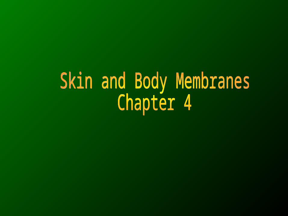
• Body membranes– Cover body surfaces– Line body cavities– Form protective sheets around
organs
• Two types of body membranes:
• Epithelial membranes
• Connective tissue membranes
Classification of Body Membranes
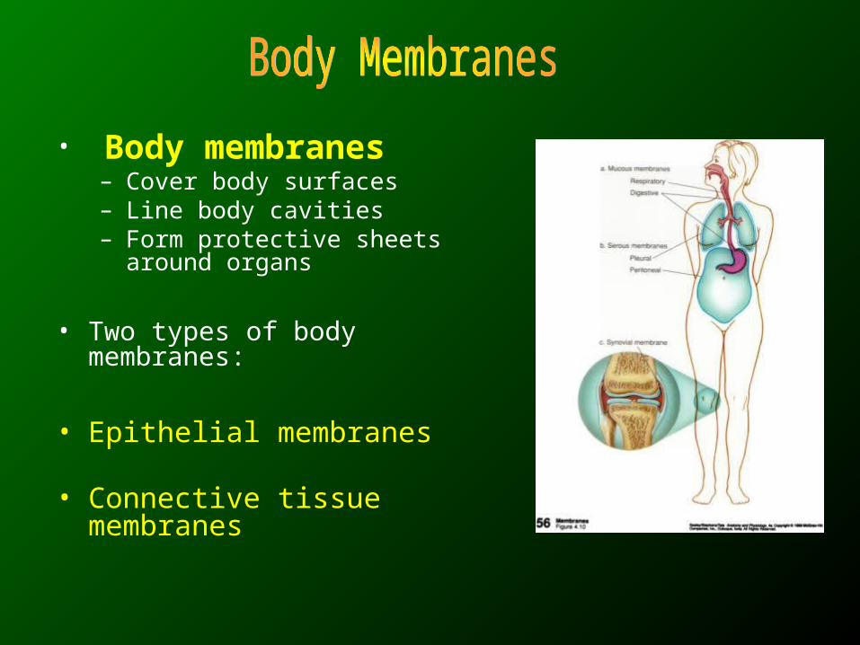
• Epithelial membranes:
– cover and lines the internal or external cavities
– Composed of epithelial cells attaching to
• underling tissue with the help of connective tissue layer
– E.g. • Cutaneous membrane• Mucous membrane• Serous membrane
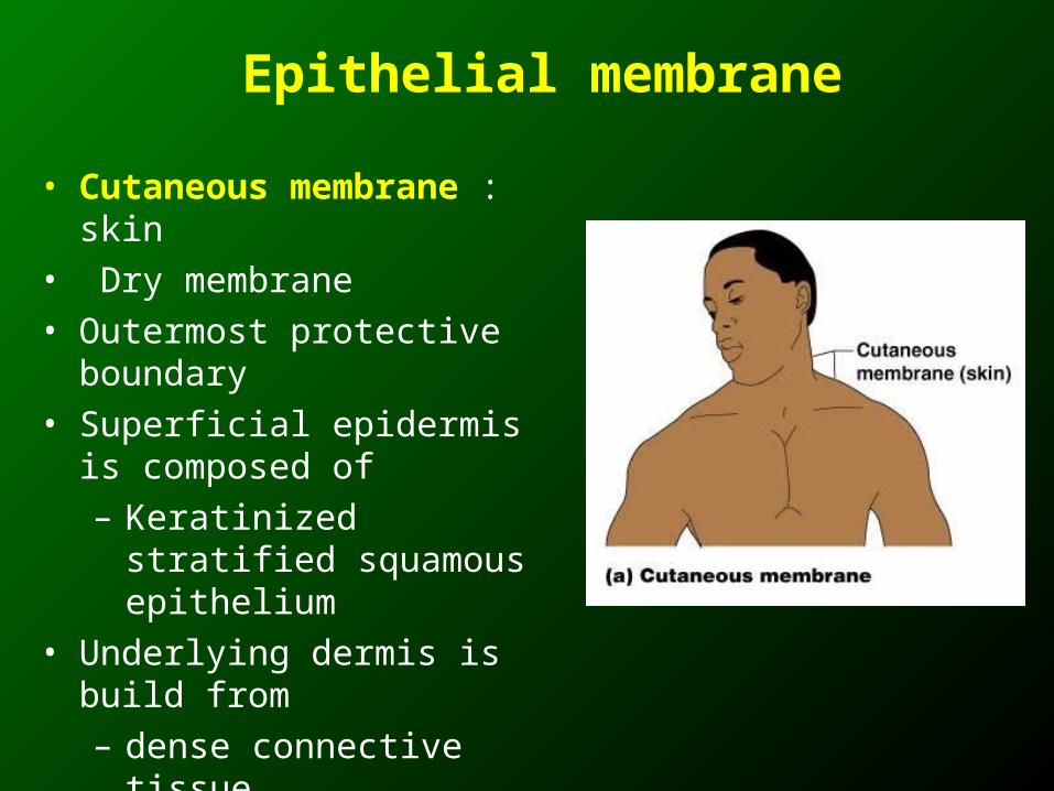
• Cutaneous membrane : skin• Dry membrane• Outermost protective boundary• Superficial epidermis is
composed of – Keratinized stratified
squamous epithelium• Underlying dermis is build from
– dense connective tissue
Epithelial membrane
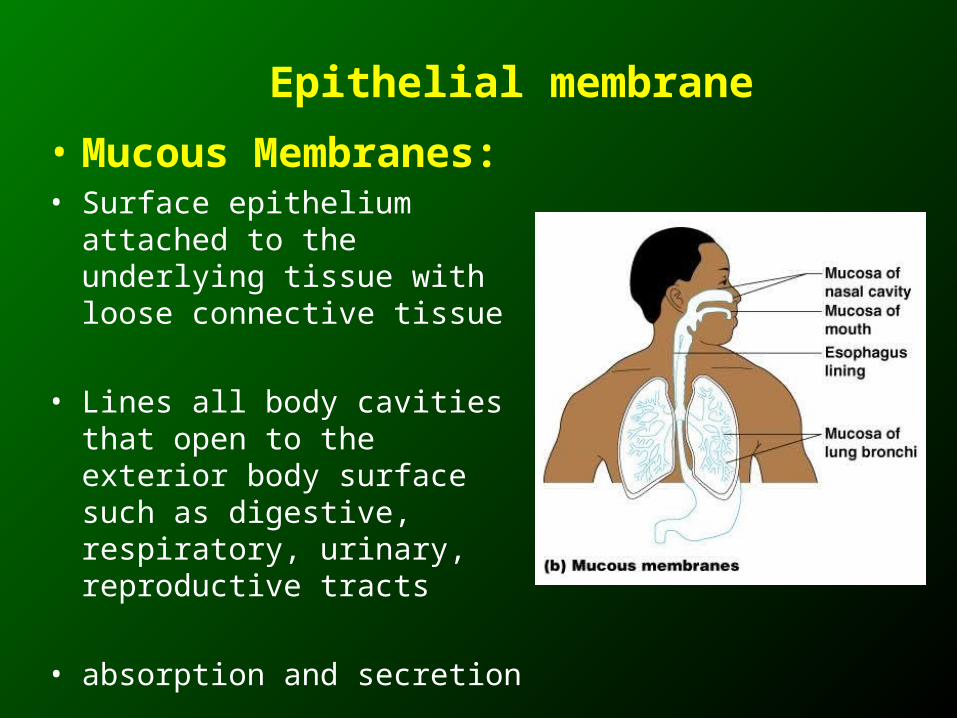
• Mucous Membranes:• Surface epithelium attached to
the underlying tissue with loose connective tissue
• Lines all body cavities that open to the exterior body surface such as digestive, respiratory, urinary, reproductive tracts
• absorption and secretion
Epithelial membrane
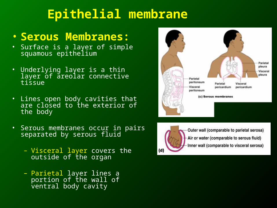
• Serous Membranes:• Surface is a layer of simple
squamous epithelium
• Underlying layer is a thin layer of areolar connective tissue
• Lines open body cavities that are closed to the exterior of the body
• Serous membranes occur in pairs separated by serous fluid
– Visceral layer covers the outside of the organ
– Parietal layer lines a portion of the wall of ventral body cavity
Epithelial membrane
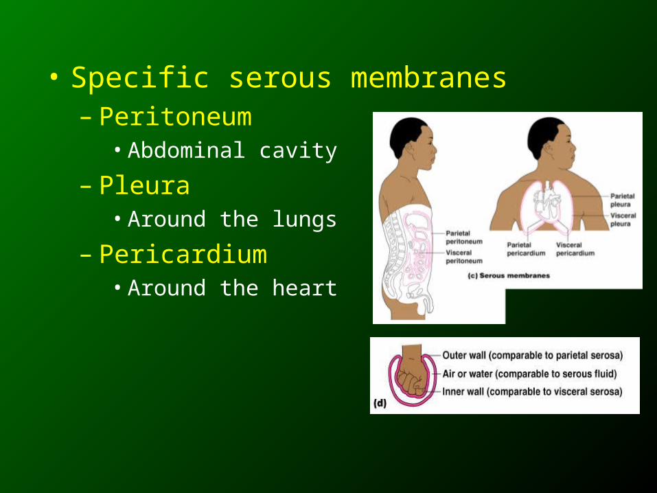
• Specific serous membranes– Peritoneum
• Abdominal cavity
– Pleura• Around the lungs
– Pericardium• Around the heart
Classification of Body Membranes
• Connective tissue membranes:
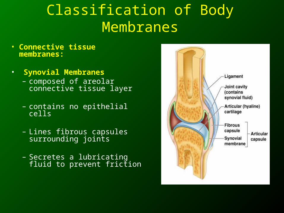
• Synovial Membranes– composed of areolar
connective tissue layer
– contains no epithelial cells
– Lines fibrous capsules surrounding joints
– Secretes a lubricating fluid to prevent friction
• Consists of skin and accessory structures
• Such as hair, nails, and glands
• Covers the outside of the body
• Forms the boundary between body and external environment
• Protects the body from the outside world
• Protection : Skin protects against abrasion, UV light, reduce water loss, prevent microorganism entry
• Sensation: Has sensory receptors, detect heat, cold, touch, pressure, pain
• Temperature regulation: By control of blood flow through skin, sweat glands
• Vitamin D production: Skin produce Vitamin D – UV light
• Excretion: Excrete through skin
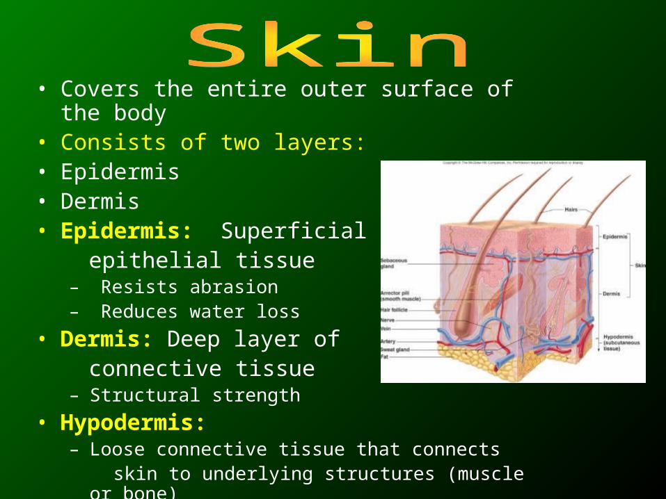
• Covers the entire outer surface of the body • Consists of two layers:• Epidermis• Dermis• Epidermis: Superficial layer of epithelial tissue
– Resists abrasion– Reduces water loss
• Dermis: Deep layer of connective tissue
– Structural strength
• Hypodermis:– Loose connective tissue that connects skin to underlying structures (muscle or bone)– Not part of skin
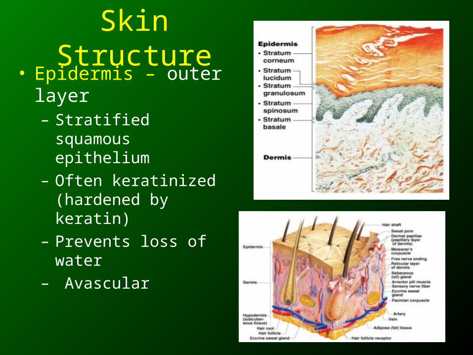
Skin Structure• Epidermis – outer layer
– Stratified squamous epithelium
– Often keratinized (hardened by keratin)
– Prevents loss of water– Avascular
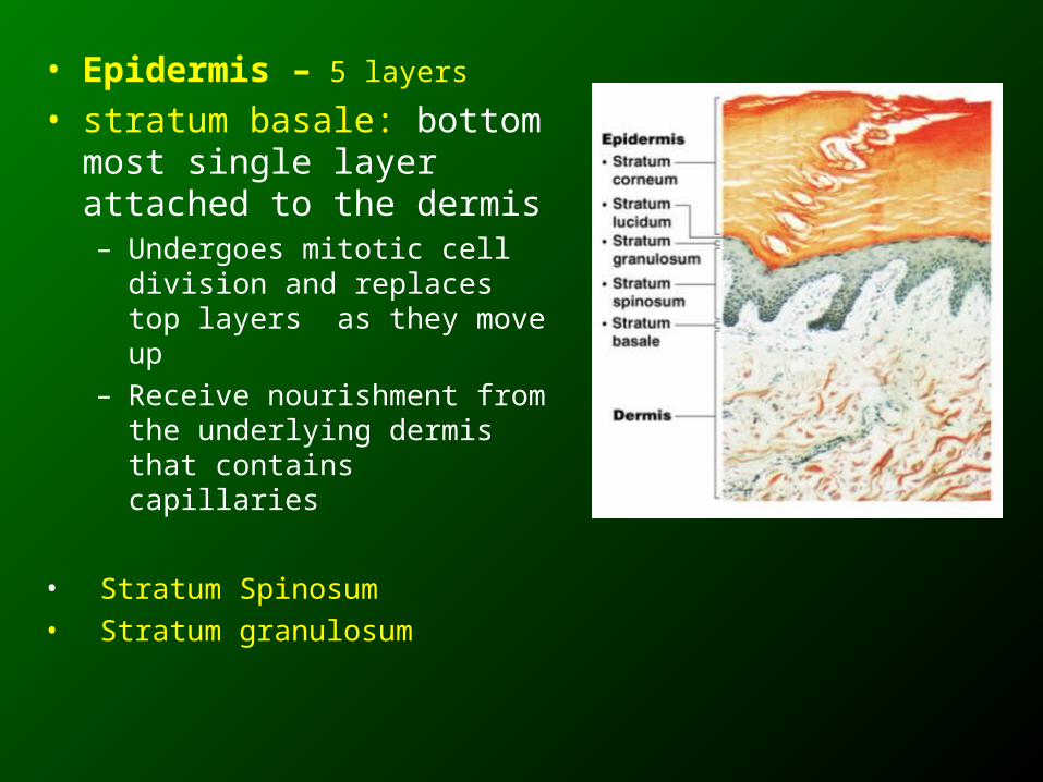
• Epidermis – 5 layers
• stratum basale: bottom most single layer attached to the dermis– Undergoes mitotic cell division
and replaces top layers as they move up
– Receive nourishment from the underlying dermis that contains capillaries
• Stratum Spinosum• Stratum granulosum
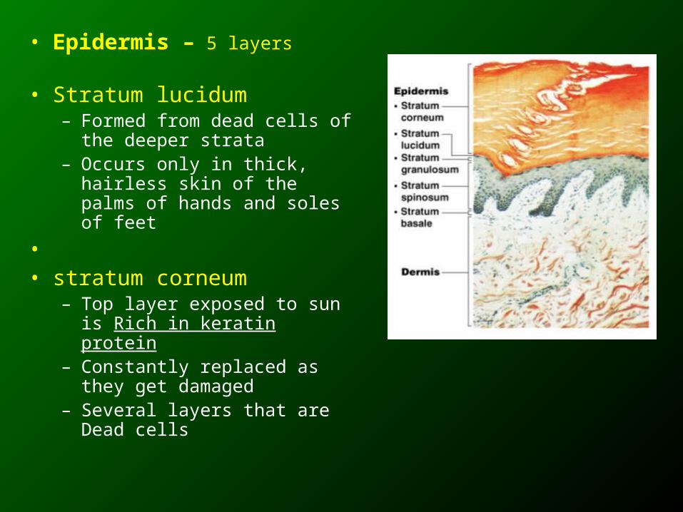
• Epidermis – 5 layers
• Stratum lucidum– Formed from dead cells of the
deeper strata– Occurs only in thick, hairless
skin of the palms of hands and soles of feet
• • stratum corneum
– Top layer exposed to sun is Rich in keratin protein
– Constantly replaced as they get damaged
– Several layers that are Dead cells
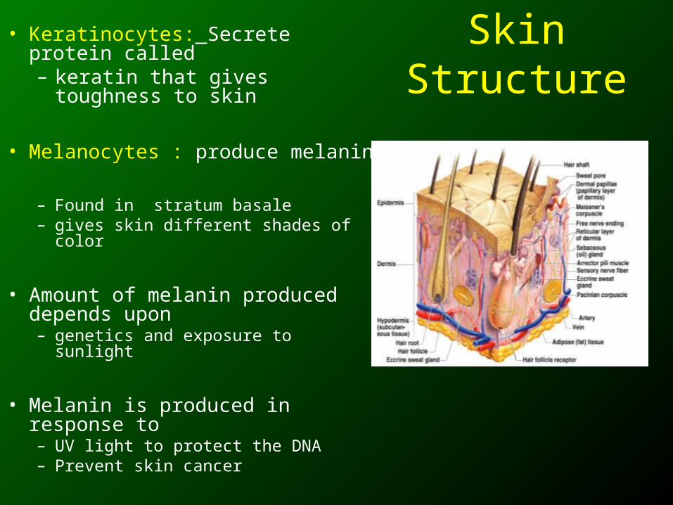
Skin Structure• Keratinocytes: Secrete protein
called – keratin that gives toughness to
skin
• Melanocytes : produce melanin
– Found in stratum basale – gives skin different shades of color
• Amount of melanin produced depends upon – genetics and exposure to sunlight
• Melanin is produced in response to – UV light to protect the DNA – Prevent skin cancer
Skin Color• Three pigments contribute to skin color:
• Melanin– Amount and kind of melanin in epidermis
– Yellow, brown or black pigments
• Carotene– Orange-yellow pigment from some vegetables
• Hemoglobin– Red coloring from blood cells in dermis capillaries
– Oxygen content determines the extent of red coloring
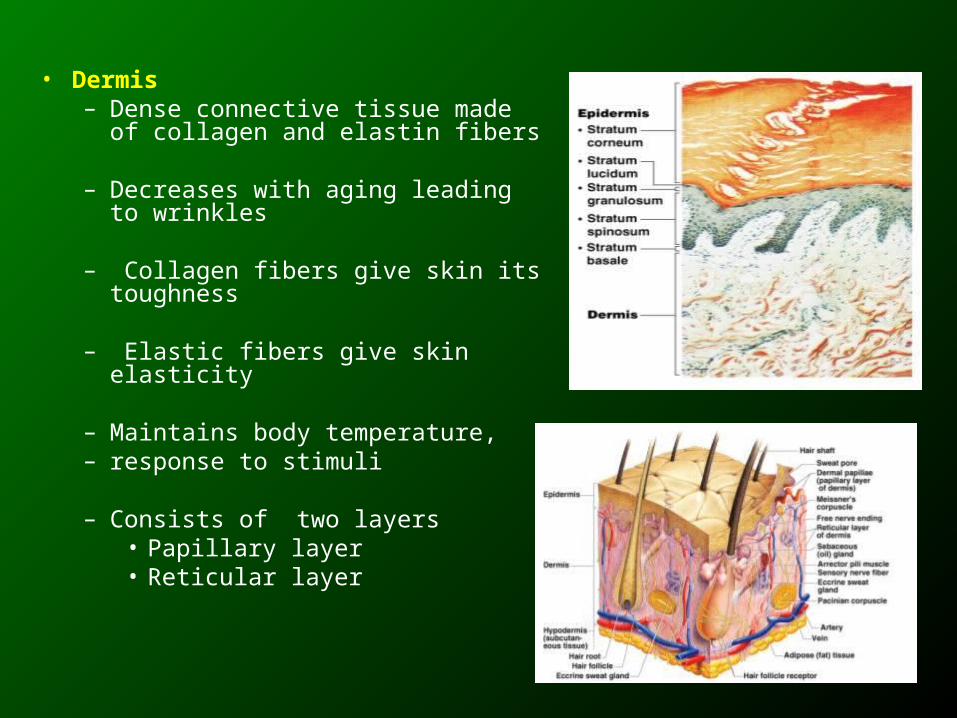
• Dermis– Dense connective tissue made of
collagen and elastin fibers
– Decreases with aging leading to wrinkles
– Collagen fibers give skin its toughness
– Elastic fibers give skin elasticity
– Maintains body temperature, – response to stimuli
– Consists of two layers• Papillary layer• Reticular layer
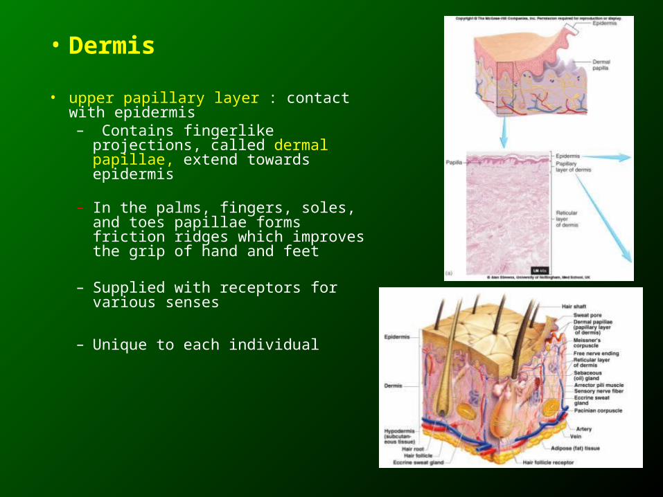
• Dermis
• upper papillary layer : contact with epidermis – Contains fingerlike projections,
called dermal papillae, extend towards epidermis
– In the palms, fingers, soles, and toes papillae forms friction ridges which improves the grip of hand and feet
– Supplied with receptors for various senses
– Unique to each individual
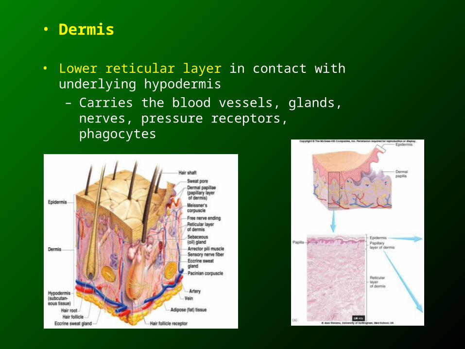
• Dermis
• Lower reticular layer in contact with underlying hypodermis– Carries the blood vessels, glands, nerves,
pressure receptors, phagocytes
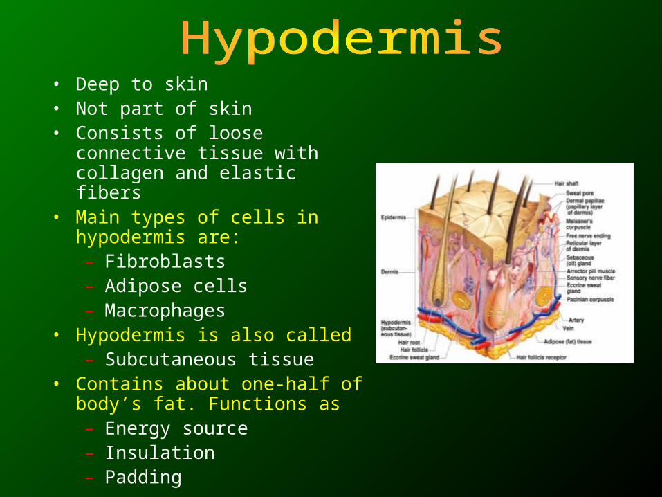
• Deep to skin• Not part of skin• Consists of loose connective tissue
with collagen and elastic fibers• Main types of cells in hypodermis
are:– Fibroblasts– Adipose cells– Macrophages
• Hypodermis is also called– Subcutaneous tissue
• Contains about one-half of body’s fat. Functions as– Energy source– Insulation– Padding
Appendages of the Skin
• Skin appendages consists of:
• Cutaneous glands : Are exocrine glands– Sebaceous glands – Sweat glands
• Hair and Hair follicles
• Nails
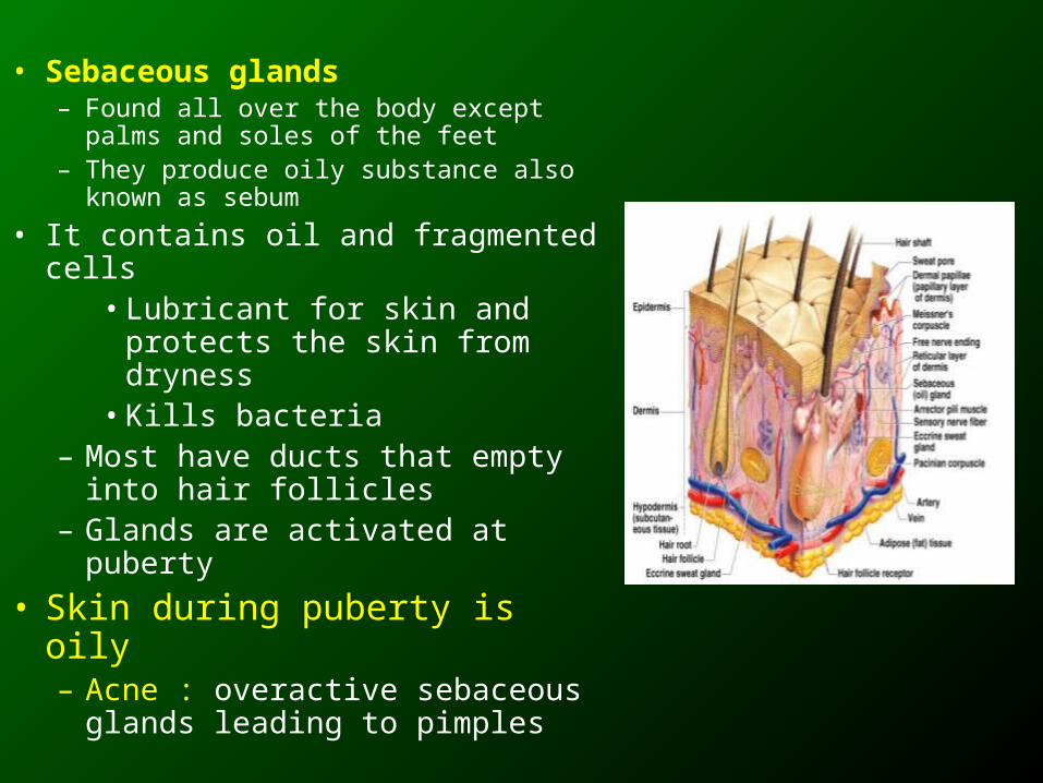
• Sebaceous glands– Found all over the body except palms
and soles of the feet– They produce oily substance also
known as sebum
• It contains oil and fragmented cells• Lubricant for skin and
protects the skin from dryness• Kills bacteria
– Most have ducts that empty into hair follicles
– Glands are activated at puberty
• Skin during puberty is oily – Acne : overactive sebaceous
glands leading to pimples
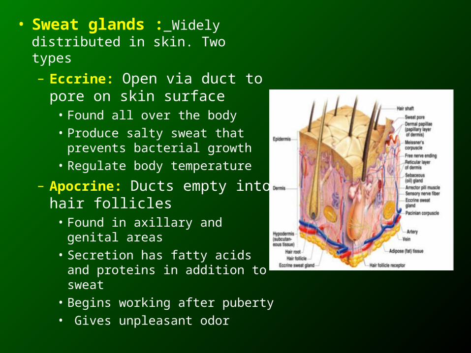
• Sweat glands : Widely distributed in skin. Two types
– Eccrine: Open via duct to pore on skin surface
• Found all over the body
• Produce salty sweat that prevents bacterial growth
• Regulate body temperature
– Apocrine: Ducts empty into hair follicles
• Found in axillary and genital areas
• Secretion has fatty acids and proteins in addition to sweat
• Begins working after puberty
• Gives unpleasant odor
Sweat and Its Function
• Composition– Mostly water– Some metabolic waste– Fatty acids and proteins (apocrine only)
• Function– Helps dissipate excess heat– Excretes waste products– Acidic nature inhibits bacteria growth
• Odor is from associated bacteria
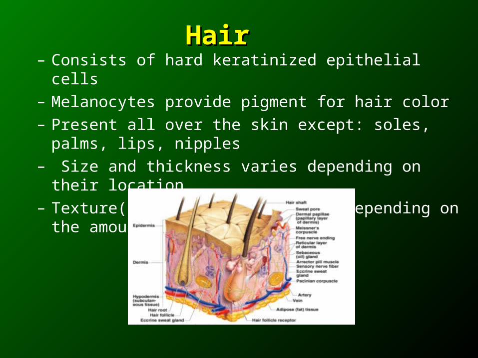
– Consists of hard keratinized epithelial cells– Melanocytes provide pigment for hair color– Present all over the skin except: soles, palms, lips, nipples– Size and thickness varies depending on their location– Texture( soft or rough) varies depending on the amount of
keratin protein
HairHair
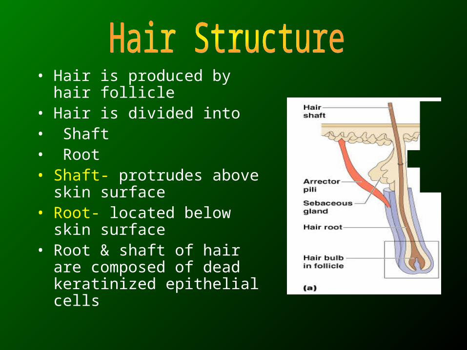
• Hair is produced by hair follicle
• Hair is divided into • Shaft• Root• Shaft- protrudes above skin
surface• Root- located below skin
surface• Root & shaft of hair are
composed of dead keratinized epithelial cells

• Each hair consists of 3 concentric layers:
– Central medulla
– Cortex surrounds medulla
– Cuticle on outside of cortex• Most heavily keratinized
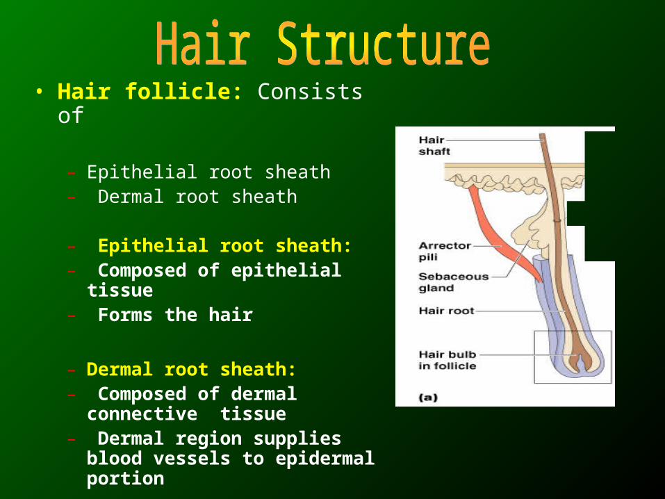
• Hair follicle: Consists of
– Epithelial root sheath– Dermal root sheath
– Epithelial root sheath:– Composed of epithelial tissue– Forms the hair
– Dermal root sheath:– Composed of dermal connective
tissue– Dermal region supplies blood
vessels to epidermal portion
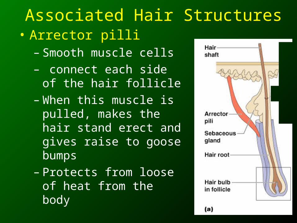
Associated Hair Structures• Arrector pilli
– Smooth muscle cells – connect each side of the
hair follicle– When this muscle is pulled,
makes the hair stand erect and gives raise to goose bumps
– Protects from loose of heat from the body
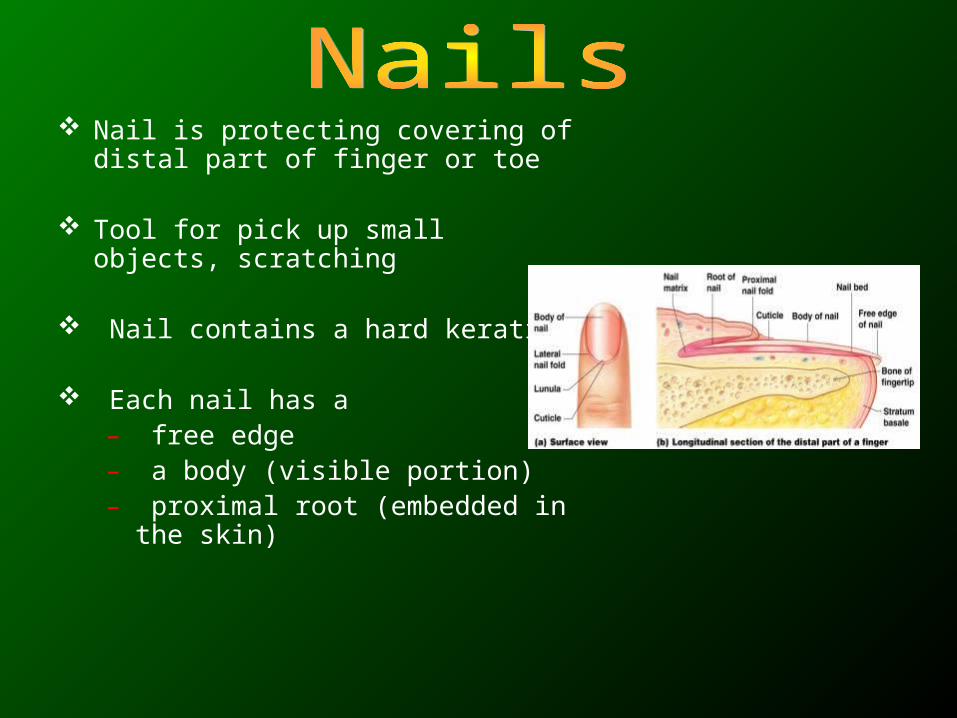
Nail is protecting covering of distal part of finger or toe
Tool for pick up small objects, scratching
Nail contains a hard keratin
Each nail has a– free edge– a body (visible portion)– proximal root (embedded in the
skin)
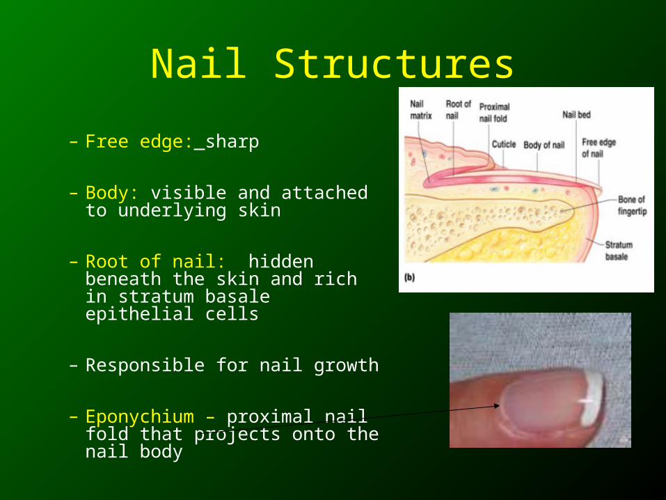
Nail Structures
– Free edge: sharp
– Body: visible and attached to underlying skin
– Root of nail: hidden beneath the skin and rich in stratum basale epithelial cells
– Responsible for nail growth
– Eponychium – proximal nail fold that projects onto the nail body
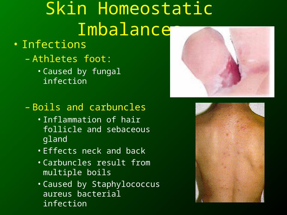
Skin Homeostatic Imbalances
• Infections– Athletes foot:
• Caused by fungal infection
– Boils and carbuncles• Inflammation of hair follicle
and sebaceous gland• Effects neck and back • Carbuncles result from
multiple boils• Caused by Staphylococcus
aureus bacterial infection
Skin Homeostatic Imbalances
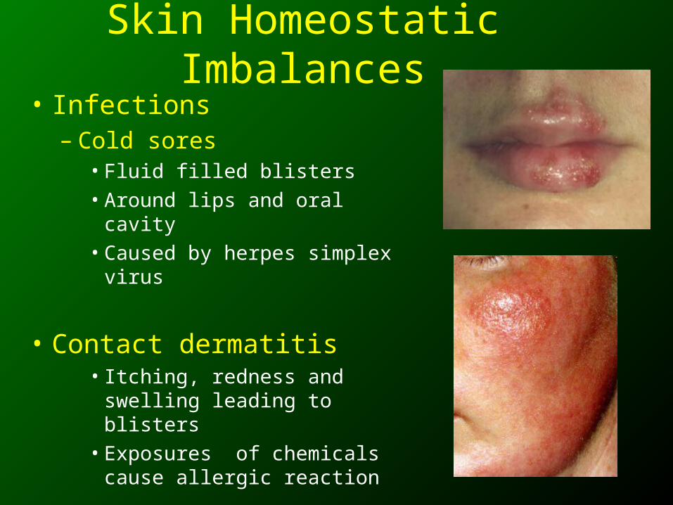
• Infections– Cold sores
• Fluid filled blisters• Around lips and oral cavity• Caused by herpes simplex
virus
• Contact dermatitis• Itching, redness and
swelling leading to blisters• Exposures of chemicals
cause allergic reaction
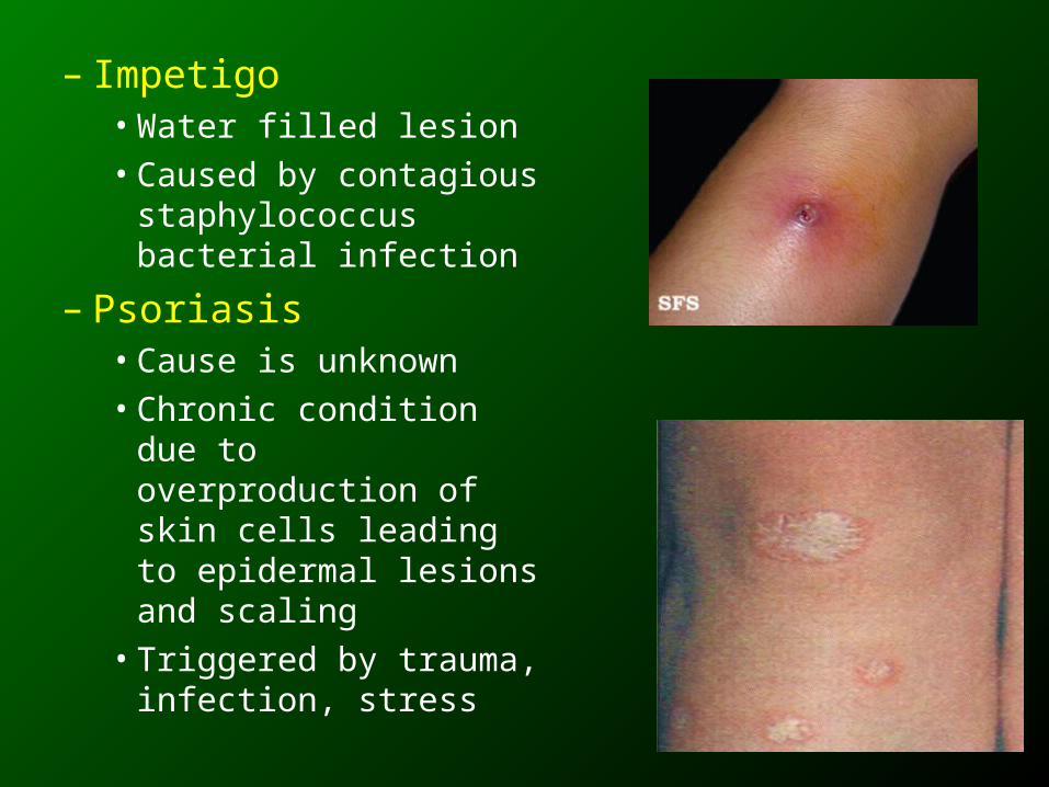
– Impetigo• Water filled lesion • Caused by contagious
staphylococcus bacterial infection
– Psoriasis• Cause is unknown• Chronic condition due to
overproduction of skin cells leading to epidermal lesions and scaling
• Triggered by trauma, infection, stress
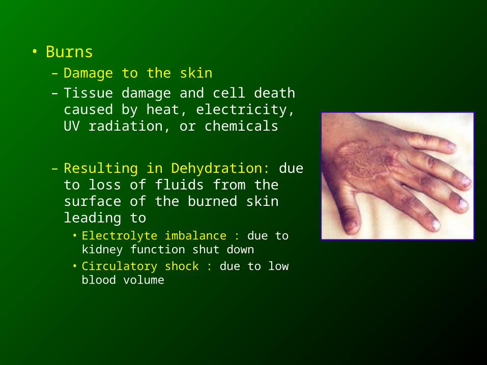
• Burns– Damage to the skin– Tissue damage and cell death
caused by heat, electricity, UV radiation, or chemicals
– Resulting in Dehydration: due to loss of fluids from the surface of the burned skin leading to
• Electrolyte imbalance : due to kidney function shut down
• Circulatory shock : due to low blood volume
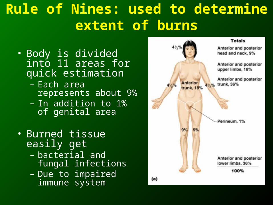
Rule of Nines: used to determine extent of burns
• Body is divided into 11 areas for quick estimation– Each area represents
about 9%– In addition to 1% of
genital area
• Burned tissue easily get – bacterial and fungal
infections– Due to impaired
immune system
Severity of Burns
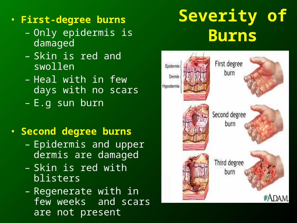
• First-degree burns– Only epidermis is
damaged– Skin is red and swollen– Heal with in few days with
no scars– E.g sun burn
• Second degree burns– Epidermis and upper
dermis are damaged– Skin is red with blisters– Regenerate with in few
weeks and scars are not present
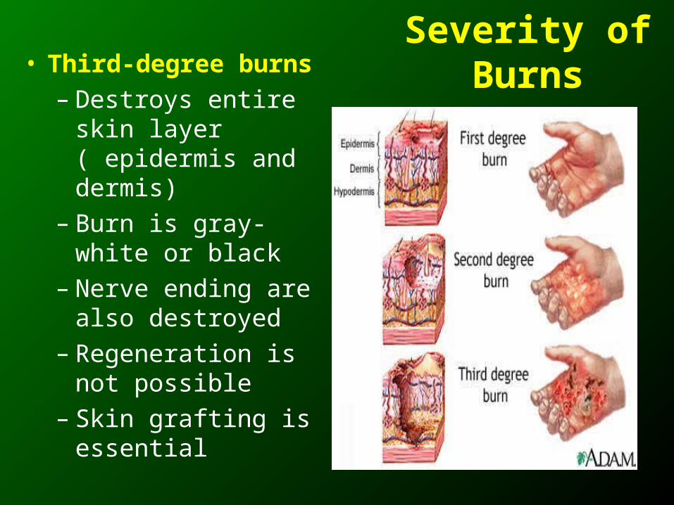
Severity of Burns• Third-degree burns
– Destroys entire skin layer ( epidermis and dermis)
– Burn is gray-white or black
– Nerve ending are also destroyed
– Regeneration is not possible
– Skin grafting is essential
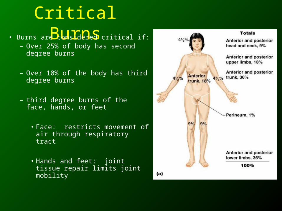
Critical Burns• Burns are considered critical if:
– Over 25% of body has second degree burns
– Over 10% of the body has third degree burns
– third degree burns of the face, hands, or feet
• Face: restricts movement of air through respiratory tract
• Hands and feet: joint tissue repair limits joint mobility
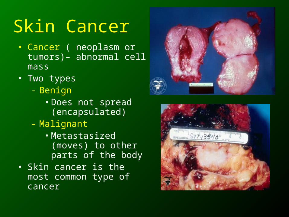
Skin Cancer• Cancer ( neoplasm or
tumors)– abnormal cell mass
• Two types– Benign
• Does not spread (encapsulated)
– Malignant• Metastasized (moves)
to other parts of the body
• Skin cancer is the most common type of cancer
Skin Cancer Types
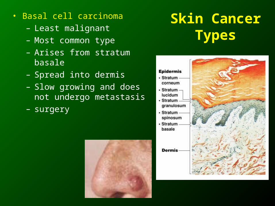
• Basal cell carcinoma
– Least malignant
– Most common type
– Arises from stratum basale
– Spread into dermis
– Slow growing and does not undergo metastasis
– surgery
Skin Cancer Types
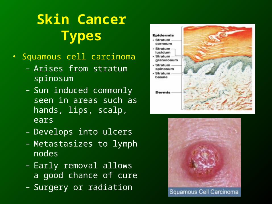
• Squamous cell carcinoma
– Arises from stratum spinosum
– Sun induced commonly seen in areas such as hands, lips, scalp, ears
– Develops into ulcers
– Metastasizes to lymph nodes
– Early removal allows a good chance of cure
– Surgery or radiation
Skin Cancer Types
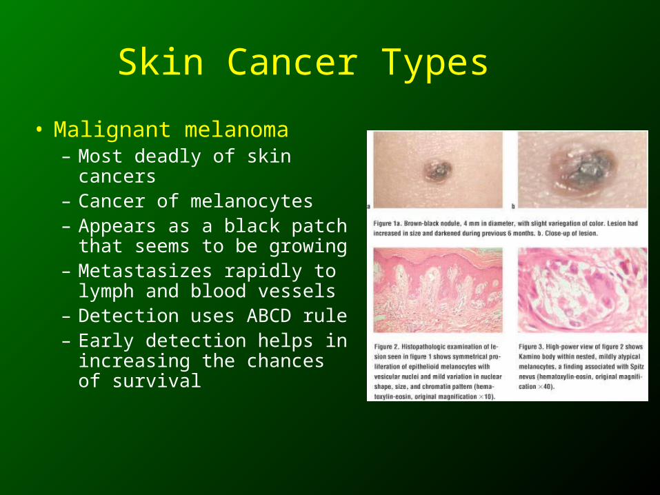
• Malignant melanoma– Most deadly of skin cancers– Cancer of melanocytes– Appears as a black patch
that seems to be growing– Metastasizes rapidly to
lymph and blood vessels– Detection uses ABCD rule– Early detection helps in
increasing the chances of survival
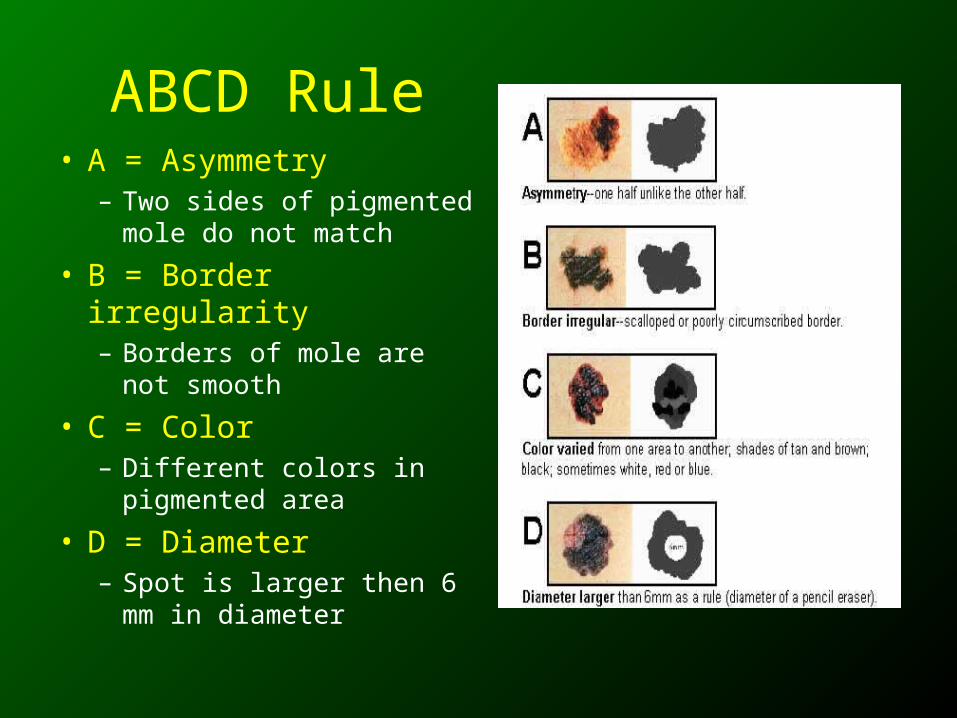
ABCD Rule• A = Asymmetry
– Two sides of pigmented mole do not match
• B = Border irregularity– Borders of mole are not
smooth
• C = Color– Different colors in
pigmented area
• D = Diameter– Spot is larger then 6 mm
in diameter