Embed Size (px)
Citation preview
BODY FLUIDS AND CIRCULATION
BYSmt.G.K.VINAYAGAM
PGT(BIO)K.V,DHARWAD.
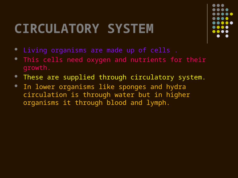
CIRCULATORY SYSTEM Living organisms are made up of cells . This cells need oxygen and nutrients for their growth. These are supplied through circulatory system. In lower organisms like sponges and hydra circulation is through
water but in higher organisms it through blood and lymph.
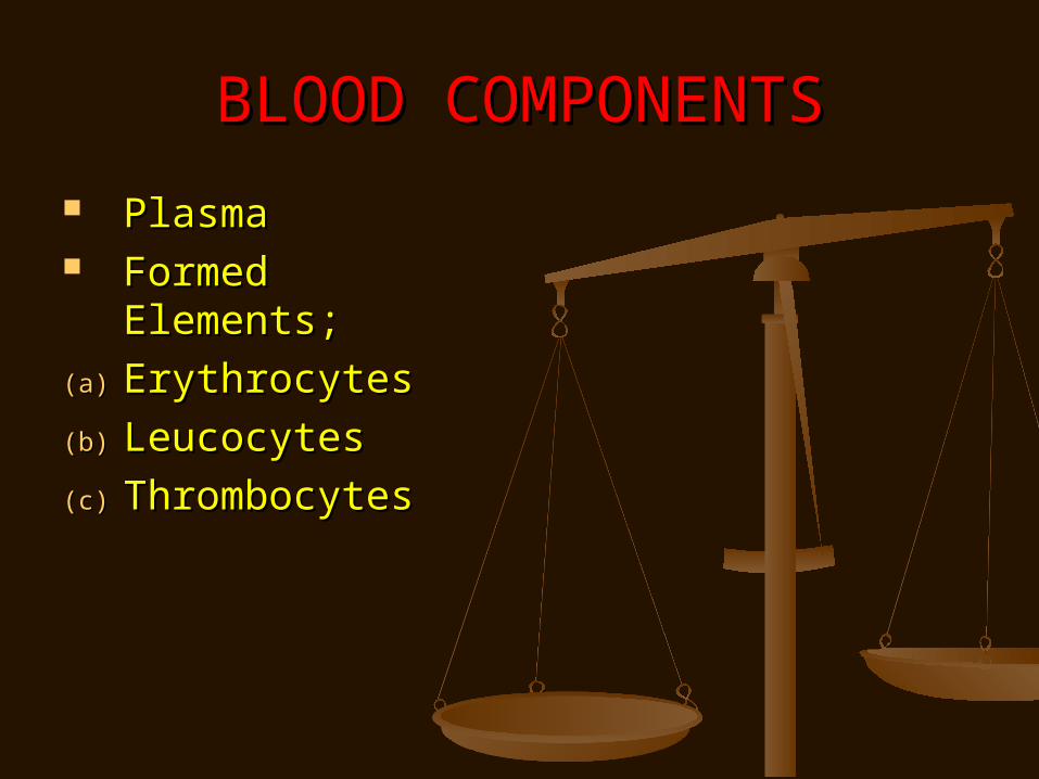
BLOOD COMPONENTSBLOOD COMPONENTS
PlasmaPlasma Formed Elements;Formed Elements;
(a)(a) ErythrocytesErythrocytes
(b)(b) LeucocytesLeucocytes
(c)(c) ThrombocytesThrombocytes
It has 90_92% of water,6_8% of proteins. There are 3 types of proteins.
Fibrinogen –Helps in coagulation of blood.
Globulin-Involved in defense mechanism.
Albumin- Helps in osmotic balance. It has minerals. Plasma without fibrinogen is called
serum.
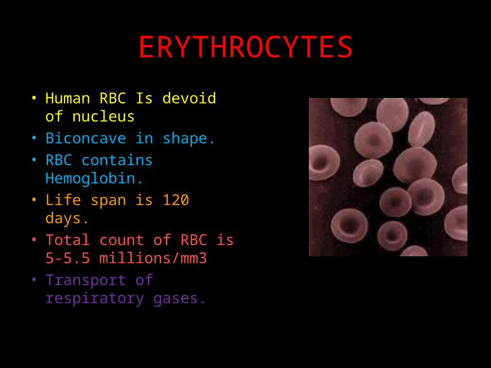
ERYTHROCYTES
• Human RBC Is devoid of nucleus
• Biconcave in shape.
• RBC contains Hemoglobin.
• Life span is 120 days.
• Total count of RBC is 5-5.5 millions/mm3
• Transport of respiratory gases.
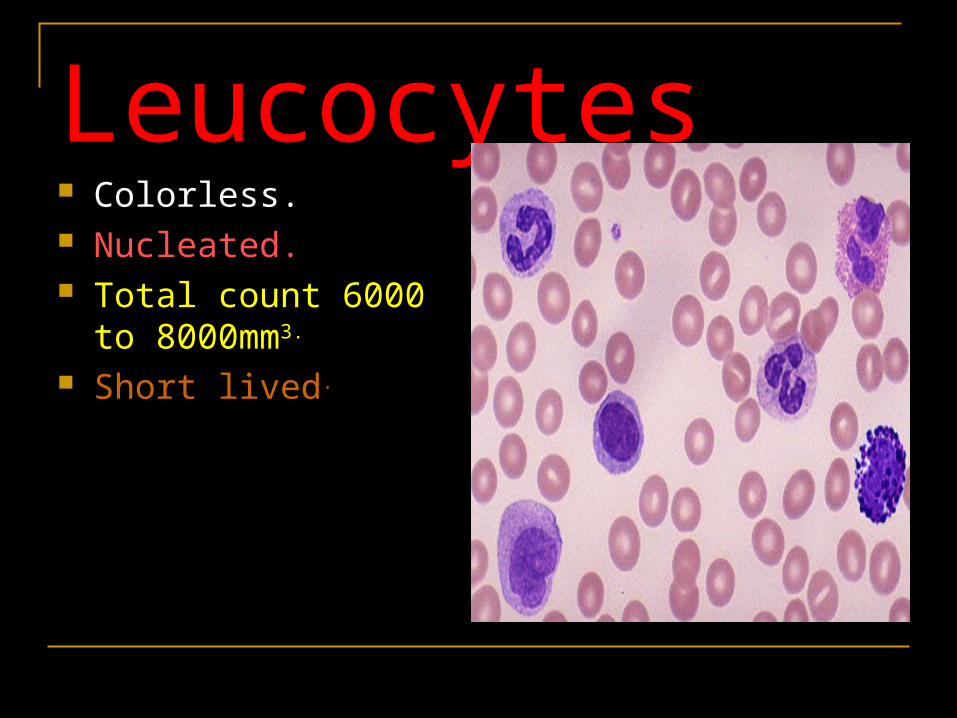
Leucocytes Colorless. Nucleated. Total count 6000 to
8000mm3.
Short lived.
TYPES OF LEUCOCYTES
Granulocytes Agranulocytes
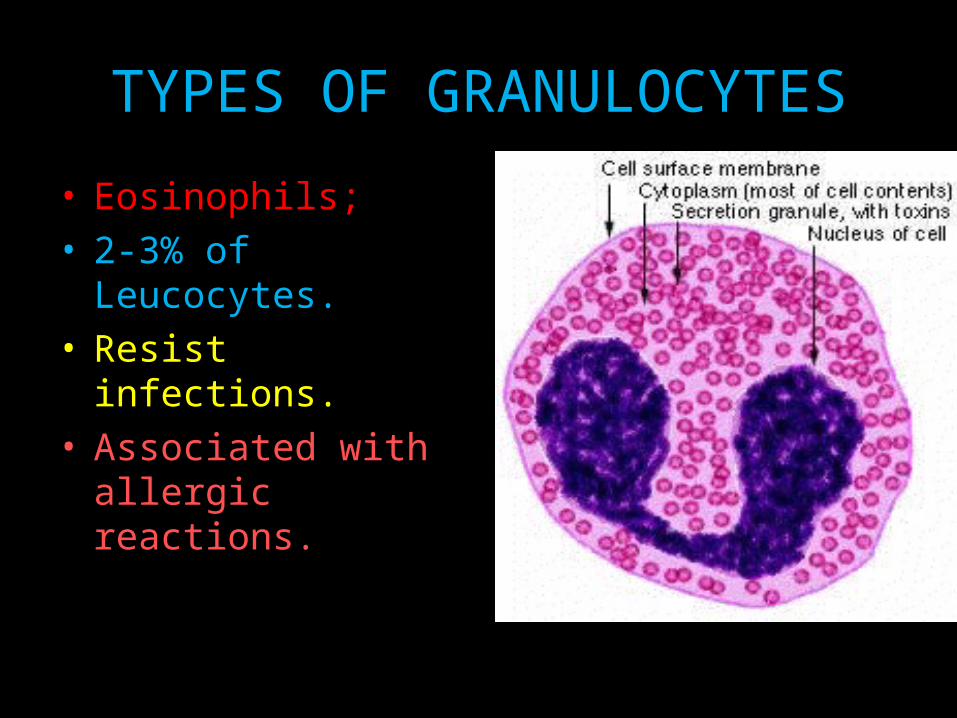
TYPES OF GRANULOCYTES
• Eosinophils;• 2-3% of Leucocytes.• Resist infections.• Associated with
allergic reactions.
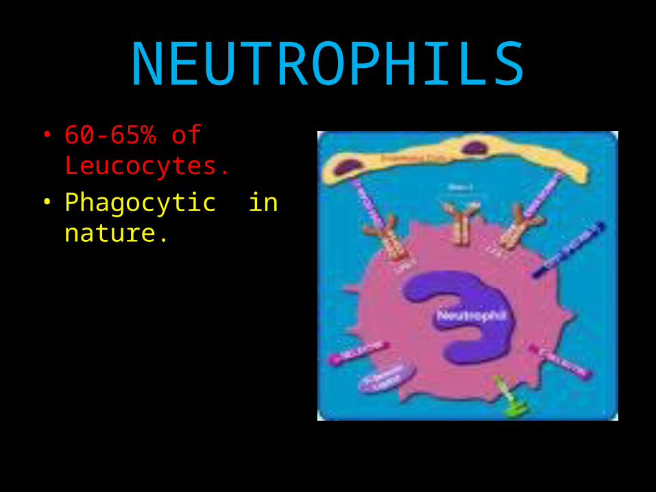
NEUTROPHILS• 60-65% of
Leucocytes.• Phagocytic in nature.
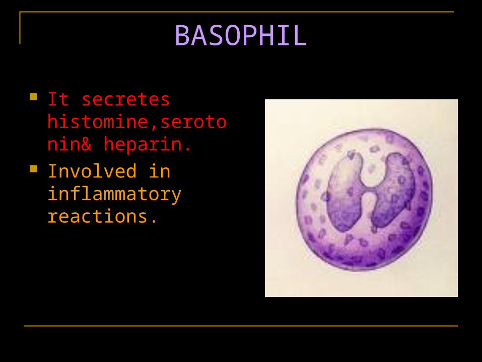
BASOPHIL
It secretes histomine,serotonin& heparin.
Involved in inflammatory reactions.
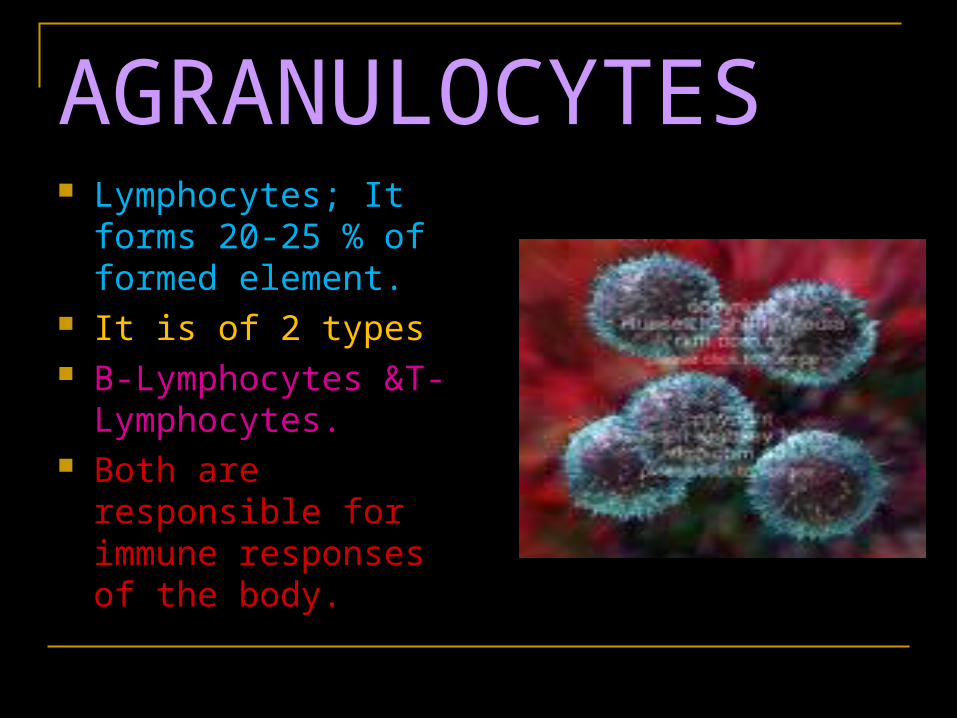
AGRANULOCYTES Lymphocytes; It forms
20-25 % of formed element.
It is of 2 types B-Lymphocytes &T-
Lymphocytes. Both are responsible
for immune responses of the body.
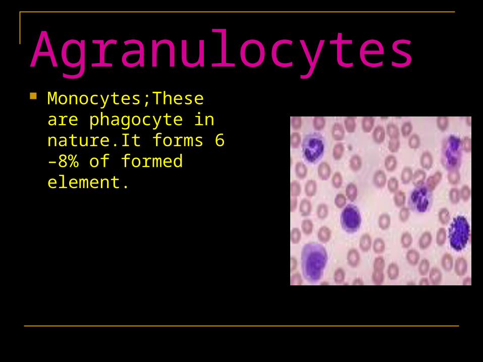
Agranulocytes Monocytes;These are
phagocyte in nature.It forms 6 –8% of formed element.
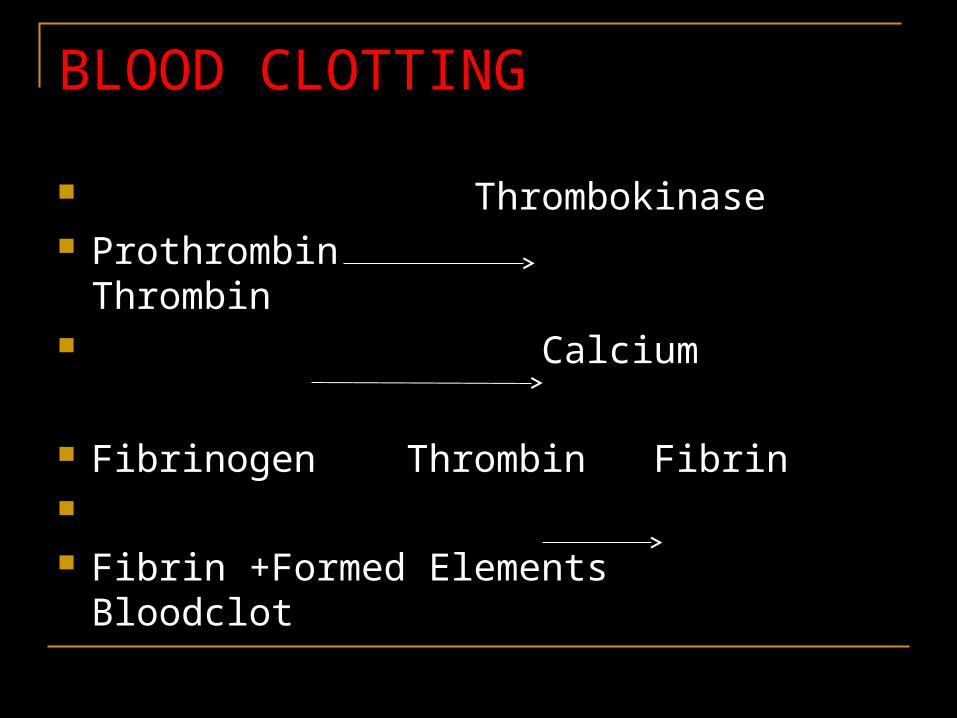
BLOOD CLOTTING
Thrombokinase Prothrombin Thrombin Calcium
Fibrinogen Thrombin Fibrin Fibrin +Formed Elements Bloodclot
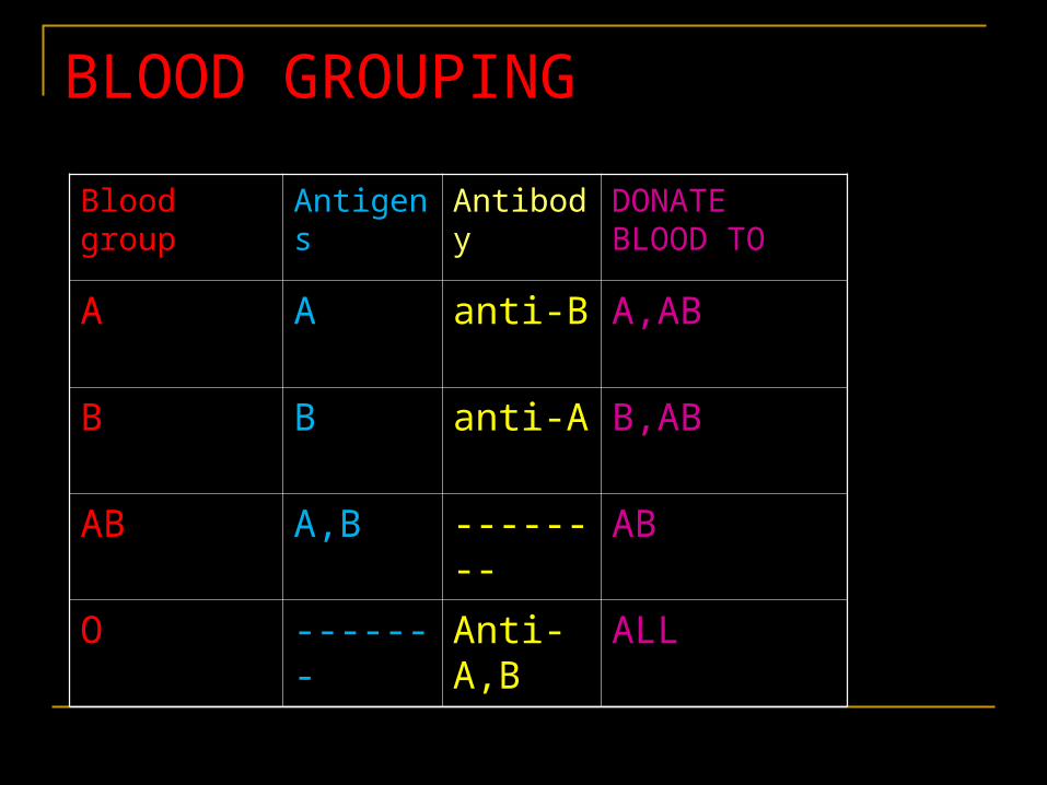
BLOOD GROUPING
Blood group Antigens Antibody DONATE BLOOD TO
A A anti-B A,AB
B B anti-A B,AB
AB A,B -------- AB
O ------- Anti-A,B
ALL
RH GROUPINGRH GROUPING
Another surface antigen is also Another surface antigen is also present in RBC.present in RBC.
This surface antigen was first This surface antigen was first reported in Rhesus Monkey. So it is reported in Rhesus Monkey. So it is called Rhesus factor or RH factor.called Rhesus factor or RH factor.
80% of people are Rh+ve ,they have 80% of people are Rh+ve ,they have Rh factor.Rh factor.

RH INCOMPATIBILIITYRH INCOMPATIBILIITY
FEMALE * MALEFEMALE * MALE Rh-ve Rh+veRh-ve Rh+ve
FOETUS (Rh+ve) safe
At child birth
Anti Rh antibody
Second Foetus
Destroy the 2nd foetus
LYMPH As the blood passes through the capillaries in tissues, some
water along with many small water soluble substances move out into the spaces between the cells of tissues leaving the larger proteins and most of the formed elements in the blood vessels.
This fluid released out is called the interstitial fluid or tissue fluid.
This fluid present in the lymphatic system is called the lymph.
It has lymphocytes. It is colourless. Fats are absorbed through lymph.
CIRCULATORY PATHWAY
OPEN CIRCULATION• In this blood pumped by the
heart enters into the open spaces called sinuses.
• Blood flow is not regulated
Closed circulation• In this blood is confined to
the blood vessels.
• Blood flow is regulated.
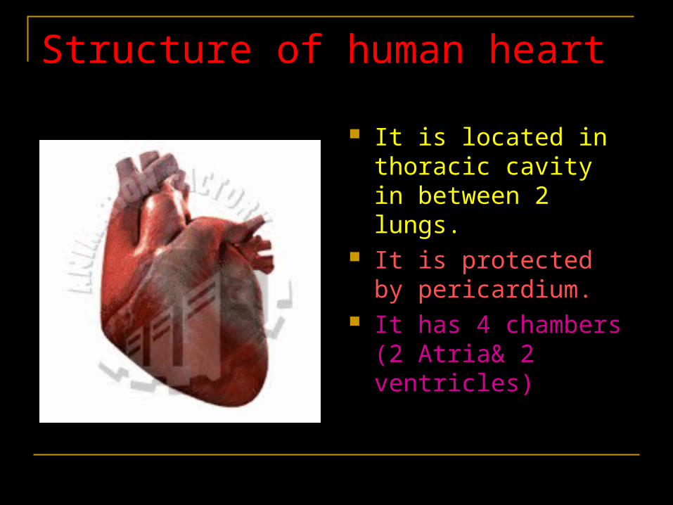
Structure of human heart
It is located in thoracic cavity in between 2 lungs.
It is protected by pericardium.
It has 4 chambers (2 Atria& 2 ventricles)
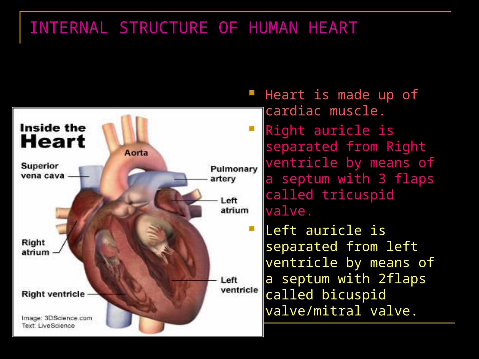
INTERNAL STRUCTURE OF HUMAN HEART
Heart is made up of cardiac muscle.
Right auricle is separated from Right ventricle by means of a septum with 3 flaps called tricuspid valve.
Left auricle is separated from left ventricle by means of a septum with 2flaps called bicuspid valve/mitral valve.
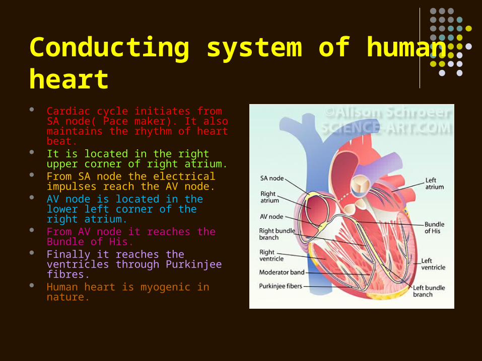
Conducting system of human heart Cardiac cycle initiates from SA
node( Pace maker). It also maintains the rhythm of heart beat.
It is located in the right upper corner of right atrium.
From SA node the electrical impulses reach the AV node.
AV node is located in the lower left corner of the right atrium.
From AV node it reaches the Bundle of His.
Finally it reaches the ventricles through Purkinjee fibres.
Human heart is myogenic in nature.
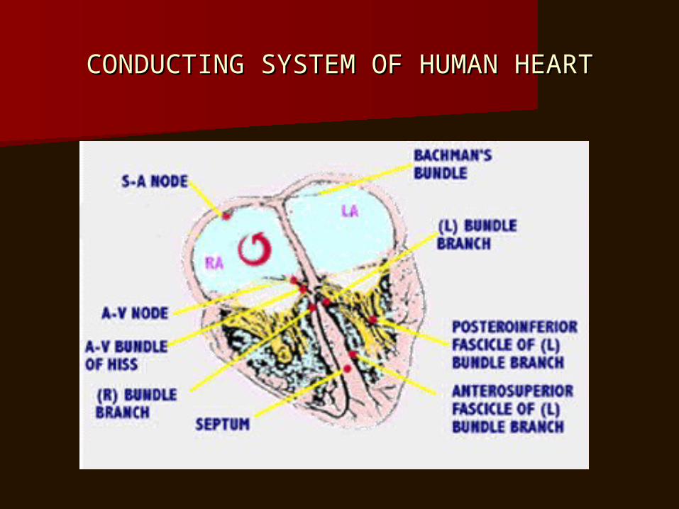
CONDUCTING SYSTEM OF HUMAN HEARTCONDUCTING SYSTEM OF HUMAN HEART
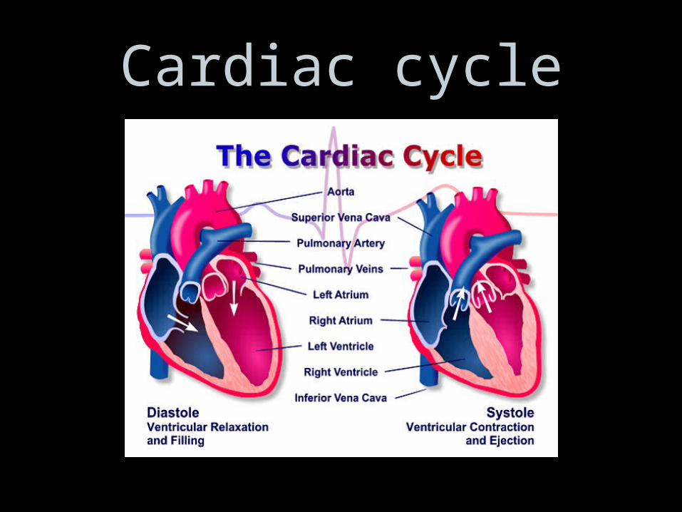
Cardiac cycle
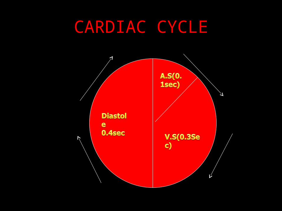
CARDIAC CYCLE
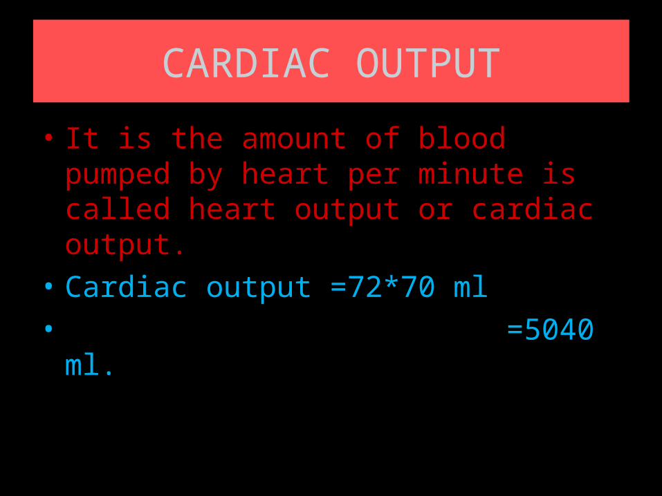
CARDIAC OUTPUT
• It is the amount of blood pumped by heart per minute is called heart output or cardiac output.
• Cardiac output =72*70 ml
• =5040 ml.
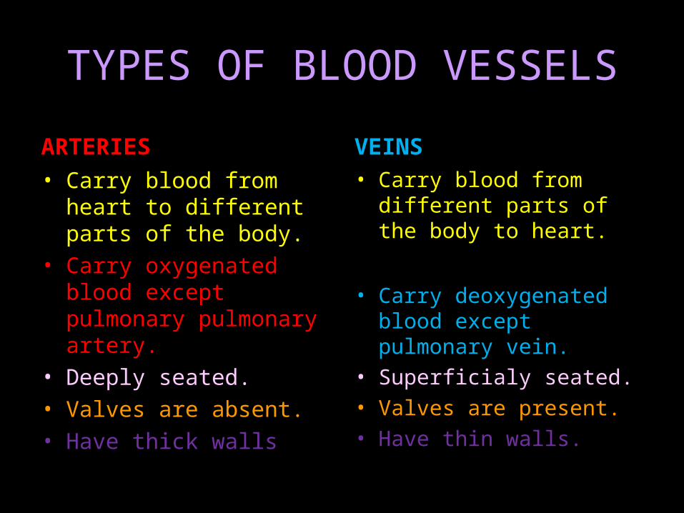
TYPES OF BLOOD VESSELS
ARTERIES
• Carry blood from heart to different parts of the body.
• Carry oxygenated blood except pulmonary pulmonary artery.
• Deeply seated.• Valves are absent.• Have thick walls
VEINS
• Carry blood from different parts of the body to heart.
• Carry deoxygenated blood except pulmonary vein.
• Superficialy seated.• Valves are present.• Have thin walls.
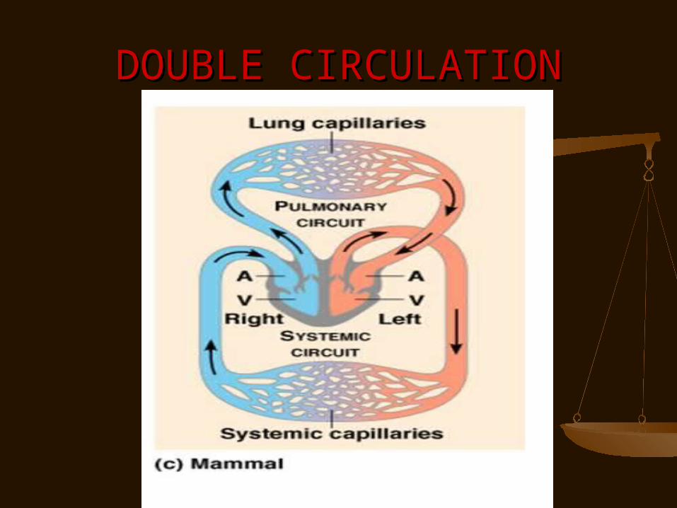
DOUBLE CIRCULATIONDOUBLE CIRCULATION
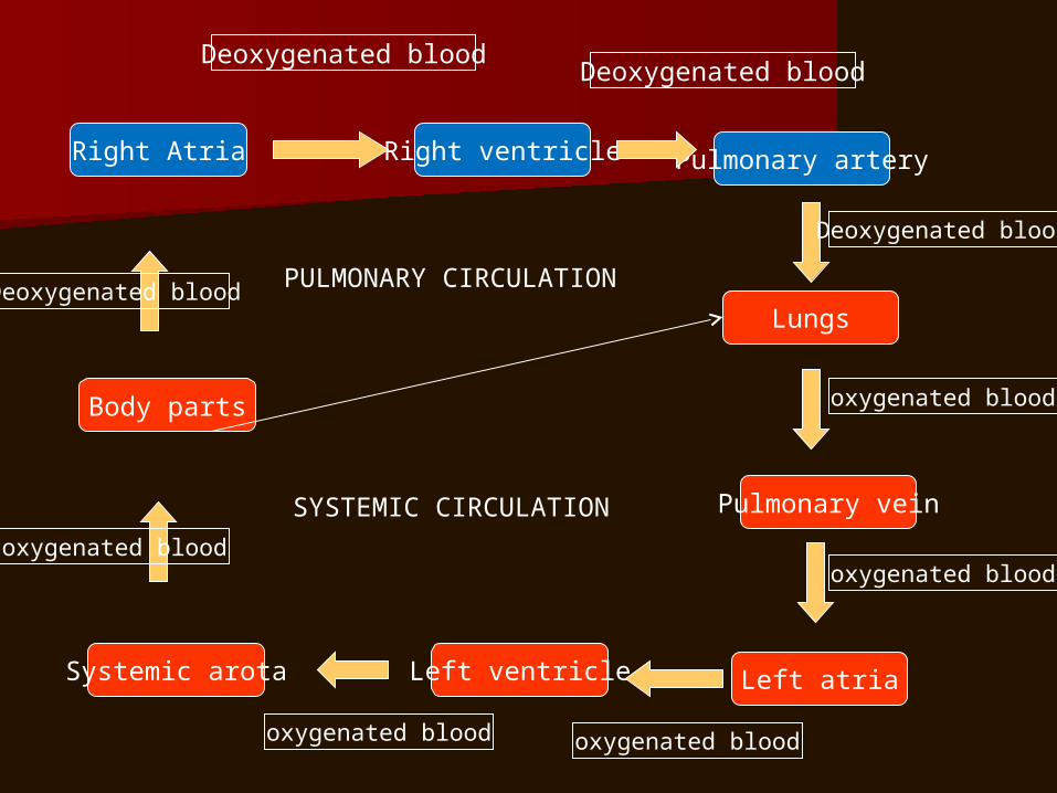
Right Atria Right ventricle Pulmonary artery
Systemic arota Left ventricle Left atria
Pulmonary vein
Body parts
Lungs
Deoxygenated bloodDeoxygenated blood
Deoxygenated blood
oxygenated blood
oxygenated blood
oxygenated bloodoxygenated blood
oxygenated blood
Deoxygenated blood PULMONARY CIRCULATION
SYSTEMIC CIRCULATION
DOUBLE CIRCULATION
Pulmonary circulation Systemic circulation
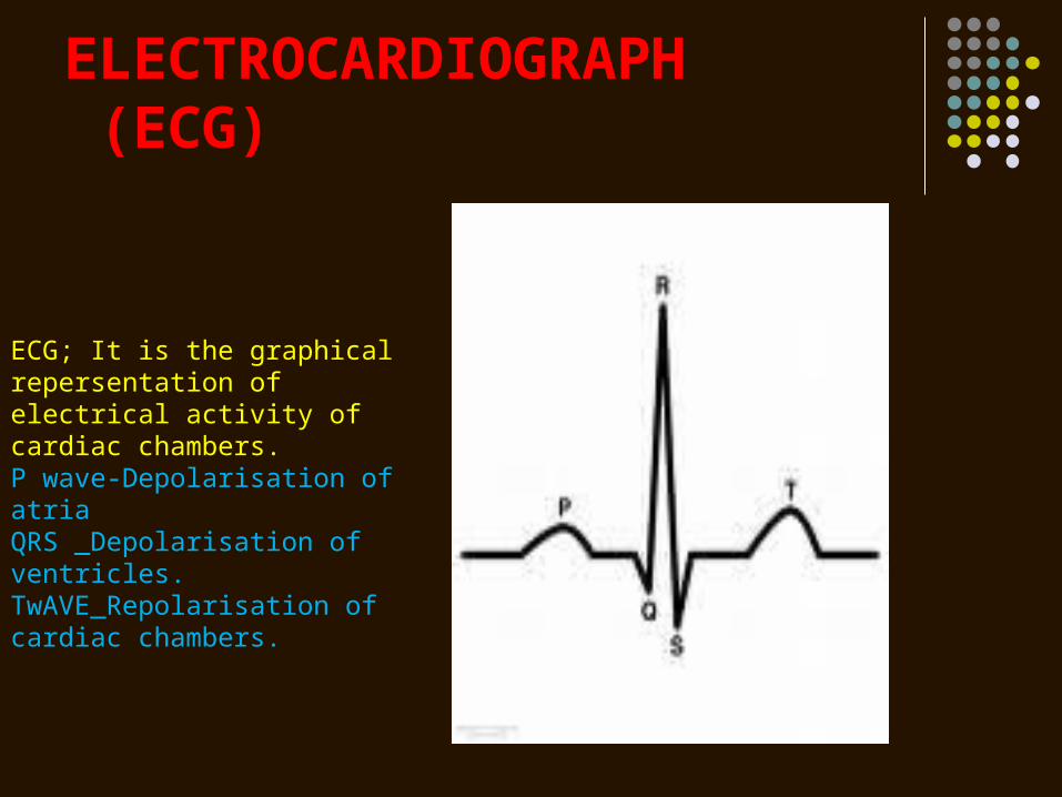
ELECTROCARDIOGRAPH (ECG)
ECG; It is the graphical repersentation of electrical activity of cardiac chambers.P wave-Depolarisation of atriaQRS _Depolarisation of ventricles.TwAVE_Repolarisation of cardiac chambers.
DISORDER OF CIRCULATORY DISORDER OF CIRCULATORY SYSTEMSYSTEM
High Blood Pressure (Hypertension)High Blood Pressure (Hypertension) Coronary Artery Disease (CAD)Coronary Artery Disease (CAD) Angina PectorisAngina Pectoris Heart FailureHeart Failure
SPHYGMOMANOMETERSPHYGMOMANOMETER
HYPER TENSIONHYPER TENSION
A persistant rise in diastolic pressure A persistant rise in diastolic pressure above 90 mmHg and or systolic above 90 mmHg and or systolic pressure above 140mmHg is termed pressure above 140mmHg is termed as hypertension.as hypertension.
ATHEROSCLEROSIS
• It is due to the deposition of cholesterol on the walls of arteries leading to narrowing of arteries.
• This also causes hypertension.
• Blood supply to the cardiac muscle is reduced.
ARTERIOSCLEROSIS
Loss of elasticity of the walls of arteries due to ageing.
MYOCARDIAL INFRACTION
Very low blood flow to the cardiac muscle. As a result cardiac muscle cannot sustain its function.
ANGINA PECTORISANGINA PECTORIS
Acute pain in the chest due to very Acute pain in the chest due to very less supply of oxygen to the heart less supply of oxygen to the heart muscle.muscle.