Embed Size (px)
Citation preview
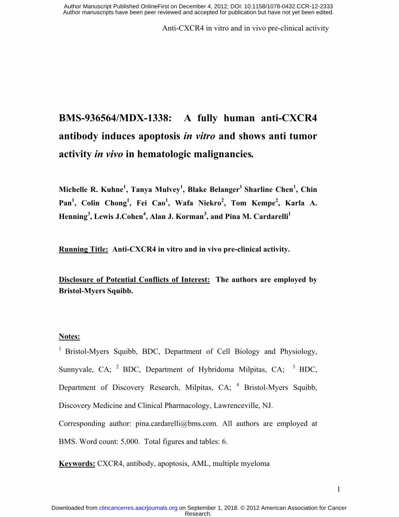
Anti-CXCR4 in vitro and in vivo pre-clinical activity
1
BMS-936564/MDX-1338: A fully human anti-CXCR4
antibody induces apoptosis in vitro and shows anti tumor
activity in vivo in hematologic malignancies.
Michelle R. Kuhne1, Tanya Mulvey1, Blake Belanger1 Sharline Chen1, Chin
Pan1, Colin Chong1, Fei Cao1, Wafa Niekro2, Tom Kempe2, Karla A.
Henning3, Lewis J.Cohen4, Alan J. Korman3, and Pina M. Cardarelli1
Running Title: Anti-CXCR4 in vitro and in vivo pre-clinical activity.
Disclosure of Potential Conflicts of Interest: The authors are employed by
Bristol-Myers Squibb.
Notes:
1 Bristol-Myers Squibb, BDC, Department of Cell Biology and Physiology,
Sunnyvale, CA; 2 BDC, Department of Hybridoma Milpitas, CA; 3 BDC,
Department of Discovery Research, Milpitas, CA; 4 Bristol-Myers Squibb,
Discovery Medicine and Clinical Pharmacology, Lawrenceville, NJ.
Corresponding author: [email protected]. All authors are employed at
BMS. Word count: 5,000. Total figures and tables: 6.
Keywords: CXCR4, antibody, apoptosis, AML, multiple myeloma
Research. on September 1, 2018. © 2012 American Association for Cancerclincancerres.aacrjournals.org Downloaded from
Author manuscripts have been peer reviewed and accepted for publication but have not yet been edited. Author Manuscript Published OnlineFirst on December 4, 2012; DOI: 10.1158/1078-0432.CCR-12-2333
Anti-CXCR4 in vitro and in vivo pre-clinical activity
2
Abstract
Purpose: CXCR4 has been identified as a prognostic marker for acute
myeloid leukemia (AML) and other malignancies. We describe the development
and characterization of a fully human antibody to CXCR4 and its application for
therapy of AML, non-Hodgkin’s lymphoma (NHL), chronic lymphoid leukemia
(CLL), and multiple myeloma (MM).
Experimental Design: Human transgenic mice were immunized with
CXCR4 expressing cells and antibodies reactive with CXCR4 were analyzed for
apoptosis induction and ability to interfere with CXCL12-induced migration and
calcium-flux. In vivo efficacy was determined in multiple AML, NHL, and MM
xenograft tumors in SCID mice.
Results: BMS-936564/MDX-1338 is a fully human IgG4 monoclonal
antibody that specifically recognizes human CXCR4. In vitro studies demonstrate
that MDX-1338 binds to CXCR4-expressing cells with low nanomolar affinity,
blocks CXCL12 binding to CXCR4 expressing cells and inhibits CXCL12
induced migration and calcium flux with low nanomolar EC50 values. When
given as monotherapy, MDX-1338 exhibits anti-tumor activity in established
tumors including AML, NHL, and MM xenograft models. Additionally, we
show that MDX-1338- induced apoptosis on a panel of cell lines and propose that
antibody induced apoptosis is one of the mechanisms of tumor growth inhibition.
Research. on September 1, 2018. © 2012 American Association for Cancerclincancerres.aacrjournals.org Downloaded from
Author manuscripts have been peer reviewed and accepted for publication but have not yet been edited. Author Manuscript Published OnlineFirst on December 4, 2012; DOI: 10.1158/1078-0432.CCR-12-2333

Anti-CXCR4 in vitro and in vivo pre-clinical activity
3
Conclusions: BMS-936564/MDX-1338 is a potent CXCR4 antagonist
which is efficacious as monotherapy in tumor bearing mice and is currently in
Phase I for the treatment of relapsed/refractory AML, NHL, CLL, and MM.
Translational Relevance: Expression of CXCR4 has been identified as a
prognostic indicator for acute myeloid leukemia (AML) and other malignancies,
in which greater expression of CXCR4 correlates with disease severity. CXCR4
plays an important role in both homing and retention of leukemic or stem cells in
the bone marrow and an antagonist of CXCR4 mobilizes these cells into the
bloodstream. In addition to mobilization, a direct apoptotic effect of the antibody
was discovered suggesting that direct killing may be a mechanism for tumor
growth inhibition. These features, together with the fact that an antibody has a
longer half life, may offer advantages over a small molecule. Consequently,
clinical trials in relapsed/refractory AML, NHL, CLL, and MM are currently
ongoing.
INTRODUCTION
CXCR4, also known as CD184, is a 7 transmembrane spanning protein
consisting of an extra-cellular N-terminal tail and three extra-cellular loops. The
intracellular carboxy terminus of CXCR4 is coupled to a heterotrimeric G-protein
consisting of β and γ subunits and a pertussis toxin-sensitive Gi α subunit.1 To
date, only one ligand for CXCR4, CXCL12, also known as SDF-1 has been
identified.2,3 CXCL12 binding to CXCR4 stimulates activation of phospholipase
C and subsequently results in an elevation of cytosolic free calcium. Ligation of
CXCR4 ultimately leads to induction of chemotaxis and migration.4,5 CXCR4 is
Research. on September 1, 2018. © 2012 American Association for Cancerclincancerres.aacrjournals.org Downloaded from
Author manuscripts have been peer reviewed and accepted for publication but have not yet been edited. Author Manuscript Published OnlineFirst on December 4, 2012; DOI: 10.1158/1078-0432.CCR-12-2333

Anti-CXCR4 in vitro and in vivo pre-clinical activity
4
found in various tissues with predominant expression on hematopoietic lineage
cells including B and T cells, monocytes, macrophages, NK, and dendritic cells,
as well as CD34+ bone marrow progenitor cells.6 Low levels of CXCR4 are also
expressed on endothelial and epithelial cells, astrocytes, and neurons.7,8 CXCL12
has been shown to induce endothelial cell migration and proliferation and together
with VEGF were shown to enhance neoangiogenesis.9
Over expression of CXCR4 has been found in 75% of cancers including
leukemias, lymphomas, pancreatic, breast, ovarian, lung, prostate and colorectal
tumors. Additionally, this pathway is implicated in stimulating the metastatic
process in multiple neoplasms.10 In clinical studies, CXCR4 has been associated
with increased propensity for metastasis and decreased survival and was identified
as a prognostic indicator for AML, breast, colorectal, non small cell lung, ovarian
and pancreatic carcinoma in which greater expression of CXCR4 correlates with
disease severity.11,12,13,14,15,16
Bone marrow stromal cells secrete CXCL12 and the interaction with
CXCR4 is essential for homing and maintaining hematopoietic stem cells within
the bone marrow microenvironment.17 Leukemic cells express high levels of
CXCR4, and the pathway plays a critical role in leukemic cell migration into the
bone marrow which in turn, supports their growth and survival. CXCR4 is
essential for metastatic spread to organs such as bone marrow where CXCL12 is
expressed. Collectively, CXCR4 plays an important role in both homing and
retention of hematopoietic stem cells in the bone marrow and an antagonist of
CXCR4 mobilizes stem cells into the bloodstream, as demonstrated with the small
Research. on September 1, 2018. © 2012 American Association for Cancerclincancerres.aacrjournals.org Downloaded from
Author manuscripts have been peer reviewed and accepted for publication but have not yet been edited. Author Manuscript Published OnlineFirst on December 4, 2012; DOI: 10.1158/1078-0432.CCR-12-2333
Anti-CXCR4 in vitro and in vivo pre-clinical activity
5
molecule CXCR4 antagonist, plerixofor (Mozobil) which was approved by the
FDA for use in combination with granulocyte-colony stimulating factor for
autologous transplants in NHL and MM patients.18
In AML, CXCR4 is highly expressed on the CD34+ fraction of bone
marrow cells. Lower levels of CXCR4 on AML cells correlate with a better
prognosis resulting in a longer relapse free and overall survival. The lower
CXCR4 receptor expression attenuates migration of primary AML cells toward
CXCL12 expressed in the chemo-protected environment of the bone marrow.19 In
addition to AML, serum levels of CXCL12 are elevated in patients with multiple
myeloma and CXCR4 expression increases in extramedullary plasmacytoma, a
manifestation of an advanced stage of multiple myeloma. Furthermore, blockade
of the CXCL12/CXCR4 axis attenuates tumor growth in MM tumor models.20
In this report we describe the generation of a fully human monoclonal
antibody specific for human CXCR4. MDX-1338 has low nM affinity for
CXCR4 and effectively blocks CXCL12 binding to CXCR4 thereby inhibiting
calcium flux and migration. MDX-1338 induces apoptosis on a panel of tumor
cell lines and significantly reduces in vivo tumor growth in several xenograft
models. These data support the development of MDX-1338 for treatment of
patients with hematologic malignancies.
MATERIALS & METHODS
Materials
Research. on September 1, 2018. © 2012 American Association for Cancerclincancerres.aacrjournals.org Downloaded from
Author manuscripts have been peer reviewed and accepted for publication but have not yet been edited. Author Manuscript Published OnlineFirst on December 4, 2012; DOI: 10.1158/1078-0432.CCR-12-2333
Anti-CXCR4 in vitro and in vivo pre-clinical activity
6
Isotype Control antibody IgG4 containing the S228P hinge mutation to
reduce half-antibody formation21 was produced at Medarex (Acquired by BMS
and currently renamed BDC), Sunnyvale, CA; The following reagents were
purchased: CXCL12 from Peprotech (Rocky Hill, NJ); 125I- CXCL12 from
PerkinElmer(Waltham, MA); Calcium dye (FLIPR Calcium 4 kit) from Molecular
Devices (Sunnyvale, CA); Bis (acetoxymethyl) 2,2':6',2''-terpyridine-6,6''-
dicarboxylate (BADTA) chemiluminescent migration reagent and DELFIA
Europium solution from PerkinElmer (Waltham, MA); Annexin V Binding Buffer
10x concentrate, 7-Amino-Actinomycin D (7-AAD), and Annexin V-APC from
BD Biosciences, (San Jose, CA); Phycoerythrin-conjugated goat anti-human
antibody from Jackson ImmunoResearch, West Grove, PA (Cat. 109-116-098).
Acute myelogenous leukemia (AML) peripheral blood mononuclear cells from
AllCells LLC. (Emeryville, CA) and Cureline Inc. (Burlingame, CA).
Cells
Ramos human B lymphoblast Burkitt's lymphoma (Cat. CRL-1596),
CCRF-CEM human T lymphoblast acute lymphoblastic leukemia (CCL-119),
HL-60 human promyeloblast (CCL-240), Namalwa human B lymphoblast Burkitt’s
lymphoma (CRL-1432), Raji human B lymphoblast Burkitt’s lymphoma (CCL-86),
RPMI 8226 human myeloma (CCL-155), MM.1S human B lymphoblast MM (CRL-
2974), U226B1 human myeloma (TIB-196), MV-4-11 human biphenotypic B
myelomonocytic leukemia (CRL-9591), MJ human T-cell lymphoma (CRL-8294),
HH human T-cell lymphoma (CRL-2105), HuT78 human lymphoblast cutaneous
Research. on September 1, 2018. © 2012 American Association for Cancerclincancerres.aacrjournals.org Downloaded from
Author manuscripts have been peer reviewed and accepted for publication but have not yet been edited. Author Manuscript Published OnlineFirst on December 4, 2012; DOI: 10.1158/1078-0432.CCR-12-2333
Anti-CXCR4 in vitro and in vivo pre-clinical activity
7
lymphoma (TIB-161), NK92 human NK cell non-Hodgkin's lymphoma (CRL-2407)
cell lines were purchased from ATCC, Manassas, VA.
NOMO-1 human acute myeloid leukemia (ACC 542), MOLP-8 MM
(ACC 569), SU-DHL6 human B cell non-Hodgkin’s lymphoma (ACC 572), L540
human Hodgkin’s lymphoma (ACC 72), KG-1 human AML (ACC 14), MOLP-8
human MM (ACC 569), OPM-2 human MM (ACC 50), L-363 human plasma cell
leukemia (ACC 49) cell lines were purchased from DSMZ, Braunschweig,
Germany.
R1610 hamster fibroblasts (CRL-1657) purchased from ATCC were
transfected with human CXCR4 and kept under selection using G418 at 500
µg/mL. JJN-3 cells (ACC 541) purchased from DSMZ were selected at BMS for
resistance to bortezomib. NKL human NK cell large granulocyte leukemia cell line
licensed from Dana-Farber Cancer Institute; KHYG-1 human NK cell leukemia cell
line (JCRB0156) was purchased from the Health Science Research Resources Bank,
Japan Health Sciences Foundation.
FACS Instrument and Software
A FACSArray or FACSCailbur (BD Biosciences, San Jose, CA)
instrument and FlowJo software v8 (TreeStar Inc, Ashland, OR) were used to
collect and analyze data.
Antibody generation
Mice from Medarex KM® transgenic mouse colonies (Milpitas, CA) were
immunized with human CXCR4 transfected R1610 cells or recombinant
Research. on September 1, 2018. © 2012 American Association for Cancerclincancerres.aacrjournals.org Downloaded from
Author manuscripts have been peer reviewed and accepted for publication but have not yet been edited. Author Manuscript Published OnlineFirst on December 4, 2012; DOI: 10.1158/1078-0432.CCR-12-2333

Anti-CXCR4 in vitro and in vivo pre-clinical activity
8
CXCL12. Spleen lysates were pooled and processed as described previously. 22
Using proprietary phage display procedures, Biosite generated antibody fragments
(Fab library). Phage which bound to CXCR4 were selected on CXCR4 magnetic
proteoliposomes (MPLs)23 which were prepared from HEK293E cells expressing
CXCR4. Phage binding to CXCL12 were selected using biotinylated-CXCL12.
Selected antigen reactive Fab were converted to full length IgG4 (S228P) and re-
expressed in CHO cells.
Functional characterization of CXCL12 and CXCR4
Serial dilutions of MDX-1338, anti-CXCL12 and control antibody were
tested for blockade of 125I-CXCL12 binding to CXCR4+ CEM cells. Competition
of 125I-CXCL12 binding to CXCR4 on CEM cells was demonstrated using a fixed
concentration of 125I-CXCL12 (100 pM) and a titration of MDX-1338 from 5 pM
to 300 nM. An isotype antibody was used as a negative control and unlabeled
CXCL12 was used as a positive control. Plates were incubated at room
temperature for 1 hour, the filters were washed, removed and counts per minute
(CPM) were read by a PerkinElmer Wizard gamma counter (Waltham, MA). For
all in vitro studies, the data was graphed and analyzed with GraphPad Prism®
software (San Diego, CA), using nonlinear regression and sigmoidal dose-
response curves.
Cells were loaded with FLIPR Calcium 4 dye (Molecular Devices,
Sunnyvale CA). A fixed concentration of CXCL12 was used to stimulate calcium
flux. A titration of MDX-1338 or anti-CXCL12 from 50 pM to 100 nM was used
to inhibit the response. A maximal calcium response was set with CXCL12 minus
Research. on September 1, 2018. © 2012 American Association for Cancerclincancerres.aacrjournals.org Downloaded from
Author manuscripts have been peer reviewed and accepted for publication but have not yet been edited. Author Manuscript Published OnlineFirst on December 4, 2012; DOI: 10.1158/1078-0432.CCR-12-2333

Anti-CXCR4 in vitro and in vivo pre-clinical activity
9
antibodies. A baseline response was established with buffer stimulation of cells
without CXCL12. Calcium fluxes were read on the Flexstation (Molecular
Devices, Sunnyvale, CA). .
Cells were loaded with BATDA. A fixed concentration of CXCL12 was
used to stimulate migration of cells through a filter containing 5 µm pores on
Migration Plates from Neuro Probe (Gaithersburg, MD; Cat. ChemoTx 106-5). A
titration of MDX-1338 or anti-CXCL12 from 20 pM to 300 nM was added to the
cells. CXCL12 without antibody was used to establish maximal migration.
Migration toward media alone without CXCL12 was used to measure background
migration. Following 2 hour incubation at 37 °C, migrated cells were detected by
addition of Europium solution to the lysed cells and detected by time resolved
fluorescence on the Fusion (Perkin Elmer).
For proliferation, cells were suspended at 1 x 10⁵ cells/mL in growth
media and incubated with antibodies and cultured for 72 hours at 37 ° C. Cell-
Titer-Glo (Promega) was added to wells, mixed and incubated at room
temperature for 10 minutes. Plate was read on GloMax Luminometer (Promega).
For apoptosis assays, cells (5 x 10⁵ cells/mL) were incubated with 10 nM -
330 nM MDX-1338 or isotype control at 37 °C for 24 hours. For a subset of cells
(see Table 1), a cross linking antibody (Goat anti-human IgG Fc specific
polyclonal Ab) was added at six-fold excess. For all cell types, camptothecin
(CPT) was added at 10 μM for 24 hours at 37º C as a positive control for
apoptosis induction. Cells were then resuspended in Annexin V binding buffer
(10 mM HEPES at pH 7.4, 140 mM NaCl, 2.5 mM CaCl2) and stained with
Research. on September 1, 2018. © 2012 American Association for Cancerclincancerres.aacrjournals.org Downloaded from
Author manuscripts have been peer reviewed and accepted for publication but have not yet been edited. Author Manuscript Published OnlineFirst on December 4, 2012; DOI: 10.1158/1078-0432.CCR-12-2333

Anti-CXCR4 in vitro and in vivo pre-clinical activity
10
Annexin V-APC and 7-Aminoactinomycin D (7-AAD) or propidium iodide (PI).
Cells were then washed, resuspended in Annexin V binding buffer, and analyzed
with a FACSArray system (BD Biosciences, San Jose, CA) and FlowJo software
(Treestar, Inc., San Carlos, CA).
Tumor Models
SCID mice were subcutaneously implanted with 10 million Ramos cells,
or HL-60 cells, or 7.5 million of Nomo-1 cells, or 2.5 million MOLP-8 cells, or 5
million JJN-3R cells in 0.1 mL phosphate-buffered saline (PBS) and 0.1 mL
Matrigel, using a 1-cm3 syringe and a 25-gauge half-inch needle. One day prior to
dosing, mice were randomized into groups of 8-10 mice each according to tumor
volume (L×W×H/2). Post implantation, mice were dosed with MDX-1338 at 3-30
mg/kg IP; human IgG4 isotype (15 or 30 mg/kg IP); bortezomib® (1.0 or 0.8
mg/kg IV); and vehicle control was dosed at 0.3 mL IP. Mice were dosed every
three to four days for five doses. Tumors and body weights were measured twice
weekly. Tumors were measured in three dimensions with a Fowler Electronic
Digital Caliper (Model 62379-531; Fred V. Fowler Co., Newton, MA), and data
was electronically recorded using StudyDirector software from Studylog Systems,
Inc. (South San Francisco, CA). Animals were checked daily for postural,
grooming, and respiratory changes, as well as lethargy. Mice were euthanized
when the tumors reached the 2000 mm3 endpoint or appeared ulcerated. All
antibody doses were well tolerated and no body weight losses were observed.
Research. on September 1, 2018. © 2012 American Association for Cancerclincancerres.aacrjournals.org Downloaded from
Author manuscripts have been peer reviewed and accepted for publication but have not yet been edited. Author Manuscript Published OnlineFirst on December 4, 2012; DOI: 10.1158/1078-0432.CCR-12-2333

Anti-CXCR4 in vitro and in vivo pre-clinical activity
11
RESULTS
CXCR4 is expressed on multiple hematopoietic cell lines and variably
expressed in AML patients.
A number of CXCR4 positive human cell lines were evaluated for MDX-
1338 binding using flow cytometry. Dose-dependent binding was seen for the cell
lines R1610-huCXCR4, Ramos, CEM, Nomo-1, HL-60, MOLP8 and JJN-3R
(Figure 1). No binding to the R1610 parental cells was detected. Based upon
geometric mean fluorescent intensity (GMFI), CXCR4 levels were highest on
R1610-huCXCR4 and Ramos cells followed by CEM (Figure 1B), Nomo-1 and
HL60 (Figure 1A). The multiple myeloma cell lines MOLP-8 and JJN-3R
expressed the lowest number of receptors (Figure 1C). The EC50 values for
binding were 2.3 nM, 4.2 nM, 10.3 nM, 40 nM , 5.3 nM 6.5 nM and 2.0 nM for
R1610-huCXCR4, Ramos, CEM, Nomo-1, HL-60 MOLP-8 and JJN-3R cells,
respectively. In addition, MDX-1338 bound to healthy donor PBMCs (data not
shown) as well as 7/8 PBMCs samples collected from AML patients with variable
GMFI (Figure 1D).
Ligand Blockade
Saturation binding studies were conducted using radiolabeled CXCL12
and CXCR4hi CEM cells. The KD of 125I-CXCL12 binding to CEM cells was
determined to be 4.3 nM (data not shown) which is similar to the reported KD of
CXCL12 for CXCR4 ranging from 3.0 to 5.4 nM.24 Using a suboptimal fixed
concentration of 125I-CXCL12 (100 pM), MDX-1338 was titrated and dose-
dependent inhibition of 125I-CXCL12 binding with an EC50 value of
Research. on September 1, 2018. © 2012 American Association for Cancerclincancerres.aacrjournals.org Downloaded from
Author manuscripts have been peer reviewed and accepted for publication but have not yet been edited. Author Manuscript Published OnlineFirst on December 4, 2012; DOI: 10.1158/1078-0432.CCR-12-2333
Anti-CXCR4 in vitro and in vivo pre-clinical activity
12
approximately 2 nM was observed (Figure 2A). Interestingly, the anti-CXCL12
antibody was more potent and induced a dose-dependent inhibition of 125I-
CXCL12 binding to CEM cells with an EC50 value of approximately 90 pM
(Figure 2B).
Blockade of CXCL12-Induced Calcium Flux
Ramos and CEM cells were used to test the capacity for MDX-1338 and
anti-CXCL12 to inhibit calcium flux. CXCL12 induces a dose dependent rise in
intracellular calcium with peak calcium flux reached at 50 nM and 5 nM with
Ramos and CEM cells, respectively. Using the optimal concentration of CXCL12
to stimulate calcium flux, a titration of MDX-1338 or anti-CXCL12 was used to
inhibit the response (Figure 2 C-D). Both MDX-1338 and anti-CXCL12 blocked
CXCL12-induced calcium flux in a dose dependent manner with an EC50 of
approximately 10 nM and 8 nM in Ramos and CEM, respectively (Figure 2C and
2D). Anti-CXCL12 blocked with an EC50 of approximately 35 nM (Ramos) and
2 nM (CEM) cells (Figure 2C and 2D).
Blockade of CXCL12-Induced Migration
The optimal concentration of CXCL12 for inducing Ramos migration was
established to be 10 ng/mL (1.25 nM) while CEM cells were more sensitive to
CXCL12 and exhibited maximal migration at 0.05 nM CXCL12. MDX-1338 was
shown to block CXCL12-induced migration with an approximate EC50 value of 1
nM in Ramos cells and 4 nM in CEM cells (Figure 2E and 2F). Anti-CXCL12
inhibited CXCL12-induced migration with an approximate EC50 value of 0.9 nM
(Ramos) and 0.13 nM (CEM) cells (Figure 2E and 2F).
Research. on September 1, 2018. © 2012 American Association for Cancerclincancerres.aacrjournals.org Downloaded from
Author manuscripts have been peer reviewed and accepted for publication but have not yet been edited. Author Manuscript Published OnlineFirst on December 4, 2012; DOI: 10.1158/1078-0432.CCR-12-2333
Anti-CXCR4 in vitro and in vivo pre-clinical activity
13
Comparison of anti-CXCR4 and anti-CXCL12 antibodies in vivo
To test the in vivo activity of MDX-1338 and anti-CXCL12, SCID mice
bearing established Ramos tumor xenografts were treated with 15 mg/kg of
antibody. Dose response studies had previously found 15 mg/kg to be an
effective dose of rituximab (data not shown). MDX-1338 and positive control,
rituximab, inhibited tumor growth when compared with vehicle and isotype
controls. Treatment with MDX-1338 resulted in a median growth inhibition of
99% on Day 21 and the inhibition was maintained for 60 days (Figure 3A). In
contrast, anti-CXCL12 did not inhibit tumor growth and performed similarly to
the isotype control antibody.
In Vitro Induction of Apoptosis
Because we observed robust in vivo activity, studies were undertaken to
understand the mechanism of action of MDX-1338. A maximum of ~50%
inhibition of Ramos cell proliferation was seen with 40 nM MDX-1338 treatment
(Figure 4A) compared to isotype control. By comparison, AMD3100, a small
molecule CXCR4 antagonist did not inhibit proliferation. A recently described
peptide antagonist, BKT140, did inhibit proliferation however at much higher
concentrations (100 μM).
Antibody-induced apoptosis was investigated using Ramos cells and
MDX-1338 for 24 hours. For comparison, the small molecule CXCR4-
antagonist, AMD3100 was investigated using 6 μM corresponding to a
concentration which inhibited CXCL12-induced calcium flux and migration.
Research. on September 1, 2018. © 2012 American Association for Cancerclincancerres.aacrjournals.org Downloaded from
Author manuscripts have been peer reviewed and accepted for publication but have not yet been edited. Author Manuscript Published OnlineFirst on December 4, 2012; DOI: 10.1158/1078-0432.CCR-12-2333
Anti-CXCR4 in vitro and in vivo pre-clinical activity
14
MDX-1338 induced an increase in Annexin V (31.2%) and in Annexin V/PI
double positive staining (27.3%) compared with cells that were either untreated
(1.7% and 4.1%), incubated with isotype control antibody (0.5% and 2.8%), or
treated with AMD3100 (2.0% and 2.7%) (Figure 4B and 4C).
To verify the specificity of the response to MDX-1338, parental R1610
which do not bind MDX-1338 (data not shown) and R1610 transfected with
human CXCR4 that do bind to MDX-1338 (Figure 1) were used to measure
apoptosis. The transfected cells R1610-hCXCR4 exhibited an increased level of
Annexin V staining and Annexin V/ PI in response to incubation with MDX-1338
(24.3% and 11.4%) while an isotype control antibody (2.5% and 0.9%) or when
untreated (2.6% and 0.9%) had minimal effects. The parental R1610 cells did not
exhibit apoptosis following MDX-1338 treatment (Figure 5) suggesting
specificity for hCXCR4. Subsequent to these findings MDX-1338 was shown to
induce apoptosis on several CXCR4 positive cell lines as well as normal PBMC
(Table 1).
MDX-1338 inhibits tumor growth of AML models.
To assess the antibody’s efficacy in AML we used two cytarabine resistant
mouse xenograft models, HL-60 and Nomo-1. The CXCR4 expression in each
cell line was confirmed by FACS staining (Figure 1A). SCID mice containing
established HL60 tumors were treated with MDX-1338 and on Day 27, the
median tumor growth inhibition was 88% and 83% when compared to isotype and
vehicle groups, respectively (Figure 3B).
Research. on September 1, 2018. © 2012 American Association for Cancerclincancerres.aacrjournals.org Downloaded from
Author manuscripts have been peer reviewed and accepted for publication but have not yet been edited. Author Manuscript Published OnlineFirst on December 4, 2012; DOI: 10.1158/1078-0432.CCR-12-2333
Anti-CXCR4 in vitro and in vivo pre-clinical activity
15
In the Nomo-1 model, the mice were dosed with MDX-1338 or cytarabine
and monitored for 57 days. On day 34, the median tumor growth inhibition of
MDX-1338 treated mice was significantly delayed by 88% compared to isotype
or vehicle control (Figure 3C). As expected, Cytarabine did not inhibit tumor
growth.
MDX-1338 inhibits tumor growth of MM models.
CXCR4+ myeloma cells, MOLP8 and JJN-3R, were tested for sensitivity
to MDX-1338 in SCID xenograft tumor models. MOLP8 cells were implanted
into SCID mice and the mice were treated with, 10 mg/kg/dose of MDX-1338
±50 mg/kg lenalidomide or ±0.8 mg/kg bortezomib (Figure 3D). MDX-1338
significantly delayed mean tumor growth by 66% and 56% when compared to
isotype control on Day 25 (last day when all mice in each cohort remained in the
study). MOLP8 tumors were relatively resistant to lenalidomide and bortezomib
and the efficacy of MDX-1338 was not improved when combined with either
drug. At the end of study on day 42, 5 out of 8 mice remained in the MDX-1338
group while no mice remained in the isotype treated group. The bortezomib
resistant, JJN3R cells were implanted into SCID mice and mice were dosed when
the tumors were established. Median tumor growth over time is shown in Figure
3E. Neither lenalidomide nor bortezomib alone inhibited tumor growth while
median tumor growth inhibition was 100% for mice treated with MDX-1338 on
day 25 compared to mice treated with isotype. At the end of study, 4 out of 7
mice were tumor free in the MDX-1338 30 mg/kg group.
Discussion
Research. on September 1, 2018. © 2012 American Association for Cancerclincancerres.aacrjournals.org Downloaded from
Author manuscripts have been peer reviewed and accepted for publication but have not yet been edited. Author Manuscript Published OnlineFirst on December 4, 2012; DOI: 10.1158/1078-0432.CCR-12-2333
Anti-CXCR4 in vitro and in vivo pre-clinical activity
16
A novel, first in class therapeutic monoclonal antibody directed to CXCR4
has been developed. In addition to blocking CXCL12-induced calcium flux and
migration, we describe antibody dependent induction of apoptosis as another
mechanism of action. Antibody-induced apoptosis resulted in robust in vivo
efficacy across multiple hematopoietic tumor xenograft models. Because CXCR4
plays a role in multiple fundamental aspects of cancer including proliferation,
migration/invasion and angiogenesis, an antagonist has potentially multiple means
to intervene in malignancies where CXCR4 is expressed. To begin to dissect the
pathway, we developed fully human monoclonal antibodies to both CXCR4 and
CXCL12. Both the anti-CXCR4 and anti-CXCL12 antibodies inhibit ligand
binding to CXCR4 resulting in inhibition of ligand-induced cellular responses
such as calcium flux and migration (Figure 2). In addition to these functions, the
CXCR4/CXCL12 axis has been implicated in promoting angiogenesis.9,25 Both
anti-CXCR4 and anti-CXCL12 antibodies also inhibited endothelial tube
formation (data not shown), an in vitro demonstration of angiogenesis.
To test our theory that disruption of CXCR4/CXCL12 interactions will
result in attenuation of tumor growth, we tested the efficacy of the antibodies in
an in vivo xenograft model. Ramos cells were engrafted into SCID mice and
Rituximab was used as a positive control. To our surprise, anti-CXCL12 antibody
did not control tumor growth and appeared indistinguishable from vehicle and
isotype control. In contrast, anti-CXCR4 antibody demonstrated nearly complete
tumor growth control with similar activity as Rituximab (Figure 3). Because in
vitro blockade of chemotaxis was similar between the two antibodies, it is
Research. on September 1, 2018. © 2012 American Association for Cancerclincancerres.aacrjournals.org Downloaded from
Author manuscripts have been peer reviewed and accepted for publication but have not yet been edited. Author Manuscript Published OnlineFirst on December 4, 2012; DOI: 10.1158/1078-0432.CCR-12-2333
Anti-CXCR4 in vitro and in vivo pre-clinical activity
17
unlikely that anti-tumor control is dependent on blockade of the CXCL12/CXCR4
axis. A direct effect of MDX-1338 was tested in a Ramos cell proliferation assay.
CXCL12 has been implicated as an autocrine factor promoting cell growth and in
a separate study CXCL12 siRNA inhibited BR5-1 growth.26 27, Though the
inhibition of growth was partial in our studies, we observed a dose-dependent
inhibition of proliferation with anti-CXCR4 while AMD3100, and anti-CXCL12
antibody had no effect. Recently, a 14-residue polypeptide reported to be a
specific CXCR4 antagonist (BKT140) was shown to inhibit proliferation of
multiple myeloma cells.28 It has been suggested that AMD3100 is a weak partial
agonist while BKT140 acts as an inverse agonist.29
Multiple agents are being developed or are approved for CXCL12/CXCR4
targeted therapy including small molecule inhibitors, AMD3100 (Plerixafor,
Mozobil, developed by Genzyme), BKT140 (Biokine Therapeutics)28,a cyclic
peptide CXCR4 antagonist (Eli Lilly) 30 and CTCE-9908 developed by
Chemokine Therapeutics31,32. In addition, an anti-CXCR4 antibody developed by
Eli-Lilly has been discontinued and an antibody developed by Pierre Fabre
Medicament33 is in pre-clinical development. Finally a first in man study of
ALX-0651, a nanobody inhibiting CXCR4 was initiated in healthy volunteers by
Ablynx.34 How these various therapies will be differentiated needs to be
determined. We have compared the activity of AMD3100 with MDX-1338 and
there was no apoptosis observed with AMD3100 suggesting the antibody binding
to CXCR4 drives a signal to induce apoptosis and is not simply antagonizing
ligand binding. Our current data supports that MDX-1338 activates the intrinsic-
Research. on September 1, 2018. © 2012 American Association for Cancerclincancerres.aacrjournals.org Downloaded from
Author manuscripts have been peer reviewed and accepted for publication but have not yet been edited. Author Manuscript Published OnlineFirst on December 4, 2012; DOI: 10.1158/1078-0432.CCR-12-2333

Anti-CXCR4 in vitro and in vivo pre-clinical activity
18
apoptotic pathway. The specific signaling pathways that CXCR4 engages upon
antibody binding is currently being investigated.
The observation of CXCR4-mediated apoptosis by binding of HIV-1
envelope glycoprotein-gp120 to CXCR4 has been reported.35 Investigation
revealed that antibodies cross-linked to CXCR4 could mimic the cell death
observed with gp120-induction.36 Those authors suggested the use of anti-
chemokine receptor antibodies to prevent HIV-1 infection might result in efficient
and rapid destruction of the receptor expressing T-cells. We measured anti-
CXCR4-induced apoptosis in over 20 different CXCR4-expressing cell lines
(Table 1) confirming that this mechanism is not restricted to one cell type.
Though MDX-1338 binds to healthy peripheral blood leukocytes, preliminary
data from our AML trial has shown that the drug is well tolerated. To date, over
40 patients have been dosed up to 10mg/kg and we have not seen any adverse
events associated with the antibody.
In vivo published data support that antagonists of CXCR4 are efficacious
in AML and MM tumor models by enhancing the sensitivity of the tumors cells to
chemotherapy.37,38 In contrast, in the studies presented here, we show that a
statistically significant tumor growth inhibition was achieved when MDX-1338
was administered as monotherapy in AML and MM models.
Research. on September 1, 2018. © 2012 American Association for Cancerclincancerres.aacrjournals.org Downloaded from
Author manuscripts have been peer reviewed and accepted for publication but have not yet been edited. Author Manuscript Published OnlineFirst on December 4, 2012; DOI: 10.1158/1078-0432.CCR-12-2333

Anti-CXCR4 in vitro and in vivo pre-clinical activity
19
Since MDX-1338 is an IgG4 antibody, the in vivo efficacy cannot be
explained by ADCC or CDC. However, it is possible that the antibody, once
bound to CXCR4-expressing cells, engages FcγR1 receptors expressed on antigen
presenting cells leading to phagocytosis. The cell lines, in which MDX-1338
efficacy was observed in vivo, required a secondary anti-Fc antibody to MDX-
1338 to induce apoptosis in vitro. This may be a consequence of lower
expression of CXCR4 on those particular cell lines. If the mechanism of
apoptosis initiation is dependent upon bringing CXCR4 molecules into close
proximity, and the density of CXCR4 on the cell surface is low relative to the
binding distance spanned by the anti-CXCR4 antibody, then a secondary high-
affinity anti-Fc antibody may be required to bridge that gap, bringing the
receptors together to drive an apoptotic signal. In vivo, this may be accomplished
through FcγR1 receptors.
In conclusion, we propose a novel mechanism of action for an anti-CXCR4
antibody in addition to its role in cellular mobilization, and propose that MDX-1338 may
be an effective therapy for AML, MM and other hematologic and possibly solid tumor
malignancies.
Research. on September 1, 2018. © 2012 American Association for Cancerclincancerres.aacrjournals.org Downloaded from
Author manuscripts have been peer reviewed and accepted for publication but have not yet been edited. Author Manuscript Published OnlineFirst on December 4, 2012; DOI: 10.1158/1078-0432.CCR-12-2333
Anti-CXCR4 in vitro and in vivo pre-clinical activity
20
1. Loetscher M., Geiser T., O’Reilly T., Zwahlen R., Baggiolini M., Moser B. Cloning of a human seven-transmembrane domain receptor, LESTR, that is highly expressed in leukocytes. J Biol Chem. 1994;269(1): 232-237.
2. Bleul C.C., Farzan M., Choe H., Parolin C., Clark-Lewis I., Sodroski J., et al. The lymphocyte chemoattractant SDF-1 is a ligand forLESTR/fusin and blocks HIV-1 entry. Nature. 1996;382(6594): 829-33.
3. Oberlin E., Amara A., Bachelerie F., Bessia C., Virelizier J.-L., Arenzana-Seisdedos F., et al. The CXC Chemokine SDF-1 is the ligand for LESTR/fusin and prevents infection by T-cell-line-adapted HIV-1. Nature. 1996;382: 833-5.
4. Tachibana K., Hirota S., Iizasa H., Yoshida H., Kawabata K., Kataoka Y., et al. The chemokine receptor CXCR4 is essential for vascularization of the gastrointestinal tract. Nature. 1998;393(6685): 591-4.
5. Zou Y.R., Kottmann A.H., Kuroda M., Taniuchi I., Littman D.R. Function of the chemokine receptor CXCR4 in haematopoiesis and in cerebellar development. Nature. 1998;393(6685): 595-9.
6. Lee B., Sharron M., Montaner L.J., Weissman D., Doms R.W. Quantification of CD4, CCR5 and CXCR4 levels on lymphocyte subsets, dendritic cells and differentially conditioned monocyte-derived macrophages. PNAS. 1999; 96(9): 5215-20.
7. Gupta, S.K., Lysko P.G., Pillarisetti K., Ohlstein E., Stadel J. M. Chemokine receptors in human endothelial cells. Functional expression of CXCR4 and its transcriptional regulation by inflammatory cytokines. J Biol Chem. 1998;273(7): 4282-7.
8. Hesselgesser J., Halks-Miller M., DelVecchio V., Peiper S.C., Hoxie J., Kolson D.L., et al. CD4-independent association between HIV-1 gp120 and CXCR4: functional chemokine receptors are expressed in human neurons. Curr Biol. 1997;7(2): 112-21.
9. Guleng B., Tateishi K., Ohta M., Kanai F., Jazaq A., Ijichi H., et al. Blockade of the stromal cell-derived factor-1/CXCR4 axis attenuates in vivo tumor growth by inhibiting angiogenesis in a vascular endothelial growth factor-independent manner. Cancer Res. 2005;65(13): 5864-71.
10. Murphy P.M. Chemokines and the molecular basis of cancer metastasis. New Engl J Med. 2001;345(11): 833-5
11. Spoo A.C., Lubbert M., Wierda W., Burger J. CXCR4 is a prognostic marker in actue myelogenous leukemia. Blood. 2007;109(2): 786-91.
12. Hiller D.J., Meschonat C., Kim R., Li B.D., Chu Q.D. Chemokine receptor CXCR4 level in primary tumors independently predicts outcome for patients with locally advanced breast cancer. Surgery. 2011;150(3): 459-65.
13. Ottaiano A., Franco R., Talamanca AA., Liguori G., Tatangelo F., Delrio P. et al. Overexpression of both CXC chemokine receptor 4 and vascular
Research. on September 1, 2018. © 2012 American Association for Cancerclincancerres.aacrjournals.org Downloaded from
Author manuscripts have been peer reviewed and accepted for publication but have not yet been edited. Author Manuscript Published OnlineFirst on December 4, 2012; DOI: 10.1158/1078-0432.CCR-12-2333
Anti-CXCR4 in vitro and in vivo pre-clinical activity
21
endothelial growth factor proteins predicts early distant relapse in stage II-III colorectal cancer patients. Clinical Cancer Research 2006;12(9): 2795-803.
14. Spano J.P., Andre F., Morat L., Sabatier L., Besse B., Combadierre C., et al. Chemokine receptor CXCR4 and early-stage non-small cell lung cancer: pattern of expression and correlation with outcome. Ann Oncol. 2004;15: 613-7.
15. Jiang Y.P., Wu X.H., Shi B., Wu W.X., Yin G.R. Expression of chemokine CXCL12 and its receptor CXCR4 in human epithelial ovarian cancer: An independent prognostic factor for tumor progression. Gynecol Oncol. 2006;103(1): 226-33.
16. Marechal R., Demetter P., Nagy N., Berton A., Decaestecker C., Polus M., et al. High expression of CXCR4 may predict poor survival in resected pancreatic adenocarcinoma. Brit J Cancer. 2009;100(9): 1444-51.
17. Mohle R., Bautz F., Rafii S., Moore M.A., Brugger W., Kanz L. The chemokine receptor CXCR4 Is Expressed on CD34+ hematopoietic progenitors and leukemic cells and mediates transendothelial migration induced by stromal cell-derived factor-1. Blood. 1998;91(12): 4523-30.
18. Dar A., Schajnovitz A., Lapid K., Kalinkovich A., Itkin T., Ludin A., et al. Rapid mobilization of hematopoietic progenitors by AMD3100 and catecholamines is mediated by CXCR4-dependent SDF-1 release from bone marrow stromal cells. Leukemia. 2011;25(8): 1286-96.
19. Tavor S., Petit I., Porozov S., Avigdor A., Dar A., Leider-Trejo L. et al.CXCR4 regulates migration and development of human acute myelogenous leukemia stem cells in transplanted NOD/SCID Mice. Cancer Research, 2004, 64(8): 2817-24.
20. Alsayed Y., Ngo H., Runnels J., Leleu X., Singha U., Pitsillides C., et al. Mechanism of regulation of CXCR4/SDF-1 (CXCL12)-dependent migration and homing in multiple myeloma. Blood. 2007;109(7): 2708-17.
21. Angal S., King D.J., Bodmer M.W., Turner A., Lawson A.D., Roberts G., et al. A single amino acid substitution abolishes the heterogeneity of chimeric mouse/human (IgG4) antibody. Mol Immunol. 1993;30(1): 105-8.
22. U.S. patent 6,794,132, Joe Buechler, Gunars Valkirs, Jeff Gray, Nils Lonberg, Human Antibodies, issued September 21, 2004.
23. Mirzabekov T., Bannert N., Farzan M., Hofmann W., Kolchinsky P., Wu L., et al. Enhanced expression, native purification, and characterization of CCR5, a principal HIV-1 coreceptor. J Biol Chem. 1999;274(40): 28745-50.
24. Di Salvo J., Koch G.E., Johnson K.E., Blake A.D., Daugherty B.L., DeMartino J.A., et al. The CXCR4 agonist ligand stromal derived factor-1 maintains high affinity for receptors in both Gai-coupled and uncoupled states. European Journal of Pharmacology 2000; 409: 143-154.
Research. on September 1, 2018. © 2012 American Association for Cancerclincancerres.aacrjournals.org Downloaded from
Author manuscripts have been peer reviewed and accepted for publication but have not yet been edited. Author Manuscript Published OnlineFirst on December 4, 2012; DOI: 10.1158/1078-0432.CCR-12-2333
Anti-CXCR4 in vitro and in vivo pre-clinical activity
22
25. Ping Y.F., Yao X.H., Jiang J.Y., Zhao L.T., Yu S.C., Jiang T., et al. The chemokine CXCL12 and its receptor CXCR4 promote glioma stem cell-mediated VEGF production and tumour angiogenesis via P13K/AKT signalling. J Pathol. 2011;224(3): 344-54.
26. Liu Z., Stanojevic V., Avadhani S., Yano T., Habener J.F. Stromal cell-derived factor-1 (SDF-1)/chemokine (C-X-C motif) receptor 4 (CXCR4) axis activation induces intra-islet glucagon-like peptide-1 (GLP-1) production and enhances beta cell survival. Diabetologia. 2011;54(8): 2067-76.
27. Righi E., Kashiwagi S., Yuan J., Santosuosso M., Leblanc P., Ingraham R., et al. CXCL12/CXCR4 blockade induces multimodal antitumor effects that prolong survival in an immunocompetent mouse model of ovarian cancer. Cancer Res. 2011;71(16): 5522-5534.
28. Beider K., Begin M., Abraham M., Wald H., Weiss I.D., Wald O., et al. CXCR4 antagonist 4F-benzoyl-TN14003 inhibits leukemia and multiple myeloma tumor growth. Exp Hematol. 2011;39(3): 282-92.
29. Zhang WB., Navenot J.-M., Haribabu B., Tamamura H., Hiramatu K., Omagari A., et al. A point mutation that confers constitutive activity to CXCR4 reveals that T140 is an inverse agonist and that AMD3100 and ALX40-4C are weak partial agonists. J Biol Chem. 2002;277(27): 24515-21.
30. United States patent application 20120052097, Oliver S. Fetzer, Jungyeon Hwang, Patrick Lim Soo, Pei-Sze Ng, Sonke Svenson, Therapeutic peptide-polymer conjugates, particles, compositions, and related methods publication date 2012 -03-01.
31. Richert M., Vaidya K., Mills C., Wong D., Korz W., Hurst D., et al. Inhibition of CXCR4 by CTCE-9908 inhibits breast cancer metastasis to lung and bone. Oncol Rep. 2009;21: 761-767.
32. Porvasnik S., Sakamoto N., Kusmartsev S., Eruslanov E., Kim W.J., Cao W., et al. Effects of CXR4 antagonist CTCE-9908 on prostate tumor growth. The Prostate. 2009;69: 1460 - 1469.
33. Corvata N., Berger S., Wirch T., Boute N., Broussas M., Beau-Larvor C., et al. 515H7, a novel anti-CXCR4 antibody: in vitro efficacy on CXCR4-associated signaling pathways and in vivo anti-tumor activity. Poster presented at 102nd Annual Meeting of the American Association for Cancer Research; 2011 Apr 2-6; Orlando, FL.
34. Clinical Trials.gov-NCT01374503
35. Garg H. and Blumenthal R. HIV gp41-induced apoptosis is mediated by caspase-3-dependent mitochondrial depolarization, which is inhibited by HIV protease inhibitor nelfinavir. J Leukocyte Biol. 2006;79: 351-62.
36. Berndt C., Mopps B., Angermuller S., Gierschik P., Krammer P.H. CXCR4 and CD4 mediate a rapid CD95-independent cell death in CD4+ T cells. PNAS. 1998;95: 12556-61.
Research. on September 1, 2018. © 2012 American Association for Cancerclincancerres.aacrjournals.org Downloaded from
Author manuscripts have been peer reviewed and accepted for publication but have not yet been edited. Author Manuscript Published OnlineFirst on December 4, 2012; DOI: 10.1158/1078-0432.CCR-12-2333
Anti-CXCR4 in vitro and in vivo pre-clinical activity
23
37. Azab A. K., Runnels J., Pitsillides C., Moreau A.S., Azab F., Leleu X., et al. CXCR4 inhibitor AMD3100 disrupts the interaction of multiple myeloma cells with the bone marrow microenvironment and enhances their sensitivity to therapy. Blood. 2009;113(18): 4341-51.
38. Zeng Z., Shi Y., Samudio I., Wang R.Y., Ling X., Frolova O., et al. Targeting the leukemia microenvironment by CXCR4 inhibition overcomes resistance to kinase inhibitors and chemotherapy in AML. Blood. 2009;113(24): 6215-24.
Research. on September 1, 2018. © 2012 American Association for Cancerclincancerres.aacrjournals.org Downloaded from
Author manuscripts have been peer reviewed and accepted for publication but have not yet been edited. Author Manuscript Published OnlineFirst on December 4, 2012; DOI: 10.1158/1078-0432.CCR-12-2333

Anti-CXCR4 in vitro and in vivo pre-clinical activity
24
Table Legends
Table 1: Apoptosis Data on a Panel of Cell Lines. Cells were incubated
with 10 nM - 330 nM MDX-1338 or isotype control at 37°C for 24 hours. For a
subset of cells, a cross linking antibody (Goat anti-human IgG Fc specific
polyclonal Ab) was added at 6-fold excess. Cells were then resuspended in
Annexin V binding buffer and stained with Annexin V-APC and 7-
Aminoactinomycin D (7-AAD) or propidium iodide (PI). Cells were then washed,
resuspended in Annexin V binding buffer, and analyzed with a FACSArray
system and FlowJo software.
Figure Legends
Figure 1. Flow Cytometric Analysis of MDX-1338 Binding.
MDX-1338 binds to AML cell lines Nomo-1 and HL-60 (A), CXCR4 Transfected R1610
Cells, CEM, and Ramos (B), MM cell lines, JJN-3R, and MOLP8 (C) and primary AML
patient blood cells (D). Cells were prepared for flow cytometry (FACS) staining by
suspending cells with the indicated concentrations of naked MDX-1338 or biotinylated
MDX-1338 before incubating the mixture of antibody and cells with goat anti-human
FCγ-PE or PE-conjugated streptavidin. Cells were analyzed by FACS by gating on the
live cell population identified by FSC and SSC.
Research. on September 1, 2018. © 2012 American Association for Cancerclincancerres.aacrjournals.org Downloaded from
Author manuscripts have been peer reviewed and accepted for publication but have not yet been edited. Author Manuscript Published OnlineFirst on December 4, 2012; DOI: 10.1158/1078-0432.CCR-12-2333
Anti-CXCR4 in vitro and in vivo pre-clinical activity
25
Figure 2: MDX-1338 blocks CXCL12 binding and cell signaling effects.
Ligand binding (A and B) assays were conducted by incubating 100pM 125I-CXCL12
with Ramos cells in the presence of increasing concentration of MDX-1338 (■) or
isotype control antibody (▲). Unlabeled CXCL12 was added at 1000 fold molar excess
(100nM) to establish non-specific binding (NSB). 125I-CXCL12 without antibody or
unlabeled competitor was added to establish total achievable binding (Total). Calcium
Flux assays were conducted by incubating either Ramos cells (C) or CEM cells (D) with
Calcium 4 ± MDX-1338 or an isotype control. Dye-loaded cells were incubated at room
temperature with 50 nM and 5 nM CXCL12, with Ramos cells and CEM, respectively.
The area under the curve of fluorescence between 20 to 200 seconds was quantitated and
an EC50 was calculated. Migration assays with Ramos (E) and CEM (F) cells was
carried out in the presence of 1.25 nM and 0.05 nM CXCL12 respectively. The number
of labeled cells, which had migrated into the lower compartment, was measured on a
Fusion (PerkinElmer) plate reader. Each point represents n = 3.
Research. on September 1, 2018. © 2012 American Association for Cancerclincancerres.aacrjournals.org Downloaded from
Author manuscripts have been peer reviewed and accepted for publication but have not yet been edited. Author Manuscript Published OnlineFirst on December 4, 2012; DOI: 10.1158/1078-0432.CCR-12-2333

Anti-CXCR4 in vitro and in vivo pre-clinical activity
26
Figure 3. A blocking CXCR4 antibody inhibits tumor growth in vivo while a
blocking CXCL12 antibody does not inhibit tumor growth.
A. Ramos cells were implanted subcutaneously and when a mean and median tumor size
of 80 mm3 was reached, the mice were randomized (n = 8). On Days 0 and 7 each animal
was injected intraperitoneally (i.p.) with ~200 μL of MDX-1338 (15 mg/kg/dose), Anti-
CXCL12 (15mg/kg/dose), human IgG4 isotype control (15 mg/kg/dose), Rituximab (15
mg/kg/dose) or PBS (vehicle control). Tumors were measured in 3 dimensions
(LxWxH/2). When the tumor was at least 2000 mm3 or appeared ulcerated, animals were
euthanized. B. HL-60 cells were implanted subcutaneously into SCID mice. When the
tumor volume reached approximately 136mm3, the mice were randomized (n = 10) and
dosed on Days 0, 3, 7, 10 and 14 and monitored for 41 days. C. Nomo-1 cells were
implanted s.c. into SCID mice. When the tumor volume reached approximately 84mm3,
the mice were randomized (n = 9) and dosed with on days 0, 3, 7, 10 and 14. D. MOLP8
cells were implanted into SCID mice. When the tumor volume reached approximately
100 mm3, the mice were randomized (n = 8) and dosed on days 0, 3, 7, 10 and 14 with
MDX-1338 alone or with 50 mg/kg lenalidomide or with 0.8 mg/kg bortezomib. E.
JJN3R cells were implanted and when the tumor volume reached approximately 100mm3,
the mice were randomized (n = 8) and dosed with MDX-1338 or 50mg/kg lenalidomide
or 0.8 mg/kg bortezomib. Dosing occurred on days 0, 4, 7, 11 and 14 and monitored for
25 days.
Research. on September 1, 2018. © 2012 American Association for Cancerclincancerres.aacrjournals.org Downloaded from
Author manuscripts have been peer reviewed and accepted for publication but have not yet been edited. Author Manuscript Published OnlineFirst on December 4, 2012; DOI: 10.1158/1078-0432.CCR-12-2333

Anti-CXCR4 in vitro and in vivo pre-clinical activity
27
Figure 4. MDX-1338 inhibits proliferation and induces apoptosis.
Ramos cells were cultured with MDX-1338 or isotype control antibody for a total of 72
hours. 3H Thymidine incorporation was measured following 24 hours of incubation (A).
In panels B and C, apoptosis assays were carried by incubating Ramos cells for 24 hours
at 37 °C with 10 μg/mL MDX-1338 or isotype control. Cells were stained with Annexin
V – FITC and PI. The percent of cells positive for Annexin V only or both Annexin V
and PI double positive was determined.
Figure 5. Induction of Apoptosis by MDX-1338 is CXCR4 specific.
MDX-1338 or isotype control were added to R1610 parental cells (B) and CXCR4
transfected cells (A) for 24 hours at 37°C then stained with Annexin V – FITC and
propidium iodide (PI). The percent of cells that are positive for Annexin V only or both
Annexin V and PI double positive was determined.
Research. on September 1, 2018. © 2012 American Association for Cancerclincancerres.aacrjournals.org Downloaded from
Author manuscripts have been peer reviewed and accepted for publication but have not yet been edited. Author Manuscript Published OnlineFirst on December 4, 2012; DOI: 10.1158/1078-0432.CCR-12-2333
Fi 1
Figure 1 A
AML Cell Lines60000
Nomo 1
Figure 1A.
20000
30000
40000
50000Nomo-1HL-60
GM
FI
0.1 1 10 100 10000
10000
Isotype Control
Antibody Concentration (nM)
Research.
on Septem
ber 1, 2018. © 2012 A
merican A
ssociation for Cancer
clincancerres.aacrjournals.org D
ownloaded from
Author m
anuscripts have been peer reviewed and accepted for publication but have not yet been edited.
Author M
anuscript Published O
nlineFirst on D
ecember 4, 2012; D
OI: 10.1158/1078-0432.C
CR
-12-2333

Figure 1 B
Transfectants, Leukemia, Lymphoma Cell Lines250000
B.
100000
150000
200000
R1610-CXCR4CEMRamos
GM
FI
0.0001 0.01 1 100 100000
50000
Isotype Control
Antibody Concentration (nM)
Research.
on Septem
ber 1, 2018. © 2012 A
merican A
ssociation for Cancer
clincancerres.aacrjournals.org D
ownloaded from
Author m
anuscripts have been peer reviewed and accepted for publication but have not yet been edited.
Author M
anuscript Published O
nlineFirst on D
ecember 4, 2012; D
OI: 10.1158/1078-0432.C
CR
-12-2333
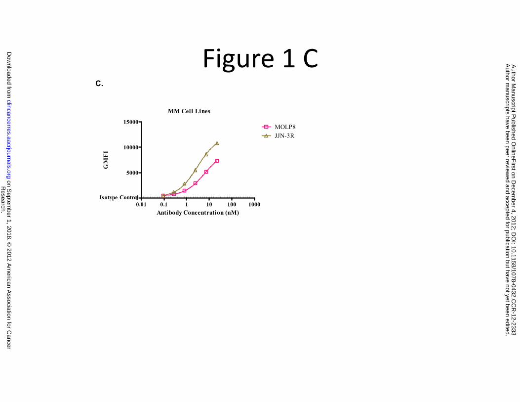
Figure 1 C
MM Cell Lines
15000MOLP8
C.
5000
10000
MOLP8JJN-3R
GM
FI
0.01 0.1 1 10 100 10000Isotype Control
Antibody Concentration (nM)
Research.
on Septem
ber 1, 2018. © 2012 A
merican A
ssociation for Cancer
clincancerres.aacrjournals.org D
ownloaded from
Author m
anuscripts have been peer reviewed and accepted for publication but have not yet been edited.
Author M
anuscript Published O
nlineFirst on D
ecember 4, 2012; D
OI: 10.1158/1078-0432.C
CR
-12-2333

Figure 1D
D
AML Patients PBMC250
D.
100
150
200
GM
FI
M4 M4 M2 M1 M5 M4 M5b M1/20
50
AML Subtype
Research.
on Septem
ber 1, 2018. © 2012 A
merican A
ssociation for Cancer
clincancerres.aacrjournals.org D
ownloaded from
Author m
anuscripts have been peer reviewed and accepted for publication but have not yet been edited.
Author M
anuscript Published O
nlineFirst on D
ecember 4, 2012; D
OI: 10.1158/1078-0432.C
CR
-12-2333
Figure 2 A
CEM Cells
A. Anti-CXCR4 Blockade of Binding
10000
15000
20000
Total
Anti-CXCR4 (MDX-1338)Isotype Control
CPM
0.01 0.1 1 10 100 10000
5000NSB
Antibody Concentration (nM)
Research.
on Septem
ber 1, 2018. © 2012 A
merican A
ssociation for Cancer
clincancerres.aacrjournals.org D
ownloaded from
Author m
anuscripts have been peer reviewed and accepted for publication but have not yet been edited.
Author M
anuscript Published O
nlineFirst on D
ecember 4, 2012; D
OI: 10.1158/1078-0432.C
CR
-12-2333
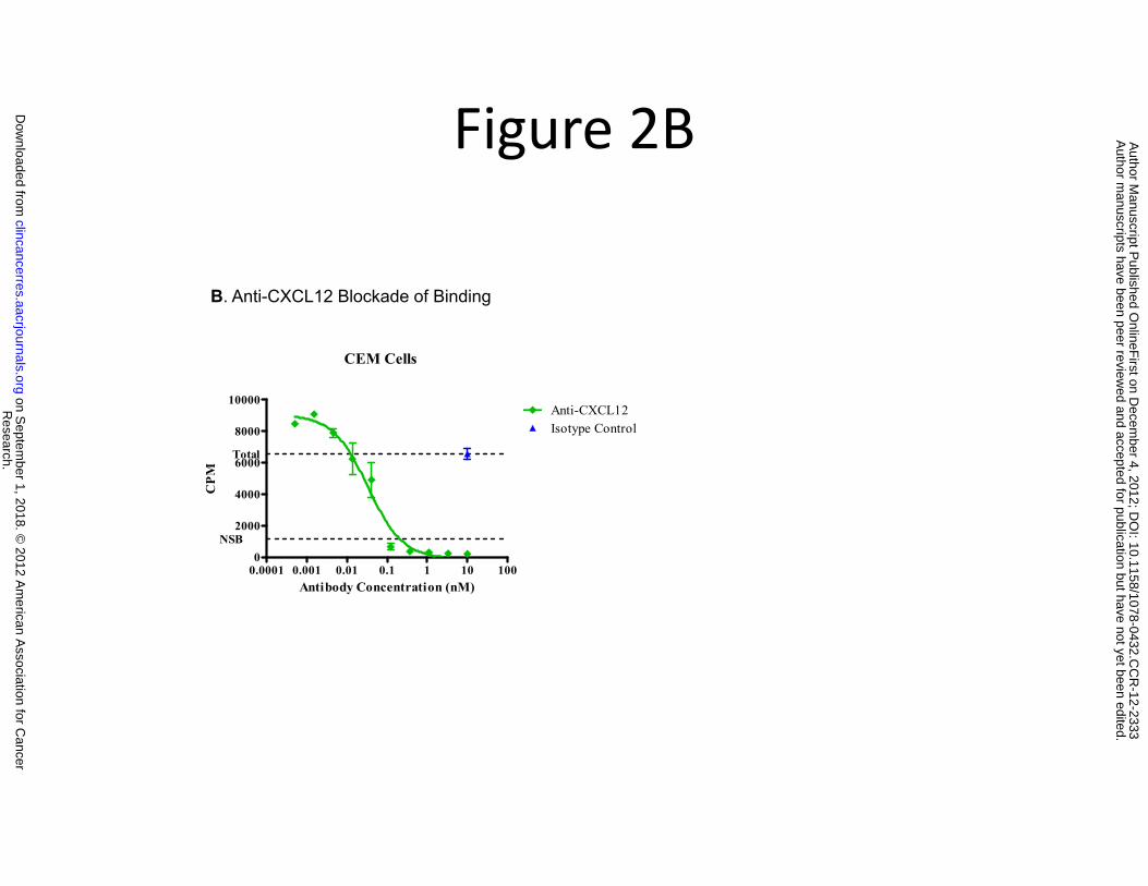
Figure 2B
CEM Cells
B. Anti-CXCL12 Blockade of Binding
4000
6000
8000
10000
Total
Anti-CXCL12Isotype Control
CPM
0.0001 0.001 0.01 0.1 1 10 1000
2000NSB
Antibody Concentration (nM)
Research.
on Septem
ber 1, 2018. © 2012 A
merican A
ssociation for Cancer
clincancerres.aacrjournals.org D
ownloaded from
Author m
anuscripts have been peer reviewed and accepted for publication but have not yet been edited.
Author M
anuscript Published O
nlineFirst on D
ecember 4, 2012; D
OI: 10.1158/1078-0432.C
CR
-12-2333
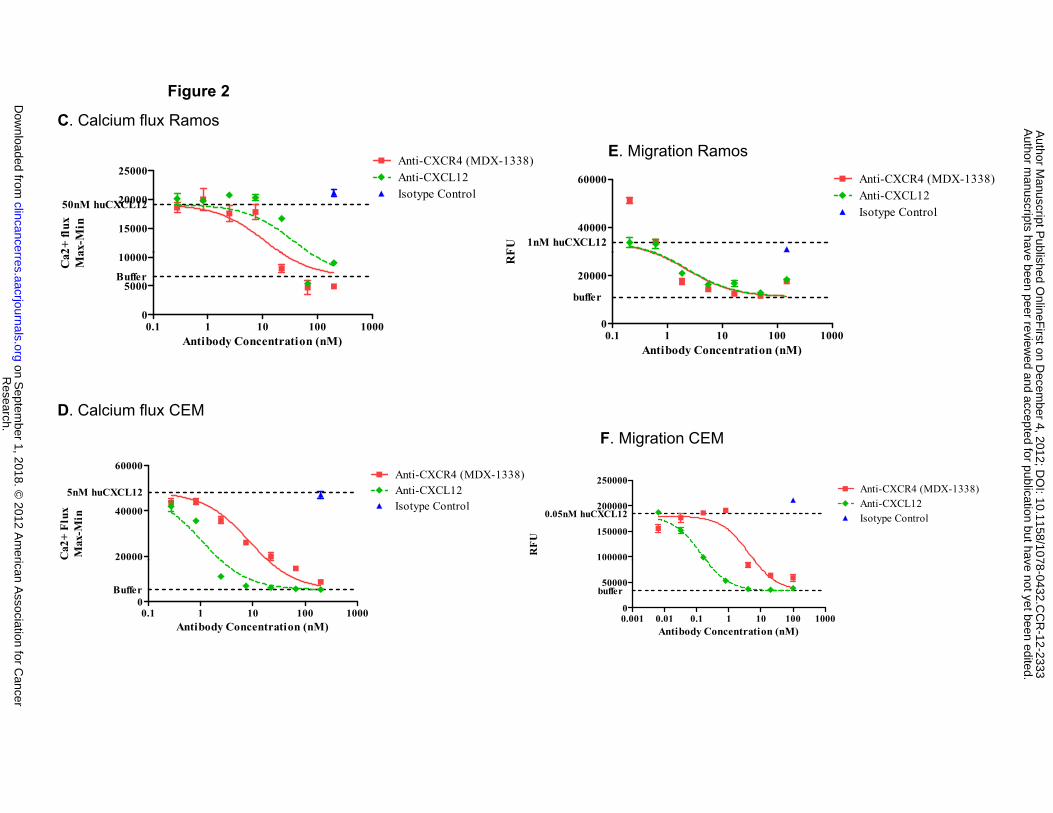
Figure 2
C. Calcium flux Ramos
E Migration Ramos
15000
20000
25000
50nM huCXCL12
Anti-CXCR4 (MDX-1338)Anti-CXCL12Isotype Control
2+ fl
uxax
-Min 40000
60000
1nM huCXCL12
Anti-CXCR4 (MDX-1338)Anti-CXCL12Isotype Control
FU
E. Migration Ramos
0.1 1 10 100 10000
5000
10000
Buffer
Antibody Concentration (nM)
Ca2 Ma
0.1 1 10 100 10000
20000
buffer
Antibody Concentration (nM)
RF
y ( )
D. Calcium flux CEM
F. Migration CEM
20000
40000
60000
5nM huCXCL12
Anti-CXCR4 (MDX-1338)Anti-CXCL12Isotype Control
Ca2
+ Fl
uxM
ax-M
in
100000
150000
200000
250000
0.05nM huCXCL12
Anti-CXCR4 (MDX-1338)Anti-CXCL12Isotype Control
RFU
0.1 1 10 100 10000
Buffer
Antibody Concentration (nM)0.001 0.01 0.1 1 10 100 10000
50000buffer
Antibody Concentration (nM)
Research.
on Septem
ber 1, 2018. © 2012 A
merican A
ssociation for Cancer
clincancerres.aacrjournals.org D
ownloaded from
Author m
anuscripts have been peer reviewed and accepted for publication but have not yet been edited.
Author M
anuscript Published O
nlineFirst on D
ecember 4, 2012; D
OI: 10.1158/1078-0432.C
CR
-12-2333
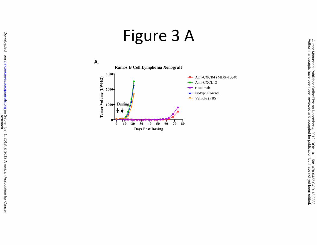
Figure 3 AA.
Ramos B Cell Lymphoma Xenograft3000
A ti CXCR4 (MDX 1338)
1000
2000
Dosing
Anti-CXCR4 (MDX-1338)Anti-CXCL12rituximabIsotype ControlVehicle (PBS)
r V
olum
e (L
WH
/2)
0 10 20 30 40 50 60 70 800
Days Post Dosing
Tum
or
Research.
on Septem
ber 1, 2018. © 2012 A
merican A
ssociation for Cancer
clincancerres.aacrjournals.org D
ownloaded from
Author m
anuscripts have been peer reviewed and accepted for publication but have not yet been edited.
Author M
anuscript Published O
nlineFirst on D
ecember 4, 2012; D
OI: 10.1158/1078-0432.C
CR
-12-2333

Figure 3 B
B.
HL60
Median Tumor Growth
0 10 20 30 40 500
1000
2000
3000MDX-1338 10mg/kg Q3-4Dx5
Isotype Control 10mg/kg Q3-4Dx5
Vehicle (PBS) Q3-4Dx5
Days Post Dosing
Tu
mo
r V
olu
me (
LW
H/2
)
Research. on September 1, 2018. © 2012 American Association for Cancerclincancerres.aacrjournals.org Downloaded from
Author manuscripts have been peer reviewed and accepted for publication but have not yet been edited. Author Manuscript Published OnlineFirst on December 4, 2012; DOI: 10.1158/1078-0432.CCR-12-2333

Figure 3 CC. Nomo-1
Median Tumor Growth3000
MDX 1338 10 /k Q3 4D 5)
1000
2000cytarabine-C 90 mg/kgcytarabine-C 60 mg/kgcytarabine-C 20 mg/kg
MDX-1338 10 mg/kg Q3-4Dx5Isotype Contro 10mg/kg Q3-4Dx5Vehicle
or V
olum
e (L
WH
/2)
0 10 20 30 40 50 600
Days Post Dosing
Tum
o
Research.
on Septem
ber 1, 2018. © 2012 A
merican A
ssociation for Cancer
clincancerres.aacrjournals.org D
ownloaded from
Author m
anuscripts have been peer reviewed and accepted for publication but have not yet been edited.
Author M
anuscript Published O
nlineFirst on D
ecember 4, 2012; D
OI: 10.1158/1078-0432.C
CR
-12-2333
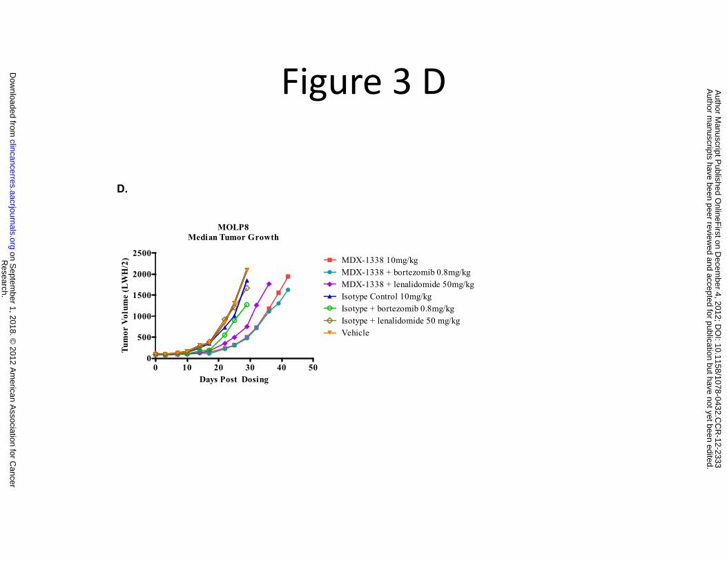
Figure 3 D
MOLP8Median Tumor Growth
D.
1000
1500
2000
2500MDX-1338 10mg/kgMDX-1338 + bortezomib 0.8mg/kgMDX-1338 + lenalidomide 50mg/kgIsotype Control 10mg/kgIsotype + bortezomib 0.8mg/kg
olum
e (L
WH
/2)
0 10 20 30 40 500
500
1000 Isotype + lenalidomide 50 mg/kgVehicle
Days Post Dosing
Tum
or V
o
Research.
on Septem
ber 1, 2018. © 2012 A
merican A
ssociation for Cancer
clincancerres.aacrjournals.org D
ownloaded from
Author m
anuscripts have been peer reviewed and accepted for publication but have not yet been edited.
Author M
anuscript Published O
nlineFirst on D
ecember 4, 2012; D
OI: 10.1158/1078-0432.C
CR
-12-2333
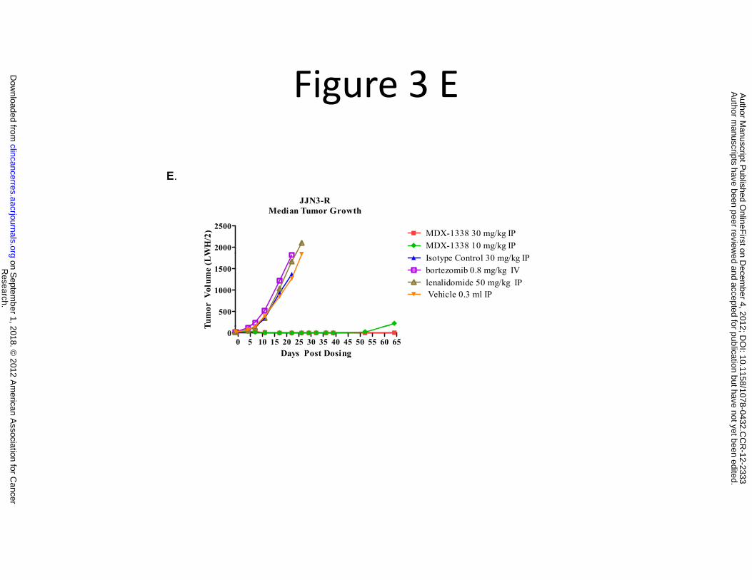
Figure 3 E
E.
JJN3-RMedian Tumor Growth
2000
2500
MDX-1338 10 mg/kg IPMDX-1338 30 mg/kg IP
WH
/2)
500
1000
1500 bortezomib 0.8 mg/kg IVlenalidomide 50 mg/kg IP Vehicle 0.3 ml IP
Isotype Control 30 mg/kg IP
Tum
or V
olum
e (L
W
0 5 10 15 20 25 30 35 40 45 50 55 60 650
Days Post Dosing
T
Research.
on Septem
ber 1, 2018. © 2012 A
merican A
ssociation for Cancer
clincancerres.aacrjournals.org D
ownloaded from
Author m
anuscripts have been peer reviewed and accepted for publication but have not yet been edited.
Author M
anuscript Published O
nlineFirst on D
ecember 4, 2012; D
OI: 10.1158/1078-0432.C
CR
-12-2333
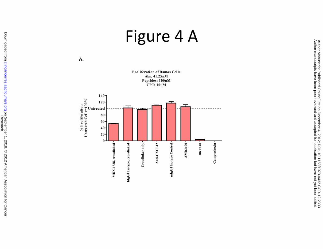
Figure 4 AA.
Proliferation of Ramos CellsAbs: 41.25nMbs: . 5
Peptides: 100uMCPT: 10uM
120
140
Untreatedion
=100
%
0
20
40
60
80
Untreated
% P
roli
fera
tiU
ntre
ated
Cel
ls=
X-1
338,
cro
sslin
ked
Isot
ype,
cro
sslin
ked
Cro
sslin
ker
only
Ant
i-CX
CL
12
gG1
Isot
ype
Con
trol
AM
D31
00
BK
T14
0
Cam
ptot
heci
n
0
MD
X
hIgG
4 I
mIg
Research.
on Septem
ber 1, 2018. © 2012 A
merican A
ssociation for Cancer
clincancerres.aacrjournals.org D
ownloaded from
Author m
anuscripts have been peer reviewed and accepted for publication but have not yet been edited.
Author M
anuscript Published O
nlineFirst on D
ecember 4, 2012; D
OI: 10.1158/1078-0432.C
CR
-12-2333

Figure 4 B
BIsotype ControlNo Treatment
10 1
10 2
10 3
10 4
FL1-
H
1.67 4.05
10 1
10 2
10 3
10 4
FL1-
H
10 1
10 2
10 3
10 4
FL1-
H
10 1
10 2
10 3
10 4
10 1
10 2
10 3
10 4
FL1-
H
1.671.67 4.05
10 1
10 2
10 3
10 4
FL1-
H
0.51 2.8
10 1
10 2
10 3
10 4
FL1-
H
10 1
10 2
10 3
10 4
FL1-
H
10 1
10 2
10 3
10 4
10 1
10 2
10 3
10 4
FL1-
H
0.510.51 2.8
An
B.
10 0 10 1 10 2 10 3 10 4
FL3-H
10 0 3.1991.1
10 0 10 1 10 2 10 3 10 4
FL3-H
10 0
10 0 10 1 10 2 10 3 10 4
FL3-H10 0 10 1 10 2 10 3 10 410 0 10 1 10 2 10 3 10 4
FL3-H
10 010 010 0 3.193.1991.191.1
31.2 27.331.231.2 27.3
10 0 10 1 10 2 10 3 10 4
FL3-H
10 0 3.1393.6
10 0 10 1 10 2 10 3 10 4
FL3-H
10 0
10 0 10 1 10 2 10 3 10 4
FL3-H10 0 10 1 10 2 10 3 10 410 0 10 1 10 2 10 3 10 4
FL3-H
10 010 010 0 3.133.1393.693.6
10 3
10 4
10 3
10 4
10 3
10 4
10 3
10 4
2.032.03 2.65
MDX-1338 AMD3100
nnexin
10 0 10 1 10 2 10 3 10 4
FL3-H
5.336.2
10 0 10 1 10 2 10 3 10 4
FL3-H10 0 10 1 10 2 10 3 10 410 0 10 1 10 2 10 3 10 4
FL3-H
5.35.336.236.2
10 0 10 1 10 2 10 3 10 4
FL3-H
10 0
10 1
10 2
FL1-
H
10 0 10 1 10 2 10 3 10 4
FL3-H10 0 10 1 10 2 10 3 10 410 0 10 1 10 2 10 3 10 4
FL3-H
10 0
10 1
10 2
FL1-
H
10 0
10 1
10 2
10 0
10 1
10 2
FL1-
H
3.233.2392.192.1
V
PIPI
Research.
on Septem
ber 1, 2018. © 2012 A
merican A
ssociation for Cancer
clincancerres.aacrjournals.org D
ownloaded from
Author m
anuscripts have been peer reviewed and accepted for publication but have not yet been edited.
Author M
anuscript Published O
nlineFirst on D
ecember 4, 2012; D
OI: 10.1158/1078-0432.C
CR
-12-2333
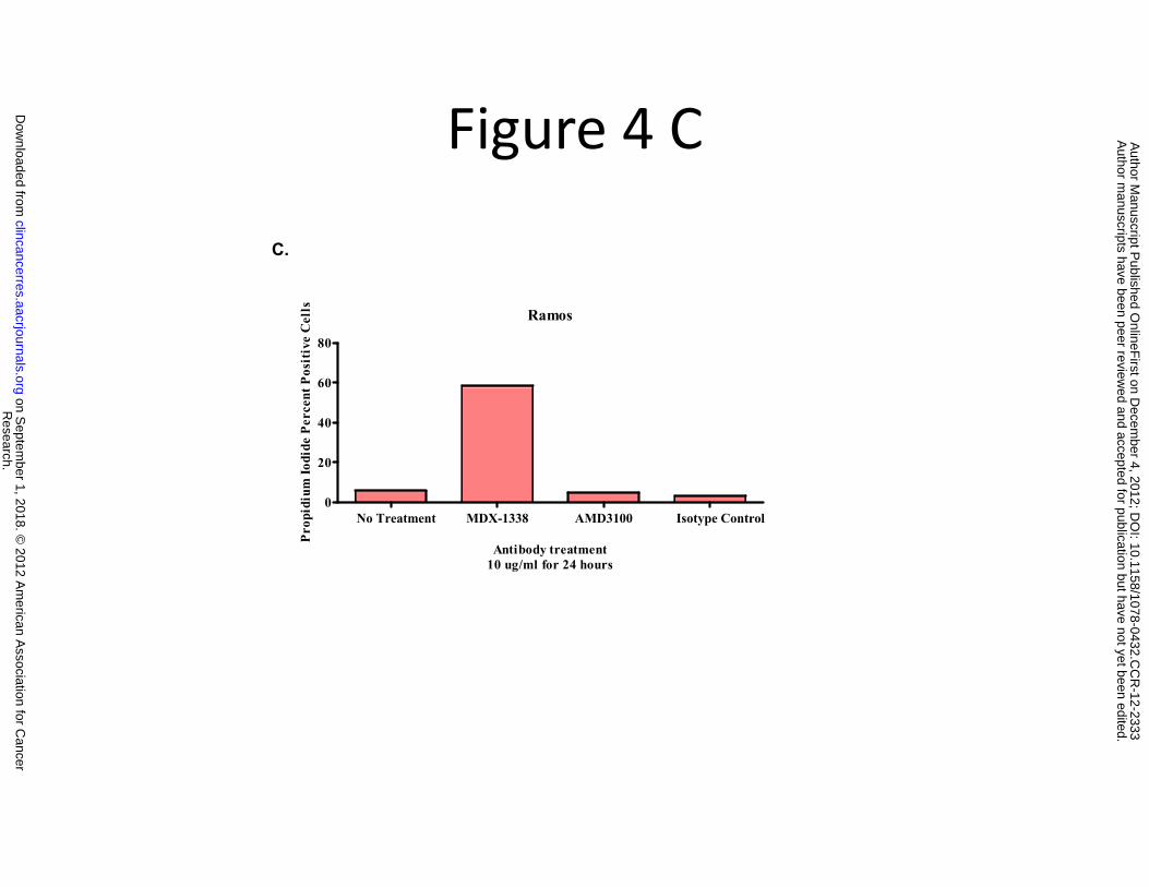
Figure 4 C
C.
Ramos
60
80
Pos
itiv
e C
ells
0
20
40
ium
Iodi
de P
erce
nt
No Treatment MDX-1338 AMD3100 Isotype Control0
Antibody treatment10 ug/ml for 24 hours
Pro
pidi
Research.
on Septem
ber 1, 2018. © 2012 A
merican A
ssociation for Cancer
clincancerres.aacrjournals.org D
ownloaded from
Author m
anuscripts have been peer reviewed and accepted for publication but have not yet been edited.
Author M
anuscript Published O
nlineFirst on D
ecember 4, 2012; D
OI: 10.1158/1078-0432.C
CR
-12-2333
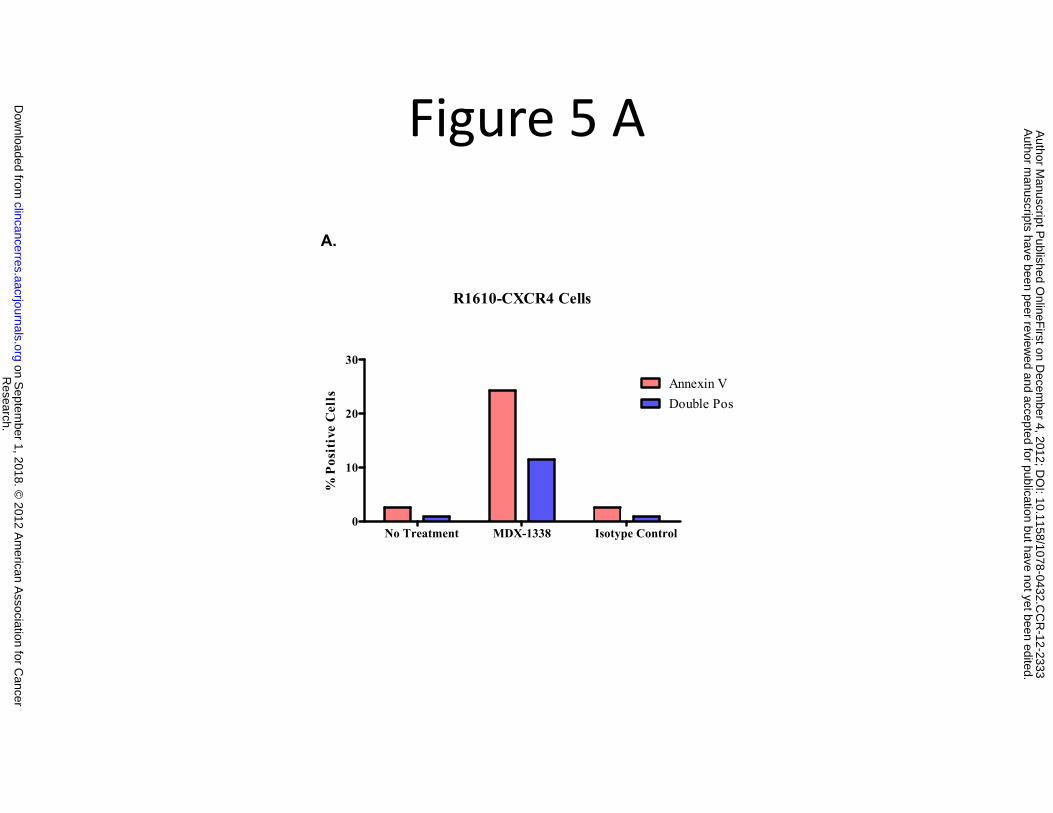
Figure 5 A
A.
R1610-CXCR4 Cells
20
30
Annexin VDouble Pos
siti
ve C
ells
No Treatment MDX-1338 Isotype Control0
10
% P
o
Research.
on Septem
ber 1, 2018. © 2012 A
merican A
ssociation for Cancer
clincancerres.aacrjournals.org D
ownloaded from
Author m
anuscripts have been peer reviewed and accepted for publication but have not yet been edited.
Author M
anuscript Published O
nlineFirst on D
ecember 4, 2012; D
OI: 10.1158/1078-0432.C
CR
-12-2333
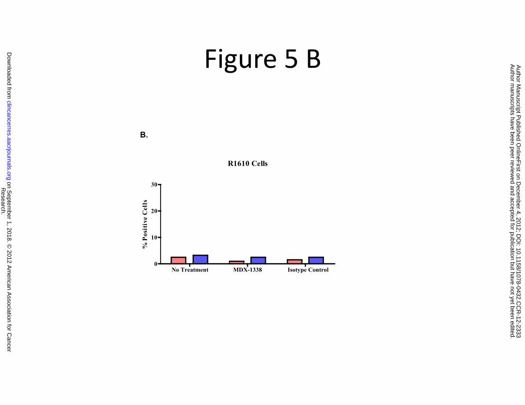
Figure 5 B
B.
R1610 Cells
20
30
tive
Cel
ls
No Treatment MDX-1338 Isotype Control0
10
% P
osi
Research.
on Septem
ber 1, 2018. © 2012 A
merican A
ssociation for Cancer
clincancerres.aacrjournals.org D
ownloaded from
Author m
anuscripts have been peer reviewed and accepted for publication but have not yet been edited.
Author M
anuscript Published O
nlineFirst on D
ecember 4, 2012; D
OI: 10.1158/1078-0432.C
CR
-12-2333
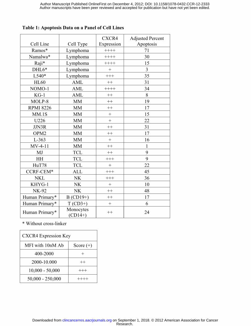
Table 1: Apoptosis Data on a Panel of Cell Lines
Cell Line Cell Type CXCR4
Expression Adjusted Percent
Apoptosis Ramos* Lymphoma ++++ 71
Namalwa* Lymphoma ++++ 30 Raji* Lymphoma ++++ 15
DHL6* Lymphoma + 3 L540* Lymphoma +++ 35 HL60 AML ++ 31
NOMO-1 AML ++++ 34 KG-1 AML ++ 8
MOLP-8 MM ++ 19 RPMI 8226 MM ++ 17
MM.1S MM + 15 U226 MM + 22 JJN3R MM ++ 31 OPM2 MM ++ 17 L-363 MM + 16
MV-4-11 MM ++ 1 MJ TCL ++ 9 HH TCL +++ 9
HuT78 TCL + 22 CCRF-CEM* ALL +++ 45
NKL NK +++ 36 KHYG-1 NK + 10 NK-92 NK ++ 48
Human Primary* B (CD19+) ++ 17 Human Primary* T (CD3+) + 6
Human Primary* Monocytes (CD14+)
++ 24
* Without cross-linker
CXCR4 Expression Key
MFI with 10nM Ab Score (+)
400-2000 +
2000-10.000 ++
10,000 - 50,000 +++
50,000 - 250,000 ++++
Research. on September 1, 2018. © 2012 American Association for Cancerclincancerres.aacrjournals.org Downloaded from
Author manuscripts have been peer reviewed and accepted for publication but have not yet been edited. Author Manuscript Published OnlineFirst on December 4, 2012; DOI: 10.1158/1078-0432.CCR-12-2333

Published OnlineFirst December 4, 2012.Clin Cancer Res Michelle R. Kuhne, Tanya Mulvey, Blake Belanger, et al. in hematologic malignancies.induces apoptosis in vitro and shows anti tumor activity in vivo BMS-936564/MDX-1338: A fully human anti-CXCR4 antibody
Updated version
10.1158/1078-0432.CCR-12-2333doi:
Access the most recent version of this article at:
Manuscript
Authoredited. Author manuscripts have been peer reviewed and accepted for publication but have not yet been
E-mail alerts related to this article or journal.Sign up to receive free email-alerts
Subscriptions
Reprints and
To order reprints of this article or to subscribe to the journal, contact the AACR Publications
Permissions
Rightslink site. Click on "Request Permissions" which will take you to the Copyright Clearance Center's (CCC)
.http://clincancerres.aacrjournals.org/content/early/2012/12/04/1078-0432.CCR-12-2333To request permission to re-use all or part of this article, use this link
Research. on September 1, 2018. © 2012 American Association for Cancerclincancerres.aacrjournals.org Downloaded from
Author manuscripts have been peer reviewed and accepted for publication but have not yet been edited. Author Manuscript Published OnlineFirst on December 4, 2012; DOI: 10.1158/1078-0432.CCR-12-2333