Embed Size (px)
Citation preview

1353
IntroductionStem cells in adult tissues have the ability to self-renew andgenerate differentiated cells that maintain tissue homeostasis.Specific regulatory microenvironments, also known as niches,are thought to regulate many stem cell types by producingsignals important for stem cell proliferation and differentiation(Watt and Hogan, 2000; Spradling et al., 2001). Stem cellsusually divide asymmetrically to generate parent stem cells anddifferentiated cells, although they can also undergo symmetriccell division to replenish lost stem cells or expand the stem cellpool (Xie and Spradling, 2000; Zhu and Xie, 2003). Eventhough many niche signals have been identified for differentstem cell types in many systems, it is still largely unknown howniche signals control stem cell self-renewal and differentiation(Spradling et al., 2001). Therefore, it is essential to reveal thelink between niche signals and intrinsic factors that areessential for stem cell self-renewal and differentiation in orderto gain a better understanding of how stem cell behavior iscontrolled.
The Drosophila ovarian germline stem cells (GSCs) havebecome an attractive system to study stem cells and theirrelationship with niches (Xie and Spradling, 2001; Lin, 2002).
Two or three GSCs are located at the tip of the ovariole, alsoknown as the germarium, and are surrounded by terminalfilament cells, cap cells and inner sheath cells that form a nichefor GSCs. GSCs and their progeny in the germarium can bereliably identified and distinguished by a germ cell-specificstructure, called the fusome, which is rich in membraneskeletal proteins, such as Hu li tai shao (Hts) and α-Spectrin(Lin et al., 1994; de Cuevas et al., 1997). In GSCs and theirimmediate differentiating daughters, cystoblasts, the fusome isspherical in shape, and is also known as the spectrosome. Acystoblast will undergo synchronous mitotic divisions withincomplete cytokinesis to generate two-, four-, eight- andsixteen-cell cysts, in which the fusome is branched tointerconnect individual cystocytes (Lin et al., 1994). GSCs areinvariably anchored to cap cells through adherens junctions(Song et al., 2002). Loss of adherens junctions between capcells and GSCs causes GSCs to migrate away from cap cellsand undergo differentiation (Song et al., 2002). Upon GSCdivision, the original GSC remains anchored to cap cells andretains stem cell identity, whereas the cystoblast moves awayfrom cap cells and undergoes differentiation. As a GSC is lost,a neighboring GSC can generate two daughter cells that bothcontact cap cells and remain as GSCs, thus replenishing a
The Drosophilaovary is an attractive system to study howniches control stem cell self-renewal and differentiation.The niche for germline stem cells (GSCs) provides aDpp/Bmp signal, which is essential for GSC maintenance.bam is both necessary and sufficient for the differentiationof immediate GSC daughters, cystoblasts. Here we showthat Bmp signals directly repress bamtranscription inGSCs in the Drosophilaovary. Similar to dpp, gbbencodesanother Bmp niche signal that is essential for maintainingGSCs. The expression of phosphorylated Mad (pMad), aBmp signaling indicator, is restricted to GSCs and somecystoblasts, which have repressed bamexpression. BothDpp and Gbb signals contribute to pMad production. bam
transcription is upregulated in GSCs mutant for dppandgbb. In marked GSCs mutant for Med and punt, twoessential Bmp signal transducers, bamtranscription is alsoelevated. Finally, we show that Med and Mad directly bindto the bamsilencer in vitro. This study demonstrates thatBmp signals maintain the undifferentiated or self-renewalstate of GSCs, and directly repress bamexpression in GSCsby functioning as short-range signals. Thus, niche signalsdirectly repress differentiation-promoting genes in stemcells in order to maintain stem cell self-renewal.
Key words: germline, stem cells, male, Bmps
Summary
Bmp signals from niche cells directly repress transcription of adifferentiation-promoting gene, bag of marbles , in germline stemcells in the Drosophila ovaryXiaoqing Song 1,*, Marco D. Wong 1,*, Eihachiro Kawase 1,*, Rongwen Xi 1, Bee C. Ding 1, John J. McCarthy 1
and Ting Xie 1,2,†
1Stowers Institute for Medical Research, 1000 East 50th Street, Kansas City, MO 64110, USA2Department of Anatomy and Cell Biology, University of Kansas School of Medicine, 3901 Rainbow Boulevard, Kansas City,KS 66160, USA*These authors contributed equally†Author for correspondence (e-mail: [email protected]).
Accepted 9 December 2003
Development 131, 1353-1364Published by The Company of Biologists 2004doi:10.1242/dev.01026
Research article

1354
vacant niche space (Xie and Spradling, 2000). Functioning asa GSC niche, terminal filament/cap cells express piwi, dpp,fs(1)Yb (also known as Yb) and hedgehog(hh), which areessential for maintaining GSC asymmetric cell division (Xieand Spradling, 1998; King and Lin, 1999; Cox et al., 2000; Xieand Spradling, 2000; King et al., 2001). Intrinsic factorsin GSCs, including pumilio, nanos, dppreceptors anddownstream components, are also important for GSCmaintenance (Lin and Spradling, 1997; Forbes and Lehmann,1998; Xie and Spradling, 1998). Two intrinsic factors, bag ofmarbles(bam) and benign gonial cell neoplasm(bgcn), arerequired in cystoblasts for their proper differentiation(McKearin and Spradling, 1990; McKearin and Ohlstein, 1995;Lavoie et al., 1999). However, in GSCs the interplay betweengenes involved in self-renewal versus differentiation remainsunclear.
The functions of dppsignaling and bamin the maintenanceof GSCs and the differentiation of cystoblasts seem to bedirectly opposing. Loss of bamfunction completely eliminatescystoblast differentiation, similar to that caused by dppoverexpression (McKearin and Spradling, 1990; Xie andSpradling, 1998). By contrast, forced overexpression of baminGSCs causes their elimination, similar to that observed whendpp signaling is disrupted in GSCs (Ohlstein and McKearin,1997; Xie and Spradling, 1998). These observations can beexplained by a simple model wherein dpp, functioning as ashort-range signal, directly promotes GSC self-renewal andsuppresses bam expression in GSCs, while allowingcystoblasts to express bamand differentiate.
Several studies have supported this model (Xie andSpradling, 1998; Chen and McKearin, 2003a; Kai andSpradling, 2003). bammRNA is absent in GSCs, but quicklyaccumulates in cystoblasts and mitotic cysts (McKearinand Spradling, 1990). Overexpression of dpp completelysuppresses the expression of BamC protein in germ cells, thuspreventing cystoblasts from differentiating (Xie and Spradling,1998). A recent study by Kai and Spradling showed that dppsignaling activity is restricted to GSCs and cystoblasts (Kai andSpradling, 2003).
The asymmetric distribution of bambetween GSCs andcystoblasts could be due to transcriptional regulation and/ormRNA stability. The recent elegant bampromoter analysis hasrevealed that its transcription is actively repressed through asilencer (Chen and McKearin, 2003a). However, whether andhow dppsignaling directly represses bamtranscription remainsunknown. In this study, we provide genetic and molecularevidence to support the model proposing that Bmp signalingrepresses bamtranscription through binding of its downstreamtranscriptional effectors, Mad and Medea (Med), to the definedbamsilencer.
Materials and methodsDrosophila stocks and geneticsThe following fly stocks used in this study were described either inFlyBase or as otherwise specified in the Results section:
punt10460;punt135; Med26; Dad-lacZ; dpphr4;dpphr56; gbb4, gbbD4;gbbD20; bam-GFP(GFP gene driven by the bampromoter);vasa-GFP; c587-gal4;
hs-gal4; UAS-dpp; andUAS-gbb; hsFLP; FRT82B armadillo-lacZ.Most stocks were cultured at room temperature. To maximize their
mutant phenotypes, dpp, gbband puntmutant adult females werecultured at 29°C for 2-7 days. To achieve the uniform GSC-likephenotype, c587-gal4;UAS-dppfemales were also cultured at 29°Cfor 7 days.
Generating mutant GSC clones and overexpressionClones of mutant GSCs were generated by Flp-mediated mitoticrecombination, as described previously (Xu and Rubin, 1993; Xie andSpradling, 1998). To generate the stocks for making mutant GSCclones and examining bam-GFPexpression, 2-day old hsFLP; bam-GFP/+; FRT82B punt135/FRT82B armadillo-lacZ and hsFLP; bam-GFP/+; FRT82B Med26/FRT82B armadillo-lacZ females were heat-shocked at 37°C for 3 consecutive days with two one-hour heat-shocktreatments separated by 8-12 hours. The ovaries were removed 3 daysafter the last heat-shock treatment and then processed for antibodystaining.
To construct the stocks for overexpressing dppor gbb, the femalesthat carried hs-gal4, and either UAS-dppor UAS-gbb, were heat-shocked at 37°C for different lengths of time for particularexperiments, as indicated in the Results. The females that carriedc587-gal4 and UAS-dpp or UAS-gbb were cultured at roomtemperature or at 29°C for 7 days. For examining the expression ofbam-GFPin the ovary overexpressing dpp or gbb, the females thatcarried c587-gal4 or hs-gal4, andUAS-dpp or UAS-gbb, also carrieda bam-GFP transgene.
Measuring GSC loss in gbb mutants and examining bam-GFP expression in gbb , dpp or punt mutant germariaTo measure stem cell loss in gbbmutant and control ovaries, thegermaria with different numbers of GSCs, ranging from three to none,were counted from the ovaries of 2-day- and one-week-old bam-GFPgbb4/gbbD4, bam-GFP gbb4/gbbD20 or bam-GFP(control) females.The 2-day-old control and gbbmutant females were cultured at roomtemperature after they eclosed at 18°C, whereas the one-week-oldcontrol and gbbmutant females were cultured at 29°C. Values areexpressed as the average GSC number per germarium and thepercentage of germaria with no GSCs.
To examine bam-GFPexpression in dpp, gbbor punt mutantgermaria, we generated females with the following genotypes at18°C: bam-GFP gbb4/gbbD4, bam-GFP gbb4/gbbD20, bam-GFPdpphr56/dpphr4, bam-GFP; punt10460/punt135 or bam-GFP (control)females. All the control and mutant females were cultured at 29°C for4 days before their ovaries were isolated and immunostained, tocomparebam-GFP expression under identical conditions.
ImmunohistochemistryThe following antisera were used: polyclonal anti-Vasa antibody(1:2000) (Liang et al., 1994); monoclonal anti-Hts antibody (1:3);polyclonal anti-β-galactosidase antibody (1:100; Cappel); polyclonalanti-GFP antibody (1:200; Molecular Probes); and polyclonal anti-pMad antibody (1:200) (Tanimoto et al., 2000). The immunostainingprotocol used in this study was described previously (Song et al.,2002). All micrographs were taken using a Leica SPII confocalmicroscope.
Examining gene expression using the Affymetrixmicroarray Total RNA from the ovaries of different genotypes or treatments wasisolated using Trizol (Invitrogen), and biotin-labeled cRNA probeswere produced using an RNA transcript labeling kit (Enzo BioArray).The Drosophila GeneChips were purchased from Affymetrix, andwere hybridized, stained and detected according to the manufacturer’sinstructions.
Development 131 (6) Research article

1355Bmps repress bam transcription in GSCs
Detecting gene expression in purified component cellsusing RT-PCRAfter sorting GFP-positive cells by using Cytomation MoFlo, totalRNA was prepared using Trizol (Invitrogen) from these isolated cells.The RNA samples were further amplified using the GeneChipEukaryotic Small Sample Target Labeling Assay Version II(Affymetrix). After the RNA amplification, 100 ng of total RNAwas reverse-transcribed (RT) using the SuperScriptIII First-StrandSynthesis System for RT-PCR, according to manufacturer’s protocol(Invitrogen). The following primers were used in this study:
dpp, 5′-AGCCGATGAAGAAGCTCTACG-3′ and 5′-ATGTCG-TAGACAAGCACCTGGTA-3′;
vasa, 5′-ATCGAGGAGGAAATCGAGATGGA-3′ and 5′-GGAA-GCTATGCCACTGCTGAATA-3′;
gbb, 5′-AGATGCAGACCCTGTACATAGAC-3′ and 5′-CTCGTC-GTTCAGGTGGTACAGAA-3′; and
rp49, 5′-GTATCGACAACAGAGTCGGTCGC-3′and 5′-TTGGT-GAGCGGACCGACAGCTGC-3′.
PCR was performed as follows: 94°C for 2 minutes; 35 cycles of94°C for 30 seconds, 45°C for 30 seconds and 72°C for 45 seconds;and 72°C for 7 minutes. RT-PCR products were electrophoresed on a2% agarose gel in the presence of ethidium bromide.
Electrophoretic mobility shift assays for the binding ofMad and Med to the bam silencerThe GST-Mad construct was described previously (Kim et al., 1997).Med was PCR-amplified from its cDNA, with the introduction ofXhoI sites at both ends, then subcloned into a pGEX-4T2 vector(Amersham Pharmacia Biotech). Its sequence was confirmed bysequencing. GST-Mad, GST-Med and GST proteins were purified byaffinity chromatography using Glutathione Sepharose™ 4B accordingto the manufacturer’s protocol (Amersham Pharmacia Biotech), andconfirmed by western blots.
A Cy5 5′-modified oligonucleotide containing the bipartite bamsilencer element (+17 to +54) was used as a probe. Binding reactionswere performed according to the published protocol (Kim et al.,1997). Specificity of binding was determined by the addition of 100-fold molar excess of unlabeled competitor DNA corresponding to thebamsilencer element, with site A and/or B. DNA-protein complexeswere resolved on a 5% (w/v) non-denaturing polyacrylamide gel using0.5×TBE running buffer at 150 V for 3 hours at 4°C. Gels wereimaged on a Typhoon 8700 (Amersham Biosciences).
Resultsdpp signaling activity is correlated with bamtranscriptional repression in GSCs and cystoblastsPrevious studies have shown that a dpp signal produced bysomatic cells is essential for maintaining GSCs but not forcystoblast development (Xie and Spradling, 1998; Xie andSpradling, 2000). bam transcription is active in young,differentiating germ cells but is repressed specifically in GSCsin the ovary (Chen and McKearin, 2003a). This raises theinteresting possibility that dpp signaling and bamexpressiondirectly oppose each other. To investigate this possibility, weexamined the correlation between dppsignaling activity andbam expression in the germarium. dppsignaling activity isusually monitored by Dad and phosphorylated Mad (pMad)expression (Tsuneizumi et al., 1997; Tanimoto et al., 2000).Dad is a dpptarget gene, and a Dad-lacZline recapitulates itsexpression (Tsuneizumi et al., 1997). A bam-GFPtransgene(with the GFP gene driven by the bam promoter) has beengenerated to study bam transcription (Chen and McKearin,2003a). Throughout this study, an anti-Hts antibody was usedto label spectrosomes and fusomes, and a DNA dye, DAPI, wasused to label nuclei. Cap cells can be reliably identified bybright DAPI staining, and by their unique position and nuclearmorphology. GSCs are identified by the presence of aspectrosome (a spherical fusome) on their anterior side and bytheir direct contact with cap cells; cystoblasts also contain aspectrosome but fail to be associated with cap cells (Fig. 1A).
Similar to the observations recently made by Kai andSpradling (Kai and Spradling, 2003), Dad-lacZwas expressedin GSCs and some cystoblasts at high levels, but in the othercystoblasts and mitotic cysts at much lower levels (Fig. 1B).As reported by Chen and McKearin (Chen and McKearin,2003a), bam transcription was repressed in GSCs and somecystoblasts, but was active in the other cystoblasts and dividingcystocytes (Fig. 1C). In germaria carrying bam-GFPand Dad-lacZ, GSCs and the cystoblasts that had strong Dad expressiondid not show bam-GFPexpression (Fig. 1D), whereas thecystoblasts and mitotic cysts that had weak or no Dad
Fig. 1.dppsignaling activity is restricted to GSCs and somecystoblasts where bamtranscription is actively repressed.(A) Diagram showing GSCs, their differentiated progenyand surrounding somatic cells. In panels B-F, cap cells arehighlighted by circles, whereas GSCs are indicated byasterisks. (B) Tip of the Dad-lacZgermarium labeled fornuclear β-Gal (red), Hts (green, fusomes) and DAPI (blue),showing high Dadexpression in GSCs and in a cystoblast(arrow), but not in another cystoblast (arrowhead). (C) Tipof the bam-GFPgermarium labeled for GFP (green), Hts(red, fusomes) and DAPI (blue), showing bamexpression ina cystoblast (arrowhead) and cysts, but not in GSCs. (D) Tipof the bam-GFP;Dad-lacZgermarium labeled for β-Gal(red), GFP (green) and DAPI (blue), showing that GSCsand two cystoblasts (arrows) express high Dadbut no bam,and that a cystoblast (arrowhead) has low Dadand begins toexpress bam. (E,F) Tip of the bam-GFPgermarium labeledfor pMad (red), GFP (green), Hts (blue, fusomes) and DAPI(white, F), showing high pMad accumulation but no bam
expression in GSCs, and low pMad but bamexpression in a cystoblast (arrowhead, E). TF, terminal filament; GSCs, germline stem cells; SS,spectrosome; Cpc, cap cells; CB, cystoblast; FS, fusome; IGS, inner sheath cells; CS, cysts. All micrographs are shown at the same scale. Scalebar: 10 µm.

1356
expression showed obvious bam-GFPexpression (Fig. 1D).Similarly, GSCs and the cystoblasts that showed strong pMadexpression did not express bam-GFP, whereas the cystoblastsand mitotic cysts that showed weak or no pMad expressionexpressed bam-GFP(Fig. 1E,F). These results further supportthe idea that the dppsignaling pathway is activated in GSCs athigh levels, whereas bamtranscription is actively repressed.
dpp signaling is essential for repressing bamtranscription in GSCsThe dpphr56/dpphr4 temperature-sensitive mutant was chosen toinvestigate the expression of bam-GFPin dpp mutant GSCsbecause it shows gradual loss of GSCs within two weeks at arestrictive temperature (29°C) (Xie and Spradling, 1998). Afterbam-GFPand bam-GFP dpphr56/dpphr4 females were culturedat 29°C for 2, 4 or 7 days, the ovaries were immunostainedwith anti-GFP and anti-Hts antibodies to visualize bam-GFPand fusomes, respectively. In the germaria from the bam-GFPfemales, the bam-GFPexpression pattern was completelynormal, and was absent in GSCs even one week after beingcultured at 29°C (Fig. 2A). However, even two days after beingcultured at 29°C, 28% of the bam-GFP dpphr56/dpphr4
germaria that contained GSCs started to express bam-GFPinone or more GSCs (n=283; Fig. 2B). After 4 days and 7 days,66% (n=35) and 89% (n=19) of the mutant germaria that stillhad at least one GSC expressed bam-GFP in one or moreGSCs, respectively (Fig. 2C,D). To further confirm the role ofdpp signaling in repressing bamtranscription, we alsocompared the levels of bammRNA in wild-type and dppmutant ovaries using a microarray approach. bammRNA wasdramatically upregulated in dpphr4/dpphr56 mutant ovaries incomparison with wild type (Table 1; samples were normalizedwith an internal control, the Actin 42Agene). These resultsdemonstrate that the dppsignal is required to repress bamtranscription in GSCs.
Next, we investigated whether elevated bamtranscription indpp mutant GSCs can be correlated with reduction of pMadexpression. After 4 days at 29°C, control germaria maintained
the normal number of GSCs and showed the normal pMadexpression pattern (Fig. 2E,F). By contrast, many dpp mutantgermaria completely lost their GSCs, and in the remainingGSC-containing germaria in which bam-GFPwas alsoupregulated in GSCs, pMad was severely reduced but notcompletely eradicated in the GSCs (Fig. 2G,H). In the germariain which bam-GFPwas not obviously upregulated, levels ofpMad were relatively higher but less than normal (data notshown). These results indicate that dpp signaling contributes,at least in part, to pMad production in GSCs, and could beresponsible for repressing bam transcription.
dpp overexpression is sufficient for repressing bamtranscription in the cystoblast Our previous study showed that overexpression of dppthroughout the germarium completely inhibits cystoblastdifferentiation and causes the accumulation of GSC-like cellsthat fail to express BamC (Xie and Spradling, 1998). Ourexperiments described above suggest that the dpp signal islikely to be restricted to the tip of the germarium, adjacent tocap cells. To test whether GSCs are competent to respond todpp signaling outside their niches, dpp was specificallyoverexpressed in somatic cells other than cap cells, using thec587-gal4line to drive a UAS-dpptransgene. The c587-gal4
Development 131 (6) Research article
Fig. 2.dpp is essential forrepressing bamtranscription inGSCs. Germaria in panels A-D arelabeled for Hts (red, fusomes), GFP(green) and DAPI (blue), whereasthe germaria in E-H are labeled forpMad (red), GFP (green), Hts (blue,fusomes) and DAPI (white; F,H).All the GSCs are indicated byasterisks, and cap cells in all thepanels are marked by circles.(A) A germarial tip from a bam-GFP female cultured at 29°C forone week, showing no bamexpression in GSCs. (B) Agermarial tip from a bamGFPdpphr56/dpphr4 female cultured at29°C for 2 days, showing that oneof the two GSCs begins to expressbam. (C,D) Germarial tips from
bamGFP dpphr56/dpphr4 females cultured at 29°C for 4 (C) or 7 (D) days, showing that the only remaining GSC starts to express bam.(E,F) Germarial tip from a bam-GFPfemale cultured at 29°C for 4 days, showing high pMad accumulation and no bamexpression in GSCs.(G,H) A germarial tip from a bamGFP dpphr56/dpphr4 female cultured at 29°C for 4 days, showing that two mutant GSCs have low pMad levelsand begin to express bam. All micrographs are shown at the same scale. Scale bar: 10 µm.
Table 1. dppsignaling is necessary and sufficient forrepressing bamexpression in GSCs in the Drosophila
ovaryGenes Wild typehs-gal4/UAS-dpp C587-gal4/UAS-dpp dpphr4/dpphr56
Actin 42A 1028 2476* (1028)† 4686 (1028) 1554 (1028)dpp 4.7 354.2 (147.6) 271.9 (59.6) –3.8 (–2.5)Dad 24.0 103.0 (42.9) 296.0 (64.9) 4.9 (3.2)bam 110.0 –9.3 (–3.8) –3.2 (–0.7) 326.2 (217.5)
aThe numbers shown in this table are the arbitrary ones that were quantifiedby the Affymetrix scanner.
†The numbers in parentheses are normalized based on the number of thehouse-keeping gene Actin 42Ain wild-type ovaries.

1357Bmps repress bam transcription in GSCs
line can drive expression of a UAS-GFP transgene in innersheath cells and early follicle cells (Fig. 3A). When UAS-dppexpression was driven in inner sheath cells and follicle cells,germaria were filled with single germ cells with a spectrosome,suggesting that germ cells distant from their niche are stillcapable of responding to dpp(Fig. 3B). To further test whetherdpp overexpression is sufficient to inhibit bamexpression, weexamined bam-GFP expression in dpp-induced GSC-liketumors. In dpp-overexpressing ovaries, bam-GFPwas notexpressed in the single germ cells either close to (Fig. 3C) oraway from (Fig. 3D) the germarial tip. These results indicatethat dppsignaling is sufficient to inhibit bamtranscription.
The c587-gal4driver is expressed in somatic cells duringearly gonadal development, and overexpression of dpp alsoinhibits germ cell differentiation at early developmental stages(Zhu and Xie, 2003). To exclude the possibility that early dppoverexpression produces abnormal GSCs whose progenycannot differentiate normally and thus fail to express bam, weexamined bam-GFPexpression at the adult stage when dppwas overexpressed using UAS-dppdriven by the hs-gal4driver(the promoter of a heat-shock protein 70 gene fused with thegal4gene). Without any heat-shock treatments, all the germariahad the normal GSC number and the normal bam-GFPexpression pattern (Fig. 3E). After three consecutive days of 2-hour heat-shock treatments, the anterior half of the germariawere filled with single spectrosome-containing germ cells, andshowed no obvious bam-GFP expression (Fig. 3F). Theseresults further support the idea that dppsignaling is sufficientfor directly or indirectly repressing bamtranscription. Owingto the fact that GFP protein is stable, we could not determinehow fast dppoverexpression can diminish bammRNA usingthe bam-GFPtransgene. Thus, we measured the quantity ofbammRNA 2 hours after a pulse of heat-shock-induced dpp
overexpression using the microarray approach. Interestingly, 2hours after a pulse of dppoverexpression, bam mRNA wasbelow detection (Table 1), indicating that dppsignaling rapidlyrepresses bamtranscription and/or causes rapid degradation ofbam mRNA. This result further suggests that dpp signalingmight directly repress bamtranscription.
gbb is expressed in the somatic cells of thegermarium and is essential for maintaining GSCsand repressing bam transcription in GSCs in theDrosophila ovaryIn addition to Dpp, another Bmp-like molecule, Glass bottomboat (Gbb), exists in Drosophilaand resembles human BMPs5, 6, 7 and 8 (Wharton et al., 1991; Doctor et al., 1992). It hasbeen shown that synergistic signaling by dppand gbbcontrolswing growth and patterning in Drosophila (Haerry et al., 1998;Khalsa et al., 1998). To investigate the possibility that gbbcould also be involved in the regulation of GSCs, we first usedRT-PCR to determine whether gbbmRNA was present indifferent cell types of the germarium. Inner sheath cells andearly follicle cells were isolated from c587-gal4;UAS-GFPfemales using fluorescent-activated cell sorting (FACS).Agametic ovaries were isolated from newly eclosed femalesthat developed from ovoD1rS1 homozygous embryos lackinggerm cells (Oliver et al., 1990). The agametic ovary iscomposed of terminal filament cells, cap cells and early folliclecells but lacks inner sheath cells (Margolis and Spradling,1995). Single germ cells, resembling GSCs, were isolated fromc587-gal4; vasa-GFP/UAS-dppfemales using FACS. vasais agerm cell-specific gene (Hay et al., 1988; Lasko andAshburner, 1988), and vasa-GFPis specifically expressed inthe germ cells (Nakmura et al., 2001). dpp is expressed in thesomatic cells of the germarium but not in germ cells (Xie and
Fig. 3.dppoverexpression issufficient for repressing bamtranscription in single germ cells.Germaria in panels A and C-F arelabeled for Hts (red, fusomes),GFP (green) and DAPI (blue),whereas the germarium in B islabeled for Vasa (red, germ cells)and Hts (green, fusomes). Circleshighlight cap cells and asterisksindicate GSCs. (A) A germarialtip showing c587-gal4-drivenUAS-GFPexpression in innersheath cells but not in cap cells.(B) A c587-gal4;UAS-dppgermarium resulting from dppoverexpression is filled withsingle germ cells with aspectrosome. The inset shows thetip of the germarium (highlightedby a rectangle in B) at a highermagnification (4×), containingonly germ cells with a
spectrosome (arrows). (C) Tip of the c587-gal4;bam-GFP;UAS-dppgermarium showing that the accumulated spectrosome-containing germcells (two indicated by arrows) a few cells away from the tip of the germarium fail to express bam-GFP. (D) Middle portion of the c587-gal4;bam-GFP;UAS-dppgermarium showing that spectrosome-containing germ cells (two indicated by arrows) fail to express bam-GFP.(E) Tip of the hs-gal4;UAS-dppgermarium showing no bamexpression in GSCs, but expression in differentiated germ cells without any heat-shock treatments. (F) Tip of the hs-gal4;UAS-dppgermarium showing no bamexpression in GSCs and in spectrosome-containing germ cells(two indicated by arrows) distant from the tip after three days of heat-shock treatments. Scale bars: in A, 10 µm for A,C-F; in B, 60 µm.

1358
Spradling, 2000). vasamRNA was present in germ cells butnot in inner sheath cells and agametic ovaries (Fig. 4A),whereas dpp mRNA was present in inner sheath cells andagametic ovaries but not in germ cells (Fig. 4A), indicating thatthe different cell types in germaria were properly isolated. gbbmRNA was detected in inner sheath cells and agametic ovariesbut not in the GSC-like germ cells (Fig. 4A), indicating thatgbb is expressed in the somatic cells. These results indicatethat gbbcould be another somatic signal for controlling GSCs.
We next determined whether mutations in gbb cause GSCloss in the ovary. Two allelic combinations of gbb, bam-GFPgbb4/gbbD4 and bam-GFP gbb4/gbbD20, and a wild-type straincarrying bam-GFPwere allowed to develop to adulthood at18°C and were then shifted to room temperature or 29°C. Thegermaria from the wild-type females 2 days after beingcultured at room temperature, or 7 days after being cultured at29°C, had a normal number of GSCs, two or three GSCs (Fig.4B). However, 2 days after being shifted to room temperature,the germaria from the gbb4/gbbD4 and gbb4/gbbD20 femaleshad an average of 1.0 and 1.5 GSCs, respectively (Table 2).One week after being shifted to 29°C, 88% of the gbb4/gbbD4
mutant germaria and 60% of the gbb4/gbbD20 mutant germariacompletely lost their GSCs in comparison with 36% and 7% 2days after being cultured at room temperature, although therest usually had one GSC left (Fig. 4C-E). These resultsdemonstrate that gbbis essential for maintaining GSCs in theDrosophilaovary.
As dpp and gbb can function synergistically in otherdevelopmental processes, we examined whether bam-GFPexpression was upregulated in gbb mutant germaria. As
described earlier, bam-GFPwas not expressed in GSCs inthe wild-type females after being cultured either at roomtemperature or at 29°C (Fig. 2A). Two days after being culturedat room temperature, the GSCs rarely expressed bam-GFPinthe mutant gbb4/gbbD4 germaria (one out of the total 49germaria) and in the gbb4/gbbD20 mutant germaria (two out ofthe total 55 germaria) (Fig. 4F). By contrast, one week afterbeing cultured at 29°C, most of the GSCs expressed bam-GFPin the gbb4/gbbD4 (five out the six germaria carrying one ormore GSCs) and gbb4/gbbD20 (13 out of the 16 germariacarrying one or more GSCs) mutant germaria (Fig. 4G,H).These results demonstrate that gbbis also essential forrepressing bamtranscription in GSCs.
Having established that dppis sufficient to repressbam expression in GSCs, we then asked whether gbb
Development 131 (6) Research article
Fig. 4.gbb is expressed in the somatic cells ofthe germarium and is essential formaintaining GSCs and for repressing bamtranscription in GSCs. (A) A DNA gel withRT-PCR products showing that gbb isexpressed in the somatic cells of thegermarium but not in GSCs. In this gel,mRNA for whole ovaries, agametic ovaries,inner sheath cells and GSC-like germ cells aremarked by templates 1, 2, 3 and 4,respectively. vasaand dppgenes are positivecontrols, whereas rp49is an internal control.Germaria in B-E are labeled for Hts (red,fusomes) and DAPI (blue), whereas germariain F-I are labeled for Hts (red, fusomes), GFP(green) and DAPI (blue). Circles highlightcap cells, whereas asterisks indicate GSCs.(B) Germarial tip from a wild-type bam-GFPfemale cultured at 29°C for 1 week showingtwo GSCs. (C-E) Germarial tips from thebam-GFP gbb4/gbbD4 females cultured at29°C for one week showing one GSC (C), noGSC but 16-cell cysts (one indicated byarrow; D) and no GSCs and no cysts (E).(F) Germarial tip from a bam-GFPgbb4/gbbD4 female cultured at roomtemperature for 2 days showing that theremaining GSC does not express bam.(G,H) Germarial tips from bam-GFPgbb4/gbbD4 (G) and bam-GFP gbb4/gbbD20
(H) females cultured at 29°C for one week, showing that the remaining single GSC expresses bam. (I) A germarial tip from a c587-gal4/UAS-gbb; bam-GFPfemale showing a normal number of GSCs, and normal bam-GFPexpression in cystoblasts (arrow) and other differentiatedgerm cells. Scale bar in B: 10 µm for B-I.
Table 2. gbbis essential for maintaining GSCs in theDrosophilaovary
GSCs
Genotypes 2 days (room temperature) 7 days (29°C)
bam-GFP 2.5±0.5* (0%)† (66)‡ 2.5±0.5 (0%) (56)bam-GFP gbb4/gbbD4 1.0±0.8 (36.5%) (76) 0.2±0.6 (88.2%) (34)bam-GFP gbb4/gbbD20 1.5±0.7 (7.0%) (86) 0.6±0.8 (60.0%) (45)
*Means and standard deviations were calculated using the Microsoft Excelprogram.
†The percentage of the germaria that carry no GSCs was calculated bydividing the number of the germaria that carry no GSCs by the number of thetotal germaria examined.
‡The number of the total germaria examined for a given genotype at aparticular treatment.

1359Bmps repress bam transcription in GSCs
overexpression was sufficient to repress bam transcription ingerm cells. Similarly, bam-GFPexpression was studied ingermaria overexpressing gbbusing the C587 driver and theUAS-gbb transgene, which has been used to effectivelyoverexpress gbb in the wing disc (Khalsa et al., 1998). Thegermaria overexpressing gbbhad the normal number of GSCsand cysts (Fig. 4I), indicating that GSC maintenance anddivision, and germ cell differentiation, appeared to be normal.Similarly, the bam-GFPexpression pattern was also normal inthe gbb-overexpressing germaria (Fig. 4I). These resultssuggest that gbboverexpression, unlike that of dpp, is notsufficient to inhibit bamtranscription.
Loss of gbb signaling results in a reduction of pMadin GSCs that is related to bam upregulation in GSCsIt appears that gbbuses the same downstream components asdppdoes in regulating wing development (Haerry et al., 1998;Khalsa et al., 1998). dppsignaling results in the production ofpMad (Newfeld et al., 1997; Tanimoto et al., 2000). Toinvestigate whether gbb is also involved in the production ofpMad in GSCs, we examined pMad accumulation in gbbmutant GSCs, and the relationship between pMadaccumulation and bamtranscription. As expected, pMad andbam-GFP expression patterns in GSCs and cystoblastsremained normal four days after the control females werecultured at 29°C (Fig. 5A,A′). Four days after being culturedat 29°C, the expression of pMad in the GSCs in bothgbb4/gbbD4 and gbb4/gbbD20 females was generally reduced(Fig. 5B-F′). Some of the mutant gbbgermaria that hadmoderately reduced pMad expression in GSCs showed nobam-GFP expression in GSCs (Fig. 5B-D′), whereas thegermaria that had severely reduced levels of pMad in GSCsshowed significant bam-GFPupregulation in GSCs (Fig. 5E-F′). There appeared to be a good correlation between levels of
pMad and bam-GFPexpression in gbbmutant GSCs. Theseresults indicate that gbbsignaling also results in thephosphorylation of Mad and that levels of pMad in GSCs seemto correlate with levels of bamrepression.
punt and Med are required cell-autonomously torepress bam transcription in GSCspuntencodes a type II serine/threonine kinase receptor for dppand also possibly for gbb (Letsou et al., 1995; Ruberte et al.,1995). A temperature-sensitive punt allelic combination,punt10460/punt135, can develop to adulthood at 18°C and exhibitsmutant phenotypes at 29°C (Theisen et al., 1996). Newly eclosedpunt10460/punt135females at 18°C had a normal number of GSCsand a normal bam-GFPexpression pattern in their germaria (Fig.6A). Some punt10460/punt135 mutant GSCs started to expressbam-GFPtwo days after being shifted to 29°C (Fig. 6B). Oneweek after being cultured at 29°C, the GSCs in 75% of themutant germaria (a total of 97 germaria were examined) that stillcarried one or more GSCs had already expressed bam-GFP(Fig.6C), and 53% of the mutant germaria (a total of 123 germariawere examined) had only one or no GSC (Fig. 6C,D). After fourdays at the restrictive temperature, pMad in most punt mutantGSCs was severely reduced and bam-GFPwas upregulated (Fig.6E-H). These results further show that defective Bmp signalingresults in the derepression of bamtranscription in GSCs and thatlevels of pMad are correlated with the repression status of bamtranscription in GSCs.
So far, bamexpression has been examined only in dpp, gbband puntmutant germaria in which Bmp signaling is defectivein both somatic cells and germ cells. To determine whetherdirect Bmp signaling in GSCs is necessary for repressing bamtranscription, we used the FLP-mediated FRT recombinationtechnique to generate puntand Medmutant GSCs marked byloss of expression of the armadillo (arm)-lacZ transgene, and
Fig. 5.Reduction of pMad iscorrelated with upregulated bamtranscription in gbbmutant GSCs. Allthe germaria are labeled for pMad(red), GFP (green), Hts (blue,fusomes) and DAPI (white). Thepanels A′-F′ represent thecorresponding DAPI images forpanels A-F. Circles highlight capcells, whereas asterisks indicateGSCs. (A) A germarial tip from abam-GFPfemale cultured at 29°C for4 days showing normal pMadexpression and no bam-GFPexpression in GSCs. (B-F) Germarialtips from either bam-GFP gbb4/gbbD4
(C,F) or bam-GFP gbb4/gbbD20
(B,D,E) females cultured at 29°C for4 days, showing reduced pMadexpression. gbbmutant GSCs witheasily detected pMad do not expressbam-GFP(B,C; one indicated by anarrow in D), whereas the other GSCswith severely reduced pMad showbam-GFPexpression (one indicatedby an arrowhead in D). All themicrographs are shown at the samescale. Scale bar: 10 µm.

1360
then examined bam-GFPexpression in the marked mutantGSCs (Xu and Rubin, 1993; Xie and Spradling, 1998). Medencodes a common Smad 4 for Tgfβ-like signaling pathways,and Med26 is a strong Medmutant (Das et al., 1998; Wisotzkeyet al., 1998). 54% of the marked three-day old punt135 GSCsexpressed bam-GFP(a total of 37 marked GSC clones wereexamined), and 65% of the marked three-day old Med26 mutantGSCs showed obvious bam-GFPupregulation (a total of 48marked GSC clones were examined) (Fig. 6I-L). These resultsdemonstrate that direct Bmp signaling is necessary forrepressing bamtranscription.
Mad and Med directly bind to the silencer in the bampromoter in vitroWe have so far shown that Bmp signaling mediated by Dppand Gbb is essential for repressing bamtranscription in GSCs.This bam transcriptional repression could be directly orindirectly controlled by Bmp signaling. As shown recently byChen and McKearin, a silencer located at the 5′ UTR of thebam gene is both necessary and sufficient for repressingbam transcription in GSCs (Chen and McKearin, 2003a). InDrosophila, the brinker (brk) gene is actively repressed by dppsignaling through a transcriptional silencer (Campbell andTomlinson, 1999; Jazwinska et al., 1999; Minami et al., 1999;Marty et al., 2000; Muller et al., 2003). Interestingly, bamandbrk silencers show remarkably similar sequences: 13 out of 19base pairs are identical in A and B sites (Fig. 7A). The brksilencer has been shown to be directly occupied by a complexcontaining Mad, Med and Schnurri (Shn), and its repressionrequires shnand functional dppsignaling (Muller et al., 2003).shn is known to be required in GSCs for their maintenance,
and loss of shnfunction results in GSC loss (Xie and Spradling,2000). All the evidence suggests that the bam silencer couldbe directly occupied by a complex containing Mad, Med andpossibly Shn.
We performed electrophoretic mobility shift assays to testwhether Med and Mad can bind directly to the bamsilencer invitro using a Cy5-labeled bam silencer element (Fig. 7B), andpurified bacterially expressed GST-Mad and GST-Med (Fig.7C). GST-Mad (a fusion between GST and the N-terminalDNA-binding domain and linker region of Mad) was shown tobind to the dppresponsive elements in vitro (Kim et al., 1997),whereas GST-Med is a fusion of GST with the full-length Med.Interestingly, both Mad and Med could bind to the silencer butwith different affinities. It appeared that Med bound to thesilencer with a higher affinity than Mad (Fig. 7D). The bindingspecificity of Mad and Med to the silencer was demonstratedby a competition experiment with an unlabelled DNA fragmentcontaining A and B sites (Fig. 7B,D). The unlabeled DNAfragment containing either an A or a B site could almostcompletely compete for binding of Mad to the labeled silencer.However, the unlabeled DNA fragment with the A site, but tomuch less extent, with the B site could compete for binding ofMed to the labeled silencer. These data suggest that Madoccupies both the A and B sites, whereas Med preferentiallybinds to the A site. pMad accumulates in the GSC nucleus athigh levels (Kai and Spradling, 2003) (this study). As describedearlier, Med is also required in GSCs for repressing bamtranscription. These in vitro binding results suggest that aprotein complex containing Mad and Med, stimulated by Bmpsignaling, directly binds to the bam silencer to repress itstranscription in GSCs.
Development 131 (6) Research article
Fig. 6.puntand Medare requiredcell-autonomously in GSCs to repressbamtranscription. Germaria frompunt10460/punt135 mutant femalescultured at 18°C for 2 days (A), or at29°C for 2 (B) or 7 (C,D) days, arelabeled for Hts (red, fusomes andsomatic follicle cells), bam-GFP(green) and DAPI (blue). Germaria inE-H from the punt10460/punt135
mutant females cultured at 29°C for 4days are labeled for pMad (red), bam-GFP (green), Hts (blue, fusomes) andDAPI (white). Germaria in I-L arelabeled for arm-lacZ(red), GFP(green), Hts (blue) and DAPI (white).F,H,J and L represent correspondingDAPI stainings for E,G,I and K,respectively. Circles highlight capcells, whereas asterisks indicateGSCs. (A-D) Germarial tips showingtwo GFP-negative GSCs (A), oneGFP-positive and one GFP-negativeGSC (B), one GFP-positive GSC(C) and no GSC (D). (E-H) Germarialtips showing two GFP-positive GSCswith severely reduced pMad(E,F) and one GFP-positive GSC withseverely reduced pMad (G,H). (I-
L) Germarial tips showing a bam-GFP-positive marked punt135 GSC (outlined by a dashed line; I) and a bam-GFP-positive marked Med26 GSC(dashed line; K). The marked GSCs are identified by loss of arm-lacZexpression. All micrographs are shown at the same scale. Scale bar: 10 µm.

1361Bmps repress bam transcription in GSCs
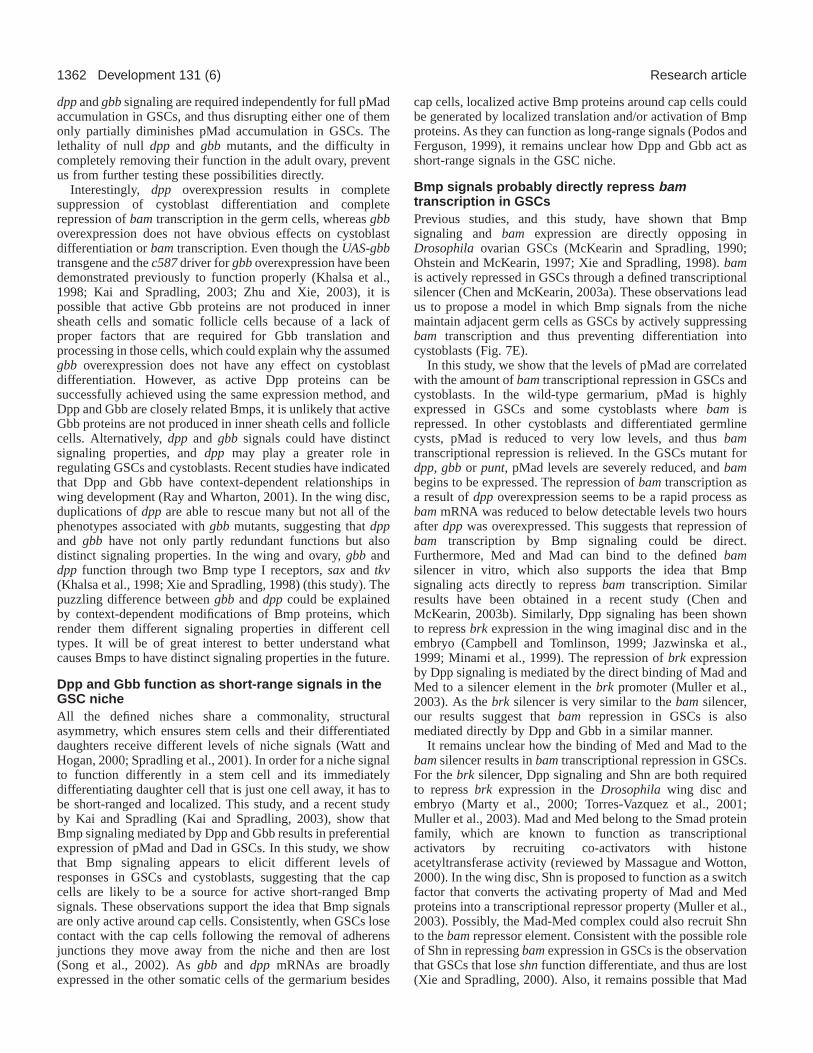
DiscussionStem cells are located in a niche, which provides extracellularcues that control stem cell self-renewal, division anddifferentiation. Dpp is a signaling molecule that originatesfrom the niche and is necessary for maintaining GSCs in theDrosophila ovary (Xie and Spradling, 1998; Xie and Spradling,2000). This study has identified another Bmp-like molecule,Gbb, as an essential niche signal for maintaining GSCs. Itis likely that Dpp and Gbb function cooperatively as short-range signals in the GSC niche, as their signaling activities,monitored by pMad and Dad expression, are restricted to GSCsand some cystoblasts. Previous studies have demonstrated thatbam is known to be necessary and sufficient for cystoblastdifferentiation (McKearin and Spradling, 1990; Ohlstein andMcKearin, 1997). This study shows that upregulation of bamin GSCs is associated with stem cell loss in dpp and gbbmutants. Here we propose a model to possibly explain howBmp signaling directly represses bamtranscription inDrosophilaovarian GSCs (Fig. 7E). The Bmp signals from theGSC niche activate their signaling cascade in GSCs, whichleads to Mad phosphorylation, and then the translocation of theMad and Med complex into the nucleus, which probablydirectly binds to the bampromoter and represses bam
transcription in GSCs. This study demonstrates that Bmpsignals maintain GSCs, at least in part, by repressing bamtranscription in GSCs in the Drosophilaovary.
Gbb is another niche signal that is essential formaintaining GSCsIn this study, a new function of gbb in the regulation of GSCsin the Drosophila ovary is revealed. Loss of gbbfunction leadsto GSC differentiation and stem cell loss, similar to dppmutants. gbbis expressed in somatic cells but not in germ cells,suggesting that gbb is another niche signal that controls GSCmaintenance. Like dpp, gbbcontributes to the production ofpMad in GSCs and also functions to repress bamexpressionin GSCs. As in the wing imaginal disc (Haerry et al., 1998;Khalsa et al., 1998; Ray and Wharton, 2001),gbb alsoprobably functions to augment the dpp signal in the regulationof GSCs through common receptors in the Drosophilaovary.In both dppand gbbmutants, pMad accumulation in GSCs isseverely reduced but not completely diminished. As the dpporgbb mutants used in this study do not carry complete loss-of-function mutations, it remains possible that completeelimination of either dppor gbb function is sufficient foreradicating pMad accumulation in GSCs. Alternatively, both
Fig. 7.Mad and Med bind directly to the bamsilencer invitro. (A) Sequence alignment of the bipartite bamand brksilencers, in which conserved base pairs are boxed. Sites A(red) and B (green) of the bamsilencer are as previouslydescribed (Chen and McKearin, 2003a). (B) A Cy5 5′-modified oligonucleotide containing the bamsilencer andunlabeled competitors in electrophoretic mobility shiftassays (competitor A+B, the unlabeled silencer with sites Aand B; competitor A, same flanking sequences with site Aonly; competitor B, same flanking sequences with site Bonly). (C) Immunoblot analysis of purified recombinantGST-tagged proteins with a mouse anti-GST antibody.Lane 1, 50 ng of GST; lane 2, 200 ng of GST-Mad; lane 3,200 ng GST-Med. The ~30 kDa bands in lane 3 areprobably C-terminal degradation products of GST-Medprotein. (D) Gel shift assay showing that Mad or Med bindto the bamsilencer in vitro. Approximately 10 nmol ofprotein was used in each binding reaction. The double shiftbands for Med may result from partially degraded proteins.The labeled probe without protein (lane 1) or with GSTprotein (lane 2) serve as negative controls. The labeledprobe binds to Mad (lane 3) and Med (lane 7). Theunlabeled competitors A+B (lane 4), A (lane 5) or B (lane6) could effectively compete away Mad binding. Theunlabeled competitors A+B (lane 8) or A (lane 9) couldeffectively compete for Med binding but B (lane 10) couldonly partially compete. (E) Current model for how Bmpniche signals control GSC identity by directly repressingbamtranscription. Bmp signals from cap cells produce thehighest levels of pMad, which associates with Med anddirectly occupies the bamsilencer to repress itstranscription in GSCs. As a cystoblast moves away fromcap cells, levels of pMad are reduced to below the criticalthreshold level, bamtranscription is then derepressed andactivated by an unknown activator (indicated by ‘?’).
1362
dppand gbbsignaling are required independently for full pMadaccumulation in GSCs, and thus disrupting either one of themonly partially diminishes pMad accumulation in GSCs. Thelethality of null dppand gbbmutants, and the difficulty incompletely removing their function in the adult ovary, preventus from further testing these possibilities directly.
Interestingly, dpp overexpression results in completesuppression of cystoblast differentiation and completerepression of bamtranscription in the germ cells, whereas gbboverexpression does not have obvious effects on cystoblastdifferentiation or bamtranscription. Even though the UAS-gbbtransgene and the c587driver for gbboverexpression have beendemonstrated previously to function properly (Khalsa et al.,1998; Kai and Spradling, 2003; Zhu and Xie, 2003), it ispossible that active Gbb proteins are not produced in innersheath cells and somatic follicle cells because of a lack ofproper factors that are required for Gbb translation andprocessing in those cells, which could explain why the assumedgbb overexpression does not have any effect on cystoblastdifferentiation. However, as active Dpp proteins can besuccessfully achieved using the same expression method, andDpp and Gbb are closely related Bmps, it is unlikely that activeGbb proteins are not produced in inner sheath cells and folliclecells. Alternatively, dppand gbbsignals could have distinctsignaling properties, and dpp may play a greater role inregulating GSCs and cystoblasts. Recent studies have indicatedthat Dpp and Gbb have context-dependent relationships inwing development (Ray and Wharton, 2001). In the wing disc,duplications of dppare able to rescue many but not all of thephenotypes associated with gbb mutants, suggesting that dppand gbb have not only partly redundant functions but alsodistinct signaling properties. In the wing and ovary, gbb anddpp function through two Bmp type I receptors, sax and tkv(Khalsa et al., 1998; Xie and Spradling, 1998) (this study). Thepuzzling difference between gbband dppcould be explainedby context-dependent modifications of Bmp proteins, whichrender them different signaling properties in different celltypes. It will be of great interest to better understand whatcauses Bmps to have distinct signaling properties in the future.
Dpp and Gbb function as short-range signals in theGSC nicheAll the defined niches share a commonality, structuralasymmetry, which ensures stem cells and their differentiateddaughters receive different levels of niche signals (Watt andHogan, 2000; Spradling et al., 2001). In order for a niche signalto function differently in a stem cell and its immediatelydifferentiating daughter cell that is just one cell away, it has tobe short-ranged and localized. This study, and a recent studyby Kai and Spradling (Kai and Spradling, 2003), show thatBmp signaling mediated by Dpp and Gbb results in preferentialexpression of pMad and Dad in GSCs. In this study, we showthat Bmp signaling appears to elicit different levels ofresponses in GSCs and cystoblasts, suggesting that the capcells are likely to be a source for active short-ranged Bmpsignals. These observations support the idea that Bmp signalsare only active around cap cells. Consistently, when GSCs losecontact with the cap cells following the removal of adherensjunctions they move away from the niche and then are lost(Song et al., 2002). As gbb and dppmRNAs are broadlyexpressed in the other somatic cells of the germarium besides
cap cells, localized active Bmp proteins around cap cells couldbe generated by localized translation and/or activation of Bmpproteins. As they can function as long-range signals (Podos andFerguson, 1999), it remains unclear how Dpp and Gbb act asshort-range signals in the GSC niche.
Bmp signals probably directly repress bamtranscription in GSCsPrevious studies, and this study, have shown that Bmpsignaling and bam expression are directly opposing inDrosophila ovarian GSCs (McKearin and Spradling, 1990;Ohstein and McKearin, 1997; Xie and Spradling, 1998). bamis actively repressed in GSCs through a defined transcriptionalsilencer (Chen and McKearin, 2003a). These observations leadus to propose a model in which Bmp signals from the nichemaintain adjacent germ cells as GSCs by actively suppressingbam transcription and thus preventing differentiation intocystoblasts (Fig. 7E).
In this study, we show that the levels of pMad are correlatedwith the amount of bamtranscriptional repression in GSCs andcystoblasts. In the wild-type germarium, pMad is highlyexpressed in GSCs and some cystoblasts where bam isrepressed. In other cystoblasts and differentiated germlinecysts, pMad is reduced to very low levels, and thus bamtranscriptional repression is relieved. In the GSCs mutant fordpp, gbb or punt, pMad levels are severely reduced, and bambegins to be expressed. The repression of bamtranscription asa result of dppoverexpression seems to be a rapid process asbammRNA was reduced to below detectable levels two hoursafter dpp was overexpressed. This suggests that repression ofbam transcription by Bmp signaling could be direct.Furthermore, Med and Mad can bind to the defined bamsilencer in vitro, which also supports the idea that Bmpsignaling acts directly to repress bamtranscription. Similarresults have been obtained in a recent study (Chen andMcKearin, 2003b). Similarly, Dpp signaling has been shownto repress brkexpression in the wing imaginal disc and in theembryo (Campbell and Tomlinson, 1999; Jazwinska et al.,1999; Minami et al., 1999). The repression of brk expressionby Dpp signaling is mediated by the direct binding of Mad andMed to a silencer element in the brk promoter (Muller et al.,2003). As the brksilencer is very similar to the bamsilencer,our results suggest that bamrepression in GSCs is alsomediated directly by Dpp and Gbb in a similar manner.
It remains unclear how the binding of Med and Mad to thebamsilencer results in bamtranscriptional repression in GSCs.For the brksilencer, Dpp signaling and Shn are both requiredto repress brkexpression in the Drosophilawing disc andembryo (Marty et al., 2000; Torres-Vazquez et al., 2001;Muller et al., 2003). Mad and Med belong to the Smad proteinfamily, which are known to function as transcriptionalactivators by recruiting co-activators with histoneacetyltransferase activity (reviewed by Massague and Wotton,2000). In the wing disc, Shn is proposed to function as a switchfactor that converts the activating property of Mad and Medproteins into a transcriptional repressor property (Muller et al.,2003). Possibly, the Mad-Med complex could also recruit Shnto the bamrepressor element. Consistent with the possible roleof Shn in repressing bam expression in GSCs is the observationthat GSCs that lose shnfunction differentiate, and thus are lost(Xie and Spradling, 2000). Also, it remains possible that Mad
Development 131 (6) Research article

1363Bmps repress bam transcription in GSCs
and Med could recruit a repressor other than Shn when bindingto the bamrepressor element. In the future, it will be veryimportant to determine whether Shn itself is a co-repressor forMad/Med proteins or whether it directly recruits a co-repressorto repress bamtranscription in GSCs.
We thank D. Drummond-Barbosa, S. Cohen, S. Kobayashi, P. Lasko,D. McKearin, A. Spradling, A. Laughon, K. Wharton, M. Hoffmann,P. ten Dijke, and the Drosophila stock center for reagents. We wouldalso like to thank G. Call, J. Coffman, S. Hawley, S. Page, A. Spradlingfor critical comments on manuscripts, the Xie laboratory members forstimulating discussions, Jo Haynes for help with the manuscriptpreparation, C. Sonnenbrot, J. Haug, H. Newkirk and K. Zueckert-Gaudenz for technical help. This work is supported by the StowersInstitute for Medical Research and NIH (1R01 GM64428-01).
ReferencesCampbell, G. and Tomlinson, A. (1999). Transducing the Dpp morphogen
gradient in the wing of Drosophila: regulation of Dpp targets by brinker.Cell 96, 553-562.
Chen, D. and McKearin, D. M. (2003a). A discrete transcriptional silencerin the bamgene determines asymmetric division of the Drosophila germlinestem cell. Development130, 1159-1170.
Chen, D. and McKearin, D. M. (2003b). Dpp signaling silences bamtranscription directly to establish asymmetric divisions of germline stemcells. Curr. Biol. 13, 1786-1791.
Cox, D. N., Chao, A. and Lin, H.(2000). piwiencodes a nucleoplasmic factorwhose activity modulates the number and division rate of germline stemcells. Development127, 503-514.
Das, P., Maduzia, L. L., Wang, H., Finelli, A. L., Cho, S. H., Smith, M. M.and Padgett, R. W.(1998). The Drosophila gene Medea demonstrates therequirement for different classes of Smads in dppsignaling. Development125, 1519-1528.
de Cuevas, M., Lilly, M. A. and Spradling, A. C.(1997). Germline cystformation in Drosophila. Annu. Rev. Genet.31, 405-428.
Doctor, J. S., Jackson, P. D., Rashka, K. E., Visalli, M. and Hoffmann, F.M. (1992). Sequence, biochemical characterization, and developmentalexpression of a new member of the TGF-beta superfamily in Drosophilamelanogaster. Dev. Biol.151, 491-505.
Forbes, A. and Lehmann, R.(1998). Nanos and Pumilio have critical rolesin the development and function of Drosophila germline stem cells.Development125, 679-690.
Haerry, T. E., Khalsa, O., O’Connor, M. B. and Wharton, K. A. (1998).Synergistic signaling by two BMP ligands through the SAX and TKVreceptors controls wing growth and patterning in Drosophila. Development125, 3977-3987.
Hay, B., Jan, L. Y. and Jan, Y. N.(1988). A protein component of Drosophilapolar granules is encoded by vasaand has extensive sequence similarity toATP-dependent helicases. Cell55, 577-587.
Jazwinska, A., Kirov, N., Wieschaus, E., Roth, S. and Rushlow, C.(1999).The Drosophilagene brinker reveals a novel mechanism of Dpp target generegulation. Cell 96, 563-573.
Kai, T. and Spradling, A. C. (2003). An empty Drosophilastem cell nichereactivates the proliferation of ectopic cells. Proc. Natl. Acad. Sci. USA100,4633-4638.
Khalsa, O., Yoon, J. W., Torres-Schumann, S. and Wharton, K. A.(1998).TGF-β/BMP superfamily members, Gbb-60A and Dpp, cooperate toprovide pattern information and establish cell identity in the Drosophilawing. Development125, 2723-2734.
Kim, J., Johnson, K., Chen, H. J., Carroll S. and Laughon, A.(1997).DrosophilaMad binds to DNA and directly mediates activation of vestigialby Decapentaplegic. Nature388, 304-308.
King, F. J. and Lin, H. (1999). Somatic signaling mediated by fs(1)Yb isessential for germline stem cell maintenance during Drosophilaoogenesis.Development126, 1833-1844.
King, F. J., Szakmary, A., Cox, D. N. and Lin, H.(2001). Ybmodulates thedivisions of both germline and somatic stem cells through piwi- and hh-mediated mechanisms in the Drosophilaovary. Mol. Cell 7, 497-508.
Lasko, P. F. and Ashburner, M.(1988). The product of the Drosophilagenevasais very similar to eukaryotic initiation factor-4A. Nature335, 611-617.
Lavoie, C. A., Ohlstein, B. and McKearin, D. M.(1999). Localization andfunction of Bam protein require the benign gonial cellneoplasm geneproduct. Dev. Biol.212, 405-413.
Letsou, A., Arora, K., Wrana, J. L., Simin, K., Twombly, V., Jamal, J.,Staehling-Hampton, K., Hoffmann, F. M., Gelbart, W. M., Massague, J.and O’Connor, M. B. (1995). DrosophilaDpp signaling is mediated by thepunt gene product: a dual ligand-binding type II receptor of the TGF betareceptor family. Cell 80, 899-908.
Liang, L., Diehl-Jones, W. and Lasko, P. F.(1994). Localization of vasaprotein to the Drosophilapole plasm is independent of its RNA-binding andhelicase activities. Development120, 1201-1211.
Lin, H. (2002). The stem-cell niche theory: lessons from flies. Nat. Rev. Genet.3, 931-940.
Lin, H. and Spradling, A. C. (1997). A novel group of pumilio mutationsaffects the asymmetric division of germline stem cells in the Drosophilaovary. Development124, 2463-2476.
Lin, H., Yue, L. and Spradling, A. C. (1994). TheDrosophila fusome, agermline-specific organelle, contains membrane skeletal proteins andfunctions in cyst formation. Development120, 947-956.
Margolis, J. and Spradling, A. (1995). Identification and behavior ofepithelial stem cells in the Drosophilaovary. Development121, 3797-3807.
Marty T., Muller, B., Basler, K. and Affolter, M. (2000). Schnurri mediatesDpp-dependent repression of brinker transcription. Nat. Cell Biol.2, 745-749.
Massague, J. and Wotton, D.(2000). Transcriptional control by the TGF-beta/Smad signaling system. EMBO J.19, 1745-1754.
McKearin, D. and Ohlstein, B. (1995). A role for the Drosophila bag-of-marbles protein in the differentiation of cystoblasts from germline stemcells. Development121, 2937-2947.
McKearin, D. M. and Spradling, A. C. (1990). bag-of-marbles: a Drosophilagene required to initiate both male and female gametogenesis. Genes Dev.4, 2242-2251.
Minami, M., Kinoshita, N., Kamoshida, Y., Tanimoto, H. and Tabata, T.(1999). brinker is a target of Dpp in Drosophila that negatively regulatesDpp-dependent genes. Nature 398, 242-246.
Muller, B., Hartmann, B., Pyrowolakis, G., Affolter M. and Basler, K.(2003). Conversion of an extracellular Dpp/BMP morphogen gradient intoan inverse transcriptional gradient. Cell 113, 221-233.
Nakamura, A., Amikura, R., Hanyu, K. and Kobayashi, S.(2001). Me31Bsilences translation of oocyte-localizing RNAs through the formation ofcytoplasmic RNP complex during Drosophilaoogenesis. Development128,3233-3242.
Newfeld, S. J., Mehra, A., Singer, M. A., Wrana, J. L., Attisano, L. andGelbart, W. M. (1997). Mothers against dppparticipates in a DDP/TGF-βresponsive serine-threonine kinase signal transduction cascade.Development124, 3167-3176.
Ohlstein, B. and McKearin, D. M. (1997). Ectopic expression of theDrosophila Bam protein eliminates oogenic germline stem cells.Development124, 3651-3662.
Oliver, B., Pauli, D. and Mahowald, A. P.(1990). Genetic evidence that theovo locus is involved in Drosophilagerm line sex determination. Genetics125, 535-550.
Podos, S. D. and Ferguson, E. L.(1999). Morphogen gradients: new insightsfrom DPP. Trends Genet.15, 396-402.
Ray, R. P. and Wharton, K. A. (2001). Context-dependent relationshipsbetween the BMPs gbband dppduring development of the Drosophilawingimaginal disk. Development128, 3913-3925.
Ruberte, E., Marty, T., Nellen, D., Affolter, M. and Basler, K.(1995). Anabsolute requirement for both the type II and type I receptors, puntand thickveins, for dppsignaling in vivo. Cell 80, 889-897.
Song, X., Zhu, C. H., Doan, C. and Xie, T.(2002). Germline stem cellsanchored by adherens junctions in the Drosophilaovary niches. Science296,1855-1857.
Spradling, A., Drummond-Barbosa, D. and Kai, T. (2001). Stem cells findtheir niche. Nature414, 98-104.
Tanimoto, H., Itoh, S., ten Dijke, P. and Tabata, T.(2000). Hedgehog createsa gradient of DPP activity in Drosophilawing imaginal discs. Mol. Cell 5,59-71.
Theisen, H., Haerry, T. E., O’Connor, M. B. and Marsh, J. L.(1996).Developmental territories created by mutual antagonism between Winglessand Decapentaplegic. Development122, 3939-3948.
Torres-Vazquez, J., Park, S., Warrior R. and Arora, K. (2001). Thetranscription factor Schnurri plays a dual role in mediating Dpp signalingduring embryogenesis. Development128, 1657-1670.

1364
Tsuneizumi, K., Nakayama, T., Kamoshida, Y., Kornberg, T. B., Christian,J. L. and Tabata, T. (1997). Daughters against dppmodulates dpporganizing activity in Drosophilawing development. Nature389, 627-631.
Watt, F. M. and Hogan, B. L.(2000). Out of Eden: stem cells and their niches.Science287, 1427-1430.
Wharton, K. A., Thomsen, G. H. and Gelbart, W. M.(1991). Drosophila60A gene, another transforming growth factor β family member, is closelyrelated to human bone morphogenetic proteins. Proc. Natl. Acad. Sci. USA88, 9214-9218.
Wisotzkey, R. G., Mehra, A., Sutherland, D. J., Dobens, L. L., Liu, X.,Dohrmann, C., Attisano, L. and Raftery, L. A. (1998). Medeais aDrosophila Smad4homolog that is differentially required to potentiate DPPresponses. Development125, 1433-1445.
Xie, T. and Spradling, A. C. (1998). decapentaplegicis essential for themaintenance and division of germline stem cells in the Drosophilaovary.Cell 94, 251-260.
Xie, T. and Spradling, A. C. (2000). A niche maintaining germ line stem cellsin the Drosophilaovary. Science290, 328-330.
Xie, T. and Spradling, A. C.(2001). The Drosophilaovary: an in vitro stemcell system. In Stem Cell Biology(ed. D. R. Marshak, R. L. Gardner and D.Gottlieb), pp. 129-148. Cold Spring Harbor, NY: Cold Spring HarborLaboratory Press.
Xu, T. and Rubin, G. M. (1993). Analysis of genetic mosaics in developingand adult Drosophilatissues. Development117, 1223-1237.
Zhu, C. H. and Xie, T. (2003). Clonal expansion of ovarian germline stemcells during niche formation in Drosophila. Development130, 2579-2588.
Development 131 (6) Research article