Embed Size (px)
Citation preview

BioMed CentralBMC Structural Biology
ss
Open AcceResearch articleLigand-induced conformational changes in a thermophilic ribose-binding proteinMatthew J Cuneo, Lorena S Beese and Homme W Hellinga*Address: The Department of Biochemistry, Duke University Medical Center, Durham, North Carolina, 27710, USA
Email: Matthew J Cuneo - [email protected]; Lorena S Beese - [email protected]; Homme W Hellinga* - [email protected]
* Corresponding author
AbstractBackground: Members of the periplasmic binding protein (PBP) superfamily are involved intransport and signaling processes in both prokaryotes and eukaryotes. Biological responses aretypically mediated by ligand-induced conformational changes in which the binding event is coupledto a hinge-bending motion that brings together two domains in a closed form. In all PBP-mediatedbiological processes, downstream partners recognize the closed form of the protein. This motionhas also been exploited in protein engineering experiments to construct biosensors that transduceligand binding to a variety of physical signals. Understanding the mechanistic details of PBPconformational changes, both global (hinge bending, twisting, shear movements) and local (rotamerchanges, backbone motion), therefore is not only important for understanding their biologicalfunction but also for protein engineering experiments.
Results: Here we present biochemical characterization and crystal structure determination of theperiplasmic ribose-binding protein (RBP) from the hyperthermophile Thermotoga maritima in itsribose-bound and unliganded state. The T. maritima RBP (tmRBP) has 39% sequence identity and isconsiderably more resistant to thermal denaturation (appTm value is 108°C) than the mesophilicEscherichia coli homolog (ecRBP) (appTm value is 56°C). Polar ligand interactions and ligand-inducedglobal conformational changes are conserved among ecRBP and tmRBP; however local structuralrearrangements involving side-chain motions in the ligand-binding site are not conserved.
Conclusion: Although the large-scale ligand-induced changes are mediated through similarregions, and are produced by similar backbone movements in tmRBP and ecRBP, the small-scaleligand-induced structural rearrangements differentiate the mesophile and thermophile. Thissuggests there are mechanistic differences in the manner by which these two proteins bind theirligands and are an example of how two structurally similar proteins utilize different mechanisms toform a ligand-bound state.
BackgroundBacterial periplasmic binding proteins (PBP) are receptorsfor extracellular solutes in metabolite uptake [1], chemo-taxis [2], and intercellular communication [3] processes.The PBPs collectively constitute a structural protein super-
family characterized by two pseudo-symmetric domainsthat are linked by a hinge formed by two or three β-strands connecting the domains; a ligand-binding site issituated at the interface between the two domains [4].Each domain adopts a three-layered α/β/α sandwich fold
Published: 19 November 2008
BMC Structural Biology 2008, 8:50 doi:10.1186/1472-6807-8-50
Received: 20 August 2008Accepted: 19 November 2008
This article is available from: http://www.biomedcentral.com/1472-6807/8/50
© 2008 Cuneo et al; licensee BioMed Central Ltd. This is an Open Access article distributed under the terms of the Creative Commons Attribution License (http://creativecommons.org/licenses/by/2.0), which permits unrestricted use, distribution, and reproduction in any medium, provided the original work is properly cited.
Page 1 of 14(page number not for citation purposes)
BMC Structural Biology 2008, 8:50 http://www.biomedcentral.com/1472-6807/8/50
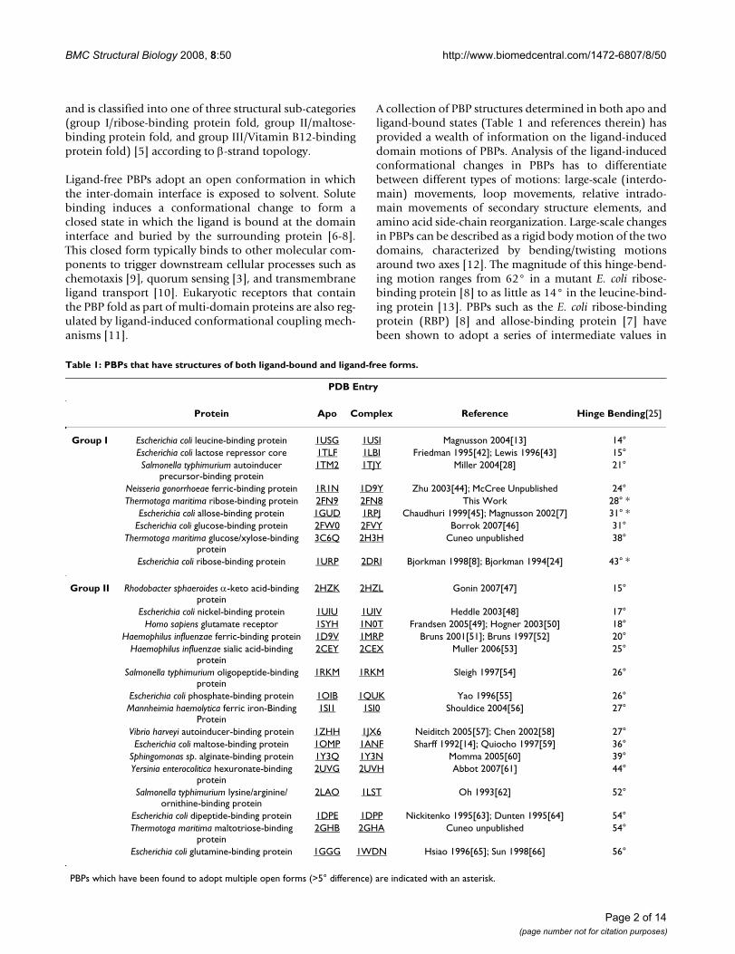
and is classified into one of three structural sub-categories(group I/ribose-binding protein fold, group II/maltose-binding protein fold, and group III/Vitamin B12-bindingprotein fold) [5] according to β-strand topology.
Ligand-free PBPs adopt an open conformation in whichthe inter-domain interface is exposed to solvent. Solutebinding induces a conformational change to form aclosed state in which the ligand is bound at the domaininterface and buried by the surrounding protein [6-8].This closed form typically binds to other molecular com-ponents to trigger downstream cellular processes such aschemotaxis [9], quorum sensing [3], and transmembraneligand transport [10]. Eukaryotic receptors that containthe PBP fold as part of multi-domain proteins are also reg-ulated by ligand-induced conformational coupling mech-anisms [11].
A collection of PBP structures determined in both apo andligand-bound states (Table 1 and references therein) hasprovided a wealth of information on the ligand-induceddomain motions of PBPs. Analysis of the ligand-inducedconformational changes in PBPs has to differentiatebetween different types of motions: large-scale (interdo-main) movements, loop movements, relative intrado-main movements of secondary structure elements, andamino acid side-chain reorganization. Large-scale changesin PBPs can be described as a rigid body motion of the twodomains, characterized by bending/twisting motionsaround two axes [12]. The magnitude of this hinge-bend-ing motion ranges from 62° in a mutant E. coli ribose-binding protein [8] to as little as 14° in the leucine-bind-ing protein [13]. PBPs such as the E. coli ribose-bindingprotein (RBP) [8] and allose-binding protein [7] havebeen shown to adopt a series of intermediate values in
Table 1: PBPs that have structures of both ligand-bound and ligand-free forms.
PDB Entry
Protein Apo Complex Reference Hinge Bending[25]
Group I Escherichia coli leucine-binding protein 1USG 1USI Magnusson 2004[13] 14°Escherichia coli lactose repressor core 1TLF 1LBI Friedman 1995[42]; Lewis 1996[43] 15°Salmonella typhimurium autoinducer
precursor-binding protein1TM2 1TJY Miller 2004[28] 21°
Neisseria gonorrhoeae ferric-binding protein 1R1N 1D9Y Zhu 2003[44]; McCree Unpublished 24°Thermotoga maritima ribose-binding protein 2FN9 2FN8 This Work 28° *
Escherichia coli allose-binding protein 1GUD 1RPJ Chaudhuri 1999[45]; Magnusson 2002[7] 31° *Escherichia coli glucose-binding protein 2FW0 2FVY Borrok 2007[46] 31°
Thermotoga maritima glucose/xylose-binding protein
3C6Q 2H3H Cuneo unpublished 38°
Escherichia coli ribose-binding protein 1URP 2DRI Bjorkman 1998[8]; Bjorkman 1994[24] 43° *
Group II Rhodobacter sphaeroides α-keto acid-binding protein
2HZK 2HZL Gonin 2007[47] 15°
Escherichia coli nickel-binding protein 1UIU 1UIV Heddle 2003[48] 17°Homo sapiens glutamate receptor 1SYH 1N0T Frandsen 2005[49]; Hogner 2003[50] 18°
Haemophilus influenzae ferric-binding protein 1D9V 1MRP Bruns 2001[51]; Bruns 1997[52] 20°Haemophilus influenzae sialic acid-binding
protein2CEY 2CEX Muller 2006[53] 25°
Salmonella typhimurium oligopeptide-binding protein
1RKM 1RKM Sleigh 1997[54] 26°
Escherichia coli phosphate-binding protein 1OIB 1QUK Yao 1996[55] 26°Mannheimia haemolytica ferric iron-Binding
Protein1SI1 1SI0 Shouldice 2004[56] 27°
Vibrio harveyi autoinducer-binding protein 1ZHH 1JX6 Neiditch 2005[57]; Chen 2002[58] 27°Escherichia coli maltose-binding protein 1OMP 1ANF Sharff 1992[14]; Quiocho 1997[59] 36°
Sphingomonas sp. alginate-binding protein 1Y3Q 1Y3N Momma 2005[60] 39°Yersinia enterocolitica hexuronate-binding
protein2UVG 2UVH Abbot 2007[61] 44°
Salmonella typhimurium lysine/arginine/ornithine-binding protein
2LAO 1LST Oh 1993[62] 52°
Escherichia coli dipeptide-binding protein 1DPE 1DPP Nickitenko 1995[63]; Dunten 1995[64] 54°Thermotoga maritima maltotriose-binding
protein2GHB 2GHA Cuneo unpublished 54°
Escherichia coli glutamine-binding protein 1GGG 1WDN Hsiao 1996[65]; Sun 1998[66] 56°
PBPs which have been found to adopt multiple open forms (>5° difference) are indicated with an asterisk.
Page 2 of 14(page number not for citation purposes)

BMC Structural Biology 2008, 8:50 http://www.biomedcentral.com/1472-6807/8/50
their apo state suggesting that the observed states repre-sent snapshots of a continuum between two extremes: thedefined closed form, and a less precisely defined fullyopen conformation.
In E. coli RBP (ecRBP) small-scale backbone movementsare restricted to the hinge region, whereas the secondarystructure elements in the two domains and the aminoacids in the binding pocket adopt essentially the sameconformations in both the apo and ribose-bound forms[8]. However, in E. coli leucine-binding protein, not onlythe hinge region, but also loops and amino acid side-chains in the binding pocket show ligand-inducedchanges [13], many of which are restricted to one domain.This difference in conformational changes between thedomains has been postulated to imply ordered interac-tions between the protein and ligand [8,13,14].
The ligand-induced conformational changes have notbeen described previously in a thermophilic PBP. We havecharacterized the stability, determined the ligand-bindingproperties, and solved the X-ray crystal structures of theapo and ligand-bound forms of a thermophilic periplas-mic ribose-binding protein from the hyperthermophileThermotoga maritima (tmRBP), the mesophilic homolog,ecRBP, of which has been studied in detail [8,15,16]. TheecRBP and tmRBP proteins share 39% amino acidsequence identity, but differ by 52°C in apparent thermalstability. We find that the interdomain motions, althoughnot of the same magnitude, exhibit similar movements.The amino acids in the tmRBP sugar-binding pocketundergo ligand-induced conformational changes,whereas their conformations in apo ecRBP are essentiallypre-formed for ligand binding.
Results and discussionExpressionThe RBP gene was identified in the T. maritima genomesequence [17] as open reading frame (ORF) tm0958, basedon sequence similarity to the E. coli RBP, and genetic link-age of this ORF within a putative operon that containssequences for ABC transporters characteristic of a ribosetransport system [18]. ORF tm0958 was amplified from T.maritima genomic DNA using the polymerase chain reac-tion. The resulting DNA fragment was cloned into a pET21avector with a C-terminal hexa-histidine tag preceded by aglycine-serine linker. The nucleotide sequence of therecombinant was confirmed by DNA sequencing. Over-expression of this ORF in E. coli produced ~50 mg of pureprotein per liter of growth medium, which was purified byimmobilized metal affinity chromatography [19] followedby gel filtration chromatography.
The gel filtration elution profile of tmRBP consists of twopeaks, one of which is consistent with a monomeric
tmRBP (34 kDa), the other consistent with a ~55 kDa pro-tein (Figure 1). SDS-PAGE of the resulting fractionsrevealed that both peaks contain tmRBP. The fractionscorresponding to the 55 kDa protein also contain signifi-cant amount of a ~20 kDa species (Figure 1). Trypticdigestion of this 20 kDa protein, followed by MALDI massspectrometry peptide mapping [20], revealed that it corre-sponds to a truncated form of the full-length tmRBP (Fig-ure 2). The 55 kDa protein is therefore a heterodimerconsisting of one full-length and one truncated copy oftmRBP. Neither full-length, nor truncated homodimerswere observed. Analysis of the tm0958 DNA sequence sug-gests that this truncation may result from translation ini-tiation at methionine 142 (numbering according to NCBINP 228766), which is preceded by a ribosome bindingsite (Figure 2). This interpretation is further supported bythe M142A mutant tmRBP in which the 20 kDa trunca-tion is absent (data not shown).
Thermal StabilityThe apparent thermal stability (appTm) of full-length mon-omeric wild-type tmRBP was determined by thermaldenaturation using circular dichroism (CD) [21]. In the
Expression and purification of the tm0958 ORFFigure 1Expression and purification of the tm0958 ORF. (A) Gel-filtration (Superdex S75) chromatogram of the immobi-lized metal affinity purified tmRBP. Fractions (10 mL) and the void volume of the S75 column (Vo) are indicated. (B) SDS-PAGE of column fractions. Lane 1 is a molecular mass ladder.
Page 3 of 14(page number not for citation purposes)

BMC Structural Biology 2008, 8:50 http://www.biomedcentral.com/1472-6807/8/50
absence of denaturant, no significant change in the CDsignal could be observed as a function of temperature(data not shown). All measurements were therefore car-ried out in the presence of the chemical denaturant guani-dine hydrochloride (GdCl) to bring thermal denaturationinto a measurable range. Melting curves were found to fita two-state model [21,22]. An appTm in the absence of GdClwas determined by linear extrapolation of a series of melt-ing point determinations carried out at different GdClconcentrations [23] (Figure 3) and was found to be108°C. tmRBP is significantly more stable than the mes-ophilic ecRBP (appTm value is 56°C (Figure 3)). Addition of
the 20 kDa truncation has no effect on the appTm value ofthe full-length wild-type monomeric protein (data notshown).
Ligand BindingRibose binding was observed as a ligand-mediated changein the appTm of full-length wild-type monomeric tmRBP inthe presence of 5.5 M GdCl. Under these conditions theappTm is 71°C in the absence of sugar and 97°C in the pres-ence of 1 mM ribose, indicating that tmRBP is a ribose-binding protein, as predicted from sequence homology(Figure 3). For the ligand-bound form (1 mM ribose), an
Peptide mapping of the tm0958 ORF gene productsFigure 2Peptide mapping of the tm0958 ORF gene products. MALDI mass spectra of the in-gel tryptic digests of the (A) 32 kDa and (B) 20 kDa products of the tm0958 ORF. Peptides observed in (A) that were not observed in (B) are indicated. (C) Map-ping of the peptides from (A) onto the tmRBP amino acid and DNA sequence. Mapped peptides are underlined in black, met142 is underlined in green, and the alternate ribosome binding site is underlined in red.
2388
2662
3212
ATGAAGAAGAGCTTGTTTGTGGTGTTGGTATTGGTTGGATTGTTACTGGTTTCCTTCACAGGTTTGGCACAGGAACAGCAGAAACCAAAAGGAAAGATGGCT M K K S L F V V L V L V G L L L V S F T G L A Q E Q Q K P K G K M A ATTGTGATCTCCACACTTAACAATCCATGGTTTGTTGTCCTCGCTGAAACAGCGAAGCAAAGAGCAGAACAACTCGGCTATGAAGCTACTATCTTTGATTCT I V I S T L N N P W F V V L A E T A K Q R A E Q L G Y E A T I F D S CAGAATGACACAGCTAAGGAGTCGGCTCACTTCGATGCGATCATAGCTGCCGGATATGATGCCATCATCTTCAATCCCACCGATGCGGATGGATCGATAGCA Q N D T A K E S A H F D A I I A A G Y D A I I F N P T D A D G S I A AACGTGAAGAGAGCGAAAGAAGCAGGCATACCTGTCTTCTGTGTAGACAGGGGAATTAACGCAAGAGGACTGGCGGTAGCACAAATTTATTCAGATAACTAC N V K R A K E A G I P V F C V D R G I N A R G L A V A Q I Y S D N Y TATGGTGGTGTGCTTATGGGTGAATACTTTGTAAAGTTCCTCAAAGAGAAATATCCAGATGCAAAAGAAATCCCATATGCGGAGCTTCTCGGAATACTCAGT Y G G V L M G E Y F V K F L K E K Y P D A K E I P Y A E L L G I L S GCACAACCCACCTGGGATAGATCAAATGGATTCCACAGCGTTGTAGATCAATATCCCGAGTTCAAGATGGTGGCACAGCAATCCGCAGAATTTGACAGAGAC A Q P T W D R S N G F H S V V D Q Y P E F K M V A Q Q S A E F D R D ACAGCTTACAAAGTCACAGAACAGATTCTCCAGGCACATCCTGAAATTAAAGCCATATGGTGCGGAAACGATGCTATGGCACTCGGTGCTATGAAAGCATGT T A Y K V T E Q I L Q A H P E I K A I W C G N D A M A L G A M K A C GAAGCTGCAGGAAGAACCGATATCTACATTTTTGGATTCGATGGAGCAGAAGACGTGATAAATGCCATCAAAGAAGGAAAGCAGATCGTAGCAACTATCATG E A A G R T D I Y I F G F D G A E D V I N A I K E G K Q I V A T I M
CAATTCCCGAAACTTATGGCAAGATTGGCAGTTGAATGGGCTGACCAGTACCTCAGAGGTGAAAGAAGCTTCCCGGAGATTGTACCTGTCACTGTTGAGCTG Q F P K L M A R L A V E W A D Q Y L R G E R S F P E I V P V T V E L GTGACAAGAGAAAACATCGATAAGTACACTGCTTACGGCAGAAAAGAAGAATAA V T R E N I D K Y T A Y G R K E E *
1000 1500 2000 2500 30000
1000
2000
3000
4000
Inte
nsity
M/z1000 1500 2000 2500 3000
0
1000
2000
3000
4000
5000
6000
7000
8000
9000
10000
Inte
nsity
M/z
A B
C
Page 4 of 14(page number not for citation purposes)

BMC Structural Biology 2008, 8:50 http://www.biomedcentral.com/1472-6807/8/50
appTm of 131°C in the absence of GdCl was determined bylinear extrapolation of a series of melting point determi-nations carried out at different GdCl concentrations [23](Figure 3).
Structure DeterminationCrystals of ribose-complexed tmRBP were grown using afull-length wild-type construct (residues 30–323) thatlacks the periplasmic signal sequence (residues 1–29). Theapo-protein was crystallized using a construct that con-sisted of residues 30–310 (numbering according to NCBINP 228766), containing a M142A mutation to preventexpression of the in-frame ORF. We were unable to obtaincrystals of the heterodimeric form. The apo-protein andribose-complex diffract to 1.4 Å and 2.15 Å resolution andwere refined to Rcryst/Rfree values of 18.0/20.3 and 19.3/22.3 respectively. The X-ray crystal structure of ribose-bound tmRBP was solved by molecular replacement usingecRBP as the search model [24]. The apo-form of tmRBPwas solved by separately searching with the amino- andcarboxy-terminal domains of the ribose-bound form oftmRBP. Data collection, refinement, and stereochemistrystatistics are summarized in Table 2.
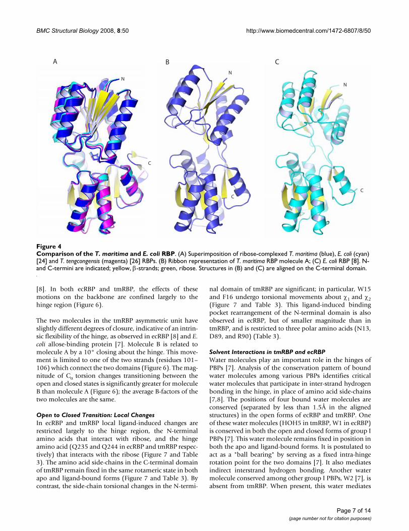
Overall Structure and Comparison of the E. coli and T. maritima apo proteinsThe apo forms of ecRBP [8] and tmRBP adopt the sameoverall fold. However, the relative inter-domain angles[25] differ significantly (43° for ecRBP; 28° and 20° for
the two molecules in the tmRBP unit) (Figure 4). Thehinge in ecRBP is very flexible as evidenced by the numberof crystal forms that differ in the inter-domain closureangle [8]. The two molecules found in the tmRBP asym-metric unit differ in the inter-domain closure angle by10°, analogous to the conformational heterogeneityobserved in ecRBP [8].
The construct used to crystallize the apo-form of tmRBPwas a C-terminally truncated form of the protein (13amino acids). It is possible that the absence of this regioncould in some way influence the observed conformationof apo form of tmRBP. However, superimposition of thetmRBP ribose complex C-terminal domain onto the C-ter-minal region of the apo protein suggests that these thisregion does not form interdomain interactions in theabsence of ligand.
Overall Structure and Comparison of the E. coli and T. maritima ribose complexesThe structure of the tmRBP ribose-complex is similar tothe ribose complexes observed in ecRBP [24] and a ther-mophilic RBP obtained from Thermoanaerobacter tengcon-gensis [26] (tteRBP). Both structures superimpose ontmRBP with a 1.2 Å RMSD calculated over Cα atoms (Fig-ure 4). The structures tteRBP and ecRBP are almost identi-cal [26]; comparisons are described therefore only forecRBP. The largest differences between ecRBP and tmRBPare at the C-termini, where tmRBP is extended by an addi-
Thermal stability of tmRBPFigure 3Thermal stability of tmRBP. (A) Thermal denaturation of tmRBP in 5.5 M GdCl (squares), tmRBP in 1 mM ribose and 5.5 M GdCl (circles), apo ecRBP (triangles), ecRBP in 1 mM ribose (inverted triangles). Solid lines in (A) are fit to a two-state model which takes into account the native and denatured baseline slopes [21,22]. (B) Extrapolated appTm value of apo (squares) and ribose-bound (1 mM) (circles) tmRBP obtained from the series of thermal melting curves at different GdCl concentrations [23,26]. Solid line represents a linear fit to the observations.
0.0 0.5 1.0 1.5 2.0 2.5 3.0 3.5 4.0 4.5 5.0 5.5 6.0 6.5 7.06065707580859095
100105110115120125130135
T m (°
C)[GdCl]
A B
20 30 40 50 60 70 80 90 100 110-0.1
0.0
0.1
0.2
0.3
0.4
0.5
0.6
0.7
0.8
0.9
1.0
1.1
Norm
alize
d θ
x10-3
(deg
cm
2 mol
-1)
Temp (°C)
Page 5 of 14(page number not for citation purposes)

BMC Structural Biology 2008, 8:50 http://www.biomedcentral.com/1472-6807/8/50
tional 13 residues that are not present in ecRBP. This seg-ment forms a short α-helix terminated by a β-hairpin(Figure 4). One of the amino acids in this region (Y289)forms extensive van der Waals interactions with theamino acids in the N-terminal domain (P14, W15 andV18). As similar extensions are found interacting with theN-terminal domain in both open and closed forms ofother PBPs [27,28]. We postulate that these C-terminalextensions form inter-domain interactions that may beimportant for modulating the intrinsic free energy differ-ence between the apo and closed forms in the absence ofligand (Miklos, Cuneo and Hellinga; in preparation).
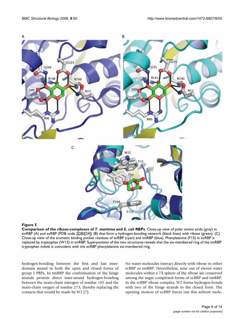
Although ribose is commonly found as a furanose carbo-hydrate in biological molecules (e.g. nucleic acids), allperiplasmic RBPs, including tmRBP, bind the β-anomer ofD-pyranose ribose [24,26] (as initially postulated byKoshland [29]). β-D-pyranose ribose the most prevalentform in solution under ambient conditions (59%) [30].The ligand-binding site of tmRBP is composed of a net-work of polar amino acids which is identical in sequenceand hydrogen-bonding pattern to the E. coli protein [24](Figure 5). Seven polar amino acids make a total of eleven
hydrogen-bonds with the ribose. One residue in ecRBP(Q235) has been postulated to be important for both lig-and-binding and hinge-bending; in the closed form itforms hydrogen-bonds with the ligand and amino acidsfrom both domains [8,15]. The equivalent residue(Q244) and the amino acids which it interacts with areconserved in tmRBP. This pattern of conservation suggeststhat similar mechanisms couple ligand-binding to confor-mational changes in both proteins [8,14].
The ribose is wedged between three aromatic amino acids(W15, F16 and F172) which make extensive van der Waalsinteractions with the sugar ring. In ecRBP the equivalentaromatic binding pocket residues are all phenylalanines.Alignment of tmRBP and ecRBP structures indicates thatthe six-membered ring of W15 in tmRBP is equivalent toF15 in ecRBP (Figure 5).
Open to Closed Transition: Global ChangesThe addition of ribose to tmRBP induces a 28° hinge-bending motion [25] mediated about residues 102–105,244–249, and 271–275. The hinge-bending motion oftmRBP is smaller than the 43° change observed in ecRBP
Table 2: Data collection and refinement statistics.
tmRBP-apo tmRBP-ribose
Data CollectionWavelength (Å) 0.997 0.979Resolution (Å) 1.40 2.15Unique reflections 115460 25783Mean I/σ(I)a 34.2 (1.7) 25.7 (3.6)Completeness (%)a 99.0 (88.8) 80.9 (21.0)Rsym (%)a 5.0 (51.5) 5.6 (28.4)Redundancya 5.8 (3.4) 5.8 (1.6)
RefinementResolution (Å) 50.0–1.40 50.0–2.15Num. of Reflections (working set/test set) 115460/5767 23715/1354Rcryst (%) 18.0 (28.0) 19.3 (25.4)Rfree
b (%) 20.3 (32.9) 22.3 (29.2)Number of atoms
Protein 4326 2286Water 627 142Ligand 0 10
r.m.s.d.Bond lengths (Å) 0.009 0.012Bond angles (°) 1.2 1.2
Average B-factor (Å2)Main Chain 15.3 34.5Side Chain 17.3 35.8Solvent 29 37.7Ligand 24.8
Protein GeometryRamachandran outliers (%) 0.4 0.3Ramachandran favored (%) 98.7 97.6Rotamer outliers (%) 2.2 3.0
aNumber in parentheses represent values in the highest resolution shell.bRfree is the R-factor based on 5% of the data excluded from refinement.
Page 6 of 14(page number not for citation purposes)
BMC Structural Biology 2008, 8:50 http://www.biomedcentral.com/1472-6807/8/50
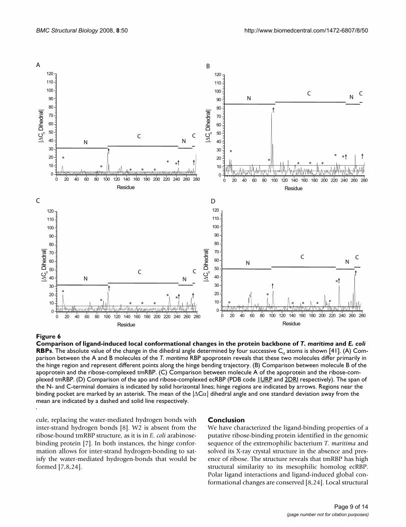
[8]. In both ecRBP and tmRBP, the effects of thesemotions on the backbone are confined largely to thehinge region (Figure 6).
The two molecules in the tmRBP asymmetric unit haveslightly different degrees of closure, indicative of an intrin-sic flexibility of the hinge, as observed in ecRBP [8] and E.coli allose-binding protein [7]. Molecule B is related tomolecule A by a 10° closing about the hinge. This move-ment is limited to one of the two strands (residues 101–106) which connect the two domains (Figure 6). The mag-nitude of Cα torsion changes transitioning between theopen and closed states is significantly greater for moleculeB than molecule A (Figure 6); the average B-factors of thetwo molecules are the same.
Open to Closed Transition: Local ChangesIn ecRBP and tmRBP local ligand-induced changes arerestricted largely to the hinge region, the N-terminalamino acids that interact with ribose, and the hingeamino acid (Q235 and Q244 in ecRBP and tmRBP respec-tively) that interacts with the ribose (Figure 7 and Table3). The amino acid side-chains in the C-terminal domainof tmRBP remain fixed in the same rotameric state in bothapo and ligand-bound forms (Figure 7 and Table 3). Bycontrast, the side-chain torsional changes in the N-termi-
nal domain of tmRBP are significant; in particular, W15and F16 undergo torsional movements about χ1 and χ2(Figure 7 and Table 3). This ligand-induced bindingpocket rearrangement of the N-terminal domain is alsoobserved in ecRBP, but of smaller magnitude than intmRBP, and is restricted to three polar amino acids (N13,D89, and R90) (Table 3).
Solvent Interactions in tmRBP and ecRBPWater molecules play an important role in the hinges ofPBPs [7]. Analysis of the conservation pattern of boundwater molecules among various PBPs identifies criticalwater molecules that participate in inter-strand hydrogenbonding in the hinge, in place of amino acid side-chains[7,8]. The positions of four bound water molecules areconserved (separated by less than 1.5Å in the alignedstructures) in the open forms of ecRBP and tmRBP. Oneof these water molecules (HOH5 in tmRBP, W1 in ecRBP)is conserved in both the open and closed forms of group IPBPs [7]. This water molecule remains fixed in position inboth the apo and ligand-bound forms. It is postulated toact as a "ball bearing" by serving as a fixed intra-hingerotation point for the two domains [7]. It also mediatesindirect interstrand hydrogen bonding. Another watermolecule conserved among other group I PBPs, W2 [7], isabsent from tmRBP. When present, this water mediates
Comparison of the T. maritima and E. coli RBPFigure 4Comparison of the T. maritima and E. coli RBP. (A) Superimposition of ribose-complexed T. maritima (blue), E. coli (cyan) [24] and T. tengcongensis (magenta) [26] RBPs. (B) Ribbon representation of T. maritima RBP molecule A; (C) E. coli RBP [8]. N- and C-termini are indicated; yellow, β-strands; green, ribose. Structures in (B) and (C) are aligned on the C-terminal domain.
A B
N
C
N
C
C
N
C
Page 7 of 14(page number not for citation purposes)
BMC Structural Biology 2008, 8:50 http://www.biomedcentral.com/1472-6807/8/50
hydrogen-bonding between the first and last inter-domain strand in both the open and closed forms ofgroup I PBPs. In tmRBP the conformation of the hingestrands permits direct inter-strand hydrogen-bondingbetween the main-chain nitrogen of residue 105 and themain-chain oxygen of residue 273, thereby replacing thecontacts that would be made by W2 [7].
No water molecules interact directly with ribose in eitherecRBP or tmRBP. Nevertheless, nine out of eleven watermolecules within a 7Å sphere of the ribose are conservedamong the sugar complexed forms of ecRBP and tmRBP.In the ecRBP ribose complex, W2 forms hydrogen-bondswith two of the hinge strands in the closed form. Theopening motion of ecRBP forces out this solvent mole-
Comparison of the ribose-complexes of T. maritima and E. coli RBPsFigure 5Comparison of the ribose-complexes of T. maritima and E. coli RBPs. Close-up view of polar amino acids (gray) in tmRBP (A) and ecRBP (PDB code 2DRI[24]) (B) that form a hydrogen-bonding network (black lines) with ribose (green). (C) Close-up view of the aromatic binding pocket residues of ecRBP (cyan) and tmRBP (blue). Phenylalanine (F15) in ecRBP is replaced by tryptophan (W15) in tmRBP. Superposition of the two structures reveals that the six-membered ring of the tmRBP tryptophan indole is coincident with the ecRBP phenylalanine six-membered ring.
Page 8 of 14(page number not for citation purposes)
BMC Structural Biology 2008, 8:50 http://www.biomedcentral.com/1472-6807/8/50
cule, replacing the water-mediated hydrogen bonds withinter-strand hydrogen bonds [8]. W2 is absent from theribose-bound tmRBP structure, as it is in E. coli arabinose-binding protein [7]. In both instances, the hinge confor-mation allows for inter-strand hydrogen-bonding to sat-isfy the water-mediated hydrogen-bonds that would beformed [7,8,24].
ConclusionWe have characterized the ligand-binding properties of aputative ribose-binding protein identified in the genomicsequence of the extremophilic bacterium T. maritima andsolved its X-ray crystal structure in the absence and pres-ence of ribose. The structure reveals that tmRBP has highstructural similarity to its mesophilic homolog ecRBP.Polar ligand interactions and ligand-induced global con-formational changes are conserved [8,24]. Local structural
Comparison of ligand-induced local conformational changes in the protein backbone of T. maritima and E. coli RBPsFigure 6Comparison of ligand-induced local conformational changes in the protein backbone of T. maritima and E. coli RBPs. The absolute value of the change in the dihedral angle determined by four successive Cα atoms is shown [41]. (A) Com-parison between the A and B molecules of the T. maritima RBP apoprotein reveals that these two molecules differ primarily in the hinge region and represent different points along the hinge bending trajectory. (B) Comparison between molecule B of the apoprotein and the ribose-complexed tmRBP. (C) Comparison between molecule A of the apoprotein and the ribose-com-plexed tmRBP. (D) Comparison of the apo and ribose-complexed ecRBP (PDB code 1URP and 2DRI respectively). The span of the N- and C-terminal domains is indicated by solid horizontal lines; hinge regions are indicated by arrows. Regions near the binding pocket are marked by an asterisk. The mean of the |ΔCα| dihedral angle and one standard deviation away from the mean are indicated by a dashed and solid line respectively.
0 20 40 60 80 100 120 140 160 180 200 220 240 260 2800
10
20
30
40
50
60
70
80
90
100
110
120
Residue
0 20 40 60 80 100 120 140 160 180 200 220 240 260 2800
10
20
30
40
50
60
70
80
90
100
110
120
Residue
0 20 40 60 80 100 120 140 160 180 200 220 240 260 2800
10
20
30
40
50
60
70
80
90
100
110
120
Residue
0 20 40 60 80 100 120 140 160 180 200 220 240 260 2800
10
20
30
40
50
60
70
80
90
100
110
120α D
ihed
ral|
Residue
A
C D
B
N
N
N
N
N
N
N
N
C
C
C
C
C
C
C
C
*
** * *
*
*
** * *
*
*
** * *
*
**
* * * *
*
*
*
*
|ΔCα D
ihed
ral|
|ΔC
α Dih
edra
l||Δ
Cα Dih
edra
l||Δ
C
Page 9 of 14(page number not for citation purposes)

BMC Structural Biology 2008, 8:50 http://www.biomedcentral.com/1472-6807/8/50
rearrangements involving side-chain motions in the lig-and-binding site differ in the mesophilic and ther-mophilic RBPs. In ecRBP the conformation of the bindingpocket undergoes little ligand-induced rearrangement.The amino acids in the N-terminal domain of the tmRBPbinding pocket undergo large χ1 and χ2 torsional changes,
whereas the C-terminal domain remains fixed. Based onhydrogen-bonding pattern (6 and 5 hydrogen-bonds withthe N- and C-terminal domains respectively) and buriedsurface area (55Å 2 and 35Å 2 with the N- and C-terminaldomains respectively) it has been postulated that orderedbinding occurs and ribose initially interacts with N-termi-
Table 3: Rotamers changes in the ecRBP (1URP molecule A/2DRI) and tmRBP (apoprotein molecule A/ribose-bound form) binding pocket residues.
ecRBP tmRBP
Δχ1 (°) Δχ2 (°) Δχ3 (°) Δχ4 (°) (Σ|Δχ|)/Nχ(°) Δχ1 (°) Δχ2 (°) Δχ3 (°) Δχ4 (°) (Σ|Δχ|)/Nχ(°)
ASN13 12 -13 13 13 -1 7PHE15 -1 16 8 26 -173 100PHE16 -7 10 8 -17 26 22ASP89 -18 17 18 -1 19 10ARG90 26 -17 -27 70 35 -1 -5 5 0 3
ARG141/148 -1 -12 -7 -6 6 0 -12 4 -19 9PHE164/172 0 0 0 1 4 3ASN190/198 -7 -4 5 4 -8 6ASP215/223 14 4 9 -1 6 3↑GLN235/244 -20 -7 45 24 -3 9 -29 14
C-terminal amino acids are in bold face type; the hinge amino acid that interacts with ribose is indicated by an arrow. Where amino acid numbering differs, ecRBP residues are listed first.
Binding pocket organization of the apo and ribose-bound tmRBPFigure 7Binding pocket organization of the apo and ribose-bound tmRBP. Stereo-view of the ribose-bound tmRBP (blue) binding pocket superimposed with the binding pocket amino acids of apo tmRBP (magenta). The C-terminal residues of the apoprotein have similar rotamers as the ribose-bound form while the rotamers of the N-terminal domain apoprotein and ribose-bound forms are in different states. The C-terminal binding pocket residues of the apoprotein interact (black lines) with bulk solvent (red spheres) in a similar manner as the ligand-bound form does with the ribose ligand, pre-organizing the apo form.
Q244
D223
N198F172
R148
W15
D89
R90
F16
Q244
D223
N198F172
R148
W15
D89
R90
F16
Page 10 of 14(page number not for citation purposes)
BMC Structural Biology 2008, 8:50 http://www.biomedcentral.com/1472-6807/8/50
nal domain of ecRBP [8]. If an order of interaction can beestablished from analysis of structure, it is likely to pro-ceed with ribose initially interacting with the C-terminaldomain of the apo tmRBP, as the entropic costs of fixingthe side-chains for ligand binding should be reduced for apre-ordered binding site.
Water molecules have been suggested to play an impor-tant mechanistic role in the evolution and adaptation ofthe PBP hinge [7]. In particular, two water molecules, (W1and W2), are closely associated with the hinges of group IPBPs [7]. In tmRBP, W1, which is postulated to act as a"ball bearing" in the ligand-mediated conformationalchange, is conserved in both the apo- and ribose-boundforms. On the other hand, W2, which is involved in medi-ating important inter-hinge contacts in apo- and ligand-bound group I PBPs, is absent in both forms of tmRBP. IntmRBP the inter-strand hydrogen bonds form directly inthe hinge. These differences in water interactions in thehinges of PBPs suggest local structural differences can sup-plant the need for W2, whereas the role of W1 cannot beaccommodated through differences in main-chain geom-etry or side-chain identity.
Ligand-induced hinge bending motion is a key character-istic of the periplasmic binding protein superfamily. Anal-ysis of PBP structures has provided a detailed descriptionof this class of conformational change [7,12-14]. Thedetailed comparative analysis of the open to closed transi-tion of the thermophilic tmRBP and mesophilic ecRBPpresented here illustrates the subtle differences in themechanism and magnitude of the ligand-induced confor-mational changes, and the interplay between global andlocal conformational changes in this protein superfamily.
MethodsCloning Over-expression and PurificationThe tm0958 gene was amplified from T. maritima genomicDNA (American Type Culture Collection) by the sticky-end PCR method [31] using the following primers tomake the full-length tmRBP (residues 30–323) and theconstruct used to crystallize the apo form of tmRBP (resi-dues 30–310) (numbering according to NCBI ProteinDatabase NP 228766: PO4--TATGAAAGGAA AGAT-GGCTATTGTGATCTCC and for the 5'-TGAAAGGAAAGATGGCTAT TGTGATCTCC end of the genes; PO4--AATTCTA ATGGTGATGGTGATGGTGACTGCCTTCT-TCTTTTCTGCCGTAAGCAGTG andCTAATGGTGATGGTGATGGTGACTGCCTTCTTCTTT-TCTGCCGTAAGCAGTG for the 3'end of the full-lengthtmRBP gene, PO4-AATTCTAATGGTGATGGTGATGGT-GACTGCCTTCTCTTGTCACCAGCTCAACAGTGAC andCTAATGGTGATGGTG ATGGTGACTGCCTTCTCTTGT-CACCAGCTCAACAGTGA C for the 3' end of the tmRBP-apo gene [31]. The 30–323 construct which was used to
crystallize the apo-form additionally contains an M142Amutation to prevent translation of the truncated form oftmRBP. The resulting fragments were cloned into theNdeI/EcoRI sites of a pET21a (Novagen) plasmid for over-expression in E. coli. This ORF lacks the periplasmic signalsequence. The coding sequence starting at lysine 30 wascloned in-frame with an ATG start codon. A hexa-histidineaffinity tag, preceded by a glycine-serine linker, was fusedin-frame at the carboxy terminus to facilitate purificationby immobilized metal affinity chromatography (IMAC).Protein concentration was determined spectrophotomet-rically (ε 280 = 41,000 M-1cm-1) [32]. The resulting geneproduct was expressed and purified by IMAC and gel fil-tration as described [23]. Pooled IMAC fractions wereconcentrated to 12 ml and were loaded onto a Superdex26/60 S75 (Amersham) gel filtration column that was pre-viously that was previously calibrated with blue dextran,bovine serum albumin, chicken serum albumin, chymot-rypsin and lysozyme.
Tryptic Digest and Mass SpectrometryProteins were excised from a 12% Tris-HCl SDS-PAGE geland were digested in-gel using the Pierce In-gel TrypticDigest Kit. Mass spectra were acquired on an Applied Bio-systems Voyager DE MALDI-TOF mass spectrometer usingan α-cyano-4-hydroxycinnamic acid matrix with a 300 nsdelay time.
Circular DichroismCircular dichroism (CD) measurements were carried outon an Aviv Model 202 CD spectrophotometer. Thermaldenaturations were determined by measuring the CD sig-nal at 222 nm (1 cm path length) as a function of temper-ature, using 1.0 μM of full-length wild-type monomerictmRBP (10 mM Tris-HCl pH 7.8, 150 mM NaCl) in thepresence or absence of 1 mM ribose at several GdCl con-centrations extrapolated to 0 M GdCl [23]. Protein sam-ples were incubated for 15 minutes prior to collectingdata. Each measurement includes a 3-second averagingtime for data collection and a 60 second equilibrationperiod at each temperature. Data were fit to a two-statemodel [22].
Crystallization and Data CollectionCrystals of full-length wild type ribose-complexed tmRBPwere grown using 3:1 stoichiometric ribose:protein ratioby micro-batch under paraffin oil in drops that contained2 μl of the protein solution (15 mg/ml in 10 mM Tris pH7.8, 20 mM NaCl, 1.5 mM ribose) mixed with 2 μl of 0.1M MES pH 6.0, 20% (w/v) PEG 8000 and 0.1 M RbCl.Crystals of the C-terminally truncated M142A apoproteinwere grown in micro-batch drops containing 2 μl of theprotein solution (15 mg/ml in 10 mM Tris pH 7.8, 20 mMNaCl) mixed with 2 μl of 0.1 M Bis-Tris pH 5.9, 25% (w/v) PEG 3350, 0.2 M NaCl. Diffraction quality crystals typ-
Page 11 of 14(page number not for citation purposes)
BMC Structural Biology 2008, 8:50 http://www.biomedcentral.com/1472-6807/8/50
ically grew within two weeks at 17.0°C. The ribose-com-plexed crystals diffract to 2.15 Å resolution and belong tothe I222 space group (a = 72.1 Å, b = 98.2 Å, c = 131.1 Å)(Table 2). The apo tmRBP crystals diffract to 1.4 Å resolu-tion and belong to the F222 space group (a = 120.9 Å, b =136.8 Å, c = 144.5 Å) (Table 2). Crystals were transferredstepwise to a cryoprotectant solution consisting of theoriginal precipitant solution with an additional 15% eth-ylene glycol or glycerol, after which they were mounted ina nylon loop and flash cooled in liquid nitrogen. All datawere collected at 100 K on the SER-CAT 22ID beam line atthe Advanced Photon Source. Diffraction data were scaledand integrated using HKL2000 [33].
Structure Determination Methods, Model Building and RefinementThe structure of ribose-complexed tmRBP was determinedby molecular replacement utilizing the AMore program,where the ligand-bound form of the E. coli ribose-bindingprotein was used as the search model [34]. The N- and C-terminal domains of ribose-complexed tmRBP were usedas a search model in Phaser to solve the apoprotein struc-ture [35]. In both cases, rotation, translation, and fittingfunctions revealed a single clear solution yielding highercorrelation coefficients and a lower R factor than all theothers. Manual model building was carried out in the pro-grams O and COOT and refined using REFMAC5 [36-38].
Structural AnalysisThe final model for ribose-complexed tmRBP includesone intact monomer (residues 30–323), one ribose mole-cule, and 142 water molecules. The final model for theapoprotein includes two intact monomers (residues 30–310) and 627 water molecules. The models exhibit goodstereochemistry as determined by PROCHECK [39] andMolProbity [40]; final refinement statistics are listed inTable 2. PDB coordinates and structure factors of ribose-complexed tmRBP and apoprotein have been deposited inthe RCSB Protein Data Bank under the accession codes2FN8 and 2FN9 respectively.
Large-scale hinge bending motions were analyzed withthe DynDom web server [25]. Local C-alpha torsionalchanges were analyzed with LSQMAN [41].
Authors' contributionsMJC purified, crystallized, solved the structure of tmRBP,and carried out circular dichroism experiments. MJC, LSBand HWH undertook sequence and structural analysis ofthe tmRBP and ecRBP structures. MJC and HWH wrote themanuscript. All authors have read and approved the finalmanuscript.
AcknowledgementsThis study was funded by a grant from HSARPA (W81XWH-05-C-0161) to HWH, a Pioneer Award from the NIH (5 DP1 OD000122-02) to HWH.
The authors would like to acknowledge G. Shirman for protein expression and purification, and A. Changela for helpful discussions on structure deter-mination/analysis. Data were collected at the Southeast Regional Collabo-rative Access Team 22-ID and the Structural Biology Center 19-ID beam lines at the Advanced Photon Source, Argonne National Laboratory. Sup-porting institutions may be found at http://www.ser-cat.org/members.html. Use of the Advanced Photon Source was supported by the U. S. Depart-ment of Energy, Office of Science, Office of Basic Energy Sciences, under Contract No. W-31-109-Eng-38.
References1. Boos W, Shuman H: Maltose/maltodextrin system of
Escherichia coli: transport, metabolism, and regulation.Microbiol Mol Biol Rev 1998, 62:204-229.
2. Davidson AL, Shuman HA, Nikaido H: Mechanism of maltosetransport in Escherichia coli: transmembrane signaling byperiplasmic binding proteins. Proc Natl Acad Sci USA 1992,89:2360-2364.
3. Neiditch MB, Federle MJ, Pompeani AJ, Kelly RC, Swem DL, JeffreyPD, Bassler BL, Hughson FM: Ligand-induced asymmetry in his-tidine sensor kinase complex regulates quorum sensing. Cell2006, 126:1095-1108.
4. Tam R, Saier MH Jr: Structural, functional, and evolutionaryrelationships among extracellular solute-binding receptorsof bacteria. Microbiol Rev 1993, 57:320-346.
5. Fukami-Kobayashi K, Tateno Y, Nishikawa K: Domain dislocation:a change of core structure in periplasmic binding proteins intheir evolutionary history. J Mol Biol 1999, 286:279-290.
6. Evenas J, Tugarinov V, Skrynnikov NR, Goto NK, Muhandiram R, KayLE: Ligand-induced structural changes to maltodextrin-bind-ing protein as studied by solution NMR spectroscopy. J MolBiol 2001, 309:961-974.
7. Magnusson U, Chaudhuri BN, Ko J, Park C, Jones TA, Mowbray SL:Hinge-bending motion of D-allose-binding protein fromEscherichia coli: three open conformations. J Biol Chem 2002,277:14077-14084.
8. Bjorkman AJ, Mowbray SL: Multiple open forms of ribose-bind-ing protein trace the path of its conformational change. J MolBiol 1998, 279:651-664.
9. Zhang Y, Gardina PJ, Kuebler AS, Kang HS, Christopher JA, MansonMD: Model of maltose-binding protein/chemoreceptor com-plex supports intrasubunit signaling mechanism. Proc NatlAcad Sci USA 1999, 96:939-944.
10. Nikaido H: Maltose transport system of Escherichia coli: anABC-type transporter. FEBS Lett 1994, 346:55-58.
11. Mayer ML, Olson R, Gouaux E: Mechanisms for ligand binding toGluR0 ion channels: crystal structures of the glutamate andserine complexes and a closed apo state. J Mol Biol 2001,311:815-836.
12. Gerstein M, Lesk AM, Chothia C: Structural mechanisms fordomain movements in proteins. Biochemistry 1994,33:6739-6749.
13. Magnusson U, Salopek-Sondi B, Luck LA, Mowbray SL: X-ray struc-tures of the leucine-binding protein illustrate conforma-tional changes and the basis of ligand specificity. J Biol Chem2004, 279:8747-8752.
14. Sharff AJ, Rodseth LE, Spurlino JC, Quiocho FA: Crystallographicevidence of a large ligand-induced hinge-twist motionbetween the two domains of the maltodextrin binding pro-tein involved in active transport and chemotaxis. Biochemistry1992, 31:10657-10663.
15. Vercillo NC, Herald KJ, Fox JM, Der BS, Dattelbaum JD: Analysis ofligand binding to a ribose biosensor using site-directed muta-genesis and fluorescence spectroscopy. Protein Sci 2007,16:362-368.
16. Shilton BH, Flocco MM, Nilsson M, Mowbray SL: Conformationalchanges of three periplasmic receptors for bacterial chemo-taxis and transport: the maltose-, glucose/galactose- andribose-binding proteins. J Mol Biol 1996, 264:350-363.
17. Nelson KE, Clayton RA, Gill SR, Gwinn ML, Dodson RJ, Haft DH,Hickey EK, Peterson JD, Nelson WC, Ketchum KA, et al.: Evidencefor lateral gene transfer between Archaea and bacteria fromgenome sequence of Thermotoga maritima. Nature 1999,399:323-329.
Page 12 of 14(page number not for citation purposes)

BMC Structural Biology 2008, 8:50 http://www.biomedcentral.com/1472-6807/8/50
18. Iida A, Harayama S, Iino T, Hazelbauer GL: Molecular cloning andcharacterization of genes required for ribose transport andutilization in Escherichia coli K-12. J Bacteriol 1984,158:674-682.
19. Yip TT, Hutchens TW: Immobilized metal-ion affinity chroma-tography. Methods Mol Biol 2004, 244:179-190.
20. Webster J, Oxley D: Peptide mass fingerprinting: protein iden-tification using MALDI-TOF mass spectrometry. Methods MolBiol 2005, 310:227-240.
21. Cohen DS, Pielak GJ: Stability of yeast iso-1-ferricytochrome cas a function of pH and temperature. Protein Sci 1994,3:1253-1260.
22. Schellman JA: The thermodynamic stability of proteins. AnnuRev Biophys Biophys Chem 1987, 16:115-137.
23. Cuneo MJ, Changela A, Warren JJ, Beese LS, Hellinga HW: The crys-tal structure of a thermophilic glucose binding proteinreveals adaptations that interconvert mono and di-saccha-ride binding sites. J Mol Biol 2006, 362:259-270.
24. Bjorkman AJ, Binnie RA, Zhang H, Cole LB, Hermodson MA, Mow-bray SL: Probing protein-protein interactions. The ribose-binding protein in bacterial transport and chemotaxis. J BiolChem 1994, 269:30206-30211.
25. Hayward S, Lee RA: Improvements in the analysis of domainmotions in proteins from conformational change: DynDomversion 1.50. J Mol Graph Model 2002, 21:181-183.
26. Cuneo MJ, Tian Y, Allert M, Hellinga HW: The backbone structureof the thermophilic Thermoanaerobacter tengcongensisribose binding protein is essentially identical to its mes-ophilic E. coli homolog. BMC Struct Biol 2008, 8:20.
27. Quiocho FA, Vyas NK: Novel stereospecificity of the L-arab-inose-binding protein. Nature 1984, 310:381-386.
28. Miller ST, Xavier KB, Campagna SR, Taga ME, Semmelhack MF,Bassler BL, Hughson FM: Salmonella typhimurium recognizes achemically distinct form of the bacterial quorum-sensing sig-nal AI-2. Mol Cell 2004, 15:677-687.
29. Aksamit RR, Koshland DE Jr: Identification of the ribose bindingprotein as the receptor for ribose chemotaxis in Salmonellatyphimurium. Biochemistry 1974, 13:4473-4478.
30. Wu J, Serianni AS: D-Penturonic acids: solution studies of sta-ble-isotopically enriched compounds by 1H- and 13C-n.m.r.spectroscopy. Carbohydr Res 1991, 210:51-70.
31. Zeng G: Sticky-end PCR: new method for subcloning. Biotech-niques 1998, 25:206-208.
32. Gill SC, von Hippel PH: Calculation of protein extinction coeffi-cients from amino acid sequence data. Anal Biochem 1989,182:319-326.
33. Otwinowski ZaM W: Processing of X-ray diffaction data col-lected in oscillation mode. Methods Enzymol 1997,276A:307-326.
34. Navaza J: AMoRe: an automated package for molecularreplacement. Acta Cryst 1994, A50:157-163.
35. Collaborative Computational Project N: The CCP4 suite: pro-grams for protein crystallography. Acta Crystallogr D Biol Crystal-logr 1994, 50:760-763.
36. Jones TA, Zou JY, Cowan SW, Kjeldgaard : Improved methods forbuilding protein models in electron density maps and thelocation of errors in these models. Acta Crystallogr A 1991, 47(Pt2):110-119.
37. Emsley P, Cowtan K: Coot: model-building tools for moleculargraphics. Acta Crystallogr D Biol Crystallogr 2004, 60:2126-2132.
38. Murshudov GN, Vagin AA, Dodson EJ: Refinement of macromo-lecular structures by the maximum-likelihood method. ActaCrystallogr D Biol Crystallogr 1997, 53:240-255.
39. Laskowski RA, MacArthur MW, Moss DS, Thornton JM: PRO-CHECK: a program to check the stereochemical quality ofprotein structures. J Appl Cryst 1993, 26:283-291.
40. Davis IW, Murray LW, Richardson JS, Richardson DC: MOLPRO-BITY: structure validation and all-atom contact analysis fornucleic acids and their complexes. Nucleic Acids Res 2004,32:W615-619.
41. Kleywegt GJ, Jones TA: Detecting folding motifs and similaritiesin protein structures. Methods Enzymol 1997, 277:525-545.
42. Friedman AM, Fischmann TO, Steitz TA: Crystal structure of lacrepressor core tetramer and its implications for DNA loop-ing. Science 1995, 268:1721-1727.
43. Lewis M, Chang G, Horton NC, Kercher MA, Pace HC, SchumacherMA, Brennan RG, Lu P: Crystal structure of the lactose operonrepressor and its complexes with DNA and inducer. Science1996, 271:1247-1254.
44. Zhu H, Alexeev D, Hunter DJ, Campopiano DJ, Sadler PJ: Oxo-ironclusters in a bacterial iron-trafficking protein: new roles fora conserved motif. Biochem J 2003, 376:35-41.
45. Chaudhuri BN, Ko J, Park C, Jones TA, Mowbray SL: Structure ofD-allose binding protein from Escherichia coli bound to D-allose at 1.8 A resolution. J Mol Biol 1999, 286:1519-1531.
46. Borrok MJ, Kiessling LL, Forest KT: Conformational changes ofglucose/galactose-binding protein illuminated by open, unli-ganded, and ultra-high-resolution ligand-bound structures.Protein Sci 2007, 16:1032-1041.
47. Gonin S, Arnoux P, Pierru B, Lavergne J, Alonso B, Sabaty M, PignolD: Crystal structures of an Extracytoplasmic Solute Recep-tor from a TRAP transporter in its open and closed formsreveal a helix-swapped dimer requiring a cation for alpha-keto acid binding. BMC Struct Biol 2007, 7:11.
48. Heddle J, Scott DJ, Unzai S, Park SY, Tame JR: Crystal structuresof the liganded and unliganded nickel-binding protein NikAfrom Escherichia coli. J Biol Chem 2003, 278:50322-50329.
49. Frandsen A, Pickering DS, Vestergaard B, Kasper C, Nielsen BB,Greenwood JR, Campiani G, Fattorusso C, Gajhede M, Schousboe A,et al.: Tyr702 is an important determinant of agonist bindingand domain closure of the ligand-binding core of GluR2. MolPharmacol 2005, 67:703-713.
50. Hogner A, Greenwood JR, Liljefors T, Lunn ML, Egebjerg J, Larsen IK,Gouaux E, Kastrup JS: Competitive antagonism of AMPAreceptors by ligands of different classes: crystal structure ofATPO bound to the GluR2 ligand-binding core, in compari-son with DNQX. J Med Chem 2003, 46:214-221.
51. Bruns CM, Anderson DS, Vaughan KG, Williams PA, Nowalk AJ,McRee DE, Mietzner TA: Crystallographic and biochemicalanalyses of the metal-free Haemophilus influenzae Fe3+-binding protein. Biochemistry 2001, 40:15631-15637.
52. Bruns CM, Nowalk AJ, Arvai AS, McTigue MA, Vaughan KG, MietznerTA, McRee DE: Structure of Haemophilus influenzae Fe(+3)-binding protein reveals convergent evolution within a super-family. Nat Struct Biol 1997, 4:919-924.
53. Muller A, Severi E, Mulligan C, Watts AG, Kelly DJ, Wilson KS,Wilkinson AJ, Thomas GH: Conservation of structure andmechanism in primary and secondary transporters exempli-fied by SiaP, a sialic acid binding virulence factor from Hae-mophilus influenzae. J Biol Chem 2006, 281:22212-22222.
54. Sleigh SH, Tame JR, Dodson EJ, Wilkinson AJ: Peptide binding inOppA, the crystal structures of the periplasmic oligopeptidebinding protein in the unliganded form and in complex withlysyllysine. Biochemistry 1997, 36:9747-9758.
55. Yao N, Ledvina PS, Choudhary A, Quiocho FA: Modulation of a saltlink does not affect binding of phosphate to its specific activetransport receptor. Biochemistry 1996, 35:2079-2085.
56. Shouldice SR, Skene RJ, Dougan DR, Snell G, McRee DE, SchryversAB, Tari LW: Structural basis for iron binding and release by anovel class of periplasmic iron-binding proteins found ingram-negative pathogens. J Bacteriol 2004, 186:3903-3910.
57. Nanavati DM, Nguyen TN, Noll KM: Substrate Specificities andExpression Patterns Reflect the Evolutionary Divergence ofMaltose ABC Transporters in Thermotoga maritima. J Bac-teriol 2005, 187:2002-2009.
58. Chen X, Schauder S, Potier N, Van Dorsselaer A, Pelczer I, BasslerBL, Hughson FM: Structural identification of a bacterial quo-rum-sensing signal containing boron. Nature 2002,415:545-549.
59. Quiocho FA, Spurlino JC, Rodseth LE: Extensive features of tightoligosaccharide binding revealed in high-resolution struc-tures of the maltodextrin transport/chemosensory receptor.Structure 1997, 5:997-1015.
60. Momma K, Mikami B, Mishima Y, Hashimoto W, Murata K: Crystalstructure of AlgQ2, a macromolecule (alginate)-binding pro-tein of Sphingomonas sp. A1 at 2.0A resolution. J Mol Biol2002, 316:1051-1059.
61. Abbott DW, Boraston AB: Specific recognition of saturated and4,5-unsaturated hexuronate sugars by a periplasmic bindingprotein involved in pectin catabolism. J Mol Biol 2007,369:759-770.
Page 13 of 14(page number not for citation purposes)

BMC Structural Biology 2008, 8:50 http://www.biomedcentral.com/1472-6807/8/50
Publish with BioMed Central and every scientist can read your work free of charge
"BioMed Central will be the most significant development for disseminating the results of biomedical research in our lifetime."
Sir Paul Nurse, Cancer Research UK
Your research papers will be:
available free of charge to the entire biomedical community
peer reviewed and published immediately upon acceptance
cited in PubMed and archived on PubMed Central
yours — you keep the copyright
Submit your manuscript here:http://www.biomedcentral.com/info/publishing_adv.asp
BioMedcentral
62. Oh BH, Pandit J, Kang CH, Nikaido K, Gokcen S, Ames GF, Kim SH:Three-dimensional structures of the periplasmic lysine/arginine/ornithine-binding protein with and without a ligand.J Biol Chem 1993, 268:11348-11355.
63. Nickitenko AV, Trakhanov S, Quiocho FA: 2 A resolution struc-ture of DppA, a periplasmic dipeptide transport/chemosen-sory receptor. Biochemistry 1995, 34:16585-16595.
64. Dunten P, Mowbray SL: Crystal structure of the dipeptide bind-ing protein from Escherichia coli involved in active transportand chemotaxis. Protein Sci 1995, 4:2327-2334.
65. Hsiao CD, Sun YJ, Rose J, Wang BC: The crystal structure ofglutamine-binding protein from Escherichia coli. J Mol Biol1996, 262:225-242.
66. Sun YJ, Rose J, Wang BC, Hsiao CD: The structure of glutamine-binding protein complexed with glutamine at 1.94 A resolu-tion: comparisons with other amino acid binding proteins. JMol Biol 1998, 278:219-229.
Page 14 of 14(page number not for citation purposes)