Embed Size (px)
Citation preview
BioMed CentralBMC Molecular Biology
ss
Open AcceResearch articleIdentifying the most suitable endogenous control for determining gene expression in hearts from organ donorsSilvia Pérez*1, Luis J Royo2, Aurora Astudillo3, Dolores Escudero1,4, Francisco Álvarez5, Aida Rodríguez2, Enrique Gómez2 and Jesús Otero1Address: 1Unidad de Coordinación de Trasplantes y Terapia Celular, Hospital Universitario Central de Asturias, C/Celestino Villamil s/n, 33006 Oviedo, Spain, 2Área de Genética y Reproducción Animal, SERIDA-Somió, C/Camino de los Claveles 604, 33203 Gijón, Spain, 3Servicio de Anatomía Patológica, Hospital Universitario Central de Asturias, Oviedo, Spain, 4Servicio de Medicina Intensiva, Hospital Universitario Central de Asturias, Oviedo, Spain and 5Servicio de Bioquímica Clínica, Hospital Universitario Central de Asturias, Oviedo, Spain
Email: Silvia Pérez* - [email protected]; Luis J Royo - [email protected]; Aurora Astudillo - [email protected]; Dolores Escudero - [email protected]; Francisco Álvarez - [email protected]; Aida Rodríguez - [email protected]; Enrique Gómez - [email protected]; Jesús Otero - [email protected]
* Corresponding author
AbstractBackground: Quantitative real-time reverse transcription PCR (qRT-PCR) is a useful tool forassessing gene expression in different tissues, but the choice of adequate controls is critical tonormalise the results, thereby avoiding differences and maximizing sensitivity and accuracy. So far,many genes have been used as a single reference gene, without having previously verified their valueas controls. This practice can lead to incorrect conclusions and recent evidence indicates a needto use the geometric mean of data from several control genes. Here, we identified an appropriateset of genes to be used as an endogenous reference for quantifying gene expression in human hearttissue.
Results: Our findings indicate that out of ten commonly used reference genes (GADPH, PPIA, ACTB,YWHAZ, RRN18S, B2M, UBC, TBP, RPLP and HPRT), PPIA, RPLP and GADPH show the most stablegene transcription levels in left ventricle specimens obtained from organ donors, as assessed usinggeNorm and Normfinder software. The expression of TBP was found to be highly regulated.
Conclusion: We propose the use of PPIA, RPLP and GADPH as reference genes for the accuratenormalisation of qRT-PCR performed on heart tissue. TBP should not be used as a control in thistype of tissue.
BackgroundGene expression analysis is a useful technique for deter-mining and comparing gene expression levels in healthyand diseased tissues. One of the most commonly usedtools in the area of gene expression quantification is quan-titative real-time reverse transcription PCR (qRT-PCR).When small amounts of nucleic acids are available, qRT-
PCR is especially suitable and provides simultaneousmeasurement of gene expression in many different sam-ples. If we compare this technique with others such as insitu hybridisation, qRT-PCR offers several advantages: it isnot time-consuming, only a small amount of tissue isrequired, it can be used in high throughput systems andno post-reaction manipulation is needed. However, the
Published: 20 December 2007
BMC Molecular Biology 2007, 8:114 doi:10.1186/1471-2199-8-114
Received: 8 May 2007Accepted: 20 December 2007
This article is available from: http://www.biomedcentral.com/1471-2199/8/114
© 2007 Pérez et al; licensee BioMed Central Ltd. This is an Open Access article distributed under the terms of the Creative Commons Attribution License (http://creativecommons.org/licenses/by/2.0), which permits unrestricted use, distribution, and reproduction in any medium, provided the original work is properly cited.
Page 1 of 7(page number not for citation purposes)
BMC Molecular Biology 2007, 8:114 http://www.biomedcentral.com/1471-2199/8/114
use of qRT-PCR requires compensation for differencesbetween samples, arising from the varying quality andquantity of the starting material, especially when startingwith solid tissue, due to the method of RNA extractionand cDNA synthesis [1]. Normalisation should includeendogenous control genes (reference genes), and some ofthe most frequently used reference genes are housekeep-ing genes. The ideal endogenous control should beexpressed at a constant level in the different tissues of anorganism at all stages of development and should be unaf-fected by experimental treatments. It should also be con-stitutively expressed in the same tissue under differentcircumstances. There is, however, no universal controlgene that is expressed at a constant level under all condi-tions and in all tissues. Hence, experimental treatments[2] and hormonal stimulation [3], as well as the differentmethods used to process tissue specimens [4] can inducechanges in the expression of typical housekeeping genes.
Consequently, the success of the technique used dependson the adequate choice of the appropriate reference genes.Despite many qRT-PCR studies having reported the use ofa single endogenous control [5], a normalisation strategybased on a single housekeeping gene can lead to errone-ous results [6,7]. Vandesompele et al. [6] propose the useof a panel of putative reference genes on a representativenumber of samples, to identify the most stable of theseand then establish the optimal number of genes requiredfor the reliable normalisation of RT-PCR data. In thepresent study, we tested ten commonly used referencegenes (GADPH, PPIA, ACTB, YWHAZ, RRN18S, B2M,UBC, TBP, RPLP and HPRT) of different functional classes(significantly reducing the chance that the genes will beco-regulated) in heart tissue obtained from organ donors.Using geNorm software [6], we were able to assess geneexpression stability under our experimental conditionsand determine how many reference genes were needed foraccurate normalisation. Then, by comparing these resultswith those generated by a similar programme,Normfinder [8,9], we identified a set of reference genesthat offers reliable results for qRT-PCR data normalisationfor use in gene expression studies involving heart tissuefrom brain-dead multiorgan donors.
ResultsThirty five samples of left ventricular tissue were obtainedfrom 35 multiorgan donors. RNA was successfully iso-lated and cDNA synthesised from all these specimens. Allthe samples analysed showed a single β-actin band in the2% agarose gel stained with ethidium bromide at theexpected size (data not shown), confirming the totalabsence of residual DNA.
In each sample, qRT-PCR using Sybr Green was performedfor ten frequently-used reference genes (GADPH, PPIA,
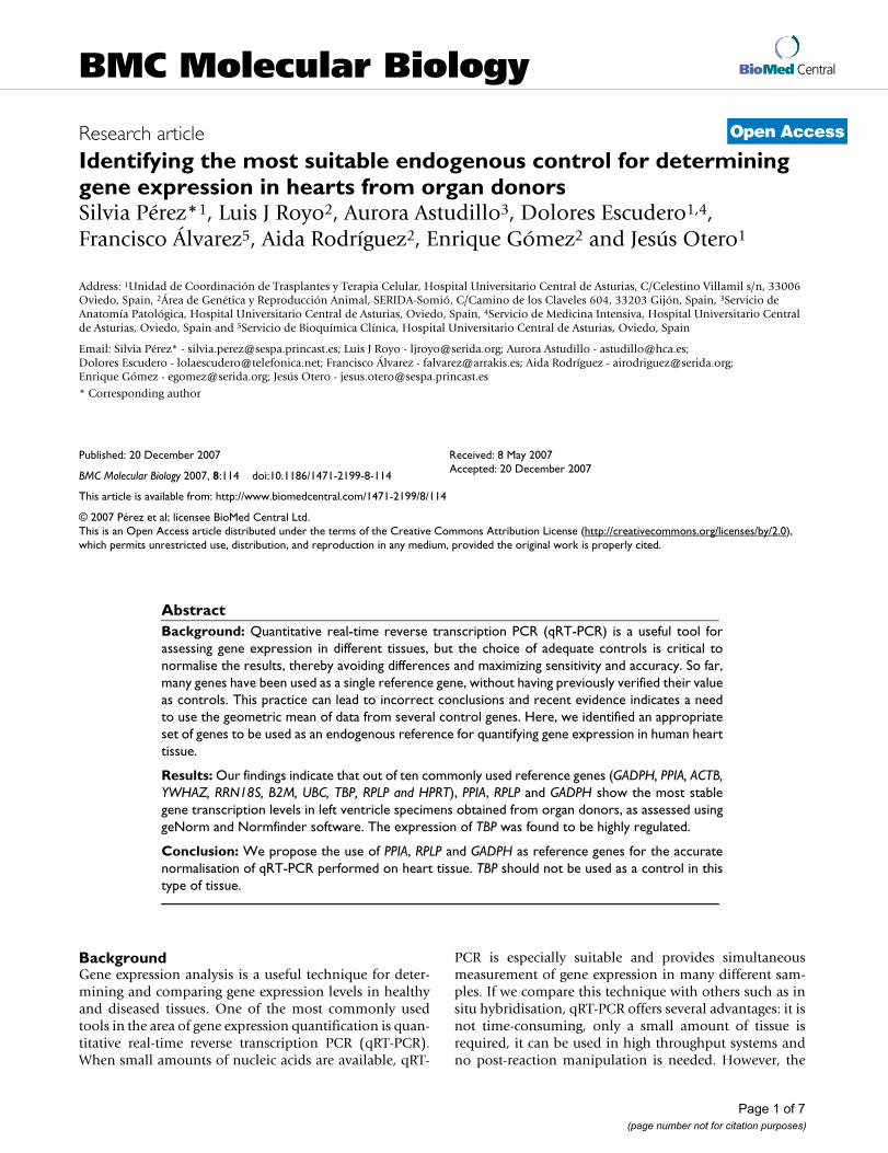
ACTB, YWHAZ, RRN18S, B2M, UBC, TBP, RPLP andHPRT). The accuracy of the qRT-PCR was assessed by meltcurve analysis and gel electrophoresis. Gene-specificamplification was verified, by both a single peak in themelt curve and a single band in the agarose gel, for the 10genes analysed in the 35 cDNA samples. Correlation coef-ficients (R2) ranged from 0.995 to 0.999 and PCR efficien-cies from 89.7% to 104%. Using the Proc VARCOMP inthe SAS/STAT™ software [10], the reproducibility of theassay was assessed using as control material samplesobtained by pooling the whole set of samples analysed inthe present study. The high average correlation coefficientobserved of 0.998 indicated good intra-sample reproduc-ibility. Observed Ct values for each gene were similaracross different samples indicating low variability (21.66± 1.39 for GADPH, 24.69 ± 1.22 for PPIA, 22.32 ± 2.03 forACTB, 26.39 ± 1.36 for YWHAZ, 11.71 ± 1.10 for RRN18S,23.87 ± 1.37 for B2M, 22.71 ± 1.88 for UBC, 30.98 ± 3.45for TBP, 22.27 ± 1.42 for RPLP and 28.08 ± 0.94 forHPRT) (Figure 1).
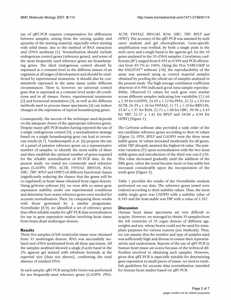
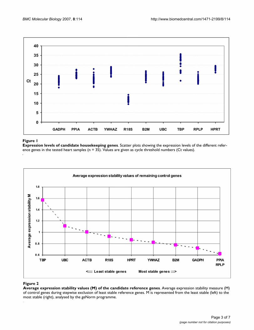
The GeNorm software also provided a rank order of theten candidate reference genes according to their M values(Figure 2). PPIA, RPLP and GADPH were the three moststable genes. M values increased moderately for all genes,while TBP abruptly attained the highest M value. The pair-wise variation (V) upon normalisation with the two moststable genes and introduction of the third one was 0.236.This value decreased gradually until the addition of thefifth gene, when the trend became more or less stable butincreased considerably upon the incorporation of thetenth gene (Figure 3).
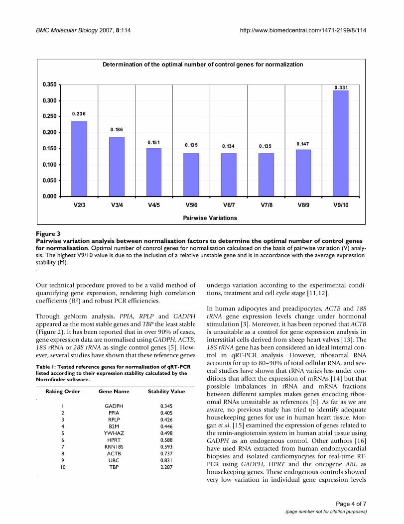
Table 1 provides the results of the Normfinder analysisperformed on our data. The reference genes tested wereordered according to their stability values. Thus, the moststable single gene was GADPH with a stability value of0.345 and the least stable was TBP with a value of 2.287.
DiscussionHuman heart tissue specimens are very difficult toacquire. However, we managed to obtain 35 samples fromthe left ventricles of 35 organ donors of different age,weights and sex, whose hearts could not be used for trans-plant purposes for various reasons (see Methods). Thus,we can assume that the number and type of samples usedwas sufficiently high and diverse to ensure their represent-ativity and randomness. Reports of the use of qRT-PCR inhuman heart tissue are scarce because of the technical dif-ficulties involved in obtaining such samples. However,given that qRT-PCR is especially suitable for determininggene expression in small pieces of tissue, we tried to estab-lish guidelines for accurate data normalisation intendedfor human heart studies based on qRT-PCR.
Page 2 of 7(page number not for citation purposes)
BMC Molecular Biology 2007, 8:114 http://www.biomedcentral.com/1471-2199/8/114
Page 3 of 7(page number not for citation purposes)
Expression levels of candidate housekeeping genesFigure 1Expression levels of candidate housekeeping genes. Scatter plots showing the expression levels of the different refer-ence genes in the tested heart samples (n = 35). Values are given as cycle threshold numbers (Ct values).
Average expression stability values (M) of the candidate reference genesFigure 2Average expression stability values (M) of the candidate reference genes. Average expression stability measure (M) of control genes during stepwise exclusion of least stable reference genes. M is represented from the least stable (left) to the most stable (right), analysed by the geNorm programme.
BMC Molecular Biology 2007, 8:114 http://www.biomedcentral.com/1471-2199/8/114
Our technical procedure proved to be a valid method ofquantifying gene expression, rendering high correlationcoefficients (R2) and robust PCR efficiencies.
Through geNorm analysis, PPIA, RPLP and GADPHappeared as the most stable genes and TBP the least stable(Figure 2). It has been reported that in over 90% of cases,gene expression data are normalised using GADPH, ACTB,18S rRNA or 28S rRNA as single control genes [5]. How-ever, several studies have shown that these reference genes
undergo variation according to the experimental condi-tions, treatment and cell cycle stage [11,12].
In human adipocytes and preadipocytes, ACTB and 18SrRNA gene expression levels change under hormonalstimulation [3]. Moreover, it has been reported that ACTBis unsuitable as a control for gene expression analysis ininterstitial cells derived from sheep heart valves [13]. The18S rRNA gene has been considered an ideal internal con-trol in qRT-PCR analysis. However, ribosomal RNAaccounts for up to 80–90% of total cellular RNA, and sev-eral studies have shown that rRNA varies less under con-ditions that affect the expression of mRNAs [14] but thatpossible imbalances in rRNA and mRNA fractionsbetween different samples makes genes encoding ribos-omal RNAs unsuitable as references [6]. As far as we areaware, no previous study has tried to identify adequatehousekeeping genes for use in human heart tissue. Mor-gan et al. [15] examined the expression of genes related tothe renin-angiotensin system in human atrial tissue usingGADPH as an endogenous control. Other authors [16]have used RNA extracted from human endomyocardialbiopsies and isolated cardiomyocytes for real-time RT-PCR using GADPH, HPRT and the oncogene ABL ashousekeeping genes. These endogenous controls showedvery low variation in individual gene expression levels
Pairwise variation analysis between normalisation factors to determine the optimal number of control genes for normalisationFigure 3Pairwise variation analysis between normalisation factors to determine the optimal number of control genes for normalisation. Optimal number of control genes for normalisation calculated on the basis of pairwise variation (V) analy-sis. The highest V9/10 value is due to the inclusion of a relative unstable gene and is in accordance with the average expression stability (M).
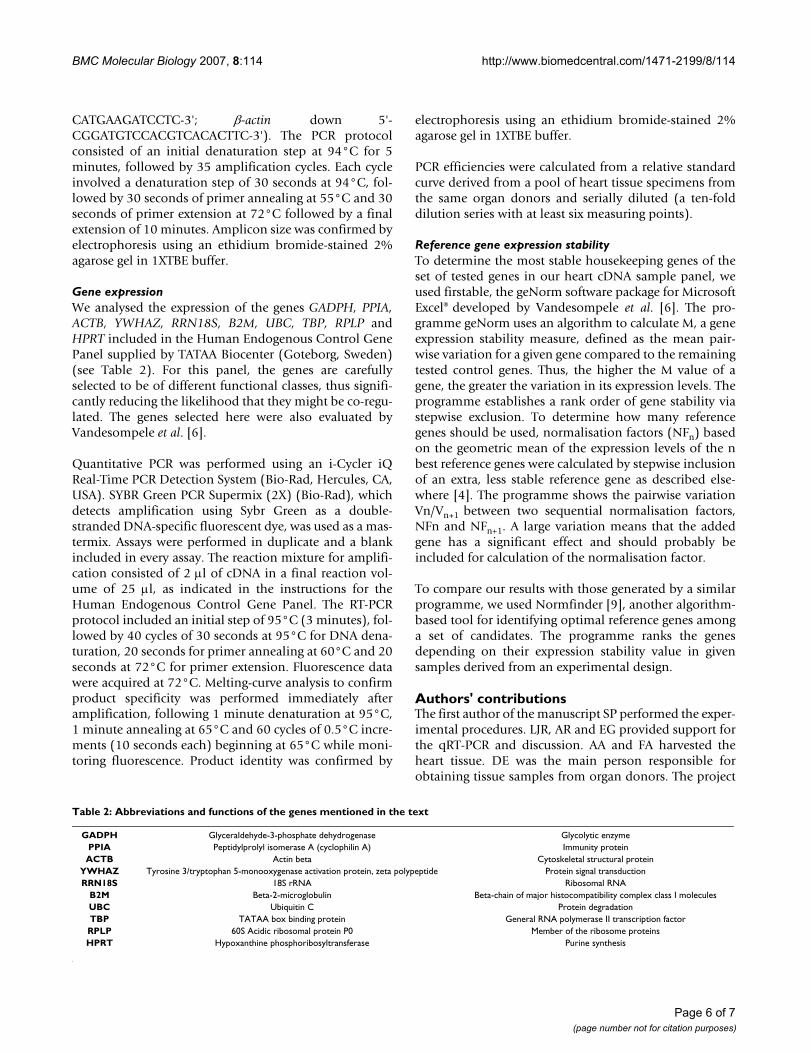
Table 1: Tested reference genes for normalisation of qRT-PCR listed according to their expression stability calculated by the Normfinder software.
Raking Order Gene Name Stability Value
1 GADPH 0.3452 PPIA 0.4053 RPLP 0.4264 B2M 0.4465 YWHAZ 0.4986 HPRT 0.5887 RRN18S 0.5938 ACTB 0.7379 UBC 0.83110 TBP 2.287
Page 4 of 7(page number not for citation purposes)
BMC Molecular Biology 2007, 8:114 http://www.biomedcentral.com/1471-2199/8/114
across cardiac pathologies, suggesting their suitable use asreference genes for quantitative PCR studies in cardiac tis-sue. In these previous studies, the specific testing of sev-eral candidate reference genes to determine the mostsuitable reference for use in cardiac tissue was notreported [15,16]. In contrast, Radonic et al. [17] deter-mined transcription levels of several housekeeping genesin different human tissues, including heart, and identifiedTBP as the gene with the lowest range of RNA transcrip-tion across tissues. This finding is in agreement with thepresent results.
We found PPIA, RPLP plus GADPH to be a reliable set ofgenes for normalising data (Figure 3) according to thegeNorm programme. As reported by Vandesompele et al.[6], geNorm proposes a pairwise variation of 0.15 as thecut-off under which the inclusion of an additional controlgene is not required. Using our set of candidate genes, thiswould mean that adding a fifth gene to the four most sta-ble genes identified would really provide the best results.Notwithstanding, this cut-off of 0.15 should not be con-sidered in a strict sense, but rather as a reference to deter-mine the optimal number of housekeeping genes.Sometimes the observed trend can be equally informative,and using the three best reference genes is, in most cases,a valid strategy for much more accurate and reliable nor-malisation compared to the use of a single housekeepinggene.
Using NormFinder software [8] as another tool to validatethe expression stability of the ten candidate referencegenes; GADPH, PPIA and RPLP also achieved the best sta-bility values. While geNorm detected the two genes whoseexpression ratios showed least variation from those of theother genes tested, NormFinder was able to identify thesingle gene with the most stable expression. Hence, themost stable candidate gene was found to be GADPH, andthe least stable TBP. Using this programme, we obtainedthe same results as with GeNorm except for the rank posi-tion ascribed to GADPH as the most stable single gene.However, the least stable genes and most stable ones iden-tified by GeNorm and Normfinder were generally well-matched.
As a limitation to our study, we should mention thatalthough we established the purity of our RNA samples,due to the amount of RNA available, we could not runelectrophoretic tests to check RNA integrity.
Finally, the set of reference genes determined here as thebest endogenous controls to be used in qRT-PCR studiesin heart tissue has applications in developing cardiovascu-lar diagnostic tests and therapeutic strategies that will sub-stantially improve human health [18].
ConclusionIt is commonly accepted that gene expression studiesshould be normalised using more than one referencegene. Based on our results, we propose the use of themean result rendered by PPIA, RPLP and GADPH ashousekeeping genes to normalise gene expression valuesobtained by qRT-PCR in heart tissue.
MethodsSample collectionTissue samples were obtained from the left ventricle of 35organ donors (19 male, 16 female; mean age 59 years,range 40 – 80 years). The organ donors had died in amultidisciplinary ICU of a tertiary university hospital. In85% of the organ donors, brain death was attributed to anon-traumatic cause and no donor had previous heartpathology. Consent for organ donation for transplant andclinical investigation was obtained from the relatives ofeach donor.
The hearts had been rejected for transplant because ofdonor age or weight, or recipient blood group incompati-bility. The cardiac tissues were freshly harvested, storedimmediately in O.C.T™ Compound (Tissue Tek, Sakura,Netherlands) and then frozen at -80°C.
RNA extraction and cDNA synthesisIsolation of total RNA was performed using the NucleoSpin RNA II Isolation Kit (Macherey-Nagel Gmb H & Co.KG) according to the manufacturer's instructions.
The quality of the RNA was assessed through absorbancemeasurements made using a NanoDrop ND-1000 UV-VisSpectrophotometer (NanoDropTechnologies, Wilming-ton, DE, USA). All the samples used in the PCR proce-dures showed a 260/280 nm absorbance ratio between1.8 and 2.2. A ratio of ≈2 is generally accepted as pure forRNA.
Each RNA sample was reverse transcribed using the Oligo-pdT and Random primers provided in the first-strandcomplementary DNA (cDNA) synthesis kit for RT-PCR(AMV; Roche Diagnostic, Switzerland) following themanufacturer's instructions. The tubes were incubated at25°C for 10 minutes to allow annealing. Reverse tran-scription was conducted at 42°C for 60 minutes, followedby 5 minutes incubation at 99°C to denature the enzyme.The cDNA samples were then cooled at 4°C and stored at-20°C until use.
To confirm the total absence of residual DNA, we per-formed a conventional PCR with β-actin [Gen-Bank:E00829] primers designed using Beacon Designersoftware (Premier Biosoft International, California) span-ning intron-exon boundaries (β-actin up 5'-GACTACCT-
Page 5 of 7(page number not for citation purposes)
BMC Molecular Biology 2007, 8:114 http://www.biomedcentral.com/1471-2199/8/114
CATGAAGATCCTC-3'; β-actin down 5'-CGGATGTCCACGTCACACTTC-3'). The PCR protocolconsisted of an initial denaturation step at 94°C for 5minutes, followed by 35 amplification cycles. Each cycleinvolved a denaturation step of 30 seconds at 94°C, fol-lowed by 30 seconds of primer annealing at 55°C and 30seconds of primer extension at 72°C followed by a finalextension of 10 minutes. Amplicon size was confirmed byelectrophoresis using an ethidium bromide-stained 2%agarose gel in 1XTBE buffer.
Gene expressionWe analysed the expression of the genes GADPH, PPIA,ACTB, YWHAZ, RRN18S, B2M, UBC, TBP, RPLP andHPRT included in the Human Endogenous Control GenePanel supplied by TATAA Biocenter (Goteborg, Sweden)(see Table 2). For this panel, the genes are carefullyselected to be of different functional classes, thus signifi-cantly reducing the likelihood that they might be co-regu-lated. The genes selected here were also evaluated byVandesompele et al. [6].
Quantitative PCR was performed using an i-Cycler iQReal-Time PCR Detection System (Bio-Rad, Hercules, CA,USA). SYBR Green PCR Supermix (2X) (Bio-Rad), whichdetects amplification using Sybr Green as a double-stranded DNA-specific fluorescent dye, was used as a mas-termix. Assays were performed in duplicate and a blankincluded in every assay. The reaction mixture for amplifi-cation consisted of 2 µl of cDNA in a final reaction vol-ume of 25 µl, as indicated in the instructions for theHuman Endogenous Control Gene Panel. The RT-PCRprotocol included an initial step of 95°C (3 minutes), fol-lowed by 40 cycles of 30 seconds at 95°C for DNA dena-turation, 20 seconds for primer annealing at 60°C and 20seconds at 72°C for primer extension. Fluorescence datawere acquired at 72°C. Melting-curve analysis to confirmproduct specificity was performed immediately afteramplification, following 1 minute denaturation at 95°C,1 minute annealing at 65°C and 60 cycles of 0.5°C incre-ments (10 seconds each) beginning at 65°C while moni-toring fluorescence. Product identity was confirmed by
electrophoresis using an ethidium bromide-stained 2%agarose gel in 1XTBE buffer.
PCR efficiencies were calculated from a relative standardcurve derived from a pool of heart tissue specimens fromthe same organ donors and serially diluted (a ten-folddilution series with at least six measuring points).
Reference gene expression stabilityTo determine the most stable housekeeping genes of theset of tested genes in our heart cDNA sample panel, weused firstable, the geNorm software package for MicrosoftExcel® developed by Vandesompele et al. [6]. The pro-gramme geNorm uses an algorithm to calculate M, a geneexpression stability measure, defined as the mean pair-wise variation for a given gene compared to the remainingtested control genes. Thus, the higher the M value of agene, the greater the variation in its expression levels. Theprogramme establishes a rank order of gene stability viastepwise exclusion. To determine how many referencegenes should be used, normalisation factors (NFn) basedon the geometric mean of the expression levels of the nbest reference genes were calculated by stepwise inclusionof an extra, less stable reference gene as described else-where [4]. The programme shows the pairwise variationVn/Vn+1 between two sequential normalisation factors,NFn and NFn+1. A large variation means that the addedgene has a significant effect and should probably beincluded for calculation of the normalisation factor.
To compare our results with those generated by a similarprogramme, we used Normfinder [9], another algorithm-based tool for identifying optimal reference genes amonga set of candidates. The programme ranks the genesdepending on their expression stability value in givensamples derived from an experimental design.
Authors' contributionsThe first author of the manuscript SP performed the exper-imental procedures. LJR, AR and EG provided support forthe qRT-PCR and discussion. AA and FA harvested theheart tissue. DE was the main person responsible forobtaining tissue samples from organ donors. The project
Table 2: Abbreviations and functions of the genes mentioned in the text
GADPH Glyceraldehyde-3-phosphate dehydrogenase Glycolytic enzymePPIA Peptidylprolyl isomerase A (cyclophilin A) Immunity proteinACTB Actin beta Cytoskeletal structural protein
YWHAZ Tyrosine 3/tryptophan 5-monooxygenase activation protein, zeta polypeptide Protein signal transductionRRN18S 18S rRNA Ribosomal RNA
B2M Beta-2-microglobulin Beta-chain of major histocompatibility complex class I moleculesUBC Ubiquitin C Protein degradationTBP TATAA box binding protein General RNA polymerase II transcription factorRPLP 60S Acidic ribosomal protein P0 Member of the ribosome proteinsHPRT Hypoxanthine phosphoribosyltransferase Purine synthesis
Page 6 of 7(page number not for citation purposes)
BMC Molecular Biology 2007, 8:114 http://www.biomedcentral.com/1471-2199/8/114
Publish with BioMed Central and every scientist can read your work free of charge
"BioMed Central will be the most significant development for disseminating the results of biomedical research in our lifetime."
Sir Paul Nurse, Cancer Research UK
Your research papers will be:
available free of charge to the entire biomedical community
peer reviewed and published immediately upon acceptance
cited in PubMed and archived on PubMed Central
yours — you keep the copyright
Submit your manuscript here:http://www.biomedcentral.com/info/publishing_adv.asp
BioMedcentral
was designed by JO. All the authors have read andapproved the final manuscript.
AcknowledgementsThis research was supported by grant IB05-135 from the Ficyt. We also thank to the Spanish Ministry of Science and Education (project NO AGL-2005-04479).
References1. Ginzinger DG: Gene quantification using real-time quantita-
tive PCR: an emerging technology hits the mainstream. ExpHematol 2002, 30(6):503-512.
2. Schmittgen TD, Zakrajsek BA: Effect of experimental treatmenton housekeeping gene expression: validation by real-time,quantitative RT-PCR. J Biochem Biophys Methods 2000, 46(1–2):69-81.
3. Gorzelniak K, Janke J, Engeli S, Sharma AM: Validation of endog-enous controls for gene expression studies in human adi-pocytes and preadipocytes. Horm Metab Res 2001,33(10):625-627.
4. von Smolinski D, Leverkoehne I, von Samson-Himmelstjerna G, Gru-ber AD: Impact of formalin-fixation and paraffin-embeddingon the ratio between mRNA copy numbers of differentlyexpressed genes. Histochem Cell Biol 2005, 124(2):177-188.
5. Suzuki T, Higgins PJ, Crawford DR: Control selection for RNAquantitation. Biotechniques 2000, 29:332-337.
6. Vandesompele J, De Preter K, Pattyn F, Poppe B, Van Roy N, DePaepe A, Speleman F: Accurate normalization of Real-Timequantitative RT-PCR by geometric averaging of multipleinternal control genes. Genome Biol 2002, 3(7):34.
7. Tricarico C, Pinzani P, Bianchi S, Paglierani M, Distante V, Pazzagli M,Bustin SA, Orlando C: Quantitative real-time reverse tran-scription polymerase chain reaction: normalization to rRNAor single housekeeping genes is inappropriate for human tis-sue biopsies. Anal Biochem 2002, 309:293-300.
8. Andersen CL, Jensen JL, Orntoft TF: Normalization of real-timequantitative reverse transcription-PCR data: a model-basedvariance estimation approach to identify genes suited fornormalization, applied to bladder and colon cancer datasets. Cancer Res 2004, 64(15):5245-5250.
9. NormFinder Software [http://www.mdl.dk/publicationsnormfinder.htm]
10. SAS/STAT™, 1999. User's Guide. Release 8.2. SAS InstituteInc, 703 Cary NC. 704. .
11. Thellin O, Zorzi W, Lakaye B, De BB, Coumans B, Hennen G, GrisarT, Igout A, Heinen E: Housekeeping genes as internal stand-ards: use and limits. J Biotechnol 1999, 75:291-295.
12. Glare EM, Divjak M, Bailey MJ, Walters EH: Beta-Actin andGADPH housekeeping gene expression in asthmatic airwaysis variable and not suitable for normalising mRNA levels.Thorax 2002, 57:765-770.
13. Yperman J, De Visscher G, Holvoet P, Flameng W: Beta-actin can-not be used as a control for gene expression in ovine intersti-tial cells derived from heart valves. J Heart Valve Dis 2004,13(5):848-853.
14. Bustin SA, Nolan T: Pitfalls of quantitative real-time reversetranscription Polymerase Chain Reaction. J Biomol Tech 2004,15:155-166.
15. Morgan K, Wharton J, Webb JC, Keogh BE, Smith PL, Taylor KM,Oakley CM, Polak JM, Cleland JG: Co-expression of renin-angi-otensin system component genes in human atrial tissue. JHypertens Suppl 1994, 12(4):S11-19.
16. Moniotte S, Vaerman J-L, Kockx MM, Larrouy D, Langin D,Noirhomme P, Balligand J-L: Real-Time RT-PCR for the detec-tion of beta-adrenoceptor messenger RNAs in small humanendomyocardial biopsies. J Mol Cell Cardiol 2001, 33:2121-2133.
17. Radonic A, Thulke S, Mackay IM, Landt O, Siegert W, Nitsche A:Guideline to reference gene selection for quantitative real-time PCR. Biochem Biophys Res Commun 2004, 313:856-862.
18. Seo D, Ginsburg GS, Goldschmidt-Clermont PJ: Gene expressionanalysis of cardiovascular diseases: novel insights into biol-ogy and clinical applications. J Am Coll Cardiol 2006,48(2):227-235.
Page 7 of 7(page number not for citation purposes)