Embed Size (px)
Citation preview

LUND UNIVERSITY
PO Box 117221 00 Lund+46 46-222 00 00
"NeuroStem Chip": a novel highly specialized tool to study neural differentiationpathways in human stem cells.
Anisimov, Sergey; Christophersen, Nicolaj; Correia, Ana S; Li, Jia-Yi; Brundin, Patrik
Published in:BMC Genomics
DOI:10.1186/1471-2164-8-46
Published: 2007-01-01
Link to publication
Citation for published version (APA):Anisimov, S., Christophersen, N., Correia, A. S., Li, J-Y., & Brundin, P. (2007). "NeuroStem Chip": a novel highlyspecialized tool to study neural differentiation pathways in human stem cells. BMC Genomics, 8(Feb 8), 46-46.DOI: 10.1186/1471-2164-8-46
General rightsCopyright and moral rights for the publications made accessible in the public portal are retained by the authorsand/or other copyright owners and it is a condition of accessing publications that users recognise and abide by thelegal requirements associated with these rights.
• Users may download and print one copy of any publication from the public portal for the purpose of privatestudy or research. • You may not further distribute the material or use it for any profit-making activity or commercial gain • You may freely distribute the URL identifying the publication in the public portal

Take down policyIf you believe that this document breaches copyright please contact us providing details, and we will removeaccess to the work immediately and investigate your claim.
Download date: 06. Jul. 2018
BioMed CentralBMC Genomics
ss
Open AcceMethodology article"NeuroStem Chip": a novel highly specialized tool to study neural differentiation pathways in human stem cellsSergey V Anisimov*, Nicolaj S Christophersen, Ana S Correia, Jia-Yi Li and Patrik BrundinAddress: Neuronal Survival Unit, Wallenberg Neuroscience Center, Lund University, 221 84 Lund, Sweden
Email: Sergey V Anisimov* - [email protected]; Nicolaj S Christophersen - [email protected]; Ana S Correia - [email protected]; Jia-Yi Li - [email protected]; Patrik Brundin - [email protected]
* Corresponding author
AbstractBackground: Human stem cells are viewed as a possible source of neurons for a cell-basedtherapy of neurodegenerative disorders, such as Parkinson's disease. Several protocols thatgenerate different types of neurons from human stem cells (hSCs) have been developed.Nevertheless, the cellular mechanisms that underlie the development of neurons in vitro as they aresubjected to the specific differentiation protocols are often poorly understood.
Results: We have designed a focused DNA (oligonucleotide-based) large-scale microarrayplatform (named "NeuroStem Chip") and used it to study gene expression patterns in hSCs as theydifferentiate into neurons. We have selected genes that are relevant to cells (i) being stem cells, (ii)becoming neurons, and (iii) being neurons. The NeuroStem Chip has over 1,300 pre-selected genetargets and multiple controls spotted in quadruplicates (~46,000 spots total). In this study, wepresent the NeuroStem Chip in detail and describe the special advantages it offers to the fields ofexperimental neurology and stem cell biology. To illustrate the utility of NeuroStem Chip platform,we have characterized an undifferentiated population of pluripotent human embryonic stem cells(hESCs, cell line SA02). In addition, we have performed a comparative gene expression analysis ofthose cells versus a heterogeneous population of hESC-derived cells committed towards neuronal/dopaminergic differentiation pathway by co-culturing with PA6 stromal cells for 16 days andcontaining a few tyrosine hydroxylase-positive dopaminergic neurons.
Conclusion: We characterized the gene expression profiles of undifferentiated and dopaminergiclineage-committed hESC-derived cells using a highly focused custom microarray platform(NeuroStem Chip) that can become an important research tool in human stem cell biology. Wepropose that the areas of application for NeuroStem microarray platform could be the following:(i) characterization of the expression of established, pre-selected gene targets in hSC lines,including newly derived ones, (ii) longitudinal quality control for maintained hSC populations, (iii)following gene expression changes during differentiation under defined cell culture conditions, and(iv) confirming the success of differentiation into specific neuronal subtypes.
Published: 8 February 2007
BMC Genomics 2007, 8:46 doi:10.1186/1471-2164-8-46
Received: 21 September 2006Accepted: 8 February 2007
This article is available from: http://www.biomedcentral.com/1471-2164/8/46
© 2007 Anisimov et al; licensee BioMed Central Ltd. This is an Open Access article distributed under the terms of the Creative Commons Attribution License (http://creativecommons.org/licenses/by/2.0), which permits unrestricted use, distribution, and reproduction in any medium, provided the original work is properly cited.
Page 1 of 15(page number not for citation purposes)

BMC Genomics 2007, 8:46 http://www.biomedcentral.com/1471-2164/8/46
BackgroundModern DNA microarrays permit a comprehensive analy-sis of quantitative and qualitative changes in RNA tran-script abundance, outlining the cross-sections of geneexpression and alterations of these in response to geneticor environmental stimuli. Genome-scale microarrays(cDNA- or oligonucleotide-based) are most valuablewhen screening populations of cells for the novel genesreflecting potential diagnostic and prognostic markers orfor an identification of novel therapeutic targets. On theother hand, custom microarray platforms that focus onspecific pre-selected subset of genes relevant to a particu-lar field of investigation can be less costly and more suita-ble for detection of smaller gene expression changes.
Microarray technology has added important informationon both normal development and pathological changesin neurons. This is well illustrated by multiple studies onsubstantia nigra dopaminergic neurons, which degeneratein Parkinson's disease (PD) [1-5]. The shortcomings ofpharmacological therapies in PD have stimulated a searchfor alternative treatment strategies. In successful cases,transplants of human embryonic mesencephalicdopaminergic neurons can both restore dopaminergicneurotransmission and provide some symptomatic relief[6-8]. A wider application of neural transplantation in PDis, however, currently not feasible due to the unpredicta-ble and variable outcome, the risks of unwanted side-effects (dyskinesias) [9,10] and ethical and practical prob-lems associated with using donor cells obtained fromaborted embryos and fetuses [11,12]. Human embryonicstem cells (hESCs) are considered a promising futuresource of cells for cell replacement therapy in PD andother neurological conditions [13]. They could constitutea virtually infinite source of self-renewing cells that can bepersuaded to differentiate into specific types of neuralcells, including dopaminergic neurons [14-16]. Themolecular mechanisms that govern development of cul-tured hESCs into specific types of neural cells are not fullyunderstood. To promote our understanding of suchmechanisms, it would be valuable to have tools that read-ily and reproducibly can help to characterize the cells asthey differentiate from pluripotent stem cells into post-mitotic neurons. This important issue was addressed inearlier studies by Luo et al. and Yang et al., who designedsmall-to-moderate scale custom microarray platforms(281 and 755 gene targets, respectively) [17,18]. In addi-tion SuperArray Bioscience Corporation (Frederick, MD,USA) have manufactured a range of small-scale arrays(263 gene targets for human array; [19]). We sought tocreate an improved and updated microarray platform forhESC/neuronal differentiation-oriented gene expressionstudies. Therefore, we generated a specialized large-scaleDNA microarray platform (the "NeuroStem Chip") thathas over 1,300 pre-selected gene targets and multiple con-
trols spotted in quadruplicates (~46,000 spots total). Herewe introduce the platform and the advantages it can offersto neuroscientists and stem cell biologists: particularly, inthe niche of gene expression-oriented characterization ofthe samples using an assay of pre-selected, already estab-lished gene targets. In the current study, we use the Neu-roStem Chip to characterize an undifferentiatedpopulation of pluripotent hESCs (cell line SA02, CellartisAB, Göteborg, Sweden) and compare the gene expressionin those cells with that of a hESC-derived cell populationrich in neurons, including tyrosine hydroxylase-positivedopaminergic neurons.
Results and DiscussionStem cells have unique biological characteristics, but onlya limited number of genes are currently recognized asestablished stem cell markers. Examples include POUdomain, class 5, transcription factor 1 (Oct3/4), signaltransducer and activator of transcription 3 (Stat3), terato-carcinoma-derived growth factor (Tdgf1), Enk-pending(Nanog), undifferentiated embryonic cell transcriptionfactor 1 (Utf1) and DNA methyltransferase 3B (Dnmt3b)[20]. At the same time, hundreds of genes are suggested ascandidate markers for "stemness", but their coupling tothe undifferentiated stem cell state is not yet fully verified[21]. The concept of "stemness" (term introduced in 1986by Grossman & Levine) is defined as "core stem cell proper-ties that underlie self-renewal and the ability to generate differ-entiated progeny" [22]. Considering the complexity of theprocesses involved, stemness can hardly be ensured by co-operation of just a few genes. Nevertheless, three stemnessgenes (namely, Oct3/4, Stat3 and Nanog) are considered"master"-genes that control the self-renewing process[23,24]. Various types of stem cells, such as hematopoi-etic, mesenchymal and neural (HSCs, MSCs and NSCs,respectively), embryonic germ and embryonic carcinomacells (EGCs and ECCs, respectively) are all characterizedby variations in gene expression profiles, and only a fewgene markers are associated with all these cell types[25,26]. We have aimed to embrace the most comprehen-sive set of those genes into a solitary array, the NeuroStemChip. Thereby, it is possible to employ it to monitor therelative expression levels of numerous known and candi-date stemness genes in a single experiment.
Similar to the genetic bases underlying stemness, cell dif-ferentiation is associated with altered expression levels ofcertain recognized or candidate genes [25]. We thereforeincorporated gene markers of development and differen-tiation in general, and that of neuronal and dopaminergicdifferentiation in particular, into the NeuroStem Chip.Examples include markers for the processes of neuronalmaturation, axonal branching, neural/neuronal survival,etc. Finally, we ensured that known markers for specifictypes of neurons, allowing identification of individual cell
Page 2 of 15(page number not for citation purposes)

BMC Genomics 2007, 8:46 http://www.biomedcentral.com/1471-2164/8/46
types, were present on the chip. We paid special attentionto genes associated with the differentiation and matura-tion of dopaminergic neurons. In many published stud-ies, the expression of only a single (tyrosine hydroxylase,TH) or 2–3 markers for dopaminergic neurons (e.g.amino acid decarboxylase (AADC), dopamine transporter(DAT), vesicular monoamine transporter 2 (VMAT2))have been used to indicate dopaminergic identity of neu-rons. In contrast, the NeuroStem Chip includes oligonu-cleotide probes for 88 genes related to dopaminergicneurons, thus being more comprehensive in this sense,compared to other existing microarray platforms, includ-ing focused ones [17,18]. Those entries encompass recog-nized and candidate markers for dopaminergic neurons(mature and early) and progenitors, as well as markers forthe maturation and differentiation of the latter (Table 1).
Table 2 represents conditional functional breakdown ofgenes targeted by the NeuroStem microarray platform. Anumber of important gene groups that are included in thechip are not mentioned in Table 2. Among these, entriesrelated to Dickkopf gene family, galanin-, melatonin-,vasoactive intestinal peptide (VIP)-, cAMP response ele-ment-binding protein (CREB)- and B cell leukemia 2(Bcl2) oncogene-related are present. Many of them playpotentially important, yet undefined, roles in the biologyof stem cells. Additionally, we included some genes impli-cated in disease mechanisms of neurodegenerative disor-ders (most importantly, Parkinson's disease andAlzheimer's disease) in the chip. Furthermore, we incor-porated a number of markers for distinct differentiationpathways (e.g. hematopoietic and pancreatic) and celltypes (e.g. cancer subtypes and a range of normal celltypes) to serve as essential controls. Taken together, we
believe that in its present form NeuroStem Chip repre-sents currently most comprehensive gene expression plat-form for studies on stem cells, neural/neuronaldifferentiation, human neurodegeneration and neuronalsurvival, both in vivo and in vitro. The complete layout ofNeuroStem Chip will be disclosed to the academic com-munity, upon request.
The microarray format we selected relies on long oligonu-cleotide molecules (69–71 nucleotides) printed over asolid surface. We spotted the synthesized oligonucleotides(Operon Biotechnologies) with a constant concentrationacross the slides, and evaluated the quality and consist-ency of spotting in a series of control experiments. Wethen illustrated the utility and technical reliability of theNeuroStem Chip by characterizing the gene expressionprofile of commonly utilized hESC line SA02 (Sahlgren-ska 2; [27]), including (i) undifferentiated cells and (ii)cells committed towards neuronal/dopaminergic differ-entiation pathway. For the first of these, we used totalRNA sample purified from hESC colonies that exhibitedmorphology consistent with cell proliferation and theabsence of spontaneous differentiation (Figure 1A). Wealso evaluated the expression of the cell cycle marker Ki67and the pluripotency marker OCT3/4 in the sample byimmunocytochemistry (Figure 1B–E). Co-culturing ofESCs with murine stromal cells (including PA6 cell line)rapidly generates dopaminergic neurons from ESCs by anunexplained mechanism termed stromal cell-derivedinducing activity (SDIA; [28,29]). We therefore commit-ted hESCs toward the neuronal/dopaminergic differentia-tion pathway by co-culturing with PA6 cells for 16 days,resulting in appearance of cells positive for early and lateneuronal markers, including nestin, β-III-tubulin, and TH,
Table 1: NeuroStem chip entries related to dopaminergic system.
Genesa Function
Aadc, Ant2 (Slc25a5), Calb1, Dat (Slc6a3), Girk2 (Kcnj6), Igf1, Ptx3, Th Markers of dopaminergic neuronsEn1, En2, Lmx1bb, Pax5, Shh, Wnt1 Markers of dopaminergic progenitorsAdh2, Lhx1 (Lim1), Lhx5 (Lim2) Markers of dopaminergic progenitors maturationDrd2b, Vmat2 Markers of mature dopaminergic neuronsGfra1, Gfra2, Gfra3, Gfra4, Otx2 Markers of early dopaminergic neuronsDlx1, Dlx2, Flj38973, Lmx1a Related to differentiation of dopaminergic neuronsAdcy7, Alcam, Art (Artn), Bcl11a, Calca, Cart, Cbln1, Cdh2, Col11a1, Cx43, D1lip (Drd1ip), Darpp32 (Ppp1r1b), Dbh, Doc2a, Drd1, Drd3, Drd4, Drd5, Drip78 (Dnajc14), Egln3 (Phd3), Egr1, Fos, Fxyd6, Galm, Ghr, Grin2c, Grp, Gsbs (C7orf16), Hs6st2, Igfbp4, Kcna5, Kcnab1, Math2, Mlp (Marcksl1), Moxd1, Mpp3, Nrip3, Nrp2, Nt (Nts), Ntn (Nrtn), Nurr1 (Nr4a2), Pac1 (Adcyap1r1), Pacap, Plagl1 (Zac), Pvrl3 (Nectin 3), Rab3c, Rcn1, Slc17a6 (Vglut2), Slc6a1, Sox6, Spp1, Tacr3, Trhr, Zfp161, Vav3, Zdhhc2
Related to dopaminergic system
The majority of genes listed may also possess other functions, not related to dopaminergic system. Four entries were omitted from the table, representing candidate markers of dopaminergic system now undergoing characterization (unpublished data).a Italics indicates genes up-regulated (Log2 ratio >1.0) in dopaminergic differentiation experiment. Rcn1, Log2 ratio >0.97; Fxyd6 and Zfp161, Log2 ratio >0.7).b May also possess other functions related to dopaminergic system.
Page 3 of 15(page number not for citation purposes)

BMC Genomics 2007, 8:46 http://www.biomedcentral.com/1471-2164/8/46
Page 4 of 15(page number not for citation purposes)
Table 2: Selected categories of NeuroStem chip entries.
Category Functional role Examples
I. Stemness Recognized markers of stemness Oct3/4, Nanog, Tdgf1Candidate markers of stemness Cpxm1, Hook1, Ddx21
Germ cell markers Rif1, Bnc1, Bnc2Hematopoietic stem cell markers Hoxb4, Cdcp1, C1qr1Mesenchymal stem cell marker Bmpr1a, Bmpr2, Cd49a
II. Proliferation Proliferation markers Ki67, Pcna, MycNeural proliferation markers Emx1, Gbx2
III. Development Differentiation Lifr, Ebaf, LyarNeuronal differentiation Ren, Rai1, Neurod2
Dopaminergic differentiation Dlx1, Dlx2, Lmx1aNeuronal maturation Mecp2, Ebf3, Sox4
Neuronal process formation Hmgb1, RageAxonal elongation and branching Pi3, Map1b, Slit1
IV. Neural markers Pan-neural markers Gap43, Nfh, Eno2Markers of dopaminergic neurons Th, Aadc, DatMarkers of cholinergic neurons Ngf
Markers of spinal neurons Hoxc6Glial markers S100β, Cd68
Astrocyte markers Gfap, Tapa1Oligodendrocyte markers Mag, Mobp, Omg
V. Distinct markers Normal tissuesLiver Gata6, G6pd, Fabp1
Pancreas Tff3, Sst, Pax4Skeletal muscle Itga7, Dmd, Tnnt3Cardiac muscle Nkx2.5, Anf, MyhcaSmooth muscle Actg2, Cnn1, Sm22αEndothelial cells Flt1, Vwf, Pecam1
Blood cell subtypes Cd4, Cd8, Cd19Cancer cells Maspin
Pancreatic cancer Kras2Colon cancer Mina53Breast cancer Klk7
Prostatic cancer Hpn, Mat8Lung cancer Rab5a, Tp63
Ovarian cancer Mgb2Hodgkin's lymphoma Ptp4a, Atf5, p21snft
VI. Relevant groups Apoptosis-related p53, Psip1, Birc2Telomere-related Tert, Terf2, Rap1
Antioxidants Sod1, Sod2, Gpx1Imprinted genes Tseb3, Gnas1, Grb10
FZD group Fzd1, Fzd3, Sfrp1WNT group Wnt1, Wnt7a, Wisp1BMP group Bmp1, Bmp2, BambiSTAT group Stat1, Stat3, Pias1FGF group Fgf1, Fgf2, Fgfr4GDF group Gdf2, Gdf3, Gdf9
Caspases group Casp1, Casp2, Hsp70Cyclins group Ccna1, Ccnc, Cdk1
Kruppel-like group Klf2, Klf9, Znf184
Due to functional redundancy, individual genes may fit into more than one category.

BMC Genomics 2007, 8:46 http://www.biomedcentral.com/1471-2164/8/46
the established marker of dopaminergic neurons (Figure2). To verify the expression of some key stem cell- andneural phenotype-associated genes we performed RT-PCRcomparing RNA samples from the undifferentiated hESCswith hESCs of the same line differentiated toward neuro-nal/dopaminergic pathway, as described above. Theexpression profile outlined by RT-PCR confirmed theidentity of the sample used (Figure 3). After performingRNA integrity tests, we incorporated fluorescent labels tothe amplified RNA samples from hESCs (Cyanine 3-CTP(Cy3) and Cyanine 5-CTP (Cy5)), hESC-derived cells con-taining TH-positive neurons (Cy3 and Cy5) and humanuniversal reference RNA (Cy5), and hybridized aliquotswith NeuroStem microarray slides using the followingconditions: hESC vs. reference, Cy3 : Cy5 = (i) 20:10pmol, and (ii) 10:5 pmol; and hESC vs. hESC-derivedcells, Cy3 : Cy5 = (iii) 30:20 pmol, respectively. Universalreference RNA has been previously established as a stand-ard reference material for microarray experiments, prov-ing an ability to effectively hybridize to a large fraction ofmicroarray spots [30].
We performed two-color hybridizations (e.g. for theexperiment vs. reference) following an established proto-col [31], and included dye-flip technical replicates in theanalysis (Figure 4). Using the online software programBASE [32] we sequentially filtered the data by backgroundsubtraction, negative flagging, negative intensities and forinconsistent data amongst replicates [33]. Figure 5Ashows a comparison of the spot intensities prior to nor-malization (M versus A plot), with the Log2 of the expres-sion ratio between Cy3/Cy5 being plotted as a function ofthe log10 of the mean of the total expression intensitiesfor Cy3 and Cy5 channels. The deviation of the line fromzero revealed a need for normalization, so prior to dataanalyses we normalized signals using a locally weightedscatterplot-smoothing regression (LOWESS) algorithm(Figure 5A–B; fitted line) implemented in BASE. Since thereproducibility of two-color microarray gene expressiondata is critically important, we calculated Pearson correla-tion coefficients of the reporters present in the filtereddatabase comparing the average expression ratios (7005for hESCs vs. universal reference; 6947 for undifferenti-ated vs. neuronal/dopaminergic lineage-committedhESCs). Results obtained revealed that data were consist-ent across technical replicates (dye-swap and amount ofloaded material), showing general high reproducibility:e.g., correlation coefficients were greater than 0.96 fortechnical replicates and 0.78 for dye-swapping samples inhESCs vs. universal reference hybridizations (Table 3). Todetect genes with high expression levels in hESC samples,we filtered data for intensity values >100 in the hESC sam-ple and performed clustering analysis using the TIGR Mul-tiExperiment Viewer (MEV; [34]). To visualize variationsof spot/reporter per technical replicate, hierarchical clus-
tering was performed by K-means classifier based on thelinear-correlation-based distance (Pearson, centred)method. The optimal number of clusters was determinedempirically to produce the most balanced ratio of entriesto cluster of highly expressed genes. A cluster of 101 genesup-regulated in the hESC sample [see Additional file 1],was plotted in a centroid graph (Figure 5C); the variationacross technical replicates was low. We merged technicalreplicates to generate a list of the most up-regulated genesexpressed in the hESC sample compared to the universalreference RNA (Table 4). Standard error of the meanexpressed as percentage was calculated for the 4 technicalreplicates, and was 6.7% for the top 25 genes up-regulatedin hESC samples, compared to universal reference RNA.We performed the analysis of microarray data, asdescribed in the Methods, and spot error values were gen-erally in the lower range, indicating high stringency of thesignals and low variance.
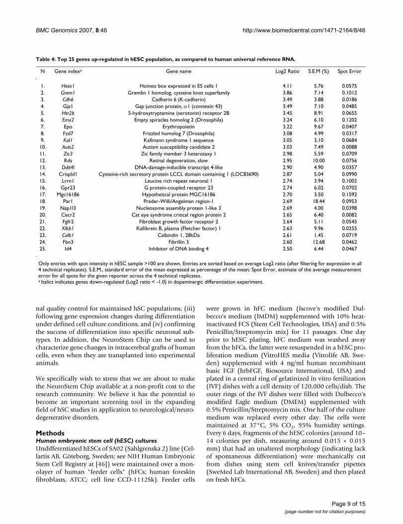
As seen in Table 4 and Table 5, the NeuroStem Chip iden-tified numerous genes associated with stem cells. In par-ticular, homeo box expressed in ES cells 1 (Hesx1) genewas identified as the most up-regulated in the ES cell prep-aration, compared to universal reference RNA. Highlyexpressed in pluripotent ESCs, Hesx1 expression is down-regulated upon embryonic stem cell differentiation[35,36], as also clearly seen in differentiation experimentof our own (Table 4). Similarly, Gremlin 1 homolog,cysteine knot superfamily gene (Grem1, also known asCktsf1b1 and Dand2) is a recognized factor of cell-fatedetermination of ESCs [37]. Many more genes highly up-regulated in the hESC sample in comparison with univer-sal reference RNA are associated with stem cells: furtherexamples include Gap junction protein α1 (Gja1) and Zicfamily member 3 heterotaxy 1 (Zic3) (Table 4) [20]. Theexpression of fibroblast growth factor receptor 2 (Fgfr2) isof particular interest. Basic fibroblast growth factor (FGF2,bFGF) supports hESC proliferation and their ability tomaintain undifferentiated phenotype when cultured invitro [38,39]. Moreover, in some hESC lines a very highconcentration of FGF2 could substitute for the need offeeder cells [40]. At the same time, genes listed in Table 4represent the most highly up-regulated entries in a rela-tively limited group of genes (Figure 5C). Many othergenes involved in maintenance of ESC phenotype (i.e.established or candidate markers of stem cells) have lowerlevels of expression (Table 5). Examples include undiffer-entiated embryonic cell transcription factor 1 (Utf1),DNA methyltransferase 3B (Dnmt3b), developmentalpluripotency associated 4 (Dppa4, a newly establishedpluripotency marker [41]) and numerous candidatemarkers of "stemness": e.g. genes for KIAA1573 protein,forkhead box O1A (Foxo1a), high-mobility group box 1(Hmgb1), C-terminal binding protein 2 (Ctbp2) and left-right determination factor 1 (Lefty1), as well as others. For
Page 5 of 15(page number not for citation purposes)

BMC Genomics 2007, 8:46 http://www.biomedcentral.com/1471-2164/8/46
Page 6 of 15(page number not for citation purposes)
Human Embryonic Stem Cells (hESCs) used in a studyFigure 1Human Embryonic Stem Cells (hESCs) used in a study. (A) Phase contrast image of unaltered hESC colony. Immunocytochem-ical analysis of (B) Ki67, (C) OCT3/4, (D)DAPI, (E) Merge. Scale bars = 100 µm.
Human Embryonic Stem Cell (hESCs)-derived cells committed toward neuronal/dopaminergic differentiation pathway by co-culturing with PA6 stromal cells for 16 daysFigure 2Human Embryonic Stem Cell (hESCs)-derived cells committed toward neuronal/dopaminergic differentiation pathway by co-culturing with PA6 stromal cells for 16 days. (A, B) Phase contrast images of structures formed in hESC colonies. (C) Immuno-cytochemical analysis of cell composition: tyrosine hydroxylase (TH), green; human nuclei marker, red. Scale bars = 100 µm.

BMC Genomics 2007, 8:46 http://www.biomedcentral.com/1471-2164/8/46
Page 7 of 15(page number not for citation purposes)
Table 3: Pearson correlation coefficients of assay comparisons.
hES (Cy3) : Ref (Cy5), 10:5 pmol hES (Cy5) : Ref (Cy3), 10:5 pmol hES (Cy3) : Ref (Cy5), 20:10 pmol
hES (Cy3) : Ref (Cy5), 10:5 pmol X X XhES (Cy5) : Ref (Cy3), 10:5 pmol 0.787 X XhES (Cy3) : Ref (Cy5), 20:10 pmol 0.966 0.821 XhES (Cy5) : Ref (Cy3), 20:10 pmol 0.816 0.961 0.846
Ref, Human Universal Reference RNA (Stratagene, USA).
RT-PCR analysis of RNA samples used and validation of microarray resultsFigure 3RT-PCR analysis of RNA samples used and validation of microarray results. Ratio, ratio of differentiated (Day 16)/undifferenti-ated (Day 0) hESC sample normalized spot intensity as detected by microarray analysis (average value from all spots). M, 100 bp DNA ladder; Day 0, undifferentiated hESCs; Day 16, hESCs committed to neuronal/dopaminergic differentiation pathway by co-culturing with PA6 stromal cells for 16 days; C-, No template control. Sox2, SRY-box 2; En1, Engrailed 1; Gapdh, glyceral-dehydes-3-phosphate dehydrogenase; Aldh1a1, aldehyde dehydrogenase 1 family, member A1; Sdha, succinate dehydrogenase 2, flavoprotein sububit; Tubb, β-tubulin; Actb, β-actin; Th, tyrosine hydroxylase; Msx1, homolog of Drosophila muscle segment homolog 1; Pitx2, paired-like homeodomain transcription factor 2.

BMC Genomics 2007, 8:46 http://www.biomedcentral.com/1471-2164/8/46
numerous established or candidate markers of stem cellsthe expression levels were not considerably higher (Log2ratio < 1) in the hESC sample compared to the universalreference RNA. For example, the expression of Nanog,DNA (cytosine-5-)-methyltransferase 3α (Dnmt3a), MutShomolog 2, colon cancer, nonpolyposis type 1 (E. coli)(Msh2), Thy-1 cell surface antigen (Thy1), high-mobilitygroup box 2 (Hmgb2), transcription factor 3 (Tcf3),Nanos homolog 1 (Nanos1), MyoD family inhibitor(Mdfi), Calumenin (Calu) and soluble thymidine kinase1 (Tk1) was detected in hES SA02 cells with Log2 ratiovalue < 1. Expression levels of those genes range frombeing inconsiderably higher to nearly equal to that in uni-versal reference RNA sample. We believe that those find-ings could be explained by cellular composition ofhuman universal reference RNA sample [42], whichincludes pooled RNA samples from proliferating cells(e.g., skin and testis cell lines). Thus, the relative differ-ence between gene expression of certain markers of stemcells in undifferentiated hESCs and universal referenceRNA is naturally decreased. Taken together, the geneexpression signature of hES SA02 cell line profiled byNeuroStem Chip is indeed characteristic for pluripotentstem cells, providing proof-of-concept.
Notably, comparison of expression profiles of undifferen-tiated hESCs and hESC-derived cells committed towarddopaminergic differentiation pathway by co-culturingwith SDIA for 16 days have revealed that many of the stemcell marker genes mentioned above were down-regulatedin differentiation (Table 5). Expectedly, Hesx1, Grem1,Dnmt3b, Utf1 and Nanog could be listed among these. Atthe same time, numerous other genes, including Pitx2,Dlk1 and Msx1 were up-regulated in the latter sample([see Additional file 2], Figure 3). Table 1 lists 24dopaminergic system-related entries (e.g., Ptx3, Th, Lhx1)with gene expression up-regulated by Day 16 of hESC dif-ferentiation protocol; few more genes have demonstratedless prominent up-regulation (Log2 ratio values in therange of 0.7/0.97–1.0). The gene expression profiles gen-erated are therefore consistent with the results of earlierstudies utilizing hSC-derived samples with similar charac-teristics [43,44]. Diversity of NeuroStem Chip entriesresponsive to hESC commitment toward neuronal/dopaminergic differentiation pathway clearly illustratesthe complexity of that pathway. The cell populationobtained after 16 day exposure to SDIA is highly heteroge-neous. Only around 0.2% of the cells are TH-positive cells(Figure 2). This heterogeneity, with an apparent presenceof residual pluripotent cells explains the presence of stemcell marker genes, including homeobox transcription fac-tor Nanog, as revealed by RT-PCR data (Figure 3). It wouldbe therefore impossible to apply the platform to identifynovel genes associated with the process of differentiation;for that application, the genome-scale microarray plat-
forms (e.g., Affymetrix) are clearly superior. Nevertheless,being based upon a moderate assay of pre-selected specificgene targets, the comparative analysis of microarray dataderived from undifferentiated and dopaminergic differen-tiate pathway-committed hESCs provides a valuablecross-cut of complex relationship between factors drivingor indicative to neuronal/dopaminergic differentiation[see Additional file 2].
RT-PCR analyses have validated the overall reliability ofNeuroStem microarray platform: all of the entriesdetected in the hybridization experiments have demon-strated similar trends when analyzed by RT-PCR means(Figure 3). Those entries include Sox2, En1 and Nanog(ratio of differentiated/undifferentiated hESC sample nor-malized spot intensity < 0.75, down-regulated), Gadph,Aldh1a1, Sdha, Tubb and Nestin (ratio .1.0, unchanged),Actb, Th, Msx1 and Pitx2 (ratio >1.25, up-regulated).Some of the housekeeping genes (Gapdh, Sdha, Tubb,Actb) have somewhat different expression in undifferenti-ated vs. differentiated cells, consistent with previousreports on certain established housekeeping genes(including Gapdh) being variable in human samples [45].Importantly, all the observed gene expression trends weresimilar in both microarray and RT-PCR. Our experimenttherefore confirms that the NeuroStem Chip microarrayplatform can still identify gene expression changes relatedto early stages of differentiation of hESC into dopaminer-gic neurons.
ConclusionRecent technological advances have led to DNA microar-rays which contain over hundred thousand of spots ofDNA material, reaching a truly genomic scale. Highly spe-cialized DNA microarrays of smaller scale (e.g. the Neu-roStem Chip) still have an important role in the directedstudies in particular fields. Since they are significantly lessexpensive, compared to many recognized large-scale plat-forms (e.g. Affymetrix Human Genome platforms), theyhave a clear advantage in routine work involving samplesfrom, e.g., multiple cell culture conditions. While there isa risk that one will miss out on changes in genes previ-ously not believed to be relevant to neural differentiation,the restricted number of genes in the NeuroStem Chipalso simplifies analysis and adds power. NeuroStem Chipis comparable to other stem cell-related focused microar-ray platforms in regards to manufacturing costs and tech-nical simplicity of the recommended hybridizationprotocols. At the same time, it currently implies an advan-tage in both the scale and the spectrum of pre-selected,specific gene targets assayed. Some suggested areas ofapplication for NeuroStem microarray platform could bethe following: (i) characterization of the expression ofestablished, pre-selected gene targets in human stem cell(hSC) lines, including newly derived ones, (ii) longitudi-
Page 8 of 15(page number not for citation purposes)
BMC Genomics 2007, 8:46 http://www.biomedcentral.com/1471-2164/8/46
Table 4: Top 25 genes up-regulated in hESC population, as compared to human universal reference RNA.
N Gene indexa Gene name Log2 Ratio S.E.M (%) Spot Error
1. Hesx1 Homeo box expressed in ES cells 1 4.11 5.76 0.05752. Grem1 Gremlin 1 homolog, cysteine knot superfamily 3.86 7.14 0.10123. Cdh6 Cadherin 6 (K-cadherin) 3.49 3.88 0.01864. Gja1 Gap junction protein, α1 (connexin 43) 3.49 7.10 0.04855. Htr2b 5-hydroxytryptamine (serotonin) receptor 2B 3.45 8.91 0.06556. Emx2 Empty spiracles homolog 2 (Drosophila) 3.24 6.10 0.12027. Epo Erythropoietin 3.22 9.67 0.04078. Fzd7 Frizzled homolog 7 (Drosophila) 3.08 4.99 0.03179. Kal1 Kallmann syndrome 1 sequence 3.05 3.10 0.068410. Auts2 Autism susceptibility candidate 2 3.03 7.49 0.008811. Zic3 Zic family member 3 heterotaxy 1 2.98 5.59 0.070912. Rds Retinal degeneration, slow 2.95 10.00 0.075613. Ddit4l DNA-damage-inducible transcript 4-like 2.90 4.90 0.035714. Crispld1 Cysteine-rich secretory protein LCCL domain containing 1 (LOC83690) 2.87 5.04 0.099015. Lrrn1 Leucine rich repeat neuronal 1 2.74 3.94 0.100216. Gpr23 G protein-coupled receptor 23 2.74 6.02 0.070217. Mgc16186 Hypothetical protein MGC16186 2.70 3.50 0.159218. Par1 Prader-Willi/Angelman region-1 2.69 18.44 0.095319. Nap1l3 Nucleosome assembly protein 1-like 3 2.69 4.00 0.039820. Cecr2 Cat eye syndrome critical region protein 2 2.65 6.40 0.008221. Fgfr2 Fibroblast growth factor receptor 2 2.64 5.11 0.054522. Klkb1 Kallikrein B, plasma (Fletcher factor) 1 2.63 9.96 0.025523. Calb1 Calbindin 1, 28kDa 2.61 1.45 0.071924. Fbn3 Fibrillin 3 2.60 12.68 0.046225. Id4 Inhibitor of DNA binding 4 2.50 6.44 0.0467
Only entries with spot intensity in hESC sample >100 are shown. Entries are sorted based on average Log2 ratio (after filtering for expression in all 4 technical replicates). S.E.M., standard error of the mean expressed as percentage of the mean; Spot Error, estimate of the average measurement error for all spots for the given reporter across the 4 technical replicates.a Italics indicates genes down-regulated (Log2 ratio < -1.0) in dopaminergic differentiation experiment.
nal quality control for maintained hSC populations, (iii)following gene expression changes during differentiationunder defined cell culture conditions, and (iv) confirmingthe success of differentiation into specific neuronal sub-types. In addition, the NeuroStem Chip can be used tocharacterize gene changes in intracerebral grafts of humancells, even when they are transplanted into experimentalanimals.
We specifically wish to stress that we are about to makethe NeuroStem Chip available at a non-profit cost to theresearch community. We believe it has the potential tobecome an important screening tool in the expandingfield of hSC studies in application to neurological/neuro-degenerative disorders.
MethodsHuman embryonic stem cell (hESC) culturesUndifferentiated hESCs of SA02 (Sahlgrenska 2) line (Cel-lartis AB, Göteborg, Sweden; see NIH Human EmbryonicStem Cell Registry at [46]) were maintained over a mon-olayer of human "feeder cells" (hFCs; human foreskinfibroblasts, ATCC; cell line CCD-1112Sk). Feeder cells
were grown in hFC medium (Iscove's modified Dul-becco's medium (IMDM) supplemented with 10% heat-inactivated FCS (Stem Cell Technologies, USA) and 0.5%Penicillin/Streptomycin mix) for 11 passages. One dayprior to hESC plating, hFC medium was washed awayfrom the hFCs, the latter were resuspended in a hESC pro-liferation medium (VitroHES media (Vitrolife AB, Swe-den) supplemented with 4 ng/ml human recombinantbasic FGF (hrbFGF, Biosource International, USA) andplated in a central ring of gelatinized in vitro fertilization(IVF) dishes with a cell density of 120,000 cells/dish. Theouter rings of the IVF dishes were filled with Dulbecco'smodified Eagle medium (DMEM) supplemented with0.5% Penicillin/Streptomycin mix. One half of the culturemedium was replaced every other day. The cells weremaintained at 37°C, 5% CO2, 95% humidity settings.Every 6 days, fragments of the hESC colonies (around 10–14 colonies per dish, measuring around 0.015 × 0.015mm) that had an unaltered morphology (indicating lackof spontaneous differentiation) were mechanically cutfrom dishes using stem cell knives/transfer pipettes(SweMed Lab International AB, Sweden) and then platedon fresh hFCs.
Page 9 of 15(page number not for citation purposes)

BMC Genomics 2007, 8:46 http://www.biomedcentral.com/1471-2164/8/46
Commitment of hESCs towards neuronal/dopaminergic differentiation pathwayCo-culturing with the PA6 stromal cell line (MC3T3-G2/Pa6, from RIKEN Cell Bank Japan (RCB 1127), derivedfrom newborn mouse calvaria rapidly generates highnumbers of DA neurons from mouse and monkey ESCsby an unknown mechanism named stromal-derivedinducing activity (SDIA; [28,29]). For differentiationexperiments, PA6 cells were plated on gelatine-coated T25flasks at 16 × 103 cells/cm2 (400,000 cells/flask) density 2days prior to introducing hESCs into the co-culture andcultured at PA6 culturing media (containing minimumessential medium alpha (α-MEM) supplemented with10% FCS and 0.5% Penicillin/Streptomycin). Alterna-
tively, PA6 cells were plated over Type I collagen-coatedglass cover-slips placed in wells of 4-well-plates (50,000cells/well, for immunocytochemical (ICC) analysis).Three hours prior to initiation of co-culture, PA6 cellswere rinsed 3 times with PBS and media was replaced withco-culture media (Glasgow's modified Eagle's media (G-MEM) supplemented with 8% knock-out serum replace-ment (KSR), 2 mM glutamine, 0.1 mM non-essentialamino-acids (NEAA), 1 mM pyruvate, 0.1 mM β-mercap-toethanol (βME) and 4 ng/µl bFGF). Fragments of hESCcolonies (80–90 per flask; 4–5 per well of 4-well-plate)presenting undifferentiated morphology were manuallypassaged onto the confluent PA6 monolayer and cell co-cultures were maintained at 37°C, 5% CO2, 95% humid-
NeuroStem Chip layout (a fragment)Figure 4NeuroStem Chip layout (a fragment). (A) Representative block (31 × 31 spots), as hybridized with actual hESC (Cy3)/universal reference (Cy5) sample. Arrow indicates β-actin (Actb) spot, which serves as a control for grid orientation; white boxes (B) highlight repetitive patterns, illustrating quadriplication of individual spots. (B, C) Dye-swap (hESC (Cy5)/universal reference (Cy3)) indicates accuracy of fluorescent dye incorporation and hybridization chemistry. Ier5, Immediate early response 5; Rab35, RAS-associated protein RAB35; Zbtb7, Zink finger and BTB domain containing 7. Spot-to-spot center distance 140 µm; average spot size 90–110 µm.
Page 10 of 15(page number not for citation purposes)

BMC Genomics 2007, 8:46 http://www.biomedcentral.com/1471-2164/8/46
Page 11 of 15(page number not for citation purposes)
Normalization and reproducibility of the NeuroStem Chip arraysFigure 5Normalization and reproducibility of the NeuroStem Chip arrays. (A-B) Representative plots depict Log2(ratio) versus log10(intensities) prior to (A) and following (B) the normalization of one technical replicate (hESCs (Cy3) : universal reference (Cy5), 10:5 pmol). Green lines represent fitted values after normalization. (C) Centroid graph of a K-means classifier of 101 genes clustered as highly up-regulated in the 4 technical replicates, indicating high reproducibility. Pink line is an average of the Log2 ratio values of the 101 genes. See [Additional file 1] for a list of genes in this cluster.

BMC Genomics 2007, 8:46 http://www.biomedcentral.com/1471-2164/8/46
ity settings. One half of the co-culture medium wasreplaced every other day for first 10 days, and dailyonwards.
Characterization of hESCs and hESC-derived cells by immunocytochemistry (ICC) and RT-PCRIVF dishes with hESCs grown atop hFCs and 4-well platedishes with hESCs growing atop PA6 cells were fixed with4% paraformaldehyde (PFA) for 15 minutes at the day ofpassage/harvest (Day 6 of hESC/hFC co-culturing) andDay 16 of co-culturing with PA6 cells, respectively. Cellswere pre-incubated with blocking solution containingPBS, 0.5% Triton X-100 and 5% of donkey serum. Theywere then incubated with primary antibodies in blockingsolution overnight at room temperature. After threewashes with PBS, cells were incubated with the donkeyanti-rabbit IgG conjugated with FITC or anti-mouse Cy3(1:200, Jackson ImmunoResearch Laboratories). Cellswere then washed once with PBS, incubated with 1:1000DAPI in PBS for 10 minutes, followed by another washwith PBS. Coverslips were mounted onto glass slides withPVA mounting medium containing anti-fading reagentDABCO. The following primary antibodies were used:mouse anti-Oct3/4 (1:500, Santa Cruz BiotechnologyInc.); rabbit anti-Ki67 (1:200, Novocastra Ltd.); rabbitanti-TH (1:500, Chemicon). Immunostained cell cultureswere visualized using a Zeiss fluorescent microscopeattached to a Nikon digital camera.
Using RT-PCR, all RNA samples used in this study weretested negative for the presence of gDNA (data notshown). The intron-spanning primers for RT-PCR wereselected from published works or designed using Oligo
4.0 software (Molecular Biology Insight) or Clone Man-ager Suite 7.1 (Sci Ed Software, NC, USA) and orderedfrom TAG Copenhagen A/S, Denmark, as the following:Sox2, SRY-box 2: 5'-TAC CTC TTC CTC CC CTC CA-3', 5'-ACT CTC CTC TTT TGC ACC CC-3'; En1, Engrailed 1: 5'-AAG GGA CGA AAC TGC GAA CTC C-3', 5'-GAC ACGAAA GGA AAC ACA CAC TCT CG-3' [47]; Nanog: 5'-TGCTTA TTC AGG ACA GCC T-3', 5'-TCT GGT CTT CTG TTTCTT GAC T-3' [48]; Gapdh, glyceraldehydes-3-phosphatedehydrogenase: 5'-GGA AGG TGA AGG TCG GAG TCA A-3', 5'-GAT CTC GCT CCT GGA AGA TGG T-3'; Aldh1A1,aldehyde dehydrogenase 1 family, member A1: 5'-GGGCAG CCA TTT CTT CTC AC-3', 5'-CTT CTT AGC CCG CTCAAC AC-3' [49]; Sdha, succinate dehydrogenase: 5'-TGGGAA CAA GAG GGC ATC TG-3', 5'-CCA CCA CTG CATCAA ATT CAT G-3' [50]; Tubb, β-tubulin: 5'-CTC ACAAGT ACG TGC CTC GAG-3', 5'-GCA CGA CGC TGA AGGTGT TCA-3'; Nestin: 5'-AGA GGG GAA TTC CTG CT GAG-3', 5'-CTG AGG ACC AGG ACT CTC TA-3' [47]; Actb, β-actin: 5'-CAT CGA GCA CGG CAT CGT CA-3', 5'-TAGCAC AGC CTG GAT AGC AAC-3' [51]; Th, Tyrosinehydroxylase: 5'-CGA GCT GTG AAG GTG TTT G-3', 5'-TTGGTG ACC AGG TGA TGA C-3'; Msx1, homolog of Dro-sophila muscle segment homolog 1: 5'-CTC AAG CTG CCAGAA GAT GC-3', 5'-TCC AGC TCT GCC TCT TGT AG-3';Pitx2, paired-like homeodomain transcription factor 2: 5'-ACC TTA CGG AAG CCC GAG TC-3', 5'-TGG ATA GGGAGG CGG ATG TA-3' [49]. cDNA was synthesized from 1mg of total RNA using SuperScript II (Invitrogen), and RT-PCR amplifications were performed using the MiniOpti-con system (Bio-Rad) with REDTaq Polymerase (Sigma-Aldrich) essentially as described by the manufacturer. Fol-lowing initial denaturation for 5 min at 95°C, DNA
Table 5: Embryonic stem cell marker genes expressed in hES SA02 cell line, as detected by the NeuroStem Chip analysis
N Gene indexa Gene name Log2 Ratio S.E.M (%)
1. Hesx1 Homeo box expressed in ES cells 1 4.11 5.762. Grem1 Gremlin 1 homolog, cysteine knot superfamily 3.86 7.143. Gja1 Gap junction protein, α1 (connexin 43) 3.49 7.104. Zic3 Zic family member 3 heterotaxy 1 2.98 5.595. Dnmt3b DNA (cytosine-5)-methyltransferase 3β 2.26 6.256. Sfrp1 Secreted frizzled-related protein 1 2.01 8.807. Kiaa1573 KIAA1573 protein 1.98 2.488. Dppa4 Developmental pluripotency associated 4 1.97 2.809. Sox2 SRY (sex determining region Y)-box 2 1.86 12.44
10. Foxo1a Forkhead box O1A (rhabdomyosarcoma) 1.85 9.1511. Hmgb1 High-mobility group box 1 1.84 1.7112. Ctbp2 C-terminal binding protein 2 1.77 9.1613. Lin28 Lin-28 homolog (C. elegans) 1.52 20.3214. Utf1 Undifferentiated embryonic cell transcription factor 1.21 9.2815. Lefty1 Left-right determination factor 1 1.06 8.72
Only entries with spot intensity in hESC sample >100 and Log2 ratio >1 are shown. Entries are sorted based on average Log2 ratio (after filtering for expression in all 4 technical replicates). S.E.M., standard error of the mean expressed as percentage of the mean.a Italics indicates genes down-regulated (< -1.0 Log2 ratio) in dopaminergic differentiation experiment. Nanog is down-regulated with Log2 ratio < -0.6.
Page 12 of 15(page number not for citation purposes)

BMC Genomics 2007, 8:46 http://www.biomedcentral.com/1471-2164/8/46
amplifications were performed for 35 (En1, Nanog,Aldh1a1), 33 (Sox2, Nestin, Th, Msx1), 32 (Tubb, Pitx2),27 (Sdha), 25 (Actb) or 22 (Gapdh) cycles of 1 min at95°C, 1 min at 55°C (En1, Pitx2), 57°C (Sox2, Nanog,Sdha, Nestin), 58°C (Tubb), 58.5°C (Aldh1a1, Th) or59°C (Gapdh, Actb, Msx1), and 1 min at 72°C. The finalextension was 5 min at 72°C. Twenty µl volumes of RT-PCR products were analyzed by electrophoresis at 1% aga-rose gels and visualized by ethidium bromide staining.
RNA purification and fluorescent dye incorporationForRNA purification of undifferentiated hESCs, the latterwere mechanically separated from hFCs, collected in a500 µl volume of VitroHES media, rinsed in PBS bufferand spun down at 300 rcf for 5 min. hESC-derived cellsgrown atop PA6 cells were harvested using a papain disso-ciation kit (Worthington Biochemical Corporation),rinsed in PBS buffer and spun down as described above.The resulting cell pellets were resuspended in RLT buffer(Qiagen, USA), passed through the shredder column(Qiagen) and stored at -80°C until the RNA sample waspurified following the RNeasy Micro Kit (Qiagen) proto-col (without carrier RNA); with DNase I (Quiagen) treat-ment incorporated to the latter. RNA integrity was testedusing both ND-1000 specrophotometer (NanoDrop,USA) and RNA Nano LabChip/2100 Bioanalyzer system(Agilent Technologies, USA).
Fluorescent label (24 nmol of the Cyanine 3-CTP (Cy3);PerkinElmer, USA) was incorporated to 350–500 ng oftotal RNA amplified using Low RNA Input FluorescentLinear Amplification Kit (Agilent Technologies), generallyfollowing the kit manufacturer's protocol. Similarly, 24nmols of the Cyanine 5-CTP (Cy5; PerkinElmer) fluores-cent label were incorporated to 400 ng sample of HumanUniversal Reference RNA (Stratagene, USA); in addition,dye-swap replicate amplification were performed. Ampli-fied fluorescent cRNA samples were purified using RNeasymini-columns (Quiagen), and fluorescence of the elutedproducts was measured using ND-1000 specrophotome-ter (NanoDrop).
Microarray technologyLong oligonucleotide probes (69–71 nucleotides) match-ing gene targets of interest were selected from Operon V2and V3 human AROS sets (Operon Biotechnologies Inc.,USA). Arrays were produced by the SweGene DNA Micro-array Resource Centre, Department of Oncology at LundUniversity (Sweden) using a MicroGrid II 600R arrayer fit-ted with MicroSpot 10 K pins (Harvard BioRobotics,USA). Printing was performed in a temperature- (18–20°C) and humidity- (44–49% RH) controlled area onCorning UltraGAPS aminosilane slides (Corning Inc.,USA) with 140 µm spot-to-spot centerdistance and 90–110 µm average spot size. Following printing, arrays were
dried for 48 hours andstored in a dessicator until used.Microarray slides were UV-cross-linked (800 mJ/cm2),pre-hybridizedwith fluorescently labeled samples usingthe Pronto! Universal Microarray Hybridization Kit(Corning) and subsequently hybridized with test (Cy3-labeled)/reference (Cy5-labeled) RNA samples (or inreverse dye-labeling order) at 42°C for 17 h using a MAUIhybridization station (BioMicro Systems Inc., USA) andthe Pronto! Universal Microarray Hybridization Kit, gen-erally following manufacturer's instructions, with severalminor adaptations [31].
Data acquisition and statistical analysisImmediately following the washing steps, the fluores-cence intensities were measured using a confocal laserscanner (G2505B, Agilent Technologies). After image for-matting by Tiff Image Channel Splitter Utility (AgilentTechnologies) and grid annotation, a complete set ofspots was visually inspected for each slide. Using GenePixPro (Molecular Devices Corp. USA) flags for artifactualspots were annotated for each spot. Median pixel intensityminus the median local background for both dyes wasused to obtain a test over reference intensity ratio. Datanormalization was performed per array subgrid usingLOWESS curve fitting with a smoothing factor of 0.33[52,53]. All normalizations, filtering, merging of technicalreplicates and analyses were performed in the BioArraySoftware Environment database [32]. To visualize sample-dependent variation of spot intensities, data wasuploaded to the TIGR MultiExperiment Viewer (MEV;[34]).
Authors' contributionsOverall design of the project was performed by joint effortof all coauthors. S.V.A. developed the NeuroStem Chipdesign, participated in hESC growth and differentiation,and performed RNA sample purifications, fluorescentsample preparations, microarray hybridizations, microar-ray data formatting and RT-PCR experiments. N.S.C. per-formed all computer analysis of microarray data. A.S.C.participated in hESC growth and differentiation and per-formed extensive characterization of hESCs on all stagesof differentiation protocol. All authors have contributedto the writing and approved the final manuscript.
Additional material
Additional file 1Genes up-regulated in hESC population, as compared to human universal reference RNA. Lists 101 genes up-regulated in hESC cells, as compared to universal reference RNA sample; sorted based on average Log2 ratio.Click here for file[http://www.biomedcentral.com/content/supplementary/1471-2164-8-46-S1.doc]
Page 13 of 15(page number not for citation purposes)

BMC Genomics 2007, 8:46 http://www.biomedcentral.com/1471-2164/8/46
AcknowledgementsWe acknowledge the supports from the Unites States Army Medical Research Acquisition Activity (USA MRAA, Award No. W81XWH-04-1-0366), National Institute of Health (Grant Number 1 R21 NS043717-01A1), the Swedish Research Council, Crafoordska Foundation and the Swedish Parkinson Foundation. S.V.A. is supported by Marie Curie Incom-ing Fellowship (MIF1-CT-2005-008445); N.S.C is supported by a grant from the Danish Academy of Technical Sciences; A.S.C. is supported by Fundação para a Ciência e a Tecnologia from the Portuguese government (Reference Number SFRH/BD/11804/2003). Authors are most thankful to SweGene DNA Microarray Resource Centre, Department of Oncology at Lund University (Sweden), namely Professor Åke Borg, Johan Staaf and Ele-onor Olsson for technical expertise and assistance with all aspects of Neu-roStem manufacturing and applications; and to Dr. Tomas Deierborg (Neuronal Survival Unit) for his kind assistance with fluorescent micros-copy imaging.
References1. Ahn JI, Lee KH, Shin DM, Shim JW, Lee JS, Chang SY, Lee YS, Brown-
stein MJ, Lee SH, Lee YS: Comprehensive transcriptome analy-sis of differentiation of embryonic stem cells into midbrainand hindbrain neurons. Dev Biol 2004, 265:491-501.
2. Grimm J, Mueller A, Hefti F, Rosenthal A: Molecular basis for cat-echolaminergic neuron diversity. Proc Natl Acad Sci USA 2004,101:13891-13896.
3. Thuret S, Bhatt L, O'Leary DD, Simon HH: Identification anddevelopmental analysis of genes expressed by dopaminergicneurons of the substantia nigra pars compacta. Mol Cell Neu-rosci 2004, 25:394-405.
4. Chung CY, Seo H, Sonntag KC, Brooks A, Lin L, Isacson O: Celltype-specific gene expression of midbrain dopaminergic neu-rons reveals molecules involved in their vulnerability andprotection. Hum Mol Genet 2005, 14:1709-1725.
5. Greene JG, Dingledine R, Greenamyre JT: Gene expression profil-ing of rat midbrain dopamine neurons: implications forselective vulnerability in parkinsonism. Neurobiol Dis 2005,18:19-31.
6. Piccini P, Brooks DJ, Bjorklund A, Gunn RN, Grasby PM, Rimoldi O,Brundin P, Hagell P, Rehncrona S, Widner H, Lindvall O: Dopaminerelease from nigral transplants visualized in vivo in a Parkin-son's patient. Nat Neurosci 1999, 2:1137-1140.
7. Lindvall O, Hagell P: Clinical observations after neural trans-plantation in Parkinson's disease. Prog Brain Res 2000,127:299-320.
8. Polgar S, Morris ME, Reilly S, Bilney B, Sanberg PR: Reconstructiveneurosurgery for Parkinson's disease: a systematic reviewand preliminary meta-analysis. Brain Res Bull 2003, 60:1-24.
9. Freed CR, Greene PE, Breze RE, Tsai WY, DuMouchel W, Kao R, Dil-lon S, Winfield H, Culver S, Trojanowski JQ, Eidelberg D, Fahn S:Transplantation of embryonic dopamine neurons for severeParkinson's disease. N Engl J Med 2001, 344:710-719.
10. Olanow CW, Goetz CG, Kordower JH, Stoessl AJ, Sossi V, Brin MF,Shannon KM, Nauert GM, Perl DP, Godbold J, Freeman TB: A dou-ble-blind controlled trial of bilateral fetal nigral transplanta-tion in Parkinson's disease. Ann Neurol 2003, 54:403-414.
11. Roybon L, Christophersen NS, Brundin P, Li JY: Stem cell therapyfor Parkinson's disease where do we stand? Cell Tissue Res 2004,318:261-273.
12. Correia AS, Anisimov SV, Li JY, Brundin P: Stem cell-based ther-apy for Parkinson's disease. Ann Med 2005, 37:487-498.
13. Lindvall O, Kokaia Z, Martinez-Serrano A: Stem cell therapy forhuman neurodegenerative disorders-how to make it work.Nat Med 2004:S42-50.
14. Ben-Hur T: Human embryonic stem cell therapy for Parkin-son's disease. Future Neurol 2006, 1:227-236.
15. Park CH, Minn YK, Lee JY, Choi DH, Chang MY, Shim JW, Ko JY, KohHC, Kang MJ, Kang JS, Rhie DJ, Lee YS, Son H, Moon SY, Kim KS, LeeSH: In vitro and in vivo analyses of human embryonic stemcell-derived dopamine neurons. J Neurochem 2005,92:1265-1276.
16. Perrier AL, Tabar V, Barberi T, Rubio ME, Bruses J, Topf N, HarrisonNL, Studer L: Derivation of midbrain dopamine neurons fromhuman embryonic stem cells. Proc Natl Acad Sci USA 2004,101:12543-12548.
17. Luo Y, Schwartz C, Shin S, Zeng X, Chen N, Wang Y, Yu X, Rao MS:A focused microarray to assess dopaminergic and glial celldifferentiation from fetal tissue or embryonic stem cells.Stem Cells 2006, 24:865-875.
18. Yang AX, Mejido J, Luo Y, Zeng X, Schwartz C, Wu T, Thies RS, Bhat-tacharya B, Han J, Freed B, Rao M, Puri RK: Development of afocused microarray to assess human embryonic stem celldifferentiation. Stem Cells Dev 2005, 14:270-284.
19. SuperArray Bioscience Corporation, Stem Cell andDevelop-ment Arrays [http://superarray.com/ArrayList.php?application=STEMCE]
20. Bhattacharya B, Miura T, Brandenberger R, Mejido J, Luo Y, Yang AX,Joshi BH, Ginis I, Thies RS, Amit M, Lyons I, Condie BG, Itskovitz-Eldor J, Rao MS, Puri RK: Gene expression in human embryonicstem cell lines: unique molecular signature. Blood 2004,103:2956-2964.
21. Cai J, Weiss ML, Rao MS: In search of "stemness". Exp Hematol2004, 32:585-598.
22. Grossman Z, Levine RF: A non-programmatic approach tohemopoiesis. Prog Clin Biol Res 1986, 215:51-69.
23. Chambers I, Colby D, Robertson M, Nichols J, Lee S, Tweedie S,Smith A: Functional expression cloning of Nanog, a pluripo-tency sustaining factor in embryonic stem cells. Cell 2003,113:643-655.
24. Mitsui K, Tokuzawa Y, Itoh H, Segawa K, Murakami M, Takahashi K,Maruyama M, Maeda M, Yamanaka S: The homeoprotein Nanogis required for maintenance of pluripotency in mouse epi-blast and ES cells. Cell 2003, 113:631-642.
25. Anisimov SV, Tarasov KV, Riordon D, Wobus AM, Boheler KR:SAGE identification of differentiation responsive genes inP19 embryonic cells induced to form cardiomyocytes invitro. Mech Dev 2002, 117:25-74.
26. Anisimov SV, Tarasov KV, Tweedie D, Stern MD, Wobus AM,Boheler KR: SAGE identification of gene transcripts withabundances unique to pluripotent mouse R1 embryonicstem cells. Genomics 2002, 79:169-176.
27. Heins N, Englund MC, Sjoblom C, Dahl U, Tonning A, Bergh C, Lin-dahl A, Hanson C, Semb H: Derivation, characterization, anddifferentiation of human embryonic stem cells. Stem Cells2004, 22:367-376.
28. Kawasaki H, Mizuseki K, Nishikawa S, Kaneko S, Kuwana Y, NakanishiS, Nishikawa SI, Sasai Y: Induction of midbrain dopaminergicneurons from ES cells by stromal cell-derived inducing activ-ity. Neuron 2000, 28:31-40.
29. Kawasaki H, Suemori H, Mizuseki K, Watanabe K, Urano F, IchinoseH, Haruta M, Takahashi M, Yoshikawa K, Nishikawa S, Nakatsuji N,Sasai Y: Generation of dopaminergic neurons and pigmentedepithelia from primate ES cells by stromal cell-derivedinducing activity. Proc Natl Acad Sci USA 2002, 99:1580-1585.
30. Novoradovskaya N, Whitfield ML, Basehore LS, Novoradovsky A,Pesich R, Usary J, Karaca M, Wong WK, Aprelikova O, Fero M, PerouCM, Botstein D, Braman J: Universal Reference RNA as a stand-ard for microarray experiments. BMC Genomics 2004, 5:20.
31. Anisimov SV: Application of DNA microarray technology togerontological studies. Meth Mol Biol in press.
32. Saal LH, Troein C, Vallon-Christersson J, Gruvberger S, Borg A,Peterson C: BioArray Software Environment (BASE): a plat-form for comprehensive management and analysis of micro-array data. Genome Biol 2002, 3:software0003.1-0003.6.
Additional file 2Top 100 NeuroStem entries up-regulated in dopaminergic differentiation. Lists top 100 genes most up-regulated in hESC-derived cells, as compared to undifferentiated hESC sample; sorted based on average Log2 ratio.Click here for file[http://www.biomedcentral.com/content/supplementary/1471-2164-8-46-S2.doc]
Page 14 of 15(page number not for citation purposes)

BMC Genomics 2007, 8:46 http://www.biomedcentral.com/1471-2164/8/46
Publish with BioMed Central and every scientist can read your work free of charge
"BioMed Central will be the most significant development for disseminating the results of biomedical research in our lifetime."
Sir Paul Nurse, Cancer Research UK
Your research papers will be:
available free of charge to the entire biomedical community
peer reviewed and published immediately upon acceptance
cited in PubMed and archived on PubMed Central
yours — you keep the copyright
Submit your manuscript here:http://www.biomedcentral.com/info/publishing_adv.asp
BioMedcentral
33. Yang IV, Chen E, Hasseman JP, Liang W, Frank BC, Wang S, Sharov V,Saeed AI, White J, Li J, Lee NH, Yeatman TJ, Quackenbush J: Withinthe fold: assessing differential expression measures andreproducibility in microarray assays. Genome Biol 2002,3:research0062.1-0062.12.
34. TIGR, The Institute for Genomic Research [http://www.tigr.org]
35. Thomas PQ, Johnson BV, Rathjen J, Rathjen PD: Sequence,genomic organization, and expression of the novel home-obox gene Hesx1. J Biol Chem 1995, 270:3869-3875.
36. Webb GC, Thomas PQ, Ford JH, Rathjen PD: Hesx1, a homeoboxgene expressed by murine embryonic stem cells, maps tomouse chromosome 14, bands A3-B. Genomics 1993,18:464-466.
37. Katoh Y, Katoh M: Comparative genomics on BMP4 orthologs.Int J Oncol 2005, 27:581-585.
38. Dvorak P, Dvorakova D, Koskova S, Vodinska M, Najvirtova M,Krekac D, Hampl A: Expression and potential role of fibroblastgrowth factor 2 and its receptors in human embryonic stemcells. Stem Cells 2005, 23:1200-1211.
39. Mummery CL, van Rooyen M, Bracke M, van den Eijnden-van Raaij J,van Zoelen EJ, Alitalo K: Fibroblast growth factor-mediatedgrowth regulation and receptor expression in embryonalcarcinoma and embryonic stem cells and human germ celltumours. Biochem Biophys Res Commun 1993, 191:188-195.
40. Levenstein ME, Ludwig TE, Xu RH, Llanas RA, VanDenHeuvel-Kramer K, Manning D, Thomson JA: Basic fibroblast growth fac-tor support of human embryonic stem cell self-renewal. StemCells 2006, 24:568-574.
41. Skotheim RI, Lind GE, Monni O, Nesland JM, Abeler VM, Fossa SD,Duale N, Brunborg G, Kallioniemi O, Andrews PW, Lothe RA: Dif-ferentiation of human embryonal carcinomas in vitro and invivo reveals expression profiles relevant to normal develop-ment. Cancer Res 2005, 65:5588-5598.
42. Stratagene Universal Reference RNAs [http://stratagene.com/products/displayProduct.aspx?pid=439]
43. Zeng X, Cai J, Chen J, Luo Y, You ZB, Fotter E, Wang Y, Harvey B,Miura T, Backman C, Chen GJ, Rao MS, Freed WJ: Dopaminergicdifferentiation of human embryonic stem cells. Stem Cells2004, 22:925-940.
44. Schwartz CM, Spivak CE, Baker SC, McDaniel TK, Loring JF, NguyenC, Chrest FJ, Wersto R, Arenas E, Zeng X, Freed WJ, Rao MS:NTera2: a model system to study dopaminergic differentia-tion of human embryonic stem cells. Stem Cells Dev 2005,14:517-534.
45. Barber RD, Harmer DW, Coleman RA, Clark BJ: GAPDH as ahousekeeping gene: analysis of GAPDH mRNA expression ina panel of 72 human tissues. Physiol Genomics 2005, 21:389-395.
46. National Institutes of Health Stem Cell Informationresource, Provider: Cellartis AB [http://stemcells.nih.gov/research/registry/cellartis.asp]
47. Hori Y, Gu X, Xie X, Kim SK: Differentiation of insulin-produc-ing cells from human neural progenitor cells. PLoS Med 2005,2:e103.
48. Yu H, Fang D, Kumar SM, Li L, Nguyen TK, Acs G, Herlyn M, Xu X:Isolation of a novel population of multipotent adult stemcells from human hair follicles. Am J Pathol 2006, 168:1879-1888.
49. Jorgensen JR, Juliusson B, Henriksen KF, Hansen C, Knudsen S,Petersen TN, Blom N, Seiger A, Wahlberg LU: Identification ofnovel genes regulated in the developing human ventral mes-encephalon. Exp Neurol 2006, 198:427-437.
50. Vandesompele J, De Preter K, Pattyn F, Poppe B, Van Roy N, DePaepe A, Speleman F: Accurate normalization of real-timequantitative RT-PCR data by geometric averaging of multi-ple internal control genes. Genome Biol 2002, 3:research0034.
51. Zhang X, Ding L, Sandford AJ: Selection of reference genes forgene expression studies in human neutrophils by real-timePCR. BMC Mol Biol 2005, 6:4.
52. Cleveland WS, Devlin SJ: Locally weighted regression: anapproach to regression analysis by local fitting. J Am Stat Assoc1988, 83:596-610.
53. Yang YH, Dudoit S, Luu P, Lin DM, Peng V, Ngai J, Speed TP: Nor-malization for cDNA microarray data: a robust compositemethod addressing single and multiple slide systematic vari-ation. Nucleic Acids Res 2002, 30:e15.
Page 15 of 15(page number not for citation purposes)