Embed Size (px)
Citation preview
BioMed CentralBMC Cancer
ss
Open AcceResearch articleCell-cycle and suppressor proteins expression in uterine cervix in HIV/HPV co-infection: comparative study by tissue micro-array (TMA)Alcina F Nicol*1, Andréa Rodrigues Cordovil Pires2, Simone R de Souza2, Gerard J Nuovo3, Beatriz Grinsztejn4,5, Aparecida Tristão6, Fabio B Russomano6, Luciane Velasque4,5, José R Lapa e Silva7 and Claude Pirmez1Address: 1Laboratory of Immunopathology, Instituto Oswaldo Cruz – FIOCRUZ, Rio de Janeiro, Brazil, 2Fonte Medicina Diagnostica Laboratory, Niterói, and Federal Fluminense University, Niterói, Rio de Janeiro, Brazil, 3Department of Pathology, University Hospitals, Columbus, Ohio, USA, 4Department of Infectious Disease of Evandro Chagas Clinical Research Institute, FIOCRUZ, Rio de Janeiro, Brazil, 5Department of Mathematics and Statistics of Federal University of Rio de Janeiro, Rio de Janeiro, Brazil, 6Fernandes Figueira Institute, FIOCRUZ, Rio de Janeiro, Brazil and 7Multidisciplinary Laboratory, Clementino Fraga Filho University Hospital, Federal University of Rio de Janeiro, Rio de Janeiro, Brazil
Email: Alcina F Nicol* - [email protected]; Andréa Rodrigues Cordovil Pires - [email protected]; Simone R de Souza - [email protected]; Gerard J Nuovo - [email protected]; Beatriz Grinsztejn - [email protected]; Aparecida Tristão - [email protected]; Fabio B Russomano - [email protected]; Luciane Velasque - [email protected]; José R Lapa e Silva - [email protected]; Claude Pirmez - [email protected]
* Corresponding author
AbstractBackground: The oncoproteins of human papillomavirus (HPVs) directly effect cell-cycle control.We hypothesize that regulatory and cell cycle protein expression might be additionally modified inthe cervix of HIV/HPV co-infected women.
Methods: We analyzed the expression of Rb, p27, VEGF and Elf-1 transcriptor factor byimmunohistochemistry in 163 paraffin-embeded cervical samples using Tissue Micro-Array (TMA)and correlated this to HIV-1 and HPV infection.
Results: HIV/HPV co-infection was associated with a significant increase in expression (p < 0.001)of VEGF and p27 in both low and high grade CIN when compared to the cervices of womeninfected by HPV alone. Decreased Rb expression was evident with increased CIN grade in thecervices of women infected with HPV alone (p = 0.003 average of cells/mm2 in CIN I: 17.9, CIN II/III: 4.8, and tumor 3.9). Rb expression increased 3-fold for both low and high grade CIN with HPV/HIV-1 co-infection compared to HPV infection alone but did not reach statistical significance. Therewas a significant increase in Elf-1 expression in HPV+/HIV- women with CIN II/III and tumor(average of cells/mm2 in CIN I: 63.8; CIN II/III: 115.7 and tumor: 112.0, p = 0.005), in comparisonto controls.
Conclusion: Co-infection of HPV and HIV leads to significant increase in the VEGF and p27expression when compared to HPV+/HIV-negative infection that could facilitate viral persistenceand invasive tumor development.
Published: 7 October 2008
BMC Cancer 2008, 8:289 doi:10.1186/1471-2407-8-289
Received: 4 December 2007Accepted: 7 October 2008
This article is available from: http://www.biomedcentral.com/1471-2407/8/289
© 2008 Nicol et al; licensee BioMed Central Ltd. This is an Open Access article distributed under the terms of the Creative Commons Attribution License (http://creativecommons.org/licenses/by/2.0), which permits unrestricted use, distribution, and reproduction in any medium, provided the original work is properly cited.
Page 1 of 10(page number not for citation purposes)
BMC Cancer 2008, 8:289 http://www.biomedcentral.com/1471-2407/8/289
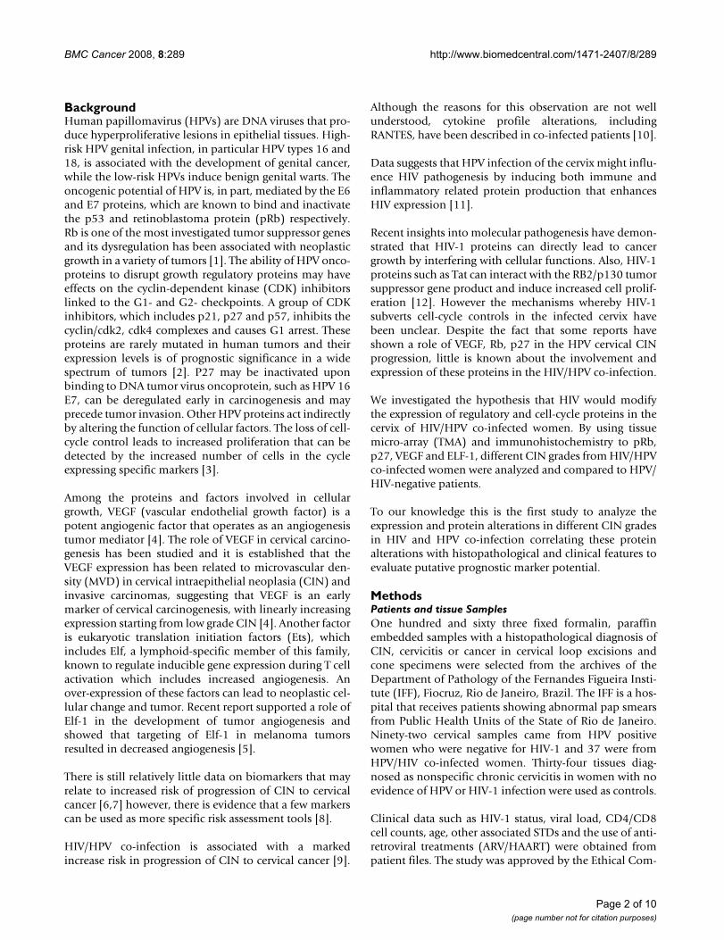
BackgroundHuman papillomavirus (HPVs) are DNA viruses that pro-duce hyperproliferative lesions in epithelial tissues. High-risk HPV genital infection, in particular HPV types 16 and18, is associated with the development of genital cancer,while the low-risk HPVs induce benign genital warts. Theoncogenic potential of HPV is, in part, mediated by the E6and E7 proteins, which are known to bind and inactivatethe p53 and retinoblastoma protein (pRb) respectively.Rb is one of the most investigated tumor suppressor genesand its dysregulation has been associated with neoplasticgrowth in a variety of tumors [1]. The ability of HPV onco-proteins to disrupt growth regulatory proteins may haveeffects on the cyclin-dependent kinase (CDK) inhibitorslinked to the G1- and G2- checkpoints. A group of CDKinhibitors, which includes p21, p27 and p57, inhibits thecyclin/cdk2, cdk4 complexes and causes G1 arrest. Theseproteins are rarely mutated in human tumors and theirexpression levels is of prognostic significance in a widespectrum of tumors [2]. P27 may be inactivated uponbinding to DNA tumor virus oncoprotein, such as HPV 16E7, can be deregulated early in carcinogenesis and mayprecede tumor invasion. Other HPV proteins act indirectlyby altering the function of cellular factors. The loss of cell-cycle control leads to increased proliferation that can bedetected by the increased number of cells in the cycleexpressing specific markers [3].
Among the proteins and factors involved in cellulargrowth, VEGF (vascular endothelial growth factor) is apotent angiogenic factor that operates as an angiogenesistumor mediator [4]. The role of VEGF in cervical carcino-genesis has been studied and it is established that theVEGF expression has been related to microvascular den-sity (MVD) in cervical intraepithelial neoplasia (CIN) andinvasive carcinomas, suggesting that VEGF is an earlymarker of cervical carcinogenesis, with linearly increasingexpression starting from low grade CIN [4]. Another factoris eukaryotic translation initiation factors (Ets), whichincludes Elf, a lymphoid-specific member of this family,known to regulate inducible gene expression during T cellactivation which includes increased angiogenesis. Anover-expression of these factors can lead to neoplastic cel-lular change and tumor. Recent report supported a role ofElf-1 in the development of tumor angiogenesis andshowed that targeting of Elf-1 in melanoma tumorsresulted in decreased angiogenesis [5].
There is still relatively little data on biomarkers that mayrelate to increased risk of progression of CIN to cervicalcancer [6,7] however, there is evidence that a few markerscan be used as more specific risk assessment tools [8].
HIV/HPV co-infection is associated with a markedincrease risk in progression of CIN to cervical cancer [9].
Although the reasons for this observation are not wellunderstood, cytokine profile alterations, includingRANTES, have been described in co-infected patients [10].
Data suggests that HPV infection of the cervix might influ-ence HIV pathogenesis by inducing both immune andinflammatory related protein production that enhancesHIV expression [11].
Recent insights into molecular pathogenesis have demon-strated that HIV-1 proteins can directly lead to cancergrowth by interfering with cellular functions. Also, HIV-1proteins such as Tat can interact with the RB2/p130 tumorsuppressor gene product and induce increased cell prolif-eration [12]. However the mechanisms whereby HIV-1subverts cell-cycle controls in the infected cervix havebeen unclear. Despite the fact that some reports haveshown a role of VEGF, Rb, p27 in the HPV cervical CINprogression, little is known about the involvement andexpression of these proteins in the HIV/HPV co-infection.
We investigated the hypothesis that HIV would modifythe expression of regulatory and cell-cycle proteins in thecervix of HIV/HPV co-infected women. By using tissuemicro-array (TMA) and immunohistochemistry to pRb,p27, VEGF and ELF-1, different CIN grades from HIV/HPVco-infected women were analyzed and compared to HPV/HIV-negative patients.
To our knowledge this is the first study to analyze theexpression and protein alterations in different CIN gradesin HIV and HPV co-infection correlating these proteinalterations with histopathological and clinical features toevaluate putative prognostic marker potential.
MethodsPatients and tissue SamplesOne hundred and sixty three fixed formalin, paraffinembedded samples with a histopathological diagnosis ofCIN, cervicitis or cancer in cervical loop excisions andcone specimens were selected from the archives of theDepartment of Pathology of the Fernandes Figueira Insti-tute (IFF), Fiocruz, Rio de Janeiro, Brazil. The IFF is a hos-pital that receives patients showing abnormal pap smearsfrom Public Health Units of the State of Rio de Janeiro.Ninety-two cervical samples came from HPV positivewomen who were negative for HIV-1 and 37 were fromHPV/HIV co-infected women. Thirty-four tissues diag-nosed as nonspecific chronic cervicitis in women with noevidence of HPV or HIV-1 infection were used as controls.
Clinical data such as HIV-1 status, viral load, CD4/CD8cell counts, age, other associated STDs and the use of anti-retroviral treatments (ARV/HAART) were obtained frompatient files. The study was approved by the Ethical Com-
Page 2 of 10(page number not for citation purposes)
BMC Cancer 2008, 8:289 http://www.biomedcentral.com/1471-2407/8/289
mittees Review Board of Evandro Chagas ClinicalResearch Institute and IFF, both from Fiocruz.
Tissue Micro-Array (TMA) block constructionThe TMA blocks were constructed as described by Pires etal, 2006 [13]. Briefly, all hematoxylin-eosin (HE) slideswere re-examined by an experienced pathologist and twomorphologic representative fields of higher CIN levelwere chosen and encircled with a marker pen. Two coresfrom each case were punched out from the donor blocks.Starting from the selected regions, the corresponding H&Eslides were overlaid with a custom-built 16 gauge Becton-Dickinson PrecisionGlide® hypodermic needle (1,1 mm2
area). Afterwards cores were attached by double-sideadhesive tape on a computer-generated paper grid afford-ing alignment on the block mould, which was then filledwith liquid paraffin. Three μm thick sections wereobtained from an American Optical standard rotatormicrotome. Each block provided 40–50 slides, containingvariable tissue core lengths. Only samples showing theoriginal lesion were considered. The design of each blockwas detailed in a TMA map (spreadsheet), indicating theposition and identification of each core. Normal tissue(placenta) and position-specific blank cores were adoptedfor orientation during microscopy analysis. A total ofthree TMA blocks were constructed, one with HPV/HIVco-infected cervical biopsies, another with controlstogether with CIN I and the third with CIN II/II, Ca andtumor.
HPV TestingThe presence of HPV DNA was determined through a pre-viously published in situ hybridization assay [14,15]. Inbrief, the probe cocktails can detect either the low riskHPV types (HPVs 6,11,42,43,44) or the high risk HPVtypes (HPVs 16,18,31,33,35,45,51,52,56,58,68) (EnzoLife Sciences, Farmingdale, NY, USA). The probe-targetcomplex is detected due to the action of alkaline phos-phatase on the chromogen nitroblue tetrazolium and bro-mochloroindolyl phosphate(NBT/BCIP) yielding a darkblue color with a pink counterstain for the HPV negativecells due to nuclear fast red.
Immunohistochemistry (IHC)IHC reactions were performed on TMA silane-coatedslides (Sigma, St. Louis, MO, USA), dried overnight at37°C and then dewaxed, rehydrated and treated in hydro-gen peroxide 3% in methanol for 10 min to eliminateendogenous peroxidase activity. The LSAB system HRP(Dakocytomation, Carpinteria, CA, USA) method wasadopted for immunolabelling. Tissue sections weresequentially incubated for 1 hour at room temperaturewith the specific antibodies (dilutions): anti-VEGF mono-clonal antibody (1/400), anti-Elf-1 polyclonal antibody(1/300), anti-Rb monoclonal antibody (1/25) and anti-
p27Kp1 monoclonal antibody (1/20) (DakoCytomation,CA USA). A secondary biotinylated multilink antibodywas incubated with the streptavidin-HRP conjugate for 30min and the signal visualized with 3.3 diaminobenzidine(Sigma Chemical Co., St. Lois, MO, USA) and 85 μl ofhydrogen peroxide 0.3%. Hematoxylin was the counter-stain and the omission of the primary antibody, whicheliminated the signal in each case, served as an additionalcontrol.
Assessment of IHC slides and cell countingThe microscopic analysis of the slides was independentlyperformed by two investigators. Positive stained cells werecounted in the epithelium and stroma at 400× magnifica-tion. The total average area counted in the epithelium/stroma was: controls- 882/766 (SD 82,02) cells/mm2;CIN I – 867/642 (SD 159,09) cells/mm2; CIN II/III – 813/538 (SD 194,45) cells/mm2; carcinoma – 1.135/589 (SD386,08) cells/mm2respectively. Digitalized photographswere taken with a Nikon COOLPIX Camera DP12, andthe images stored in a computer-based software (Image-Pro 4.5) for documentation.
Statistical AnalysisData analysis was carried out with the SAS System (SASversion 9.1, Cary, North Carolina, USA). Preliminaryanalyses verified that data did not follow a normal distri-bution, so we used non-parametric tests for comparisons.Mann-Whitney U, Kruskal Wallis and Dunn's tests wereapplied to compare means of positive cells in the epithe-lium and stroma which was then compared to peripheralCD4+T cells (< 200 and ≥ 200 cells/mm3), CIN grade, andpresence/absence of HPV and/or HIV-1 infection. Differ-ences were considered significant at p < 0.05.
ResultsHistopathological dataHPV+/HIV-negative cervices (n = 92) were classified aslow grade (CIN I; n = 33) or high grade/CIN (CIN II/III; n= 47). Twelve additional cases of invasive cancer, consist-ing of 3 cases of adenocarcinoma and 9 of squamous cellcarcinoma (SCC) were included in the study. HPV+/HIV+co-infected cervices (n = 37) were classified as low gradeCIN (CIN I; n = 14) and high grade CIN (CIN II/III; n =23).
Immunohistochemical data: OverviewThe expression of VEGF, Rb, Elf-1 and p27 in the epithe-lium of the cervical biopsies that were co-infected withHPV+/HIV+ was compared to the cervices infected by HPValone. The results are summarized in Table 1 and Graphic1 [see Additional file 1]. There was a statistically signifi-cant increase (p = 0.001) in the number of cells expressingVEGF and p27 in HIV+/HPV+ co-infection as compared tothe HPV+/HIV-. Note that this increase was evident for
Page 3 of 10(page number not for citation purposes)
BMC Cancer 2008, 8:289 http://www.biomedcentral.com/1471-2407/8/289
both CIN I and CIN II/III. Table 2 provides a compilationof the comparisons of the number of epithelial cellsexpressing VEGF, p27, Rb, and Elf-1 in the cervicitis con-trols, CIN I, CIN II/III, and invasive carcinoma in womenwith HPV infection alone.
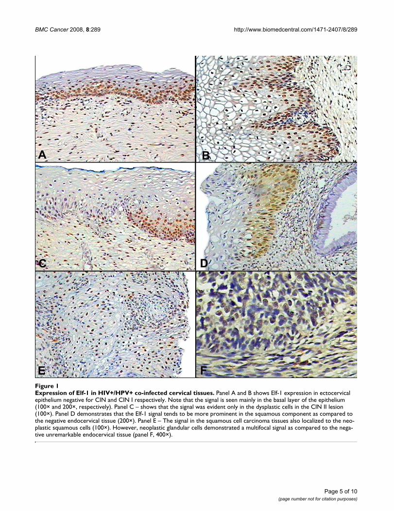
Elf-1 expressionElf-1 was evidenced in the cervical squamous epithelium,exhibiting a strong nuclear staining (Fig. 1A and 1B), par-ticularly in the areas of CIN II/III. In non-neoplastic areas
or without evidence of dysplasia, Elf-1 staining was scat-tered in the basal cell layer (Fig. 1C). In comparison, Elf-1was not evident in the endocervix (Fig. 1D). The squa-mous cell cancer cases also showed a strong nuclear-basedsignal, whereas the adenocarcinoma cases displayedmultifocal staining (Fig. 1E and 1F). HPV+/HIV- infectionshowed statistical significant difference both in the epi-thelium (p = 0.005) and stroma (p = 0.009, data notshown). However, the Dunn's test failed to show differ-ences between the controls and CIN I, CIN II/III and
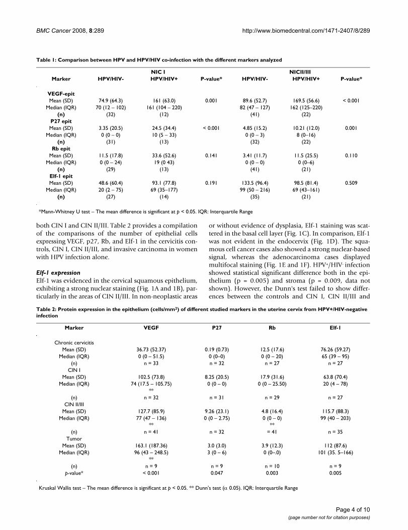
Table 1: Comparison between HPV and HPV/HIV co-infection with the different markers analyzed
NIC I NICII/IIIMarker HPV/HIV- HPV/HIV+ P-value* HPV/HIV- HPV/HIV+ P-value*
VEGF-epitMean (SD) 74.9 (64.3) 161 (63.0) 0.001 89.6 (52.7) 169.5 (56.6) < 0.001
Median (IQR) 70 (12 – 102) 161 (104 – 220) 82 (47 – 127) 162 (125–220)(n) (32) (12) (41) (22)
P27 epitMean (SD) 3.35 (20.5) 24.5 (34.4) < 0.001 4.85 (15.2) 10.21 (12.0) 0.001
Median (IQR) 0 (0 – 0) 10 (5 – 33) 0 (0 – 3) 8 (0–16)(n) (31) (13) (32) (22)
Rb epitMean (SD) 11.5 (17.8) 33.6 (52.6) 0.141 3.41 (11.7) 11.5 (25.5) 0.110
Median (IQR) 0 (0 – 24) 19 (0 43) 0 (0 – 0) 0 (0–6)(n) (29) (13) (41) (21)
Elf-1 epitMean (SD) 48.6 (60.4) 93.1 (77.8) 0.191 133.5 (96.4) 98.5 (81.4) 0.509
Median (IQR) 20 (2 – 75) 69 (35–177) 99 (50 – 216) 69 (43–161)(n) (27) (14) (35) (21)
*Mann-Whitney U test – The mean difference is significant at p < 0.05. IQR: Interquartile Range
Table 2: Protein expression in the epithelium (cells/mm2) of different studied markers in the uterine cervix from HPV+/HIV-negative infection
Marker VEGF P27 Rb Elf-1
Chronic cervicitisMean (SD) 36.73 (52.37) 0.19 (0.73) 12.5 (17.6) 76.26 (59.27)
Median (IQR) 0 (0 – 51.5) 0 (0–0) 0 (0 – 20) 65 (39 – 95)(n) n = 33 n = 32 n = 27 n = 27
CIN IMean (SD) 102.5 (73.8) 8.25 (20.5) 17.9 (31.6) 63.8 (70.4)
Median (IQR) 74 (17.5 – 105.75) 0 (0 – 0) 0 (0 – 25.50) 20 (4 – 78)**
(n) n = 32 n = 31 n = 29 n = 27CIN II/III
Mean (SD) 127.7 (85.9) 9.26 (23.1) 4.8 (16.4) 115.7 (88.3)Median (IQR) 77 (47 – 136) 0 (0 – 2.75) 0 (0 – 0) 99 (40 – 203)
** **(n) n = 41 n = 32 = 41 n = 35
TumorMean (SD) 163.1 (187.36) 3.0 (3.0) 3.9 (12.3) 112 (87.6)
Median (IQR) 96 (43 – 248.5) 3 (0 – 6) 0 (0–.0) 101 (35. 5–166)**
(n) n = 9 n = 9 n = 10 n = 9p-value* < 0.001 0.047 0.003 0.005
Kruskal Wallis test – The mean difference is significant at p < 0.05. ** Dunn's test (α 0.05). IQR: Interquartile Range
Page 4 of 10(page number not for citation purposes)
BMC Cancer 2008, 8:289 http://www.biomedcentral.com/1471-2407/8/289
Page 5 of 10(page number not for citation purposes)
Expression of Elf-1 in HIV+/HPV+ co-infected cervical tissuesFigure 1Expression of Elf-1 in HIV+/HPV+ co-infected cervical tissues. Panel A and B shows Elf-1 expression in ectocervical epithelium negative for CIN and CIN I respectively. Note that the signal is seen mainly in the basal layer of the epithelium (100× and 200×, respectively). Panel C – shows that the signal was evident only in the dysplastic cells in the CIN II lesion (100×). Panel D demonstrates that the Elf-1 signal tends to be more prominent in the squamous component as compared to the negative endocervical tissue (200×). Panel E – The signal in the squamous cell carcinoma tissues also localized to the neo-plastic squamous cells (100×). However, neoplastic glandular cells demonstrated a multifocal signal as compared to the nega-tive unremarkable endocervical tissue (panel F, 400×).
BMC Cancer 2008, 8:289 http://www.biomedcentral.com/1471-2407/8/289
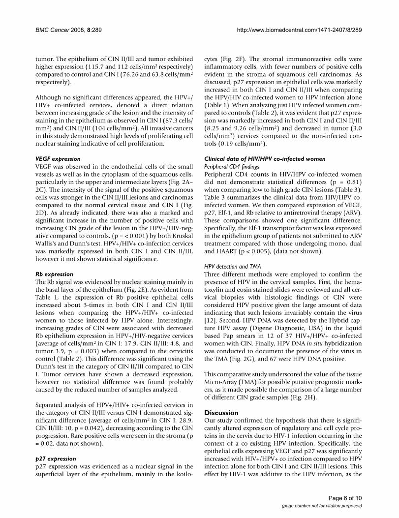
tumor. The epithelium of CIN II/III and tumor exhibitedhigher expression (115.7 and 112 cells/mm2 respectively)compared to control and CIN I (76.26 and 63.8 cells/mm2
respectively).
Although no significant differences appeared, the HPV+/HIV+ co-infected cervices, denoted a direct relationbetween increasing grade of the lesion and the intensity ofstaining in the epithelium as observed in CIN I (87.3 cells/mm2) and CIN II/III (104 cells/mm2). All invasive cancersin this study demonstrated high levels of proliferating cellnuclear staining indicative of cell proliferation.
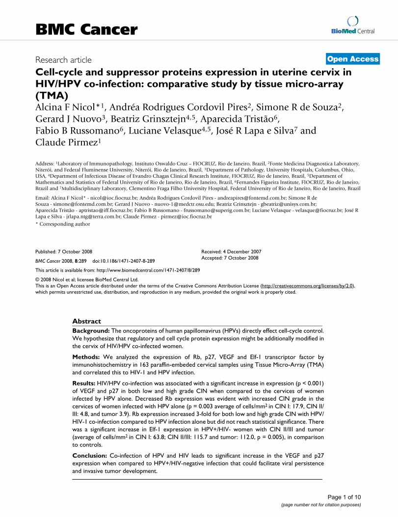
VEGF expressionVEGF was observed in the endothelial cells of the smallvessels as well as in the cytoplasm of the squamous cells,particularly in the upper and intermediate layers (Fig. 2A–2C). The intensity of the signal of the positive squamouscells was stronger in the CIN II/III lesions and carcinomascompared to the normal cervical tissue and CIN I (Fig.2D). As already indicated, there was also a marked andsignificant increase in the number of positive cells withincreasing CIN grade of the lesion in the HPV+/HIV-neg-ative compared to controls, (p = < 0.001) by both KruskalWallis's and Dunn's test. HPV+/HIV+ co-infection cerviceswas markedly expressed in both CIN I and CIN II/III,however it not shown statistical significance.
Rb expressionThe Rb signal was evidenced by nuclear staining mainly inthe basal layer of the epithelium (Fig. 2E). As evident fromTable 1, the expression of Rb positive epithelial cellsincreased about 3-times in both CIN I and CIN II/IIIlesions when comparing the HPV+/HIV+ co-infectedwomen to those infected by HPV alone. Interestingly,increasing grades of CIN were associated with decreasedRb epithelium expression in HPV+/HIV-negative cervices(average of cells/mm2 in CIN I: 17.9, CIN II/III: 4.8, andtumor 3.9, p = 0.003) when compared to the cervicitiscontrol (Table 2). This difference was significant using theDunn's test in the category of CIN II/III compared to CINI. Tumor cervices have shown a decreased expression,however no statistical difference was found probablycaused by the reduced number of samples analyzed.
Separated analysis of HPV+/HIV+ co-infected cervices inthe category of CIN II/III versus CIN I demonstrated sig-nificant difference (average of cells/mm2 in CIN I: 28.9,CIN II/III: 10, p = 0.042), decreasing according to the CINprogression. Rare positive cells were seen in the stroma (p= 0.02, data not shown).
p27 expressionp27 expression was evidenced as a nuclear signal in thesuperficial layer of the epithelium, mainly in the koilo-
cytes (Fig. 2F). The stromal immunoreactive cells wereinflammatory cells, with fewer numbers of positive cellsevident in the stroma of squamous cell carcinomas. Asdiscussed, p27 expression in epithelial cells was markedlyincreased in both CIN I and CIN II/III when comparingthe HPV/HIV co-infected women to HPV infection alone(Table 1). When analyzing just HPV infected women com-pared to controls (Table 2), it was evident that p27 expres-sion was markedly increased in both CIN I and CIN II/III(8.25 and 9.26 cells/mm2) and decreased in tumor (3.0cells/mm2) cervices compared to the non-infected con-trols (0.19 cells/mm2).
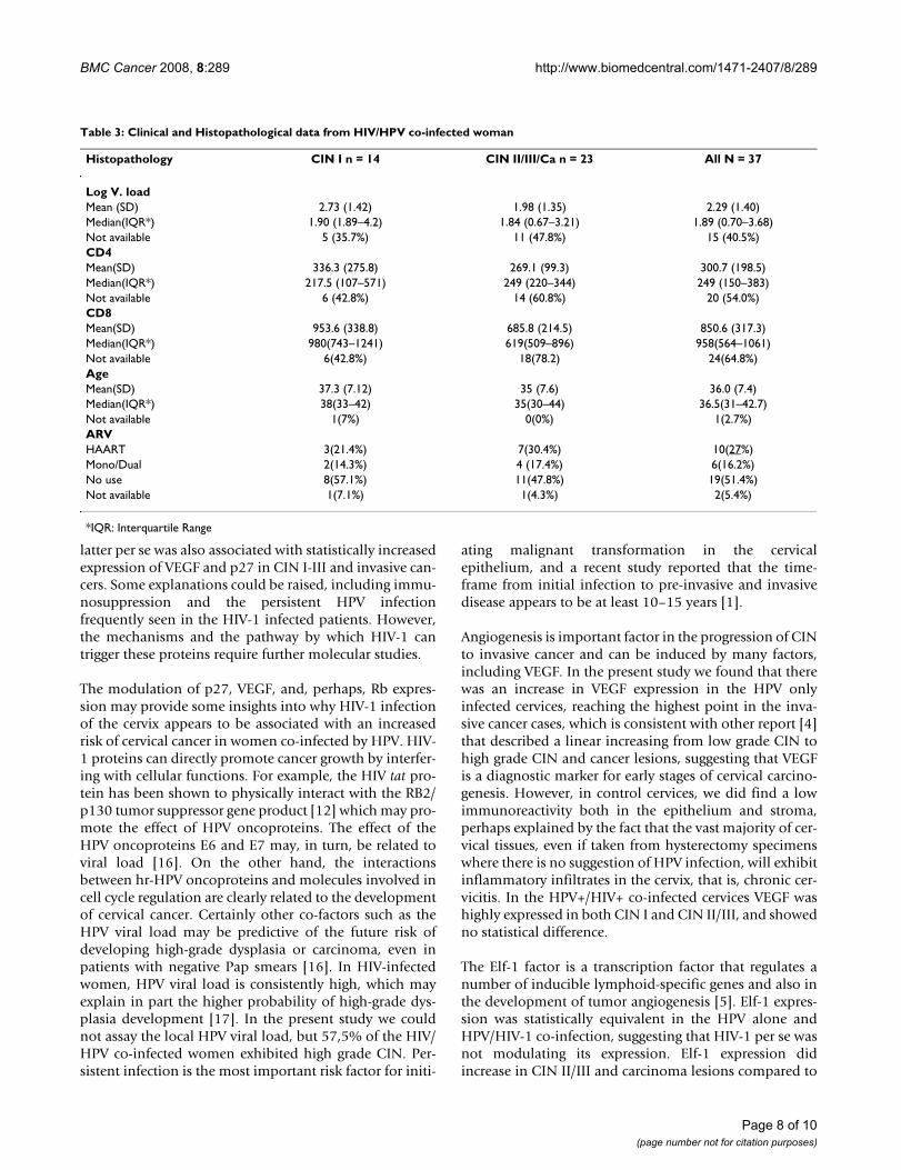
Clinical data of HIV/HPV co-infected womenPeripheral CD4 findingsPeripheral CD4 counts in HIV/HPV co-infected womendid not demonstrate statistical differences (p = 0.81)when comparing low to high grade CIN lesions (Table 3).Table 3 summarizes the clinical data from HIV/HPV co-infected women. We then compared expression of VEGF,p27, Elf-1, and Rb relative to antiretroviral therapy (ARV).These comparisons showed one significant difference.Specifically, the Elf-1 transcriptor factor was less expressedin the epithelium group of patients not submitted to ARVtreatment compared with those undergoing mono, dualand HAART (p < 0.005), (data not shown).
HPV detection and TMAThree different methods were employed to confirm thepresence of HPV in the cervical samples. First, the hema-toxylin and eosin stained slides were reviewed and all cer-vical biopsies with histologic findings of CIN wereconsidered HPV positive given the large amount of dataindicating that such lesions invariably contain the virus[12]. Second, HPV DNA was detected by the Hybrid cap-ture HPV assay (Digene Diagnostic, USA) in the liquidbased Pap smears in 12 of 37 HIV+/HPV+ co-infectedwomen with CIN. Finally, HPV DNA in situ hybridizationwas conducted to document the presence of the virus inthe TMA (Fig. 2G), and 67 were HPV DNA positive.
This comparative study underscored the value of the tissueMicro-Array (TMA) for possible putative prognostic mark-ers, as it made possible the comparison of a large numberof different CIN grade samples (Fig. 2H).
DiscussionOur study confirmed the hypothesis that there is signifi-cantly altered expression of regulatory and cell cycle pro-teins in the cervix due to HIV-1 infection occurring in thecontext of a co-existing HPV infection. Specifically, theepithelial cells expressing VEGF and p27 was significantlyincreased with HIV+/HPV+ co infection compared to HPVinfection alone for both CIN I and CIN II/III lesions. Thiseffect by HIV-1 was additive to the HPV infection, as the
Page 6 of 10(page number not for citation purposes)
BMC Cancer 2008, 8:289 http://www.biomedcentral.com/1471-2407/8/289
Page 7 of 10(page number not for citation purposes)
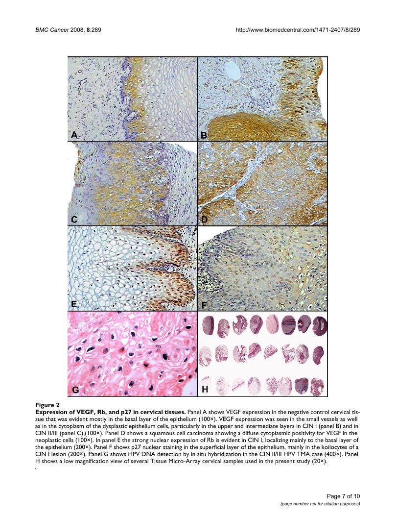
Expression of VEGF, Rb, and p27 in cervical tissuesFigure 2Expression of VEGF, Rb, and p27 in cervical tissues. Panel A shows VEGF expression in the negative control cervical tis-sue that was evident mostly in the basal layer of the epithelium (100×). VEGF expression was seen in the small vessels as well as in the cytoplasm of the dysplastic epithelium cells, particularly in the upper and intermediate layers in CIN I (panel B) and in CIN II/III (panel C),(100×). Panel D shows a squamous cell carcinoma showing a diffuse cytoplasmic positivity for VEGF in the neoplastic cells (100×). In panel E the strong nuclear expression of Rb is evident in CIN I, localizing mainly to the basal layer of the epithelium (200×). Panel F shows p27 nuclear staining in the superficial layer of the epithelium, mainly in the koilocytes of a CIN I lesion (200×). Panel G shows HPV DNA detection by in situ hybridization in the CIN II/III HPV TMA case (400×). Panel H shows a low magnification view of several Tissue Micro-Array cervical samples used in the present study (20×).
BMC Cancer 2008, 8:289 http://www.biomedcentral.com/1471-2407/8/289
latter per se was also associated with statistically increasedexpression of VEGF and p27 in CIN I-III and invasive can-cers. Some explanations could be raised, including immu-nosuppression and the persistent HPV infectionfrequently seen in the HIV-1 infected patients. However,the mechanisms and the pathway by which HIV-1 cantrigger these proteins require further molecular studies.
The modulation of p27, VEGF, and, perhaps, Rb expres-sion may provide some insights into why HIV-1 infectionof the cervix appears to be associated with an increasedrisk of cervical cancer in women co-infected by HPV. HIV-1 proteins can directly promote cancer growth by interfer-ing with cellular functions. For example, the HIV tat pro-tein has been shown to physically interact with the RB2/p130 tumor suppressor gene product [12] which may pro-mote the effect of HPV oncoproteins. The effect of theHPV oncoproteins E6 and E7 may, in turn, be related toviral load [16]. On the other hand, the interactionsbetween hr-HPV oncoproteins and molecules involved incell cycle regulation are clearly related to the developmentof cervical cancer. Certainly other co-factors such as theHPV viral load may be predictive of the future risk ofdeveloping high-grade dysplasia or carcinoma, even inpatients with negative Pap smears [16]. In HIV-infectedwomen, HPV viral load is consistently high, which mayexplain in part the higher probability of high-grade dys-plasia development [17]. In the present study we couldnot assay the local HPV viral load, but 57,5% of the HIV/HPV co-infected women exhibited high grade CIN. Per-sistent infection is the most important risk factor for initi-
ating malignant transformation in the cervicalepithelium, and a recent study reported that the time-frame from initial infection to pre-invasive and invasivedisease appears to be at least 10–15 years [1].
Angiogenesis is important factor in the progression of CINto invasive cancer and can be induced by many factors,including VEGF. In the present study we found that therewas an increase in VEGF expression in the HPV onlyinfected cervices, reaching the highest point in the inva-sive cancer cases, which is consistent with other report [4]that described a linear increasing from low grade CIN tohigh grade CIN and cancer lesions, suggesting that VEGFis a diagnostic marker for early stages of cervical carcino-genesis. However, in control cervices, we did find a lowimmunoreactivity both in the epithelium and stroma,perhaps explained by the fact that the vast majority of cer-vical tissues, even if taken from hysterectomy specimenswhere there is no suggestion of HPV infection, will exhibitinflammatory infiltrates in the cervix, that is, chronic cer-vicitis. In the HPV+/HIV+ co-infected cervices VEGF washighly expressed in both CIN I and CIN II/III, and showedno statistical difference.
The Elf-1 factor is a transcription factor that regulates anumber of inducible lymphoid-specific genes and also inthe development of tumor angiogenesis [5]. Elf-1 expres-sion was statistically equivalent in the HPV alone andHPV/HIV-1 co-infection, suggesting that HIV-1 per se wasnot modulating its expression. Elf-1 expression didincrease in CIN II/III and carcinoma lesions compared to
Table 3: Clinical and Histopathological data from HIV/HPV co-infected woman
Histopathology CIN I n = 14 CIN II/III/Ca n = 23 All N = 37
Log V. loadMean (SD) 2.73 (1.42) 1.98 (1.35) 2.29 (1.40)Median(IQR*) 1.90 (1.89–4.2) 1.84 (0.67–3.21) 1.89 (0.70–3.68)Not available 5 (35.7%) 11 (47.8%) 15 (40.5%)CD4Mean(SD) 336.3 (275.8) 269.1 (99.3) 300.7 (198.5)Median(IQR*) 217.5 (107–571) 249 (220–344) 249 (150–383)Not available 6 (42.8%) 14 (60.8%) 20 (54.0%)CD8Mean(SD) 953.6 (338.8) 685.8 (214.5) 850.6 (317.3)Median(IQR*) 980(743–1241) 619(509–896) 958(564–1061)Not available 6(42.8%) 18(78.2) 24(64.8%)AgeMean(SD) 37.3 (7.12) 35 (7.6) 36.0 (7.4)Median(IQR*) 38(33–42) 35(30–44) 36.5(31–42.7)Not available 1(7%) 0(0%) 1(2.7%)ARVHAART 3(21.4%) 7(30.4%) 10(27%)Mono/Dual 2(14.3%) 4 (17.4%) 6(16.2%)No use 8(57.1%) 11(47.8%) 19(51.4%)Not available 1(7.1%) 1(4.3%) 2(5.4%)
*IQR: Interquartile Range
Page 8 of 10(page number not for citation purposes)
BMC Cancer 2008, 8:289 http://www.biomedcentral.com/1471-2407/8/289
the negative controls in HPV alone infected cervices anddid reach statistical significance, suggesting that molecu-lar events in the more progressed HPV infected cerviceswas increasing Elf-1 expression. Interestingly, Elf-1 wasless expressed in patients without ARV treatment com-pared with those undergoing mono, dual or HAART (p <0.005), possibly justified by the fact that some cellulartranscription factors can bind to the HIV-1 5' long termi-nal repeat (LTR). However, the contribution of these fac-tors to human immunodeficiency virus type 1 replicationin infected individuals remains obscure [18].
Many tumor suppressor genes, most of them involved inthe cell cycle, have been described, the functional loss ofthese suppressor gene pathways probably being the basisfor cervical carcinogenesis. P27, an important cell cycleinhibitor protein, is involved in the growth and differen-tiation of the normal squamous epithelia of the uterinecervix, and controversial data have been described in theliterature. Some authors have found that low levels of p27are associated with poor prognosis and suggested thatdecreased p27 protein might contribute to the alteredkinetics of the dysplastic and neoplastic cervical epithe-lium [19,20], whereas others have suggested that anincreased and/or aberrant function of p27 expression mayoccur in invasive squamous cell carcinoma of the cervix[21]. Our data showed a marked increase in p27 infectionby HPV alone in all CIN grades compared to controls, anddecreased in tumor compared to any other group of CIN.One possibility to explain this increase is the blocking ofp27 activity by the HPV oncogene E7 which binds p27 viaits C terminus. However, the mechanisms by which theHIV would modify this activity is unknown. It is impor-tant to recall that cell cycle control is a multifactorial sys-tem and, for example, decreased Rb expression mayincrease cyclin E and cdk2 which, in turn, can overcomethe tumor suppressive effect of increased p27 [22]. In thisconnection, our data show that there was a 3-fold increasein Rb expression with HIV-1 co-infection versus HPVinfection alone, but this did not reach statistical signifi-cance, there may well be a stimulatory effect on Rb pro-duction due to HIV-1 co-infection in this context. Thispossibility will require additional studies. Nonetheless,since Rb expression decreased in CIN II/III and carcinomain HPV only infected cervices compared to the negativecontrols, corroborating the previously published data[23,24]. Interestingly, HIV-1 co-infection appeared to res-cue Rb expression which may, in turn, potentiate theincreased expression of p27 [25]. However, this interac-tion by itself may not be sufficient for neoplastic transfor-mation and other cofactors may be required.Nevertheless, it is likely that the deregulation of cellulargenes and function by Tat can also cause abnormalitiesthat may contribute to AIDS pathogenesis and to thedevelopment of AIDS-associated disorders [12].
This comparative study adopting Tissue Micro-Array forpossible putative prognostic markers, made possible thecomparison of a large number of different CIN grade sam-ples, and demonstrated to be a useful research tool, pri-marily for immunohistochemistry procedures, in spite ofsome limitations. First, we could not assay high risk-HPVsub-typing in all samples, which prevented comparisonbetween Hr-HPV types with the expression markers in thedifferent CIN grades. Second, we cannot accurately deter-mine the HPV type in these relatively small cores. Third,we decided to use nonspecific cervicitis as controls, asmost transformation zone biopsies, even from womenwith no evidence of HPV or HIV-1 infection, show non-specific cervicitis. Despite the limitations, we were able todocument statistically significant changes due to HIV-1co-infection in HPV infected cervices.
The neoplastic process of the uterine cervix occurs in theepithelium. In the present study we analyzed the expres-sion of different markers both in the epithelium and stro-mal tissue, however we focused our analysis in theepithelium. Although VEGF demonstrated significant dif-ferences (p < 0.001) in low grade CIN compared to con-trol in the HPV+/HIV-negative patients, this marker wasalso increased in high grade CIN, suggesting that it is nota good marker for CIN progression. Increasing grades ofCIN were associated with decreased Rb expression cervi-ces, however further studies with more infected cervicesshould be done in order to confirm these observations.Additionally, we suggest that many immune markers thathave been studied as CIN progressors should also be eval-uated in HIV/HPV co-infection, since this group ofpatients may have different clinical and immune out-comes.
ConclusionIn conclusion, this study demonstrated a significantaltered expression of regulatory and cell cycle proteinssuch as VEGF and p27 in cervical samples from HPV+/HIV+ co-infected compared to HPV+/HIV-negativewomen, suggesting that HIV-1 virus may be triggeringthese proteins which in turn could facilitate viral persist-ence and progression to an invasive tumor.
AbbreviationsHPV: Human Papillomavirus; Rb: Retinoblastoma pro-tein; VEGF: vascular endothelial growth factor; Elf: Eukar-iotic Translation Initiation Factors; TMA: Tissue Micro-Array; HIV: Human Immunodeficiency Syndrome; CIN:Cervical intra-epithelial neoplasia; ARV: antiretroviral;HAART: Highly active antiretroviral treatment; SCC: Squa-mous cell carcinoma; STD: Sexually Transmitted Disease.
Competing interestsThe authors declare that they have no competing interests.
Page 9 of 10(page number not for citation purposes)
BMC Cancer 2008, 8:289 http://www.biomedcentral.com/1471-2407/8/289
Authors' contributionsAFN conceived the study design, prepared the manuscriptand was the main investigator. ARCP and SRS carried outthe Tissue Micro-array studies and the immunohisto-chemistry analysis. GJN contributed to the immunohisto-chemical study and in the preparation of the manuscript.BG, AP and FR contributed toward the clinical patientselection and treatment. LSV participated in the statisticalanalysis, JRLS and CP participated in the discussion of theresults and in the preparation of the manuscript. Allauthors have read and approved the final manuscript.
Additional material
AcknowledgementsWe would like to thank Max Thiago F. Xavier; Karine da Conceição Pessa-nha and Rodrigo Mexas for the excellent technical assistance. This work was supported in part by grants from PN-DST/AIDS-MS- UNESCO; FAPERJ and IOC-Fiocruz, Rio de Janeiro, Brazil.
References1. Tjalma W, van Waes T, van den Eeden L, Bogers J: Role of human
papillomavirus in the carcinogenesis of squamous cell carci-noma and adenocarcinoma of the cervix. Best practice &Research Clin Obst Gynaecol 2005, 19(4):469-483.
2. Cho HN, Kim TY, Kim JW: Alteration of cell cycle in cervicaltumor associated with Human Papillomavirus – Cyclin-Dependent kinase inhibitors. Yonsei Med J 2002, 43(6):722-728.
3. Aggarwal BB, Shishodia S, Sandur SK, Pandey MK, Sethi G: Inflam-mation and cancer: how hot is the link? Biochem Pharmacol 2006,72(11):1605-21.
4. Branca M, Giorgi C, Santini D, Di Bonito L, Ciotti M, Benedetto A,Paba P, Costa S, Bonifacio D, Di Bonito P, Accardi L, Favalli C, Syr-janen K: Aberrant expression of VEGF-C is related to grade ofcervical intraepithelial neoplasia (CIN) and high risk HPV,but does not predict virus clearance after treatment of CINor prognosis of cervical cancer. J Clin Pathol 2006, 59(1):40-7.
5. Huang X, Brown C, Ni W, Maynard E, Rigby AC, Oetthen P: Criticalrole for the Ets transcription factor ELF-1 in the develop-ment of tumor angiogenesis. Blood 2006, 107(8):3153-3160.
6. Troncone G, Vetrani A, de Rosa G, Palombini L: Cyclin dependentkinase inhibitor p27 expression in normal and neoplastic cer-vical epithelium. J Clin Pathol 1999, 52:880-887.
7. Benevolo M, Mottolese M, Marandino F, Vocaturo G, Sindico R, Pip-erno G, Mariani L, Sperduti I, Canalini P, Donnorso RP, Vocaturo A:Immunohistochemical expression of p16(INK4a) is predic-tive of HR-HPV infection in cervical low-grade lesions. ModPathol 2006, 19(3):384-91.
8. Wentzensen N, von Knebel Doeberitz M: Biomarkers in cervicalcancer screening. Dis Markers 2007, 23(4):315-30.
9. Nicol AF, Fernandes AT, Grinsztejn B, Russomano F, E Silva JR,Tristao A, Perez M, Nuovo GJ, Martinez-Maza O, Bonecini-AlmeidaM: Distribution of immune cell subsets and cytokine-produc-ing cells in the uterine cervix of human papillomavirus(HPV)-infected women: influence of HIV-1 coinfection. DiagnMol Pathol 2005, 14(1):39-47.
10. Nicol AF, Nuovo GJ, Salomão-Estevez A, Grinsztein B, Tristão A,Russomano F, Lapa E Silva JR, Oliveira MP, Pirmez C: Immune fac-tors involved in the cervical immune response in the HIV/HPV co-infection. J Clin Pathol 2008, 61(1):84-8.
11. Gage JR, Sandhu AK, Nihira M, Bonecini-Almeida M da G, Cristofor-oni P, Kishimoto T, Montz FJ, Martínez-Maza O: Effects of humanpapillomavirus-associated cells on human immunodeficiencyvirus gene expression. Obstet Gynecol 2000, 96(6):879-85.
12. Nyagol J, Leucci E, Onnis A, De Falco G, Tigli C, Sanseverino F, Tor-riccelli M, Palummo N, Pacenti L, Santopietro R, Spina D, Gichangi P,Muchiri L, Lazzi S, Petraglia F, Leoncini L, Giordano A: The effectsof HIV-1 Tat protein on cell cycle during cervical carcinogen-esis. Cancer Biol Ther 2006, 5(6):684-90.
13. Pires AR, da Matta Andreiuolo F, de Souza SR: TMA for all: a newmethod for the construction of tissue microarrays withoutrecipient paraffin block using custom-built needles. DiagnPathol 2006, 25(1):14.
14. Nicol AF, Nuovo GJ, RT in situ PCR: Protocols and Applicationsin Methods in Microbiology. Volume 34. Edited by: Tor Savidge,Charalabos P. Elsevier Acad Press; 2005:239-254.
15. Nicol AF, Nuovo GJ, Wang Y, Grinsztejn B, Tristao A, Russomano F,Perez MA, Lapa e Silva JR, Fernandes AT, Gage JR, Martinez-Maza O,Bonecini-Almeida MG: In situ detection of SOCS and cytokineexpression in the uterine cervix from HIV/HPV coinfectedwomen. Exp Mol Pathol 2006, 81(1):42-7.
16. Moberg M, Gustavsson I, Gyllensten U: Type-specific associationsof human papillomavirus load with risk of developing cervicalcarcinoma in situ. Int J Cancer 2004, 112:854-859.
17. Tjalma WA, Arbyan M, Paavonen J, van Waes TR, Bogers JJ: Prophy-lactic human papillomavirus vaccines: the beginning of theend of cervical cancer. Int J Gynecol Cancer 2004, 14:751-761.
18. Estable MC, Bell B, Merzouki A, Montaner JS, O'Shaughnessy MV, Sad-owski IJ: Human immunodeficiency virus type 1 long terminalrepeat variants from 42 patients representing all stages ofinfection display a wide range of sequence polymorphismand transcription activity. J Virol 1996, 70(6):4053-62.
19. Goff BA, Sallin J, Garcia R, VanBlaricom A, Paley PJ, Muntz HG: Eval-uation of p27 in preinvasive and invasive malignancies of thecervix. Gynecol Oncol 2003, 88(1):40-4.
20. Shiozawa T, Shiohara S, Kanai M, Konishi I, Fujii S, Nikaido T: Expres-sion of the cell cycle regulator p27(Kip1) in normal squa-mous epithelium, cervical intraepithelial neoplasia, andinvasive squamous cell carcinoma of the uterine cervix.Immunohistochemistry and functional aspects of p27(Kip1).Cancer 2001, 92(12):3005-11.
21. Clarke B, Chetty R: Cell cycle aberrations in the pathogenesisof squamous cell carcinoma of the uterine cervix. GynecolOncol 2001, 82(2):238-46.
22. Nam EJ, Kim JW, Kim SW, Kim YT, Kim JH, Yoon BS, Cho NH, KimS: The expressions of the Rb pathway in cervical intraepithe-lial neoplasia; predictive and prognostic significance. GynecolOncol 2007, 104(1):207-11.
23. Tringler B, Gup CJ, Singh M, Groshong S, Shroyer AL, Heinz DA,Shroyer KR: Evaluation of p16INK4A and pRb expression incervical squamous and glandular neoplasia. Hum Pathol 2004,35(6):689-696.
24. Stiegler P, Kasten M, Giordano A: The RB family of cell cycle reg-ulatory factors. J Cell Biochem Suppl 1998, 31:30-6. Review
25. De Falco G, Bellan C, Lazzi S, Claudio P, La Sala D, Cinti C, Tosi P,Giordano A, Leoncini L: Interaction between HIV-1 Tat andpRb2/p130: a possible mechanism in the pathogenesis ofAIDS-related neoplasms. Oncogene 2003, 22(40):6214-9.
Pre-publication historyThe pre-publication history for this paper can be accessedhere:
http://www.biomedcentral.com/1471-2407/8/289/prepub
Additional file 1
Click here for file[http://www.biomedcentral.com/content/supplementary/1471-2407-8-289-S1.doc]
Page 10 of 10(page number not for citation purposes)