Embed Size (px)
Citation preview
BLUNT INJURY ABDOMEN – AND THEIR IMPACT ON
ABDOMINAL VISCERAE
Dissertation submitted in partial fulfillment of the
Requirement for the award of the Degree of
M.S DEGREE EXAMINATION
GENERAL SURGERY
TIRUNELVELI
TIRUNELVELI MEDICAL COLLEGE
THE TAMILNADU DR. M.G.R. MEDICAL UNIVERSITY
CHENNAI, TAMILNADU
April 2012
CERTIFICATE This is to certify that the dissertation titled “BLUNT INJURY ABDOMEN
AND THEIR IMPACT ON ABDOMINAL VISCERAE” is the original work done
by Dr.J.KEVIN JOSEPH, Post Graduate in Department of General Surgery ,
Tirunelveli, to be Submitted to The TamilNadu Dr.M.G.R. Medical University,
Chennai – 32 towards the partial fulfillment of the requirement for the award of M.S.
Degree in General Surgery April 2012.
Prof. S.Soundararajan M.S Prof.V.Pandy M.S
Prof & HOD Unit Chief
Department of General Surgery Department of General Surgery
Tirunelveli Medical College Hospital Tirunelveli Medical College Hospital
Tirunelveli. Tirunelveli.
DEAN
Tirunelveli Medical College Hospital
Tirunelveli.
ACKNOWLEDGEMENT
I am deeply indebted to my beloved Teacher Professor.
S.Soundararajan Professor and Head of the Department of Surgery for his
constant guidance, encouragement and untiring help throughout the period of
this study.
I express my profound gratitude to Professor. V.PANDY, M.S.,
Professor. S. SURESH, M.S., for their constant guidance and suggestions
throughout my study period.
I am very thankful to assistant professor DR. S. AMALAN M.S , DR S.
SENTHIL ARUMUGAM , M.S. , Dr.Rakesh Fernando , Dr.Sivanupandian
for their kind help and suggestions.
I express my sincere gratitude to former professor and HOD
Dr.Jeyakumar Sagayam for his constant motivation and guidance
throughout my career
I thank the DEAN, TIRUNELVELI MEDICAL COLLEGE for
permitting me to use the Hospital facilities for my study.
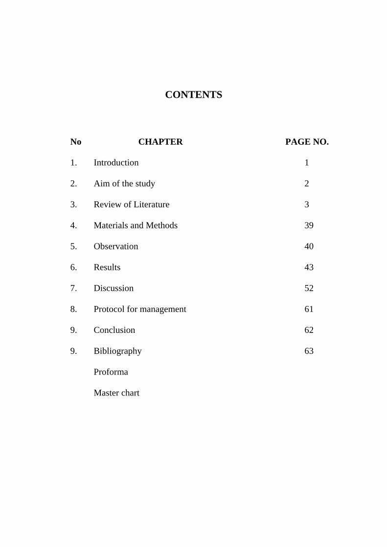
CONTENTS
No CHAPTER PAGE NO.
1. Introduction 1
2. Aim of the study 2
3. Review of Literature 3
4. Materials and Methods 39
5. Observation 40
6. Results 43
7. Discussion 52
8. Protocol for management 61
9. Conclusion 62
9. Bibliography 63
Proforma
Master chart
1
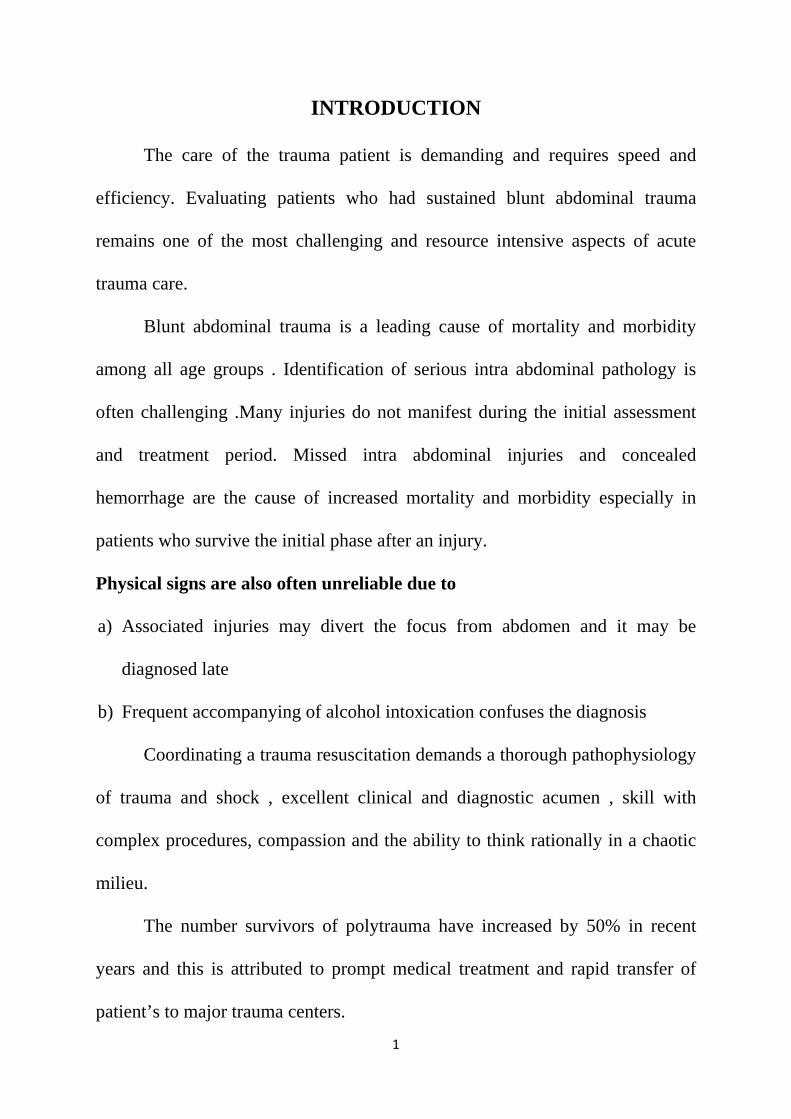
INTRODUCTION
The care of the trauma patient is demanding and requires speed and
efficiency. Evaluating patients who had sustained blunt abdominal trauma
remains one of the most challenging and resource intensive aspects of acute
trauma care.
Blunt abdominal trauma is a leading cause of mortality and morbidity
among all age groups . Identification of serious intra abdominal pathology is
often challenging .Many injuries do not manifest during the initial assessment
and treatment period. Missed intra abdominal injuries and concealed
hemorrhage are the cause of increased mortality and morbidity especially in
patients who survive the initial phase after an injury.
Physical signs are also often unreliable due to
a) Associated injuries may divert the focus from abdomen and it may be
diagnosed late
b) Frequent accompanying of alcohol intoxication confuses the diagnosis
Coordinating a trauma resuscitation demands a thorough pathophysiology
of trauma and shock , excellent clinical and diagnostic acumen , skill with
complex procedures, compassion and the ability to think rationally in a chaotic
milieu.
The number survivors of polytrauma have increased by 50% in recent
years and this is attributed to prompt medical treatment and rapid transfer of
patient’s to major trauma centers.
2
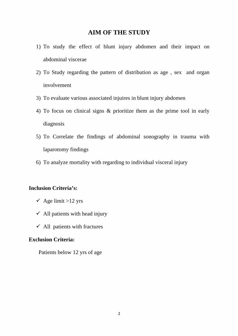
AIM OF THE STUDY
1) To study the effect of blunt injury abdomen and their impact on
abdominal viscerae
2) To Study regarding the pattern of distribution as age , sex and organ
involvement
3) To evaluate various associated injuires in blunt injury abdomen
4) To focus on clinical signs & prioritize them as the prime tool in early
diagnosis
5) To Correlate the findings of abdominal sonography in trauma with
laparotomy findings
6) To analyze mortality with regarding to individual visceral injury
Inclusion Criteria’s:
Age limit >12 yrs
All patients with head injury
All patients with fractures
Exclusion Criteria:
Patients below 12 yrs of age
3
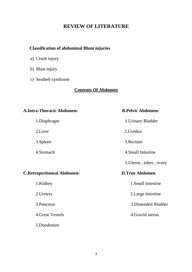
REVIEW OF LITERATURE
Classification of abdominal Blunt injuries
a) Crush injury
b) Blast injury
c) Seatbelt syndrome
Contents Of Abdomen
A.Intra-Thoracic Abdomen: B.Pelvic Abdomen:
1.Diaphragm 1.Urinary Bladder
2.Liver 2.Urethra
3.Spleen 3.Rectum
4.Stomach 4.Small Intestine
5.Uterus , tubes , ovary
C.Retroperitoneal Abdomen: D.True Abdomen
1.Kidney 1.Small Intestine
2.Ureters 2.Large Intestine
3.Pancreas 3.Distended Bladder
4.Great Vessels 4.Gravid uterus
5.Duodenum
4
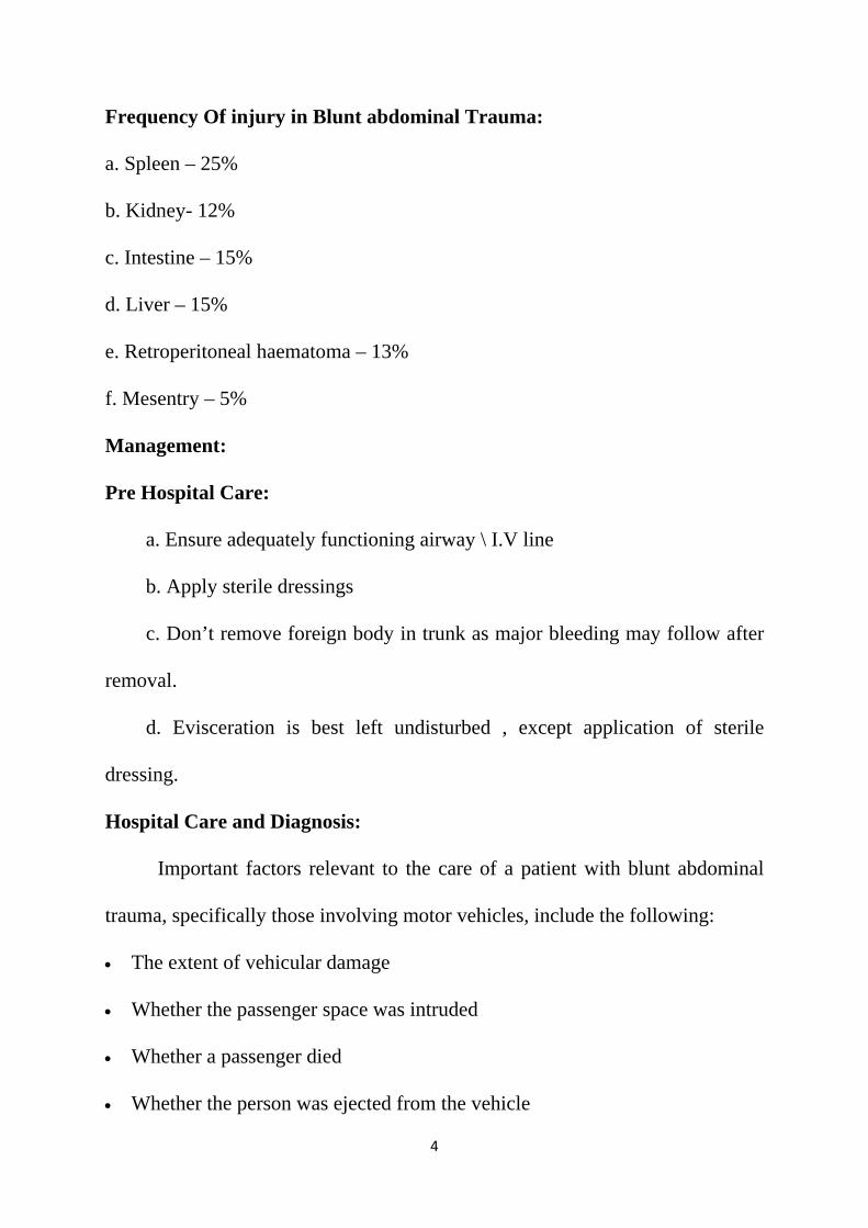
Frequency Of injury in Blunt abdominal Trauma:
a. Spleen – 25%
b. Kidney- 12%
c. Intestine – 15%
d. Liver – 15%
e. Retroperitoneal haematoma – 13%
f. Mesentry – 5%
Management:
Pre Hospital Care:
a. Ensure adequately functioning airway \ I.V line
b. Apply sterile dressings
c. Don’t remove foreign body in trunk as major bleeding may follow after
removal.
d. Evisceration is best left undisturbed , except application of sterile
dressing.
Hospital Care and Diagnosis:
Important factors relevant to the care of a patient with blunt abdominal
trauma, specifically those involving motor vehicles, include the following:
• The extent of vehicular damage
• Whether the passenger space was intruded
• Whether a passenger died
• Whether the person was ejected from the vehicle
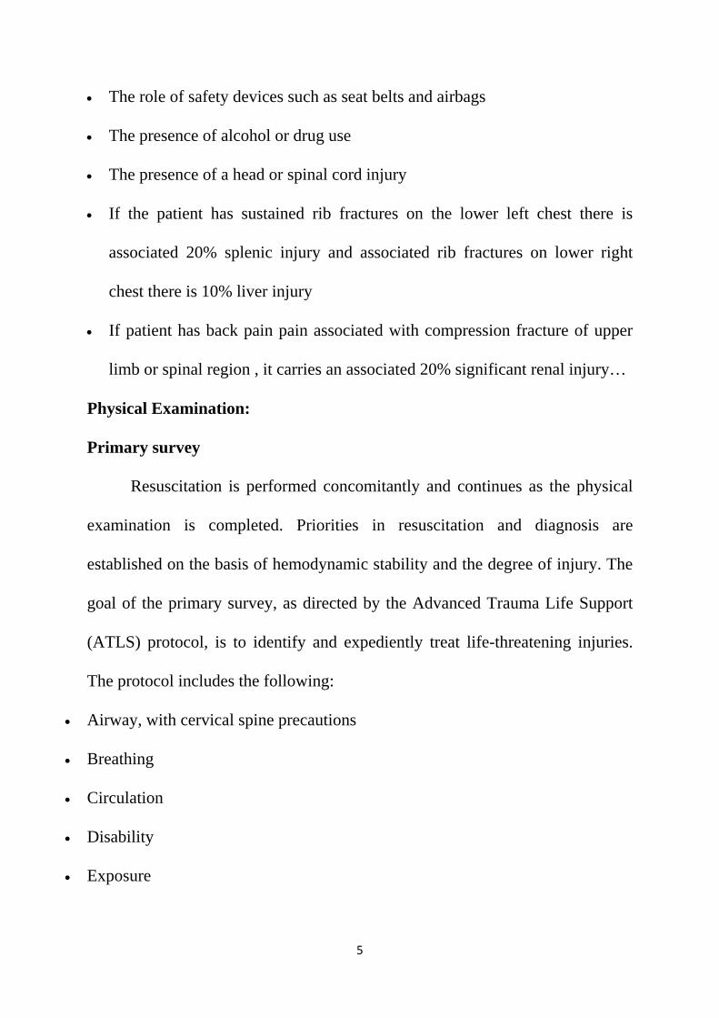
5
• The role of safety devices such as seat belts and airbags
• The presence of alcohol or drug use
• The presence of a head or spinal cord injury
• If the patient has sustained rib fractures on the lower left chest there is
associated 20% splenic injury and associated rib fractures on lower right
chest there is 10% liver injury
• If patient has back pain pain associated with compression fracture of upper
limb or spinal region , it carries an associated 20% significant renal injury…
Physical Examination:
Primary survey
Resuscitation is performed concomitantly and continues as the physical
examination is completed. Priorities in resuscitation and diagnosis are
established on the basis of hemodynamic stability and the degree of injury. The
goal of the primary survey, as directed by the Advanced Trauma Life Support
(ATLS) protocol, is to identify and expediently treat life-threatening injuries.
The protocol includes the following:
• Airway, with cervical spine precautions
• Breathing
• Circulation
• Disability
• Exposure
6
Secondary survey
After an appropriate primary survey and initiation of resuscitation,
attention should be focused on the secondary survey of the abdomen. The
secondary survey is the identification of all injuries via a head-to-toe
examination. For life-threatening injuries that necessitate emergency surgery, a
comprehensive secondary survey should be delayed until the patient has been
stabilized.
At the other end of the spectrum are victims of blunt trauma who have a
benign abdomen upon initial presentation. Many injuries initially are occult and
manifest over time. Frequent serial examinations, in conjunction with the
appropriate diagnostic studies, such as abdominal computed tomography (CT)
and bedside ultrasonography, are essential in any patient with a significant
mechanism of injury.
The evaluation of a patient with blunt abdominal trauma must be
accomplished with the entire patient in mind, with all injuries prioritized
accordingly. This implies that injuries involving the head, the respiratory
system, or the cardiovascular system may take precedence over an abdominal
injury.
The abdomen should neither be ignored nor be the sole focus of the
treating clinician and surgeon. In an unstable patient, the question of abdominal
involvement must be expediently addressed. This is accomplished by
identifying free intra-abdominal fluid with diagnostic peritoneal lavage (DPL)
7
or focused assessment with sonography for trauma (FAST). The objective is
rapid identification of those patients who need a laparotomy.
The initial clinical assessment of patients with blunt abdominal trauma is
often difficult and notably inaccurate. Associated injuries often cause tenderness
and spasms in the abdominal wall and make diagnosis difficult. Lower rib
fractures, pelvic fractures, and abdominal wall contusions may mimic the signs
of peritonitis.
In general, accuracy increases if the patient is reevaluated repeatedly and
at frequent intervals. However, repeated examinations may not be feasible in
patients who need general anesthesia and surgery for other injuries. The greatest
compromise of the physical examination occurs in the setting of neurologic
dysfunction, which may be caused by head injury or substance abuse.
The most reliable signs and symptoms in alert patients are pain,
tenderness, gastrointestinal hemorrhage, hypovolemia, and evidence of
peritoneal irritation. However, large amounts of blood can accumulate in the
peritoneal and pelvic cavities without any significant or early changes in the
physical examination findings.
The respiratory pattern should be observed because abdominal breathing
may indicate spinal cord injury. A sensory examination of the chest and
abdomen should be performed to evaluate the potential for spinal cord injury.
Spinal cord injury may interfere with the accurate assessment of the abdomen
by causing decreased or absent pain perception.
8
The abdominal examination must be systematic. The abdomen is
inspected for abrasions or ecchymosis. Particular attention should be paid to
injury patterns that predict the potential for intra-abdominal trauma (eg, lap belt
abrasions, steering wheel–shaped contusions). In most studies, lap belt marks
have been correlated with rupture of the small intestine and an increased
incidence of other intra-abdominal injuries.
Ecchymosis involving the flanks (Grey Turner sign) or the umbilicus
(Cullen sign) indicates retroperitoneal hemorrhage, but this is usually delayed
for several hours to days.
Visual inspection for abdominal distention, which may be due to pneumo
peritoneum, gastric dilatation secondary to assisted ventilation or swallowing of
air, or ileus produced by peritoneal irritation, is important.
Auscultation of bowel sounds in the thorax may indicate the presence of a
diaphragmatic injury. Abdominal bruit may indicate underlying vascular disease
or traumatic arteriovenous fistula.
Palpation may reveal local or generalized tenderness, guarding, rigidity,
or rebound tenderness, which suggests peritoneal injury. Such signs appearing
soon after an injury suggest leakage of intestinal content. Peritonitis due to
intra-abdominal hemorrhage may take several hours to develop.
Fullness and doughy consistency on palpation may indicate intra-
abdominal hemorrhage. Crepitation or instability of the lower thoracic cage
9
indicates the potential for splenic or hepatic injuries associated with lower rib
injuries.
Tenderness on percussion constitutes a peritoneal sign. Tenderness
mandates further evaluation and probably surgical consultation.
Rectal and bimanual vaginal pelvic examinations should be performed. A
rectal examination should be done to search for evidence of bony penetration
resulting from a pelvic fracture, and the stool should be evaluated for gross or
occult blood. The evaluation of rectal tone is important for determining the
patient’s neurologic status, and palpation of a high-riding prostate suggests
urethral injury.
The genitals and perineum should be examined for soft tissue injuries,
bleeding, and hematoma. Pelvic instability indicates the potential for lower
urinary tract injury, as well as pelvic and retroperitoneal hematoma. Open pelvic
fractures are associated with a mortality rate exceeding 50%.
A nasogastric tube should be placed routinely (in the absence of
contraindications, eg, basilar skull fracture) to decompress the stomach and to
assess for the presence of blood. If the patient has evidence of a maxillofacial
injury, an orogastric tube is preferred.
As the assessment continues, a Foley catheter is placed and a sample of
urine is sent for analysis for microscopic hematuria. If injury to the urethra or
bladder is suggested because of an associated pelvic fracture, then a retrograde
urethrogram is performed before catheterization.
10
Tertiary survey
The concept of the tertiary trauma survey was first introduced by
Enderson et al to assist in the diagnosis of any injuries that may have been
missed during the primary and secondary surveys. The tertiary survey involves
a repetition of the primary and secondary surveys and a revision of all
laboratory and radiographic studies. In a study, a tertiary trauma survey detected
56% of injuries missed during the initial assessment within 24 hours of
admission.
The most important initial concern in the evaluation of a patient with
blunt abdominal trauma is an assessment of hemodynamic stability. In the
hemodynamically unstable patient, a rapid evaluation must be made regarding
the presence of hemoperitoneum. This can be accomplished by means of
diagnostic peritoneal lavage (DPL) or the focused assessment with sonography
for trauma (FAST). Radiographic studies of the abdomen are indicated in stable
patients when the physical examination findings are inconclusive.
Blood Studies:
Complete blood count:
The presence of massive hemorrhage is usually obvious from
hemodynamic parameters, and an abnormal hematocrit value merely confirms
the diagnosis. Normal hemoglobin and hematocrit results do not rule out
significant hemorrhage. Patients bleed whole blood. Until blood volume is
replaced with crystalloid solution or hormonal effects (eg, adrenocorticotropic
11
hormone [ACTH], aldosterone, antidiuretic hormone [ADH]) and transcapillary
refill occurs, anemia may not develop.
Bedside diagnostic testing with rapid hemoglobin or hematocrit machines
may quickly identify patients who have physiologically significant volume
deficits and hemodilution. Reported hemoglobin from ABG measurements also
may be useful in identifying anemia. Some studies have correlated a low initial
hematocrit (ie, < 30%) with significant injuries.
Do not withhold transfusion in patients who have relatively normal
hematocrit results (ie, >30%) but have evidence of clinical shock, serious
injuries (eg, open-book pelvic fracture), or significant ongoing blood loss.
Hemodynamic instability in an adult despite the administration of 2 L of fluid
indicates ongoing blood loss and is an indication for immediate blood
transfusion. Use platelet transfusions to treat patients with thrombocytopenia
(ie, platelet count < 50,000/µL) and ongoing hemorrhage.
An elevated white blood cell (WBC) count on admission is nonspecific
and does not predict the presence of a hollow viscus injury (HVI). The
diagnostic value of serial WBC counts for predicting HVI within the first 24
hours after trauma is very limited.
Liver function tests
Liver function tests (LFTs) may be useful in the patient with blunt
abdominal trauma; however, test findings may be elevated for several reasons
(eg, alcohol abuse). One study has shown that an aspartate aminotransferase
12
(AST) or alanine aminotransferase (ALT) level more than 130 U corresponds
with significant hepatic injury. Lactate dehydrogenase (LDH) and bilirubin
levels are not specific indicators of hepatic trauma.
Serum amylase or lipase measurements
The serum lipase or amylase level is neither sensitive nor specific as a
marker for major pancreatic or enteric injury. Normal levels do not exclude a
major pancreatic injury. Elevated levels may be caused by injuries to the head
and face or by an assortment of nontraumatic causes (eg, alcohol, narcotics,
various other drugs). Amylase or lipase levels may be elevated because of
pancreatic ischemia caused by the systemic hypotension that accompanies
trauma.
However, persistent hyperamylasemia or hyperlipasemia (eg, abnormal
elevation 3-6 hours after trauma) should raise the suggestion of significant intra-
abdominal injury and is an indication for aggressive radiographic and surgical
investigation.
Blood typing, screening, and cross-matching
Blood from all trauma patients with suspected blunt abdominal injury
should be screened and typed. If an injury is identified, this practice greatly
reduces the time required for cross-matching. An initial cross-match should be
performed on a minimum of 4-6 units for those patients with clear evidence of
abdominal injury and hemodynamic instability. Until cross-matched blood is
available, O-negative or type-specific blood should be used.
13
Drug and alcohol screening
Perform drug and alcohol screens on trauma patients who have alterations
in their level of consciousness. Breath or blood testing may quantify alcohol
level.
Urine Studies
Indications for diagnostic urinalysis include significant trauma to the
abdomen and/or flank, gross hematuria, microscopic hematuria in the setting of
hypotension, and a significant deceleration mechanism.
Obtain a contrast nephrogram by utilizing intravenous pyelography (IVP)
or computed tomography (CT) scanning with intravenous (IV) contrast. Gross
hematuria indicates a workup that includes cystography and IVP or CT scanning
of the abdomen with contrast.
Perform a urine toxicologic screen as appropriate. Obtain a serum or
urine pregnancy test on all females of childbearing age.
Plain Radiography
Although their overall value in the evaluation of patients with blunt
abdominal trauma is limited, plain films can demonstrate numerous findings.
The chest radiograph may aid in the diagnosis of abdominal injuries such as
ruptured hemidiaphragm (eg, a nasogastric tube seen in the chest) or
pneumoperitoneum.
The pelvic or chest radiograph can demonstrate fractures of the
thoracolumbar spine. The presence of transverse fractures of the vertebral
14
bodies (ie, Chance fractures) suggests a higher likelihood of blunt injuries to the
bowel. In addition, free intraperitoneal air, or trapped retroperitoneal air from
duodenal perforation, may be seen.
Ultrasonography
The use of diagnostic ultrasonography to evaluate a patient with blunt
trauma for abdominal injuries has been advocated since the 1970s. European
and Asian investigators have extensive experience with this technology and are
leaders in the use of ultrasound for the diagnosis of blunt abdominal trauma.
The first American report of physician-performed abdominal ultrasonography in
the evaluation of blunt abdominal trauma was published in 1992 by Tso and
colleagues.
Bedside ultrasonography is a rapid, portable, noninvasive, and accurate
examination that can be performed by emergency clinicians and trauma
surgeons to detect hemoperitoneum. In fact, in many medical centers, the FAST
examination has virtually replaced DPL as the procedure of choice in the
evaluation of hemodynamically unstable trauma patients.
The FAST examination is based on the assumption that all clinically
significant abdominal injuries are associated with hemoperitoneum. However,
the detection of free intraperitoneal fluid is based on factors such as the body
habitus, injury location, presence of clotted blood, position of the patient, and
amount of free fluid present.
15
In a patient with isolated blunt abdominal trauma and multisystem
injuries, FAST performed by an experienced sonographer can rapidly identify
free intraperitoneal fluid (generally appearing as a black stripe). The sensitivity
for solid organ encapsulated injury is moderate in most studies. Hollow viscus
injury (HVI) rarely is identified; however, free fluid may be visualized. For
patients with persistent pain or tenderness or those developing peritoneal signs,
FAST may be considered as a complementary measure to CT scanning, DPL, or
exploration.
The minimum threshold for detecting hemoperitoneum is unknown and
remains a subject of interest. Kawaguchi and colleagues found that 70 mL of
blood could be detected,whereas Tiling et al found that 30 mL is the minimum
requirement for detection with ultrasonography. They also concluded that a
small anechoic stripe in the Morison pouch represents approximately 250 mL of
fluid, whereas 0.5-cm and 1-cm stripes represent approximately 500 mL and 1 L
of free fluid, respectively.
The current FAST examination protocol consists of 4 acoustic windows
with the patient supine. These windows are pericardiac, perihepatic, perisplenic,
and pelvic (known as the 4 P s). An examination is interpreted as positive if free
fluid is found in any of the 4 acoustic windows and as negative if no fluid is
seen. An examination is deemed indeterminate if any of the windows cannot be
adequately assessed.
16
The pericardial window is obtained via a subcostal or transthoracic
approach. It provides a 4-chamber view of the heart and can detect the presence
of hemopericardium, which is demonstrated by the separation of the visceral
and parietal pericardial layers.
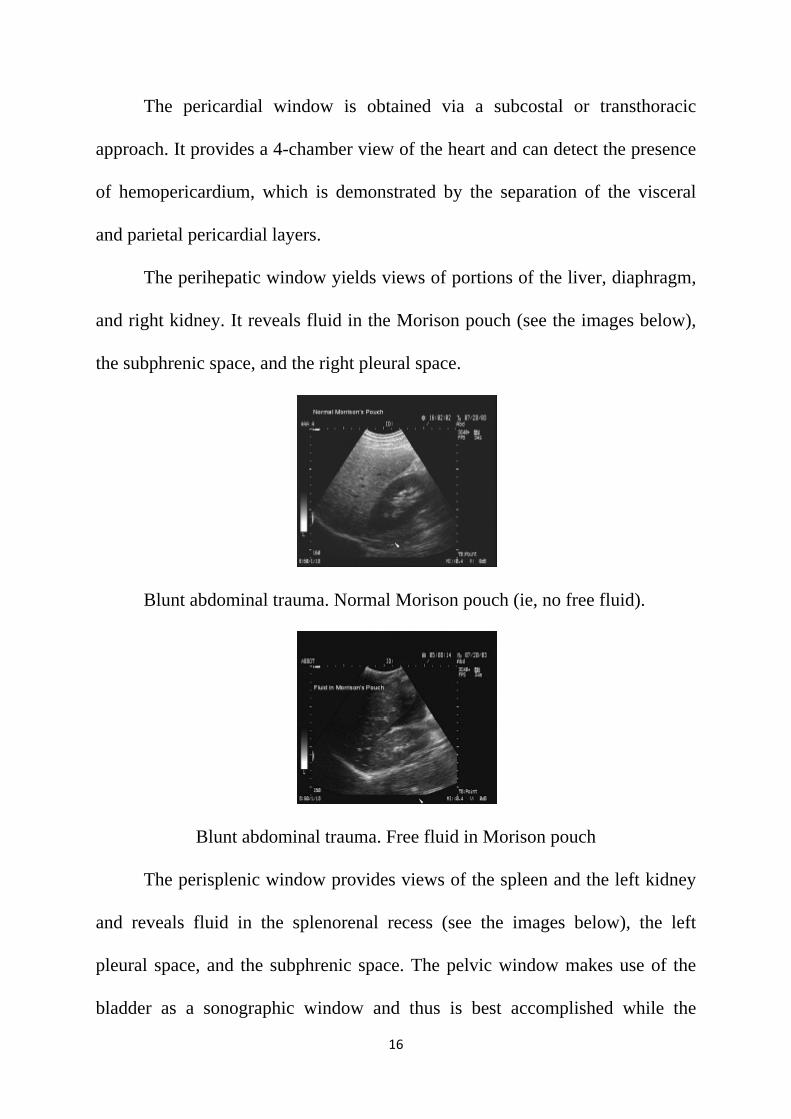
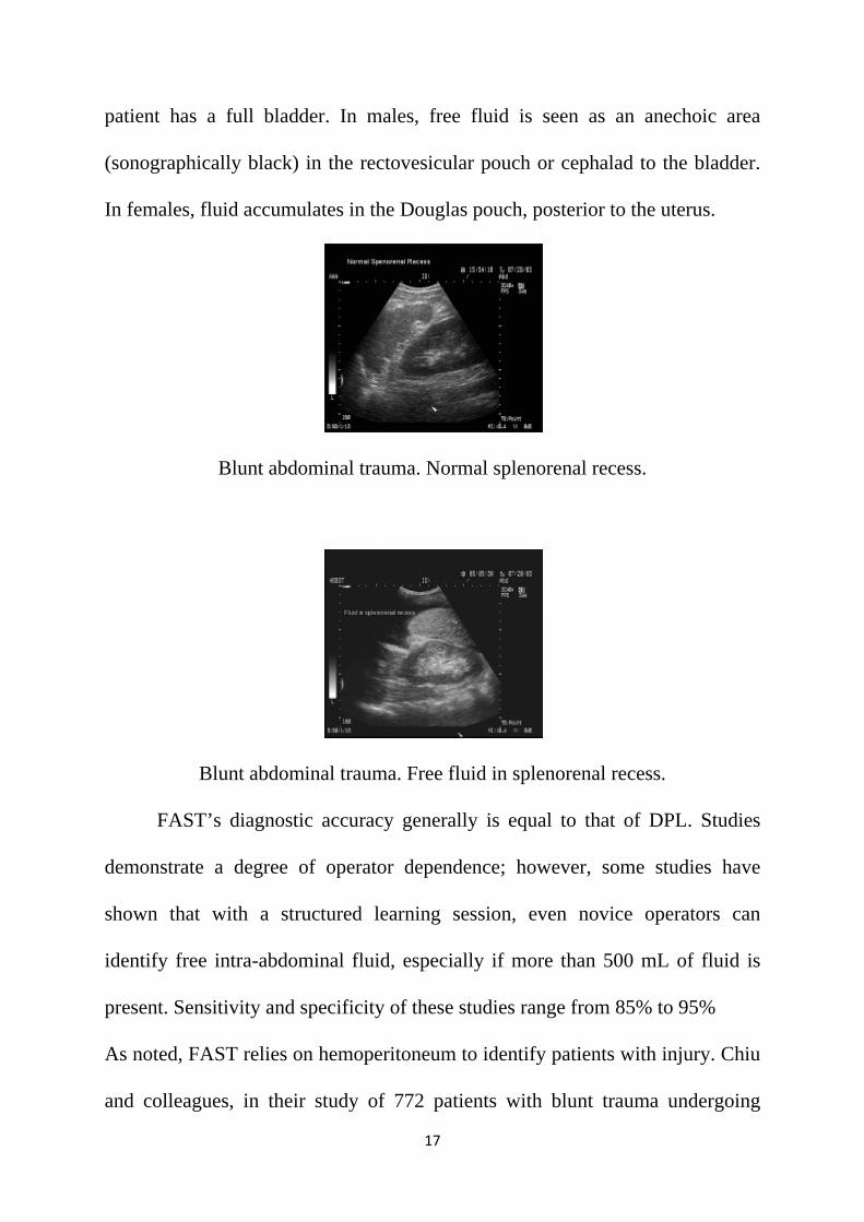
The perihepatic window yields views of portions of the liver, diaphragm,
and right kidney. It reveals fluid in the Morison pouch (see the images below),
the subphrenic space, and the right pleural space.
Blunt abdominal trauma. Normal Morison pouch (ie, no free fluid).
Blunt abdominal trauma. Free fluid in Morison pouch
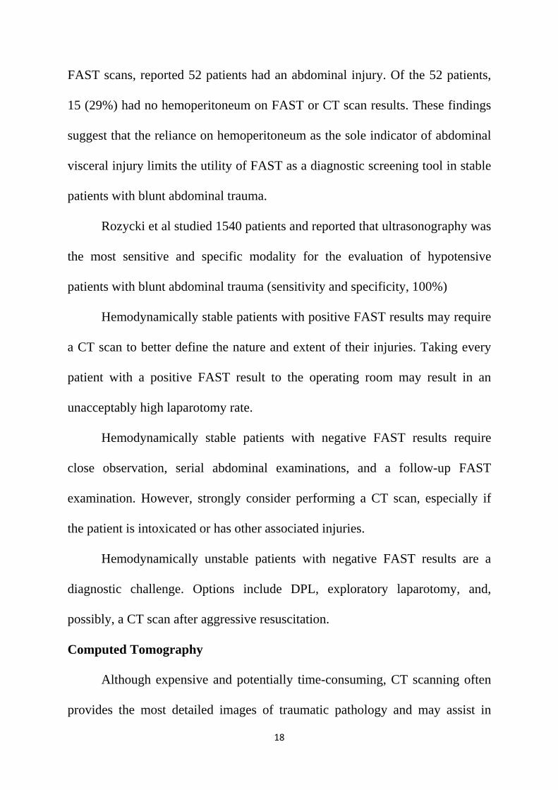
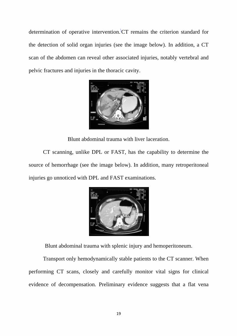
The perisplenic window provides views of the spleen and the left kidney
and reveals fluid in the splenorenal recess (see the images below), the left
pleural space, and the subphrenic space. The pelvic window makes use of the
bladder as a sonographic window and thus is best accomplished while the
17
patient has a full bladder. In males, free fluid is seen as an anechoic area
(sonographically black) in the rectovesicular pouch or cephalad to the bladder.
In females, fluid accumulates in the Douglas pouch, posterior to the uterus.
Blunt abdominal trauma. Normal splenorenal recess.
Blunt abdominal trauma. Free fluid in splenorenal recess.
FAST’s diagnostic accuracy generally is equal to that of DPL. Studies
demonstrate a degree of operator dependence; however, some studies have
shown that with a structured learning session, even novice operators can
identify free intra-abdominal fluid, especially if more than 500 mL of fluid is
present. Sensitivity and specificity of these studies range from 85% to 95%
As noted, FAST relies on hemoperitoneum to identify patients with injury. Chiu
and colleagues, in their study of 772 patients with blunt trauma undergoing
18
FAST scans, reported 52 patients had an abdominal injury. Of the 52 patients,
15 (29%) had no hemoperitoneum on FAST or CT scan results. These findings
suggest that the reliance on hemoperitoneum as the sole indicator of abdominal
visceral injury limits the utility of FAST as a diagnostic screening tool in stable
patients with blunt abdominal trauma.
Rozycki et al studied 1540 patients and reported that ultrasonography was
the most sensitive and specific modality for the evaluation of hypotensive
patients with blunt abdominal trauma (sensitivity and specificity, 100%)
Hemodynamically stable patients with positive FAST results may require
a CT scan to better define the nature and extent of their injuries. Taking every
patient with a positive FAST result to the operating room may result in an
unacceptably high laparotomy rate.
Hemodynamically stable patients with negative FAST results require
close observation, serial abdominal examinations, and a follow-up FAST
examination. However, strongly consider performing a CT scan, especially if
the patient is intoxicated or has other associated injuries.
Hemodynamically unstable patients with negative FAST results are a
diagnostic challenge. Options include DPL, exploratory laparotomy, and,
possibly, a CT scan after aggressive resuscitation.
Computed Tomography
Although expensive and potentially time-consuming, CT scanning often
provides the most detailed images of traumatic pathology and may assist in
19
determination of operative intervention.\CT remains the criterion standard for
the detection of solid organ injuries (see the image below). In addition, a CT
scan of the abdomen can reveal other associated injuries, notably vertebral and
pelvic fractures and injuries in the thoracic cavity.
Blunt abdominal trauma with liver laceration.
CT scanning, unlike DPL or FAST, has the capability to determine the
source of hemorrhage (see the image below). In addition, many retroperitoneal
injuries go unnoticed with DPL and FAST examinations.
Blunt abdominal trauma with splenic injury and hemoperitoneum.
Transport only hemodynamically stable patients to the CT scanner. When
performing CT scans, closely and carefully monitor vital signs for clinical
evidence of decompensation. Preliminary evidence suggests that a flat vena
20
cava on CT scan is a marker for underresuscitation and may be correlated with
higher mortality and hemodynamic decompensation.
CT scans provide excellent imaging of the pancreas, duodenum, and
genitourinary system. The images can help quantitate the amount of blood in the
abdomen and can reveal individual organs with precision. The primary
advantage of CT scanning is its high specificity and use for guiding
nonoperative management of solid organ injuries.
Drawbacks of CT scanning relate to the need to transport the patient from
the trauma resuscitation area and the additional time required to perform CT
scanning compared to FAST or DPL.
In addition, CT scanning may miss injuries to the diaphragm and
perforations of the gastrointestinal (GI) tract, especially when performed soon
after the injury. Although some pancreatic injuries may be missed with a CT
scan performed soon after trauma, virtually all are identified if the scan is
repeated in 36-48 hours. For selected patients, endoscopic retrograde
cholangiopancreatography (ERCP) may complement CT scanning to rule out a
ductal injury.
Finally, CT scanning is relatively expensive and time consuming and
requires oral or intravenous (IV) contrast, which may cause adverse reactions.
The best CT imagery requires both oral and IV contrast. Some controversy has
arisen over the use of oral contrast and whether the additional information it
provides negates the drawbacks of increased time to administration and risk of
21
aspiration. The value of oral contrast in diagnosing bowel injury has been
debated, but no definitive answer exists at this time.
Diagnostic Laparoscopy
The introduction of minimally invasive surgery has revolutionized many
surgical diagnostic protocols. In the late 1980s and early 1990s, there was
considerable interest in the use of laparoscopy for evaluation and management
of blunt and penetrating abdominal trauma. Subsequent studies, however,
revealed major limitations to this approach and cautioned against its widespread
use. The most important limitation is inability to reliably identify hollow viscus
and retroperitoneal injuries, even in the hands of experienced laparoscopists.
Diagnostic laparoscopy involves placing a subumbilical or subcostal trocar for
the introduction of the laparoscope and creating other ports for retractors,
clamps, and other tools necessary for visualization of the repair.
Diagnostic laparoscopy has been most useful in the evaluation of possible
diaphragmatic injuries, especially in penetrating thoracoabdominal injuries on
the left side.In blunt trauma, it has no clear advantages over less invasive
modalities such as DPL and CT scanning; furthermore, complications can result
from trocar misplacement.
Diagnostic Peritoneal Lavage
The idea of evaluating the abdomen by analyzing its contents was first
used in the diagnosis of acute abdominal conditions. In 1906, Salomon
described the passage of a urethral catheter by means of a trocar inserted
22
through the abdominal wall to obtain samples of peritoneal fluid with the aim of
establishing the diagnosis of peritonitis from infectious agents (eg,
pneumococcal or tuberculous organisms). This technique has since been refined
and is now known as abdominal paracentesis.
In 1926, Neuhof and Cohen described the sampling of peritoneal fluid in
cases of acute pancreatitis and blunt abdominal trauma by passing a spinal
needle through the abdominal wall. In 1965, Root et al reported the use of
percutaneous DPL in patients who had sustained blunt abdominal trauma.
DPL is used as a method of rapidly determining the presence of intraperitoneal
blood. It is particularly useful if the history and abdominal examination of an
unstable patient with multisystem injuries are either unreliable (eg, because of
head injury, alcohol, or drug intoxication) or equivocal (eg, because of lower rib
fractures, pelvic fractures, or confounding clinical examination).
DPL is also useful for patients in whom serial abdominal examinations
cannot be performed (eg, those in an angiographic suite or operating room
during emergency orthopedic or neurosurgical procedures).
INDICATIONS:
• Hemodynamically unstable patients with negative FAST \CT
• Patients with a spinal cord injury
• Those with multiple injuries and unexplained shock
• Obtunded patients with a possible abdominal injury
• Intoxicated patients in whom abdominal injury is suggested
23
• Patients with potential intra-abdominal injury who will undergo
prolonged anesthesia for another procedure.
Contraindications:
Absolute contraindication :
The obvious need for laparotomy.
Relative contraindication:
Morbid obesity
History of multiple abdominal surgeries
Pregnancy.
Various methods of introducing the catheter into the peritoneal space have been
described.
These include the open, semiopen, and closed methods.
The open method requires an infraumbilical skin incision that is extended
to and through the linea alba. (In pregnant patients or in patients with particular
risk for potential pelvic hematoma, the incision should be placed superior to the
umbilicus.) The peritoneum is opened, and the catheter is inserted under direct
visualization.
The semiopen method is identical, except that the peritoneum is not
opened and the catheter is delivered percutaneously through the peritoneum into
the peritoneal cavity.
The closed technique requires the catheter to be inserted blindly through
the skin, subcutaneous tissue, linea alba, and peritoneum.
24
The closed and semiopen techniques at the infraumbilical site are
preferred at most centers. The fully open method is the most technically
demanding and is restricted to those situations in which the closed or semiopen
technique is unsuccessful or is deemed unsafe (eg, patients with pelvic fractures,
pregnancy, obesity, or prior abdominal operations).
PROCEDURE:
After insertion of the catheter into the peritoneum, attempt to aspirate free
intraperitoneal blood (at least 15-20 mL).
Results are considered positive in a blunt trauma patient if ,
10 mL of grossly bloody aspirate is obtained before infusion of the
lavage fluid
If the siphoned lavage fluid contains more than 100,000 red blood cells
(RBCs)/µL, more than 500 white blood cells (WBCs)/µL,
Elevated amylase content,
Bile, bacteria, vegetable matter, or urine.
Only approximately 30 mL of blood is needed in the peritoneum to
produce a microscopically positive DPL result.
If findings are negative, infuse 1 L of crystalloid solution (eg, lactated
Ringer solution) into the peritoneum. Then, allow this fluid to drain by gravity,
and ensure that laboratory analysis is performed.
Complications :
Bleeding from the incision and catheter insertion,
25
Infection (ie, wound, peritoneal),
Injury to intra-abdominal structures (eg, urinary bladder, small bowel,
uterus).
These complications may increase the possibility of false-positive studies
Bleeding from the incision, dissection, or catheter insertion can cause
false-positive results that may lead to unnecessary laparotomy. Achieve
appropriate hemostasis prior to entering the peritoneum and placing the
catheter.
False-positive DPL results can occur if an infraumbilical approach is used
in a patient with a pelvic fracture. A pelvic x-ray film should be obtained prior
to performing DPL if a pelvic fracture is suggested. Before DPL is attempted,
the urinary bladder and stomach should be decompressed.
DPL has been shown in some studies to have a diagnostic accuracy of
98-100%, a sensitivity of 98-100%, and a specificity of 90-96%. It has some
advantages, including high sensitivity, rapidity, and immediate interpretation.
The main limitations of DPL include its potential for iatrogenic abdominal
injury and its high sensitivity, which can lead to nontherapeutic laparotomies.
With the availability of fast, noninvasive, and better imaging modalities
(eg, FAST, CT scanning), the role of DPL is now limited to the evaluation of
unstable trauma patients in whom FAST results are negative or inconclusive. In
some contexts, DPL may be complemented with a CT scan if the patient has
positive lavage results but stabilizes.
26
MANAGEMENT OF SPECIFIC INJURIES AT
LAPAROTOMY
PATHOPHYSIOLOGY OF BLUNT INJURIES
Management of patient with blunt abdominal trauma requires and
understanding of the injury mechanism. In general injuries can be classified
as high energy or low energy. Several pathophysiogical processes involved are
1) Sudden pronounced rise in intra abdominal pressure causing burst injury
of solid organs or rupture of hollow viscus.
2) Compression of abdominal viscera the applied force to the anterior wall
to the posterior thoracic cage or vertebral column.
3) Abrupt, shearing forces can cause tear of organs or vascular pedicles
SPLENIC INJURIES
Organ most frequently injured in blunt abdominal trauma
a. Compression may occur between the anterior wall and posterior rib cage
b. Clinical picture includes Left upper quadrant pain , Signs of hypovolumia ,
Pain in the left shoulder (Kehr’s Sign)
c. Xray features include-
Enlargement of splenic shadow ,
Medial displacement of gastric shadow ,
associated rib fractures..
27
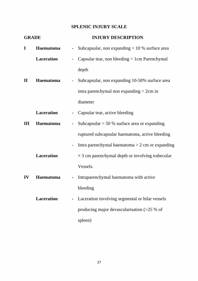
SPLENIC INJURY SCALE
GRADE INJURY DESCRIPTION
I Haematoma - Subcapsular, non expanding < 10 % surface area
Laceration - Capsular tear, non bleeding < 1cm Parenchymal
depth
II Haematoma - Subcapsular, non expanding 10-50% surface area
intra parenchymal non expanding < 2cm in
diameter
Laceration - Capsular tear, active bleeding
III Haematoma - Subcapsular > 50 % surface area or expanding
ruptured subcapsular haematoma, active bleeding
- Intra parenchymal haematoma > 2 cm or expanding
Laceration > 3 cm parenchymal depth or involving trabecular
Vessels.
IV Haematoma - Intraparenchymal haematoma with active
bleeding
Laceration - Laceration involving segmental or hilar vessels
producing major devascularisation (>25 % of
spleen)
28
Splenic Salvage:
Adequate mobilization enhances success of salvage
Capsular tear-Topical hemostatic agents
Small lacerations- Interlocking sutures
Major laceration <50% - Segmental splenic resection
Splenic Salvage Should not be pursued:
If the patient has protracted hypotension
Undue delay is anticipated in repair of laceration
Patient has other severe injuries
Splenectomy Complications:
Left pleural effusion
Left lower lobe collapse
Post splenectomy sepsis
Liver Injuries:
Liver is the largest organ in the abdominal cavity .Due to its size injuries
sufficient to lacerate the liver are associated with injuries to other organs in
about 80% cases.Spontaneous hemostatic mechanism that characterize liver
tissue may contribute pervasive observation that 85% of liver injuries do not
bleed at time of laparotomy.
A patient who has history of being in shock at the scene following blunt
trauma should be suspected of having major liver trauma.
29
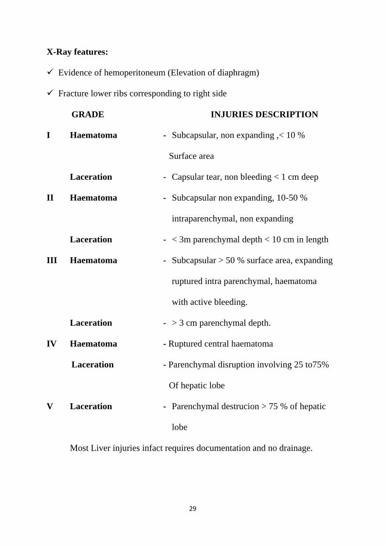
X-Ray features:
Evidence of hemoperitoneum (Elevation of diaphragm)
Fracture lower ribs corresponding to right side
GRADE INJURIES DESCRIPTION
I Haematoma - Subcapsular, non expanding ,< 10 %
Surface area
Laceration - Capsular tear, non bleeding < 1 cm deep
II Haematoma - Subcapsular non expanding, 10-50 %
intraparenchymal, non expanding
Laceration - < 3m parenchymal depth < 10 cm in length
III Haematoma - Subcapsular > 50 % surface area, expanding
ruptured intra parenchymal, haematoma
with active bleeding.
Laceration - > 3 cm parenchymal depth.
IV Haematoma - Ruptured central haematoma
Laceration - Parenchymal disruption involving 25 to75%
Of hepatic lobe
V Laceration - Parenchymal destrucion > 75 % of hepatic
lobe
Most Liver injuries infact requires documentation and no drainage.
30
Indications for laparotomy during the period of observation are
1. Deteriorating vital signs.
2. Infection
3. Progressive expansion of the haematoma.
General measures in Management:
Class I parenchymal Lacerations:
treated by compression for 5 to 10 minutes.
Topical agents. microfibrillar collagen and gel foam. can be used to stop
bleeding
Class II parenchymal lacerations:
Classically horizontal mattress sutures with 0 chromic catgut often
place with blunt needle is used
Class III & IV Lacerations:
Lacerations which continue to bleed despite initial management requires
tractotomy .The depth of wound are explored and specific blood vessels
and biliary radicals are individually suture ligated.
If bleeding continues still compression on porta hepatis (Pringle
manoeuvre)
When selective ligation fails ligation of hepatic artery is done . It
produces dramatic hemostasis without subsequent liver failure
Alternative is resectional debridement accompalisehed by finger fracture ,
removing the devitalized liver or portion of the segment
31
Bleeding from Hepatic Veins:
Unilobular – Resection and debridement is sufficient
Bilobular – placement of intracaval shunt
Still uncontrollable packing of injury and resuscitation is appropriate
followed by subsequent removal of pack in 24-72 hours
Complications following liver injury:
Pulmonary complications
Coagulopathy
Jaundice
Biliary fistula
Hemobilia
DIC
DUODENAL INJURIES
These injuries are suspected if history of trunk injury or localized blow to
epigastrium with handle bars , steering wheel or fist. Failure to recognize this
injury is associated with high mortality and morbidity caused by abscess
formation and sepsis since it’s a retroperitoneal organ.
X-Ray features:
Obliteration of psoas shadow
Absence of air in duodenal bulb
Air in retroperitoneum
32
CT with oral contrast is definite showing extravasation of dye..
Management:
Full inspection by taking down hepatic flexure of colon and performing a
kocher maneuver is mandatory….
Limited perforation or simple laceration within 6 hrs are treated by
primary closure , if more than 6 hrs has elapsed suture closure with tube
duodenostomy is indicated
If laceration is extensive Roux-en-Y jejuno duodenostomy is indicated
Distal duodenum (III &IV) parts injuries can be primarily closed if it
occurs within 6 hrs of injury , but if duration extends above 6 hrs
resection of III & IV parts and duodenal jejunostomy should be
performed.
Pancreatico duodenectomy is occasionally indicated for massive injury in
right upper quadrant cant be repaired usually due to devascularisation…
SMALL INTESTINE INJURIES
Crushing injury of the bowel between the spine and the blunt object such
as the steering wheel or handlebars
Deceleration shearing of the small bowel at fixed points such as
ileocaecal valve and around superior mesenteric artery
Closed loop rupture caused by increased intra-abdominal pressure.
33
Antibiotics should be started preoperatively
At Laparotomy:
Significant bleeding must be first priority
Apply non-crushing clamps to prevent further leakage of small bowel
contents
Examine small bowel carefully from ligament of trietz to ileocaecal valve
Single holes can be closed without debridment
Transection of small bowel is debrided and closed in routine fashion and
mesenteric defect is to be closed
Complications:
Intra-abdominal abscess
Anastamotic leakage
Enterocutaneous fistula
Intestinal obstruction
34
PANCRETIC INJURIES
Blunt trauma to the abdomen from a direct kick , blow or seat belt injury
may crush the pancreas over the vertebral column . The shared blood supply
between the pancreas and duodenum makes the likelihood of these two injuries
in combination very high
Persistant Amylasemia
Contrast duodenography revealing widening of C loop
Loss of psoas shadow , anterior displacement of stomach
Left pleural effusion
CLASSIFICATION OF PANCREATIC INJURIES
TYPE DEFINITIONS
I Contusion and laceration
II Distal transection or parenchymal
Injury with duct injury
III Proximal transection or parenchymal
Injury with probable duct injury
Management:
The entire pancreas should be visualized clearly .
Simple debridement
Injury to body and tail should be treated with partial resection
In patients with ductal transactions in the head, the treatment options are
1. Roux-en-y distal pancreatico jejunostomy
35
2. Anterior Roux-en-y distal pancreatico jejunostomy.
3. Resection.
Complications:
• Pancreatic Fistula
• Abscess in lesser sac
• Pseudo pancreatic Cyst
RETRO-PERITONEAL HEMATOMAS
Retroperitoneum can be divided into three zones,
Zone 1 –Central
Zone 2 – Flank \ Peri nephric
Zone 3 – Pelvic
Management:
Zone 3 injuries will usually have associated pelvic injuires and so
exploration of these injuries are hazardous . Incision to peritoneum destroys the
tamponade effect so non operative management is the rule in zone 3 injuries .
Also discrete bleeding points can rarely be identified as there is catastrophic
bleeding.
Zone 2 injuries can be left alone if not expanding
Zone 1 injuries are always explored due to associated injury to major
vascular structures and viscera ( Pancreatico duodenal injuries)
36
RENAL INJURIES
The kidney is the most common injured part of the Urinary Tract.
Classification:
MAJOR – 15% (Deep cortical \ Medullary lacerations , Large perinephric
hematomas,Vascular Injuries Of Vascular pedicle)
MINOR – Other Injuries
X-Ray:
a. Absent psoas muscle shadow
b. Altered kidney outline
c. Ground glass appearance suggestive of either extravasation
of urine or haemoperiotoneum.
d. Associated bony injuries like rib fractures
IVU:
a. Delay in visualization of the contract.
b. Extravasation of the contract
c. Lack of continuous renal outline
d. Enlarged renal shadow.
A normal IVU in a patient with haematuria indicates minor renal
contusion
37
MANAGEMENT:
Renal Injuries associated with blunt injury abdomen are usually not explored
unless they are pulsatile or expanding.
Shattered kidney is treated with nephrectomy to prevent haemorrhage.
Renal vein injury is repaied by venorrhaphy.
Renal artery injury is repaired by lateral arteriorrhaphy, arterial resection.
BLADDER INJURY
Majority of bladder injuries occur due to blunt abdominal trauma .
Bladder injury should be suspected strongly in persons with hematuria and
pelvic fractures.
Bladder rupture may be Extraperitoneal or Intraperitoneal
Diagnosis by IVU \ Cystography
Extra-Peritoneal Rupture:
• Non Operative management is the rule
• By prolonged Foleys catheter use
- If patient has no Intra-abdominal injuries
- No significant local hemorrhage
- No UTI
- Severe pelvic fractures \ massive Retro peritoneal bleeding

38
Delayed repair can be contemplated once the retroperitoneal bleeding is
controlled and their condition is stabilized
Intra-Pertitoneal Rupture:
Transabdominal approach – Closure with SPC and drainage..
39
MATERIALS AND METHODS
Thirty Four cases of blunt abdominal trauma admitted in all surgical units
at Tirunelveli Medical College Hospital, Tirunelveli during the period of
December 2010 to November 2011 were taken for this study.
The cases were selected in such a way that only those patients with
definitive history and clinical findings suggestive of injury to Viscerae which
were later confirmed by investigations, laparotomy and autopsy. Detailed
history regarding the mode and nature of injury were taken, The clinical features
were studies in details with special note to any associated injuries like head
injury, chest injury and fracture limbs. Basic investigations viz. blood Hb, blood
urea, blood sugar, serum creatinine and blood grouping were done in all cases.
Plain X-ray of the abdomen in erect posture was taken in most of the
cases expect in those who were admitted in a critically ill condition.
Radiographs of other parts were also taken to find out associated injuries.
Under aseptic precaution using sterile 18 G needle peritoneal tapping
done in all the four quadrants, in all patients with the history of blunt abdominal
trauma.
At laparotomy a systematic approach with examination of all intra
abdominal organs were made. After surgery the patients were continued on
nasogastric, aspiration, antibiotics.Postoperative complications were specifically
looked for, if present were treated appropriately.
40
OBSERVATION
In our study conducted at Tirunelveli Medical college hospital for 34
cases of blunt injury abdomen , the following findings were noted,
Splenic Injuries 11 -32.35%
Liver injuries 08 - 23.52%
Bladder injury 04 -11.76%
Renal injury 04 -11.76%
Mesentery 03 -8.82%
Bowel perforation 04 -11.76%
At laparotomy the commonest organ injured in most of the series is
spleen ( Denver Hospital study, Herman Hospital )
Age wise the age group of 31-40 is most commonly affected 35% . S.C
Dwivedi et al and B.C. Jain et al reported similar results. This shows that
persons in the active period of life are more susceptible for accidents and
injuries.
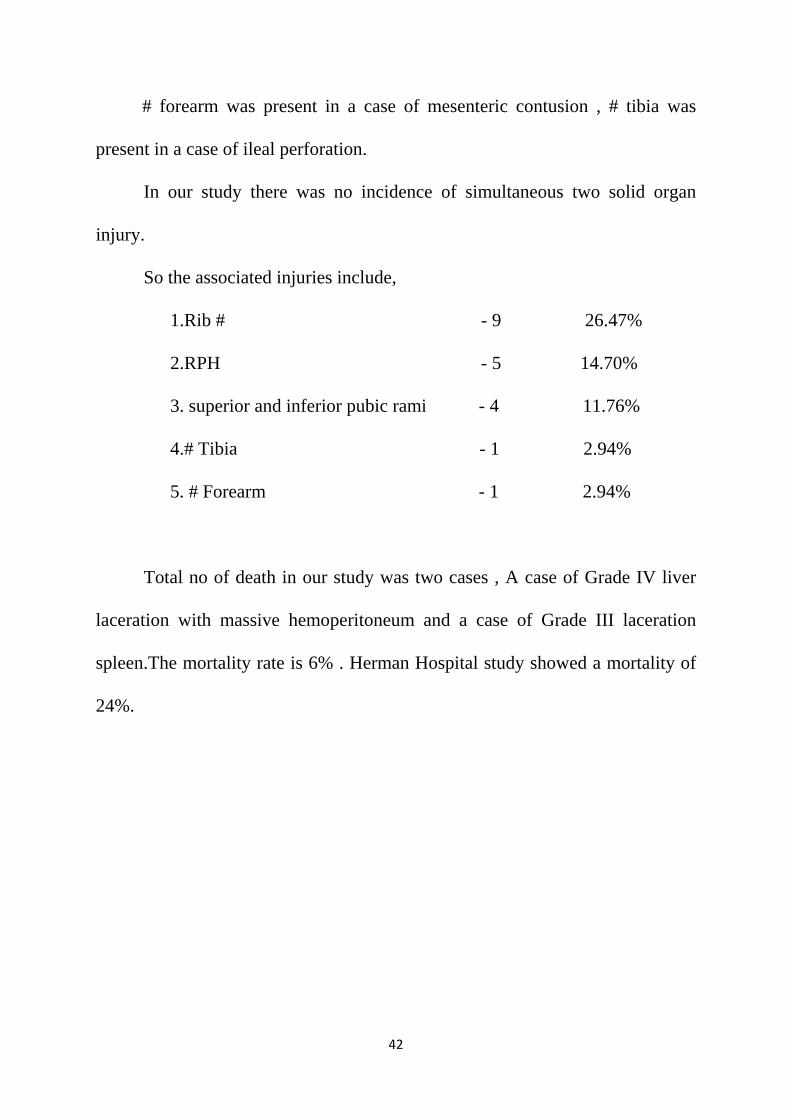
Sex wise male affection rate is 94% . B.C. Jain et al and Connecticut
society of surgeons study on abdominal trauma reported similar results. This
increased incidence in males is probably due to outdoor nature of occupation
and aggressive behaviour in males.
Diagnostic Four quadrant aspiration was done in all cases , and it was
positive for presence of blood \ fluid in 29 cases 85.29%. Abdominal
paracentesis when positive is highly predictive of significant intraabdominal
41
injuries but the accuracy varies from 50 % to 90 % in various studies, Authony
et al Showed 90 % accuracy.
All the cases were resuscitatied and subjected to FAST before exploratory
laparotomy . Of these there were 3 cases where the FAST findings did not
correlate with laparotomy findings .FAST missed a case of splenic laceration , a
case of Liver laceration , and there was a wrong interpretation of a case of Renal
injury having associated liver injury.
The incidence of False positive was 2.94% and the incidence of False
negative 5.88%.In cased of bladder injuries FAST revealed presence of clots
and mesenteric and bowel injuries it revealed presence of free fluid. Stritmatter
B. et al howed a sensitivity of 95.5 % and specificity of 97.5 % hence
ultrasonogram can be used as a initial imaging procedure.
Associated injuries include Rib fractures in 5 cases of liver lacertion
62.5% , 3 rib fractures in spleen 27.27% , 1 rib fracture in mesenteric contusion
33.33%. There is a 20 % chance of splenic injury and10 % chance of liver
injury with fracture ribs on the left or right lower six ribs (Graffin W.o et al ,
Moore E.E. )
Zone 2 RPH was present in all four cases of renal injury , the right kidney
was injured in 3 cases and the left in one case , Zone 3 RPH was present in a
case of mesenteric contusion.
# of both superior and inferior pubic rami was present in all four cases of
bladder injury
42
# forearm was present in a case of mesenteric contusion , # tibia was
present in a case of ileal perforation.
In our study there was no incidence of simultaneous two solid organ
injury.
So the associated injuries include,
1.Rib # - 9 26.47%
2.RPH - 5 14.70%
3. superior and inferior pubic rami - 4 11.76%
4.# Tibia - 1 2.94%
5. # Forearm - 1 2.94%
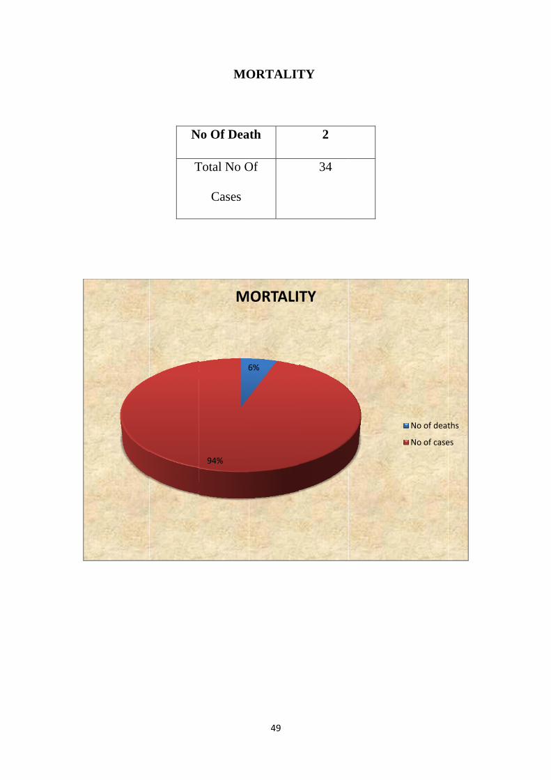
Total no of death in our study was two cases , A case of Grade IV liver
laceration with massive hemoperitoneum and a case of Grade III laceration
spleen.The mortality rate is 6% . Herman Hospital study showed a mortality of
24%.
43
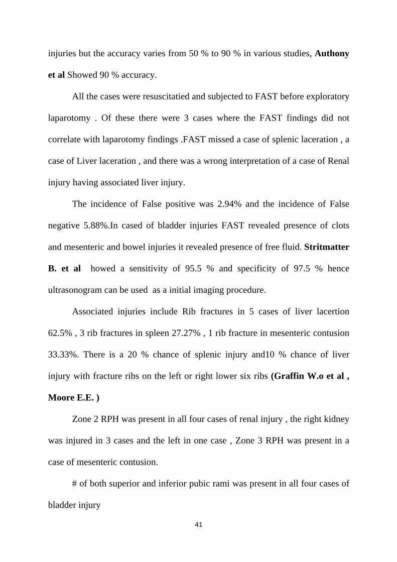
RESULTS
AGE WISE INCIDENCE OF CASES
Sl No Age of the patient Number of cases
1 13 – 20 6
2 21 – 30 10
3 31 – 40 12
4 41 – 50 4
5 51 – 60 2
0
2
4
6
8
10
12
14
13 ‐ 20 21 ‐ 30 31 ‐ 40 41 ‐ 50 51 ‐ 60
Agewise incidence of cases
SEX
M
F
T
WISE IN
Num
SEX
Male
Female
Total
44
NCIDENC
6%
mber Of MALE FEM
NO OF
32
2
34
CE OF CA
94%
CasesMALE
F CASES
ASES
S
45
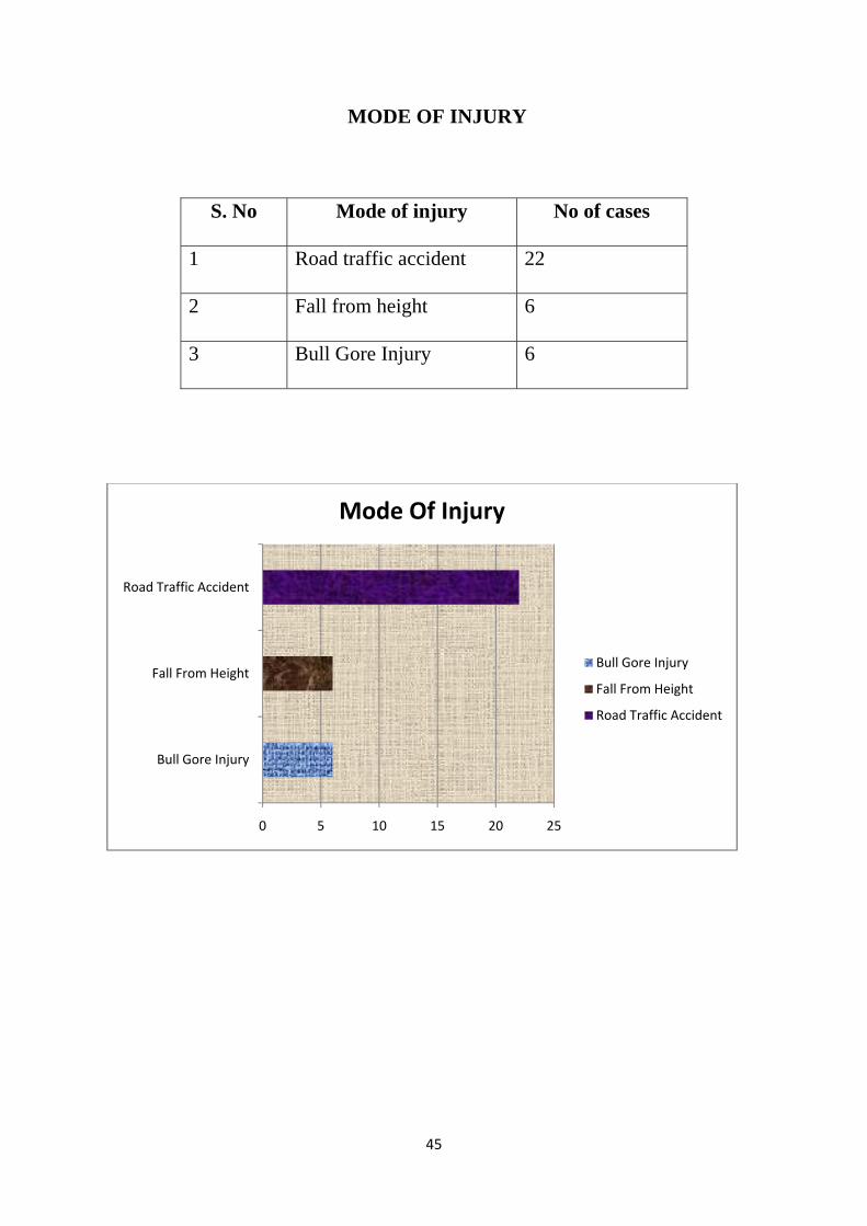
MODE OF INJURY
S. No Mode of injury No of cases
1 Road traffic accident 22
2 Fall from height 6
3 Bull Gore Injury 6
0 5 10 15 20 25
Bull Gore Injury
Fall From Height
Road Traffic Accident
Mode Of Injury
Bull Gore Injury
Fall From Height
Road Traffic Accident
46
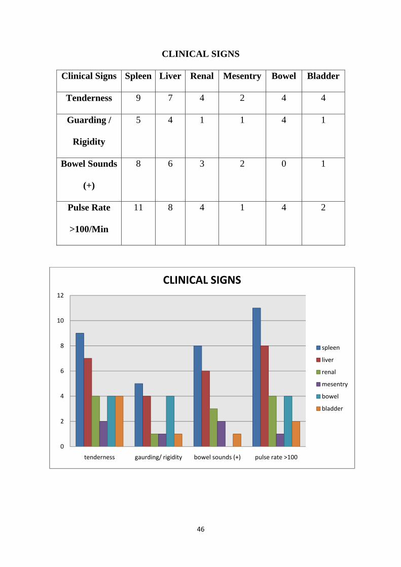
CLINICAL SIGNS
Clinical Signs Spleen Liver Renal Mesentry Bowel Bladder
Tenderness 9 7 4 2 4 4
Guarding /
Rigidity
5 4 1 1 4 1
Bowel Sounds
(+)
8 6 3 2 0 1
Pulse Rate
>100/Min
11 8 4 1 4 2
0
2
4
6
8
10
12
tenderness gaurding/ rigidity bowel sounds (+) pulse rate >100
CLINICAL SIGNS
spleen
liver
renal
mesentry
bowel
bladder
47
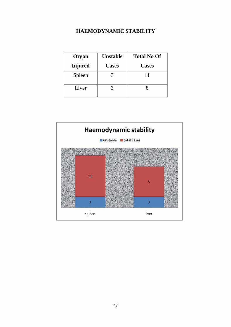
HAEMODYNAMIC STABILITY
Organ
Injured
Unstable
Cases
Total No Of
Cases
Spleen 3 11
Liver 3 8
3 3
118
spleen liver
Haemodynamic stabilityunstable total cases
48
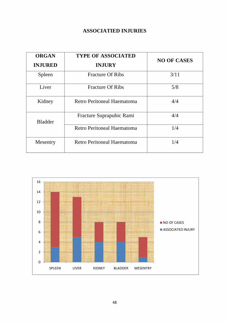
ASSOCIATIED INJURIES
ORGAN
INJURED
TYPE OF ASSOCIATED
INJURY NO OF CASES
Spleen Fracture Of Ribs 3/11
Liver Fracture Of Ribs 5/8
Kidney Retro Peritoneal Haematoma 4/4
Bladder Fracture Suprapubic Rami 4/4
Retro Peritoneal Haematoma 1/4
Mesentry Retro Peritoneal Haematoma 1/4
0
2
4
6
8
10
12
14
16
SPLEEN LIVER KIDNEY BLADDER MESENTRY
NO OF CASES
ASSOCIATED INJURY
N
T
MO
No Of Dea
Total No O
Cases
6
94%
M
49
ORTALIT
ath
Of
6%
ORTALI
TY
2
34
TY
No of deat
No of case
ths
es
50
DIAGNOSTIC PARACENTESIS
ORGAN FREE FLUID (+) FREE FLUID (-)
Spleen 11 0
Liver 8 0
Kidney 2 2
Bladder 3 1
Bowel 4 0
Mesentry 1 3
0
2
4
6
8
10
12
spleen liver kidney bladder bowel mesentry
Free Fluid (‐)
Free fluid (+)
51
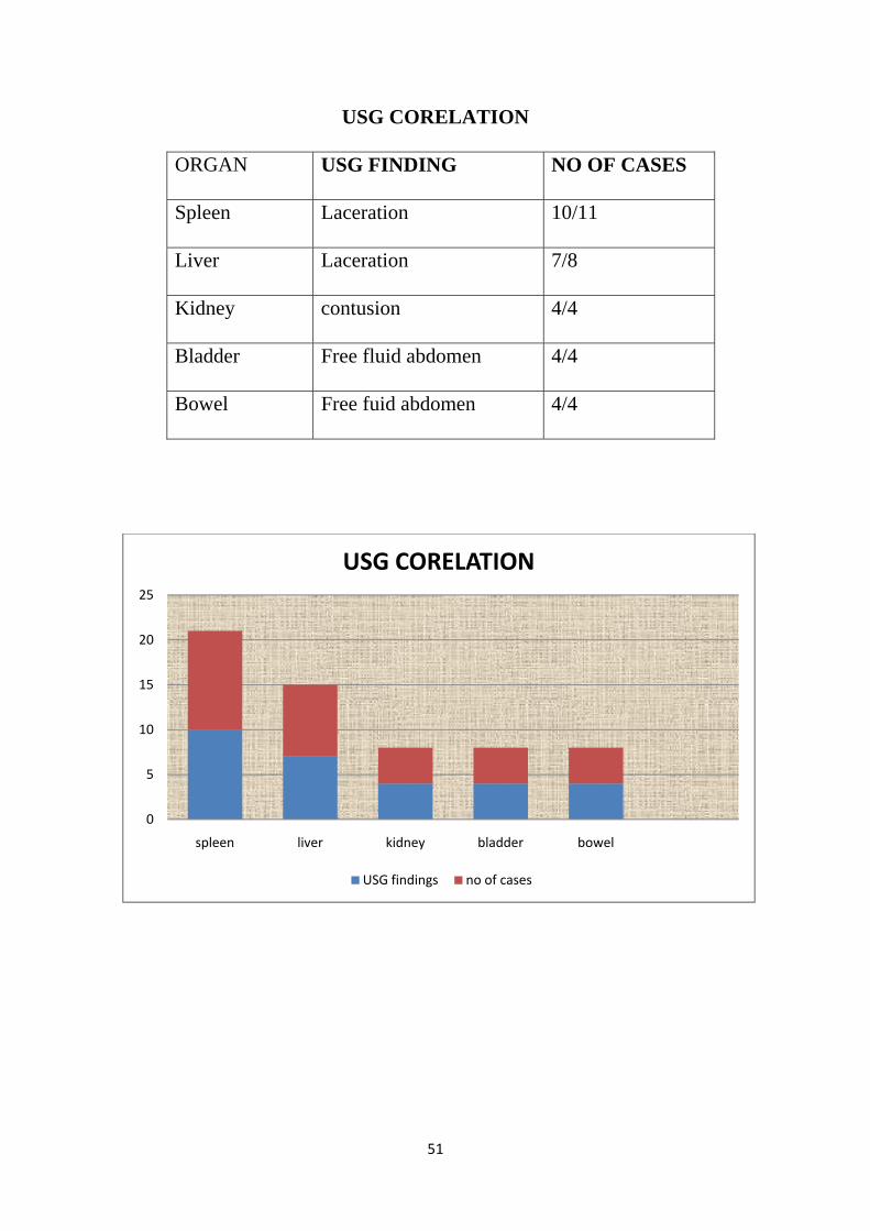
USG CORELATION
ORGAN USG FINDING NO OF CASES
Spleen Laceration 10/11
Liver Laceration 7/8
Kidney contusion 4/4
Bladder Free fluid abdomen 4/4
Bowel Free fuid abdomen 4/4
0
5
10
15
20
25
spleen liver kidney bladder bowel
USG CORELATION
USG findings no of cases
52
DISCUSSION
SPLENIC INJURY:
Spleen is the commonest organ injured following blunt abdominal
trauma. In our study 11 cases (32.35%) of cases presented with injury to
spleen.The associated injuries included fracture of Left lower ribs in 3 cases
.27.27% .
Of the patients presented 3 patients were hemodynamically unstable
with B.P systolic below 90 mm of Hg and pulse rate greater than 120\mt .They
were resuscitated appropriately and taken up for laparotomy.
The commonest finding in all the patients were tenderness in the left
Hypochondrium which was present in nine patients . Most of the patients had
contusions or abrasions over the left hypochondrium.
Kehrs sign was present in 4 patients , ballance sign was present in
none.Bowel sounds were present in 8 cases .
Abdominal paracentesis was done in all cases and was positive in all
cases. X-ray chest and abdomen was taken in all hemodynamically stable
patients . It showed fracture ribs 6-9 in two patients and # 9th rib in one patient..
The diagnosis of splenic injury was confirmed by clinical examination,
the presence of haemoperitoneum which was confirmed by abdominal
paracentesis and ultrasonogram.
At laparotomy all the cases which were operated upon had Grade III
injuries in 7 cases Grade IV in 2 cases and Grade V in 1 case .Splenectomy was

53
done in all cases, appropriate surgical procedures (ICD insertion) was done for
cases with hemothorax.
One patient expired pre operatively .He was hemodynamically unstable at
the time of admission and his condition was very poor and did not respond to
resuscitation.
54
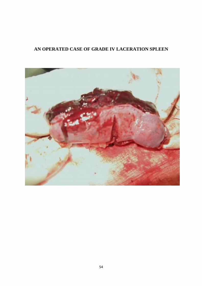
AN OPERATED CASE OF GRADE IV LACERATION SPLEEN
55
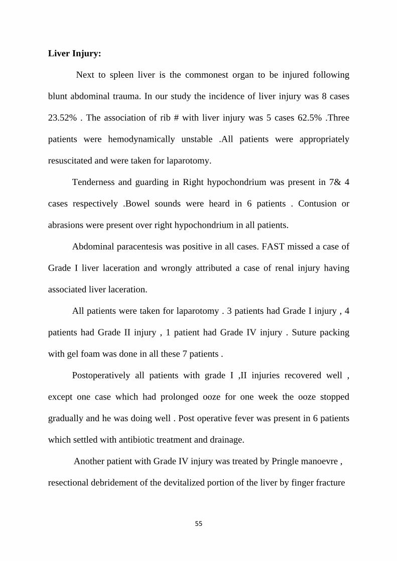
Liver Injury:
Next to spleen liver is the commonest organ to be injured following
blunt abdominal trauma. In our study the incidence of liver injury was 8 cases
23.52% . The association of rib # with liver injury was 5 cases 62.5% .Three
patients were hemodynamically unstable .All patients were appropriately
resuscitated and were taken for laparotomy.
Tenderness and guarding in Right hypochondrium was present in 7& 4
cases respectively .Bowel sounds were heard in 6 patients . Contusion or
abrasions were present over right hypochondrium in all patients.
Abdominal paracentesis was positive in all cases. FAST missed a case of
Grade I liver laceration and wrongly attributed a case of renal injury having
associated liver laceration.
All patients were taken for laparotomy . 3 patients had Grade I injury , 4
patients had Grade II injury , 1 patient had Grade IV injury . Suture packing
with gel foam was done in all these 7 patients .
Postoperatively all patients with grade I ,II injuries recovered well ,
except one case which had prolonged ooze for one week the ooze stopped
gradually and he was doing well . Post operative fever was present in 6 patients
which settled with antibiotic treatment and drainage.
Another patient with Grade IV injury was treated by Pringle manoevre ,
resectional debridement of the devitalized portion of the liver by finger fracture
56
technique. Omental pack was placed over the defect in the liver and the
peritoneal cavity was drained. Patient expired Postoperatively due to severity of
injuries.
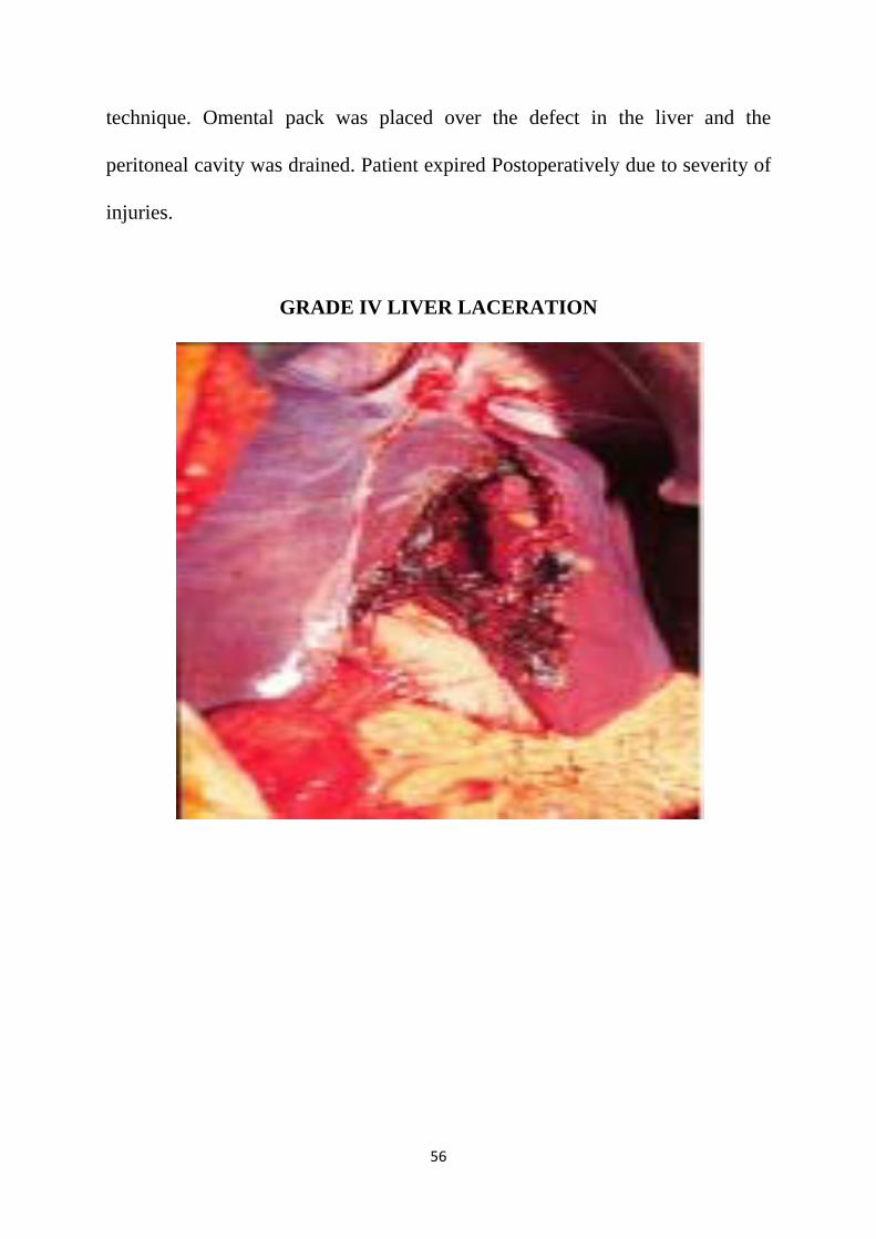
GRADE IV LIVER LACERATION
57
RENAL INJURIES:
The total no of renal injuries was 4 , right kidney was injured in three
cases and the left kidney in one case , All four cases were associated with RPH .
All patients presented with tenderness in flanks.Guarding \ Rigidity was
present in one case . Hematuria was present in all cases. Paracentesis was
positive in 2 cases. FAST revelaed Contusion of kidney in all cases & presence
of free fluid in morrisons pouch in three cases. Hence to rule out any expanding
hematoma and associated injuries patients were taken up for laparotomy
At laparotomy all renal injuries were of minor nature being contusion
involving the renal cortex . There was non expanding RPH in zone 2 right side
for 3 cases and non expanding RPH zone 2 left for one case . Since it was not
expanding Retroperitoneum was not opened . There was minor breach of
peritoneum with minimal free fluid in the pelvis in all cases.
There was no mortality in our study of four cases and in all cases
hematuria settled after 2 weeks and renal parameters were normal.
Bladder Injuries:
The total no of bladder injury in our study was 4 . All cases were
intraperitoneal rupture .These injuries were associated with fracture of superior
and inferior pubic rami. Once case was associated to have a Zone 3
Retroperitoneal hematoma .
Clinically patient presented with diffuse abdominal guarding and
tenderness in supra pubic region .Hematuria was present in three cases .
58
Diagnostic paracentesis was positive for turbid fluid in three cases. USG
abdomen revealed presence of free fluid in all cases and presence of clots in the
urinary bladder.
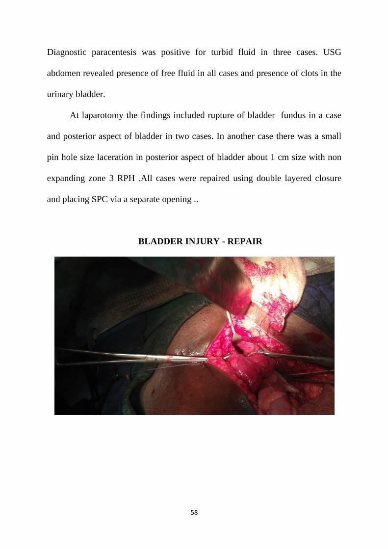
At laparotomy the findings included rupture of bladder fundus in a case
and posterior aspect of bladder in two cases. In another case there was a small
pin hole size laceration in posterior aspect of bladder about 1 cm size with non
expanding zone 3 RPH .All cases were repaired using double layered closure
and placing SPC via a separate opening ..
BLADDER INJURY - REPAIR
59
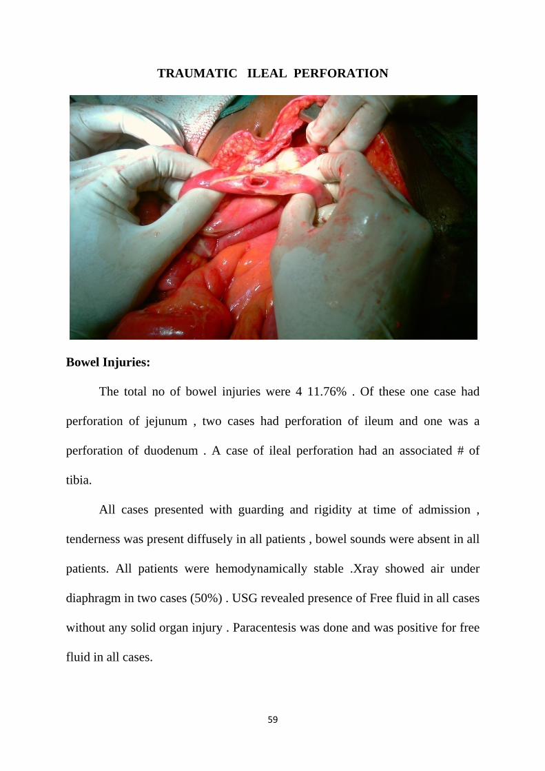
TRAUMATIC ILEAL PERFORATION
Bowel Injuries:
The total no of bowel injuries were 4 11.76% . Of these one case had
perforation of jejunum , two cases had perforation of ileum and one was a
perforation of duodenum . A case of ileal perforation had an associated # of
tibia.
All cases presented with guarding and rigidity at time of admission ,
tenderness was present diffusely in all patients , bowel sounds were absent in all
patients. All patients were hemodynamically stable .Xray showed air under
diaphragm in two cases (50%) . USG revealed presence of Free fluid in all cases
without any solid organ injury . Paracentesis was done and was positive for free
fluid in all cases.
60
At laparotomy the perforation of duodenum was closed with a live
omental patch , the jejunal perforation was closed in two layers with inner vicryl
and outer silk . The ileal perforation margins were friable so resection
anastamosis was done .
Mesenteric Injury:
The mesenteric injuries were 4 cases in total . Mesenteric contusion in
jejunum was present in three cases and ileum was present in one case . The
mesenteric contusion in ileum was associated with Retroperitoneal hematoma in
zone 3 which was non expanding.
Clinically two patients presented with tenderness over umbilical and iliac
regions.Guarding was present in one case . Bowel sounds were present in two
cases . Diagnostic paracentesis was positive in one case for presence of blood .
USG revealed presence of minimal free fluid in pelvis in all cases .
At laparotomy the contusion of mesentery in jejunum was not associated
with any bowel pathology and the the bleeding stopped spontaneously . Hence
after securing perfect hemostasis closure with a tube drain was done . The
contusion in ileum was associated with segment of gangrene of ileum hence
resection anastamosis of ileum was done . The associated Retroperitoneal
hematoma in zone 3 was non expanding and hence was left as such .All patients
recovered well without complications .
61
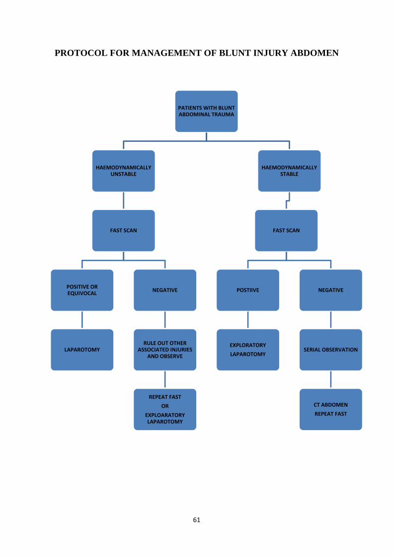
PROTOCOL FOR MANAGEMENT OF BLUNT INJURY ABDOMEN
PATIENTS WITH BLUNT ABDOMINAL TRAUMA
HAEMODYNAMICALLY UNSTABLE
FAST SCAN
POSITIVE OR EQUIVOCAL
LAPAROTOMY
NEGATIVE
RULE OUT OTHER ASSOCIATED INJURIES
AND OBSERVE
REPEAT FAST OR
EXPLOARATORY LAPAROTOMY
HAEMODYNAMICALLY STABLE
FAST SCAN
POSTIIVE
EXPLORATORY LAPAROTOMY
NEGATIVE
SERIAL OBSERVATION
CT ABDOMENREPEAT FAST
62
CONCLUSION
1) The most commonly injured organ is spleen in blunt abdominal trauma
which is similar to other studies
2) RTA accounted for majority of cases of blunt injury abdomen which is
around 64.70%
3) Similar to many large series males are more often affected in blunt
abdominal injuries than females and middle aged persons are more often
affected than extremes of age
4) Commonest associated injuries occurred in our study was chest injury in
eight cases
5) FAST is rapid cheap noninvasive procedure used for screening in the
emergency ward itself while the patient is resuscitated.
6) Biochemical investigations are not of much help. The investigations only
complimentary to clinical diagnosis.
7) In the unstable trauma patient, a positive FAST eliminates the need for
further tests and indicates the necessity for abdominal exploration the
emergency ward itself while the patient is resuscitated.
8) Diagnostic paracentesis is a rapid , bedside tool for diagnosis
immediately at the bedside arrival of the patient.
9) Thorough initial clinical evaluation, repeated clinical examinations
monitoring vital signs are essential in minimizing the chance of missing
life threatening intra abdominal injuries.
10) The mortality in this study is related to severity of injuries . Severe the
grading of injury more is the mortality.
BIBLIOGRAPHY
1. Abdomen Injuries D.B.Hoyt and A.R.Moosa, Essential Surgical Practice
Cuschieri, 5th edition
2. Blumgart L H, Surgery of the liver and and biliary tract resection forliver
vol. II: ch, 85; 1222 – 1238..
3. Das S. Clinical methods in surgery, examination of abdominal injuries
4. Herman Hospital study on Blunt injury abdomen. Maingots abdominal
operations
5. Maingot’s Abdominal operations 10th edition – Blunt abdominal trauma
6. Sabistan D C Text book of surgery.
7. Campbell’s Text book of urology. 6th edition. Genitourinary trauma
8. American College of Surgeons Committee on Trauma. Abdominal Trauma.
In: ATLS Student Course Manual. 8th. American College of Surgeons;
2008.
9. Jansen JO, Yule SR, Loudon MA. Investigation of blunt abdominal trauma.
BMJ. Apr 26 2008
10. Ong CL, Png DJ, Chan ST. Abdominal trauma--a review. Singapore Med
J. Jun
11. Enderson BL, Reath DB, Meadors J, Dallas W, DeBoo JM, Maull KI. The
tertiary trauma survey: a prospective study of missed injury. J Trauma. Jun
1990;30(6):666-9; discussion 669-70.
12. Janjua KJ, Sugrue M, Deane SA. Prospective evaluation of early missed
injuries and the role of tertiary trauma survey. J Trauma. Jun
1998;44(6):1000-6; discussion 1006-7.
13. Schnüriger B, Inaba K, Barmparas G, Eberle BM, Lustenberger T, Lam L,
et al. Serial white blood cell counts in trauma: do they predict a hollow
viscus injury?. J Trauma. Aug 2010;69(2):302-7.
14. Denver Hospital study on blunt injury abdomen, Surgicial clincs of north
america
15. Ritchie AH, Williscroft DM. Elevated liver enzymes as a predictor of liver
injury in stable blunt abdominal trauma patients: case report and systematic
review of the literature. Can J Rural Med. Fall 2006;11(4):283-7.
16. Knudson MM, McAninch JW, Gomez R, Lee P, Stubbs HA. Hematuria as
a predictor of abdominal injury after blunt trauma. Am J Surg. Nov
1992;164(5):482-5; discussion 485-6.
17. Tso P, Rodriguez A, Cooper C, Militello P, Mirvis S, Badellino MM, et al.
Sonography in blunt abdominal trauma: a preliminary progress report. J
Trauma. Jul 1992;33(1):39-43; discussion 43-4.
18. Kawaguchi S, Toyonaga J, Ikeda K. Five point method: An
ultrasonographic quantification formula of intra-abdominal fluid collection.
Jpn J Acute Med. 1987;7:993-
19. Tiling T, Boulion B, Schmid A, et al. Ultrasound in blunt
abdominothoracic trauma. In: Border JR,
20. Blaivas M, Brannam L, Hawkins M, Lyon M, Sriram K. Bedside
emergency ultrasonographic diagnosis of diaphragmatic rupture in blunt
abdominal trauma. Am J Emerg Med. Nov 2004;22(7):601-4.
21. Branney SW, Moore EE, Cantrill SV, Burch JM, Terry SJ. Ultrasound
based key clinical pathway reduces the use of hospital resources for the
evaluation of blunt abdominal trauma. J Trauma. Jun 1997;42(6):1086-90.
22. Kornezos I, Chatziioannou A, Kokkonouzis I, Nebotakis P, Moschouris H,
Yiarmenitis S, et al. Findings and limitations of focused ultrasound as a
possible screening test in stable adult patients with blunt abdominal
trauma: a Greek study. Eur Radiol. Jan 2010;20(1):234-8.
23. Kendall JL, Faragher J, Hewitt GJ, Burcham G, Haukoos JS. Emergency
Department Ultrasound Is not a Sensitive Detector of Solid Organ Injury.
West J Emerg Med. Feb 2009;10(1):1-5..
24. Sonography for trauma (FAST). J Trauma. Apr 1997;42(4):617-23
25. Neuhof H, Cohen I. ABDOMINAL PUNCTURE IN THE DIAGNOSIS
OF ACUTE INTRAPERITONEAL DISEASE. Ann Surg. Apr
1926;83(4):454-62..
26. ROOT HD, HAUSER CW, MCKINLEY CR, LAFAVE JW, MENDIOLA
RP Jr. DIAGNOSTIC PERITONEAL LAVAGE. Surgery. May
1965;57:633-7.
27. Liu M, Lee CH, P'eng FK. Prospective comparison of diagnostic peritoneal
lavage, computed tomographic scanning, and ultrasonography for the
diagnosis of blunt abdominal trauma. J Trauma. Aug 1993;35(2):267-70.
28. Cryer HG, Larmon B. Patient care phase: prehospital and resuscitation
care. In: Greenfield LJ, Mulholland MW, Oldham KT, Zelenock GB,
Lillemoe KD, eds. Surgery: Scientific Principles and Practice.
Philadelphia: Lippincott-Raven;. 1997:280-4.
29. Pryor JP, Pryor RJ, Stafford PW. Initial phase of trauma management and
fluid resuscitation. Trauma Reports. 2002;3(3):1-12.
30. Nirula R, Maier R, Moore E, Sperry J, Gentilello L. Scoop and run to the
trauma center or stay and play at the local hospital: hospital transfer's effect
on mortality. J Trauma. Sep 2010;69(3):595-9; discussion 599-601.
31. Perel P, Roberts I. Colloids versus crystalloids for fluid resuscitation in
critically ill patients. Cochrane Database Syst Rev. Oct 17
2007;CD000567.
32. M, Ratliff J, Osler T. "Never be wrong": the morbidity of negative and
delayed laparotomies after blunt trauma. J Trauma. Dec 2010;69(6):1386-
91; discussion 1391-2.
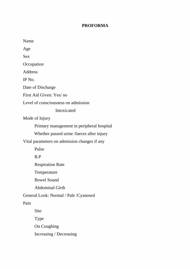
PROFORMA
Name
Age
Sex
Occupation
Address
IP No.
Date of Discharge
First Aid Given: Yes/ no
Level of consciousness on admission
Intoxicated
Mode of Injury
Primary management in peripheral hospital
Whether passed urine /faeces after injury
Vital parameters on admission changes if any
Pulse
B.P
Respiration Rate
Temperature
Bowel Sound
Abdominal Girth
General Look: Normal / Pale /Cyanosed
Pain
Site
Type
On Coughing
Increasing / Decreasing

Shoulder Tip Pain: +/ -
Vomiting: +/-
Gaurding: +/-
Rigidity: +/-
Tenderness:
Site
Rebound
Type of abdominal injury
Abrasion / Laceration / ecchymosis / Bruises
Others
Other system injury
Associated bony injury
Lower ribs
Pelvis
Spine
Long bones
Perineal heamatomas
PR
NG Aspirations
Bladder catheterization
Details of blood transfusion
DPA: +/-
Blood
TC
Hb
Grouping & Typing
Plain X ray Abdomen
Chest
FAST
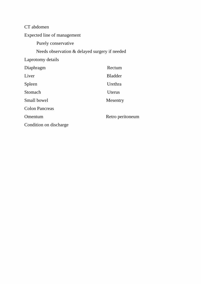
CT abdomen
Expected line of management
Purely conservative
Needs observation & delayed surgery if needed
Laprotomy details
Diaphragm Rectum
Liver Bladder
Spleen Urethra
Stomach Uterus
Small bowel Mesentry
Colon Pancreas
Omentum Retro peritoneum
Condition on discharge
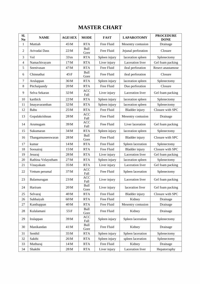
MASTER CHART Sl. No NAME AGE\SEX MODE FAST LAPAROTOMY PROCEDURE
DONE 1 Muttiah 45\M RTA Free Fluid Mesentry contusion Drainage
2 Arivudai Dass 22\M Bull Gore Free Fluid Jejunal perforation Closure
3 Vel 33\m RTA Spleen injury laceration spleen Splenectomy 4 Namachivayam 17\M RTA Liver injury Laceration liver Gel foam packing 5 Seenivasan 47\M RTA Free Fluid ileal perforation Resect anastamose
6 Chinnathai 45\F Bull Gore Free Fluid ileal perforation Closure
7 Arulappan 36\M RTA Spleen injury laceration spleen Splenectomy 8 Pitchaipandy 20\M RTA Free Fluid Duo perforation Closure
9 Selva Sekaran 32\M ACC Fall Liver injury Laceration liver Gel foam packing
10 karthick 22\M RTA Spleen injury laceration spleen Splenectomy 11 Imayavaramban 32\M RTA Spleen injury laceration spleen Splenectomy 12 Babu 25\M RTA Free Fluid Bladder injury Closure with SPC
13 Gopalakrishnan 28\M ACC Fall Free Fluid Mesentry contusion Drainage
14 Arumugam 39\M ACC Fall Free Fluid Liver laceration Gel foam packing
15 Sukumaran 34\M RTA Spleen injury laceration spleen Splenectomy
16 Thangamuneeswaran 28\M Bull Gore Free Fluid Bladder injury Closure with SPC
17 kumar 14\M RTA Free Fluid Spleen laceration Splenectomy 18 Soosairaj 15\M RTA Free Fluid Bladder injury Closure with SPC 19 Jesuraj 28\M RTA Liver injury Laceration liver Gel foam packing 20 Rathina Velayutham 27\M RTA Spleen injury laceration spleen Splenectomy 21 Vinayakam 35\M RTA Liver injury Laceration liver Gel foam packing
22 Vettum perumal 37\M ACC Fall Free Fluid Spleen laceration Splenectomy
23 Balamurugan 23\M ACC Fall Liver injury Laceration liver Gel foam packing
24 Hariram 20\M Bull Gore Liver injury laceration liver Gel foam packing
25 Selvaraj 40\M RTA Free Fluid Bladder injury Closure with SPC 26 Subbaiyah 60\M RTA Free Fluid Kidney Drainage 27 Kanthappan 40\M RTA Free Fluid Mesentry contusion Drainage
28 Kulalamani 55\F Bull Gore Free Fluid Kidney Drainage
29 Irulappan 39\M ACC Fall Spleen injury Spleen laceration Splenectomy
30 Manikandan 41\M Bull Gore Free Fluid Kidney Drainage
31 Senthil 35\M RTA Spleen injury Spleen laceration Splenectomy 32 Sakthi 26\M RTA Spleen injury spleen laceration Splenectomy 33 Muthuraj 14\M RTA Free Fluid Kidney Drainage 34 Shakthi 28\M RTA Liver injury Laceration liver Hepatorraphy