Embed Size (px)
DESCRIPTION
Quantification of cultured keloid fibroblasts after irradiation with 470nm blue LED, in vitro
Citation preview
Acta Cirúrgica Brasileira - Vol. 26 (1) 2011 - 25
6 - ORIGINAL ARTICLEWound Healing
In vitro effect of 470 nm LED (Light Emitting Diode) in keloid fibroblasts1
Efeito in vitro do LED (Light Emitting Diode) de 470 nm em fibroblastos de quelóide
Silvilena BonattiI, Bernardo HochmanII, Vanina Monique Tucci-ViegasIII, Fabianne FurtadoIII, Carlos Eduardo PinfildiIV,Ana Carolina PedroV, Lydia Masako FerreiraVI
1 Research performed at the Plastic Surgery Division, Department of Surgery, Federal University of Sao Paulo (UNIFESP), Brazil.
I PT, Fellow Improvement Program, Plastic Surgery Division, Department of Surgery, UNIFESP, Sao Paulo-SP, Brazil. Acquisition of data, helpedwith technical procedures, collection and processing of study informations, manuscript writing.II Affiliate Professor, Plastic Surgery Division, Department of Surgery, UNIFESP, Sao Paulo-SP, Brazil. Mentor, conception and design of the study,manuscript writing and critical revision.III Fellow PhD degree, Plastic Surgery Division, Department of Surgery, UNIFESP, Sao Paulo-SP, Brazil. Helped with technical procedures,collection and processing of study informations.IV Associate Professor, Plastic Surgery Division, Department of Surgery, UNIFESP, Sao Paulo-SP, Brazil. Collection of study informations.V Graduate student, UNIFESP, Sao Paulo-SP, Brazil. Processing of study informations, involved with technical procedures.VI Full Professor, Chairwoman Plastic Surgery Division, Head Department of Surgery, UNIFESP, Sao Paulo-SP, Brazil. Scientific and intellectualcontent of the study, acquisition and interpretation of data, critical revision.
ABSTRACTPurpose: To quantify keloid fibroblasts after irradiation with 470nm blue LED, in vitro. Methods: Fibroblasts from keloid and adjacentskin have been obtained from 6 patients. Cells have been cultivated and maintained in DMEM culture medium. In Petri dishes, theywere irradiated with energy doses of 6J, 12J and 18J. After 24 h, counting was done by the average of the triplicates for eachsample. Results: There were no significant differences in the number of irradiated keloid fibroblasts at the studied doses (p=0.261).In adjacent skin fibroblasts, differences were observed (p=0.025) concerning the doses of 18 J and 6 J (p=0.03). Conclusions: Therewas a reduction in the number of adjacent skin fibroblasts irradiated with 470nm blue LED at the energy dose of 18 J compared tothe ones irradiated at the energy dose of 6 J. There were no changes in keloid fibroblasts counting at any of the doses applied, 24 hafter irradiation.Key words: Fibroblasts. Keloid. Phototherapy. Lasers. Skin. Laser Therapy, Low-Level.
RESUMOObjetivo: Quantificar fibroblastos de quelóide após irradiação com LED azul de 470nm, in vitro. Métodos: Foram obtidos fibroblastosde quelóide e pele adjacente, de seis pacientes. As células foram cultivadas e mantidas em meio de cultura DMEM. Em placas de Petri,receberam irradiação com doses de energia de 6J, 12J e 18J. Após 24 horas a contagem foi feita pela média da triplicata para cadaamostra. Resultados: Não houve diferença na quantidade de fibroblastos de quelóide irradiados nas doses estudadas (p=0,261).Observou-se diferença nos fibroblastos de pele adjacente (p=0,025), com relação às doses de 18 J e 6 J (p=0,03). Conclusões: Houveredução dos fibroblastos de pele adjacente irradiados com LED azul de 470 nm na dose de energia de 18 J em relação à dose de 6 J. Nãohouve alteração na quantidade de fibroblastos de quelóide nas doses aplicadas após 24 horas da irradiação.Descritores: Fibroblastos. Quelóide. Fototerapia. Lasers. Pele. Terapia a Laser de Baixa Intensidade.
Introduction
Keloid is a wound repair disorder resulting fromexcessive collagen deposition, which occurs in susceptibleindividuals, and its pathophysiology has not yet been fullyelucidated1-3. The literature presents many options of treatment,alone or in combination, nevertheless they are not specific.Among the therapeutic modalities that have been most used,stand out the administration of topical and intralesional
corticosteroids, the use of silicon plates, pressotherapy andexeresis followed by betatherapy4,5.
Recently, high potency LASER (Light Amplificationby Stimulated Emission of Radiation) and IPL (Intense PulsedLight) were added to that arsenal. However, these resourcesof phototherapy have high cost and relative success, besidesthe fact that the applications are painful and involve risks ofburns and hypochromia6,7.
Bonatti S et al
26 - Acta Cirúrgica Brasileira - Vol. 26 (1) 2011
Low-level LASER therapy (LLLT) is a modality oftreatment that is being used for the improvement of impairedwound healing due to its biomodulatory character, with majoraction in mitochondrial metabolism8. In this sense, nowadays,LED (Light Emitting Diode) appears as an alternative to LLLTand differentiates itself by not showing collimated and coherentbeam. However, both lasertherapy and LED may have similarbiological effects due to absorption of photons by tissuechromophores. Such resources are already being used in manyskin diseases like acne9, in which blue light exerts anti-bacterialeffect stimulating bacterial membrane chromophores fromPropionum acnes and releasing reactive oxygen species.Phototherapy is also used in tissue repair treatments, acting onfibroblasts proliferation10, collagen synthesis11 andneovascularization12.
In vitro studies with red13 and infrared LASER14,15
showed that the highest doses investigated inhibited fibroblastproliferation. In hypertrophic scars and skin fibroblasts, in vitro,it was demonstrated that low intensity LASER with wavelength680 nm (red) and energy (E) 0.5 J (31 s) and 0.8 J (52 s) had astimulatory effect on both cell counts, with a more pronouncedresult obtained with the lowest dose used13. On the otherhand, 880 nm infrared LASER at doses of 0.71 J (44.5 s) and 1.18 J(74 s) resulted in reduction of cell proliferation in both skin andhypertrophic scar fibroblasts, noting that the second dose led to agreater inhibition14.
Experimental studies with LED, in wavelengthscorresponding to green and blue, reported effects in fibroblasts.In chicken embryos, it was observed that irradiation with greenspectrum (532 nm) at a dose of 0.90 J, for 1 s caused an increasein the number of fibroblasts16, compared with the group of cellsthat was not irradiated. In blue spectrum of 470 nm, irradiationwith LED for 180 s inhibited proliferation of gingival fibroblasts17.However, no studies were found using LED in blue spectrum,in keloid fibroblasts, which could be valuable as a prevention ortreatment of this disorder. Therefore, the objective of this studywas to quantify keloid fibroblasts after irradiation with 470 nmblue LED, in vitro.
Methods
This study was approved by the Research EthicsCommittee of the Federal University of Sao Paulo and thediscarded parts were used after all patients signed a free andinformed consent form.
Collection of keloid samples
Keloid samples used were obtained from six femalepatients aged 18-50 years and with Fitzpatrick skin types II to VI.The scars had at least 1-year evolution, were in clinical activity,presenting hyperemia, pruritus and/or pain, and also centralinvolution (i.e. a lower relief in the central compared with theperipheral area) and with minimum dimensions 3 cm at thelongitudinal axe and 2 cm at the transversal axe. Keloid locationwas delimited in the thoracic region, between a transverse planeat the level of the acromion and a transverse plane at the level
of xiphoid process3. Were excluded keloids previously treatedor patients with chronic dermatopathies, metabolic, collagen ordegenerative / auto-immune diseases, malignant neoplasms, orpatients submitted to systemic or topic treatment withcorticosteroids.
Keloids were excised in the subcutaneous plane, underlocal anesthesia, with exceeding skin fragments represented bythe “dog ears”. After exeresis of the keloid lesions, samples wereobtained from adjacent skin and peripheral keloid (between theexcisional margin and the central umbilication) of the surgicalspecimens using a circular punch of 3 mm diameter, from theepithelialized surface of tissue containing epithelium andconnective tissue18.
Obtainment of the fibroblast cultures
Primary keloid fibroblast culture was done by explant19,20
by which the fractions corresponding to samples of adjacentskin and keloid were obtained. Fractions were placed in 15 mlconic tubes and exhaustively rinsed (six times) with 10 mlPBS (Phosphate-Buffered Saline, Cultilab, Brazil) containingpenicillin (100Ul/ml, Gibco®) and streptomycin (100µ/ml, Gibco®)under vigorous agitation, changing tubes and PBS at eachrepetition. Then, fractions were transferred to 60 mm² diameterPetri dishes, in grid areas scratched with a scalpel, and plates wereleft semi-opened in the laminar flow for 30 min, for the fragmentsto adhere to its surface. Then, 6 ml of DMEM (Dulbecco’s ModifiedEagle’s Medium, Cultilab, Brazil) supplemented with 15% FBS(Fetal Bovine Serum, Cultilab, Brazil), penicillin (100 UI/ml,Gibco®) and streptomycin (100 µg/ml, Gibco®) were carefullyadded to each plate. Plates were kept in humidified incubator at37ºC, 95% O2 and 5% CO2.
Culture medium was changed every two days and a fewdays after establishing the primary culture, spindle-like cells wereseen proliferating from the edges of the explanted tissue, regardedas culturing fibroblasts. Fibroblast satisfactory proliferation wasobserved in approximately 7-14 days and subculturing (passage)was performed when cellular confluence reached approximately80% at the Petri dish.
For subculturing, the culture medium was aspiratedand the keloid fragments, discarded with tweezers. The platecontaining fibroblasts was washed with PBS, and then quicklyrinsed with Versene™ (PBS with 0.05M EDTA, Ethylene DiamineTetra Acetic, Sigma®). Versene™ was aspirated and 1 ml of trypsin0.25% with EDTA 0.02% was added to the plate. The plate waskept for 2 min in the incubator and taken to the microscope toconfirm fibroblast detachment from the dish surface. Trypsinwas neutralized with 3.0 ml DMEM 10% FBS and the cellularsuspension was centrifuged (100 g, 6 min). The pellet wasresuspended in DMEM 10% FBS and antibiotics (penicillin100 UI/ml and streptomycin 100 µg/ml) and 100,000 cells wereseeded in each 75 cm2 culture flask. For the experiments, cellswere seeded in 30 mm2 diameter (10 mm height) Petri dishes, intriplicates. In the first sample, 2x10⁵ cells were seeded per plateand, for better confluence, in all other samples, 5x10⁴ cells wereseeded per plate, always in triplicates. Before irradiation, cells werekept for 48 h in the incubator, at 37ºC.
Acta Cirúrgica Brasileira - Vol. 26 (1) 2011 - 27
In vitro effect of 470 nm LED (Light Emitting Diode) in keloid fibroblasts
Cell irradiation
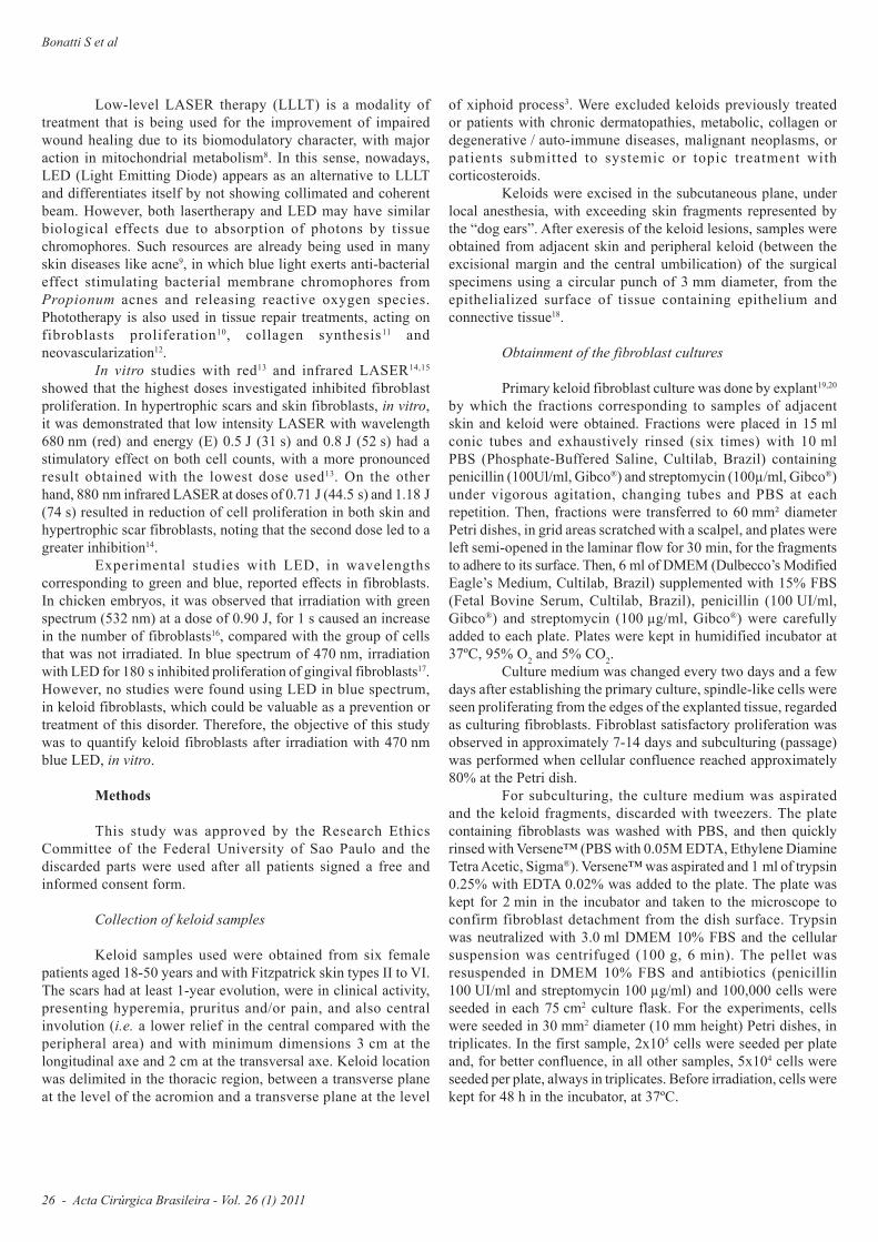
The equipment Quasar Esthetique® was used for cellirradiation. The device has a handpiece with LEDs, covering theblue spectrum (470 nm) with 100 mW power, 125 mW/cm2 powerdensity, optical fiber diameter 1 cm and beam area of 0.8 cm2.
The samples of keloid and adjacent skin fibroblastswere divided into groups according to the total energy supplied:Control Group, received no irradiation; Group 1 min, received 6 Jtotal energy (E) and energy density (ED) 59.87 J/cm2, Group 2 minreceived 12 J (E) and (ED) 122.3 J/cm2, Group 3 min, received18 J (E), and (ED) 183.43 J/cm2 (Table 1).
Group Control 1 min 2 min 3 min
ED (J/cm2) 0 59.87 122.30 183.43
E (J) 0 6 J 12 J 18 J
t (s) 0 60 120 180
TABLE 1 – Irradiation parameters
ED: energy density, E: energy; t: time
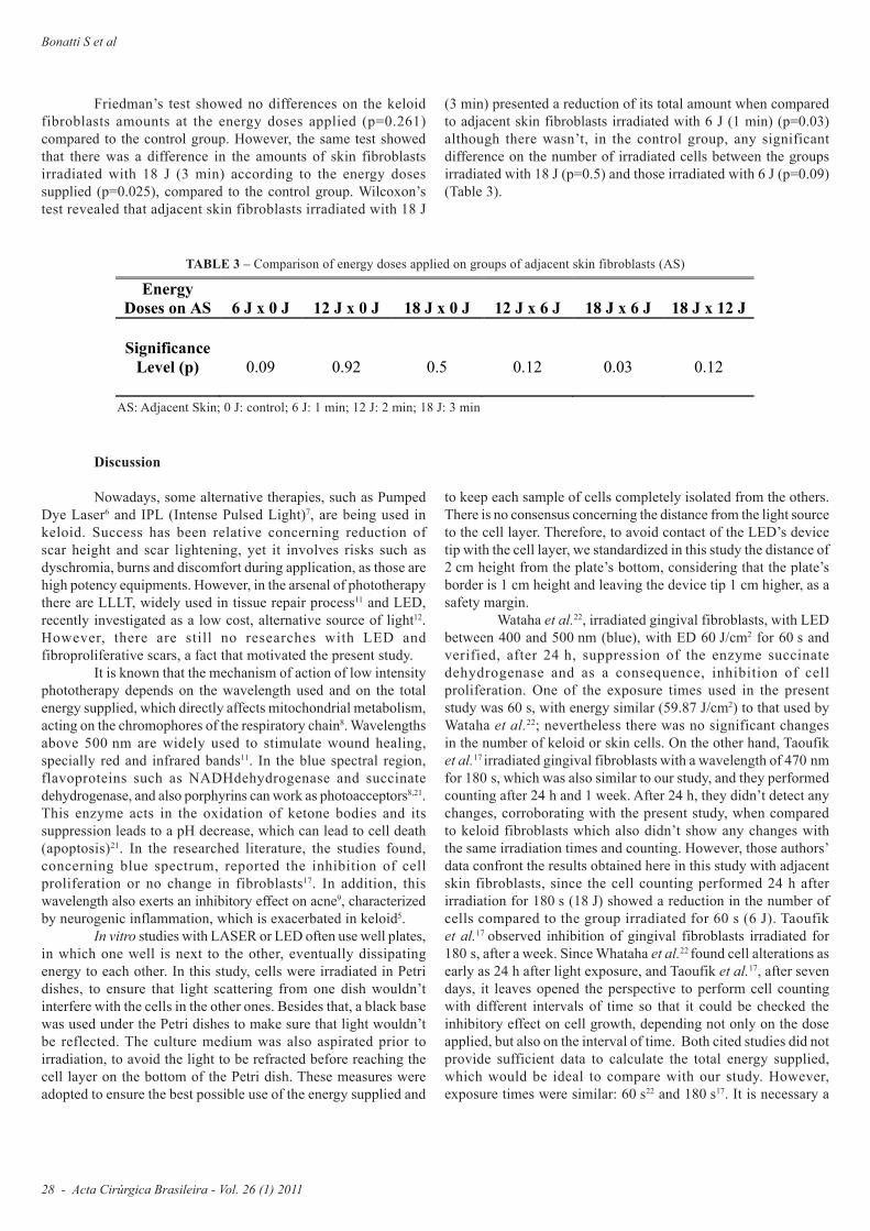
The device tip was fixed at a distance of 2 cm in heightfrom the plate’s bottom. The culture medium was aspiratedfor 3 min in all groups, for homogenization of the samples.Irradiation was performed with the LED’s collimatorperpendicular to the Petri dish, which was placed on top of ablack base (Figure 1).
FIGURE 1 – Cell irradiation with 470 nm blue LED.
After irradiation, cells were put back in incubator foranother 24 h. After this period, cell cultures were preparedfor counting in Neubauer chamber. Each sample (adjacentskin and keloid fibroblasts) was counted three times, using amanual cell counter, and data were expressed as the average ofthe triplicates. Statistical significance was set at p<0.05.
Results
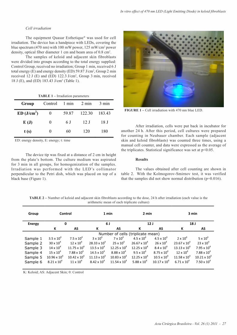
The values obtained after cell counting are shown intable 2. With the Kolmogorov-Smirnov test, it was verifiedthat the samples did not show normal distribution (p=0.016).
TABLE 2 – Number of keloid and adjacent skin fibroblasts according to the dose, 24 h after irradiation (each value is thearithmetic mean of each triplicate culture)
K: Keloid; AS: Adjacent Skin; 0: Control
Group Control 1 min 2 min 3 min
Energy 0 6 J 12 J 18 J
K AS K AS K AS K AS
Number of cells (triplicate mean)
Sample 1 3.5 x 10⁴
7.5 x 10⁴
3 x 10⁴
7 x 10⁴
4.5 x 10⁴
4.5 x 10⁴
2 x 10⁴
5 x 10⁴
Sample 2 30 x 104
12 x 104
28.33 x 10⁴
25 x 104
26.67 x 10⁴
26 x 104
23.67 x 10⁴
23 x 104
Sample 3 14 x 104
11.75 x 10⁴
13.5 x 10⁴
12.25 x 10⁴
12.25 x 10⁴
8.4 x 10⁴
13.13 x 10⁴
7.95 x 10⁴
Sample 4 15 x 104
7.88 x 10⁴
14.5 x 10⁴
8.88 x 10⁴
9.5 x 10⁴
8.75 x 10⁴
12 x 104
7.88 x 10⁴
Sample 5 10.96 x 10⁴
10.42 x 10⁴
11.13 x 10⁴
10.83 x 10⁴
12.25 x 10⁴
10.5 x 10⁴
11.58 x 10⁴
10.21 x 10⁴
Sample 6 8.21 x 10⁴
11 x 104
8.42 x 10⁴
11.54 x 10⁴
5.88 x 10⁴
10.17 x 10⁴
6.71 x 10⁴
7.50 x 10⁴
Bonatti S et al
28 - Acta Cirúrgica Brasileira - Vol. 26 (1) 2011
Discussion
Nowadays, some alternative therapies, such as PumpedDye Laser6 and IPL (Intense Pulsed Light)7, are being used inkeloid. Success has been relative concerning reduction ofscar height and scar lightening, yet it involves risks such asdyschromia, burns and discomfort during application, as those arehigh potency equipments. However, in the arsenal of phototherapythere are LLLT, widely used in tissue repair process11 and LED,recently investigated as a low cost, alternative source of light12.However, there are still no researches with LED andfibroproliferative scars, a fact that motivated the present study.
It is known that the mechanism of action of low intensityphototherapy depends on the wavelength used and on the totalenergy supplied, which directly affects mitochondrial metabolism,acting on the chromophores of the respiratory chain8. Wavelengthsabove 500 nm are widely used to stimulate wound healing,specially red and infrared bands11. In the blue spectral region,flavoproteins such as NADHdehydrogenase and succinatedehydrogenase, and also porphyrins can work as photoacceptors8,21.This enzyme acts in the oxidation of ketone bodies and itssuppression leads to a pH decrease, which can lead to cell death(apoptosis)21. In the researched literature, the studies found,concerning blue spectrum, reported the inhibition of cellproliferation or no change in fibroblasts17. In addition, thiswavelength also exerts an inhibitory effect on acne9, characterizedby neurogenic inflammation, which is exacerbated in keloid5.
In vitro studies with LASER or LED often use well plates,in which one well is next to the other, eventually dissipatingenergy to each other. In this study, cells were irradiated in Petridishes, to ensure that light scattering from one dish wouldn’tinterfere with the cells in the other ones. Besides that, a black basewas used under the Petri dishes to make sure that light wouldn’tbe reflected. The culture medium was also aspirated prior toirradiation, to avoid the light to be refracted before reaching thecell layer on the bottom of the Petri dish. These measures wereadopted to ensure the best possible use of the energy supplied and
to keep each sample of cells completely isolated from the others.There is no consensus concerning the distance from the light sourceto the cell layer. Therefore, to avoid contact of the LED’s devicetip with the cell layer, we standardized in this study the distance of2 cm height from the plate’s bottom, considering that the plate’sborder is 1 cm height and leaving the device tip 1 cm higher, as asafety margin.
Wataha et al.22, irradiated gingival fibroblasts, with LEDbetween 400 and 500 nm (blue), with ED 60 J/cm2 for 60 s andverified, after 24 h, suppression of the enzyme succinatedehydrogenase and as a consequence, inhibition of cellproliferation. One of the exposure times used in the presentstudy was 60 s, with energy similar (59.87 J/cm2) to that used byWataha et al.22; nevertheless there was no significant changesin the number of keloid or skin cells. On the other hand, Taoufiket al.17 irradiated gingival fibroblasts with a wavelength of 470 nmfor 180 s, which was also similar to our study, and they performedcounting after 24 h and 1 week. After 24 h, they didn’t detect anychanges, corroborating with the present study, when comparedto keloid fibroblasts which also didn’t show any changes withthe same irradiation times and counting. However, those authors’data confront the results obtained here in this study with adjacentskin fibroblasts, since the cell counting performed 24 h afterirradiation for 180 s (18 J) showed a reduction in the number ofcells compared to the group irradiated for 60 s (6 J). Taoufiket al.17 observed inhibition of gingival fibroblasts irradiated for180 s, after a week. Since Whataha et al.22 found cell alterations asearly as 24 h after light exposure, and Taoufik et al.17, after sevendays, it leaves opened the perspective to perform cell countingwith different intervals of time so that it could be checked theinhibitory effect on cell growth, depending not only on the doseapplied, but also on the interval of time. Both cited studies did notprovide sufficient data to calculate the total energy supplied,which would be ideal to compare with our study. However,exposure times were similar: 60 s22 and 180 s17. It is necessary a
Energy
Doses on AS
6 J x 0 J
12 J x 0 J
18 J x 0 J
12 J x 6 J
18 J x 6 J
18 J x 12 J
Significance
Level (p)
0.09
0.92
0.5
0.12
0.03
0.12
TABLE 3 – Comparison of energy doses applied on groups of adjacent skin fibroblasts (AS)
AS: Adjacent Skin; 0 J: control; 6 J: 1 min; 12 J: 2 min; 18 J: 3 min
Friedman’s test showed no differences on the keloidfibroblasts amounts at the energy doses applied (p=0.261)compared to the control group. However, the same test showedthat there was a difference in the amounts of skin fibroblastsirradiated with 18 J (3 min) according to the energy dosessupplied (p=0.025), compared to the control group. Wilcoxon’stest revealed that adjacent skin fibroblasts irradiated with 18 J
(3 min) presented a reduction of its total amount when comparedto adjacent skin fibroblasts irradiated with 6 J (1 min) (p=0.03)although there wasn’t, in the control group, any significantdifference on the number of irradiated cells between the groupsirradiated with 18 J (p=0.5) and those irradiated with 6 J (p=0.09)(Table 3).
Acta Cirúrgica Brasileira - Vol. 26 (1) 2011 - 29
In vitro effect of 470 nm LED (Light Emitting Diode) in keloid fibroblasts
better standardization of data provided by phototherapy studies,such as time, energy density, beam area and power for greaterreliability when comparing, for example, the energy doses usedin different studies.
Webb and Dyson13 irradiated hypertrophic scars andskin fibroblasts, using LLLT at a wavelength of 660 nm withenergies of 0.52 J (31 s) and 0.8 J (52 s), and performed differentialcounting during 5 consecutive days. They observed an increaseof the cell numbers in all counting periods, comparing to thegroups of non-irradiated cells. This is an expected result whenusing red spectrum, which increases mitochondrial activity andATP synthesis in respiratory chain8. Even though, despite thewavelength, Webb and Dyson13 irradiated hypertrophic scarfibroblasts using the same conditions of cultivation andpreservation of cells and obtained results after one day ofirradiation, the present study was performed under similarconditions of cultivation, irradiation and counting, but there wasno change in the number of keloid or skin fibroblasts regardlessof the dose used.
After that, Webb et al.14 investigated the effect of880 nm infrared LASER in hypertrophic scar and skin fibroblastsat doses of 0.71 J (44.5 s) and 1.18 J (74 s). 24 hours afterirradiation, they didn’t find any difference in cell counts comparedwith the controls. However, from the second day on, they reporteda reduction in the number of cells of both skin and hypertrophicscar. It can be confirmed at the literature that high doses used in invitro experiments ranging from 1.19 J14 to 145.2 J15, especially withinfrared, cause inhibition of cell proliferation15. As a result, someauthors suggest this wavelength as a possible alternative for thetreatment of hypertrophic scars14. In the present study, in additionto the different wavelength, the doses used (6 J, 12 J and 18 J)were higher than those used by Webb et al.14 and applied in keloidcells. Nevertheless, it is worth noting that there is a consensusnowadays in literature3,23 that keloid fibroblasts and hypertrophicscars fibroblasts have the same pathophysiology, i.e., they wouldbe both fibroproliferative scars with different expressions. Webbet al.14, using infrared laser observed, after 48 h, a reduction onthe cell number with a smaller energy and time of exposure andthus, differently from this study, using blue led, which observedno change in the keloid fibroblast number, regardless of the doseof energy supplied. On the other hand, the skin fibroblasts responsegoes towards those authors’ findings, since the greater dose usedcaused cell inhibition.
It becomes essential to have more studies comparing theeffects on keloid and hypertrophic scar fibroblasts, in the infraredand blue spectra, since both are the most frequently reported,according to the literature, as potential inhibitors of mithochondrialmetabolism, especially in high doses Another perspective with blueLED would be its application in preoperative and postoperativegroups in experimental studies, to verify its action in the woundhealing process, and more specifically in the neurogenicinflammation phase, and thus be able to raise hypothesis of itsuse in clinical trials in the prevention of fibroproliferative scars.
Even more, the fact that this study verified a reductionin the skin fibroblasts counting, might open the possibility ofapplying pre-operative blue LED on the adjacent skin of patientsundergoing excision of fibroproliferative scars.
Shi et al.24 recently demonstrated that the metabolicactivity of keloid fibroblasts is increased when compared to skinfibroblasts, and that their rate of apoptosis is lower. This couldexplain why keloid fibroblasts didn’t show any changes afterbeing irradiated with blue LED at the doses used in this study.Perhaps, with doses above 18 J, keloid fibroblasts could haveshowed some inhibitory response, similar to those observed inskin fibroblasts.
Conclusions
This study demonstrated that a single in vitro applicationof 470 nm blue LED caused, after 24 h, a decrease in the totalamount of adjacent skin fibroblasts at a dose of 18 J whencompared to a dose of 6 J. However, at the doses tested, therewere no significant differences in the amount of keloid fibroblasts.
References
1. Campaner AB, Ferreira LM, Gragnani A, Bruder JM, Cusick JL,Morgan JR. Upregulation of TGF-beta1 expression may be necessarybut is not sufficient for excessive scarring. J Invest Dermatol.2006;126(5):1168-76.2. Ramos ML, Gragnani A, Ferreira LM. Is there an ideal animal modelto study hypertrophic scarring? J Burn Care Res. 2008;29(2):363-8.3. Hochman B, Nahas FX, Sobral CS, Arias V, Locali RF, Juliano Y,Ferreira LM. Nerve fibers: a possible role in keloid pathogenesis. Br JDermatol. 2008;158(3):651-2.4. Shaffer JJ, Taylor SC, Cook-Bolden F. Keloidal scars: a review with acritical look at therapeutic options. J Am Acad Dermatol. 2002;46(2):S63-97.5. Hochman B, Locali RF, Matsuoka PK, Ferreira LM. Intralesionaltriamcinolone acetonide for keloid treatment: a systematic review.Aesthetic Plast Surg. 2008;32(4):705-9.6. Bouzari N, Davis SC, Nouri K. Laser treatment of keloids andhypertrophic scars. Intern J Dermatol. 2007;46(1):80-8.7. Erol OO, Gurlek A, Agaoglu G, Topcuoglu E, Oz H. Treatment ofhypertrophic scars and keloids using intense pulsed light (IPL). AestheticPlast Surg. 2008;32(6):902-9.8. Karu TI, Kolyakov SF. Exact action spectra for cellular responsesrelevant to phototherapy. Photomed Laser Surg. 2005;23(4):355-61.9. Toyoda M, Morohashi M. New aspects in acne inflammation.Dermatology. 2003;206(1):17-23.10. Hawkins DH, Abrahamse H. Time-dependent responses of woundedhuman skin fibroblasts following phototherapy. J Photochem PhotobiolB. 2007;88(2-3):147-55.11. da Silva JP, da Silva MA, Almeida AP, Lombardi Junior I, Matos AP.Laser therapy in the tissue repair process: a literature review. PhotomedLaser Surg. 2010;28(1):17-2112. Corazza AV, Jorge J, Kurachi C, Bagnato VS. Photobiomodulationon the angiogenesis of skin wounds in rats using different light sources.Photomed Laser Surg. 2007;25(2):102-6.13. Webb C, Dyson M, Lewis WHP. Stimulatory effect of 660 nmlow level laser energy on hypertrophic scar-derived fibroblasts:possible mechanisms for increase in cell counts. Laser Surg Med.1998;22(5):294-301.14. Webb C, Dyson M. The effect of 880 nm low level laser energy onhuman fibroblast cell numbers: a possible role in hypertrophic woundhealing. J Photochem Photobiol Biol B. 2003;70(1):39-44.15. Houreld NN, Abrahamse H. Laser light influences cellular viabilityand proliferation in diabetic-wounded fibroblast cells in a dose andwavelength dependent manner. Lasers Med Sci. 2008;23(1):11-8.
Bonatti S et al
30 - Acta Cirúrgica Brasileira - Vol. 26 (1) 2011
16. Vinck EM, Cagnie BJ, Cornelissen MJ, Declercq HA, Cambier DC.Increased fibroblast proliferation induced emitting diode and low powerlaser irradiation. Laser Med Sci. 2003;18(2):95-9.17. Taoufik K, Mavrogonatou E, T Eliades, L Papagiannoulis, Eliades G,Kletsas D. Effect of blue light on the proferation of human gingivalfibroblasts. Dent Mater. 2008;24(7):895-900.18. Tucci-Viegas VM, Hochman B, França JP, Ferreira LM. Keloidexplant culture: a model for keloid fibroblasts isolation and cultivationbased on the biological differences of its specific regions. Int Wound J.2010;7(5):339-48.19. Keira SM, Ferreira LM, Gragnani A, Duarte IS, Santos, IAN.Experimental model for fibroblast culture. Acta Cir Bras. 2004;19(Suppl 1):11-6.20. Gragnani A, Giannoccaro FB, Sobral CS, Moraes AA, França JP,Ferreira AT, Ferreira LM. Dimethylaminoethanol affects the viability ofhuman cultured fibroblasts. Aesthetic Plast Surg. 2007;31(6):711-8.21. Gorgidze LA, Oshemkova SA, Vorobjev IA. Blue light inhibitsmitosis in tissue culture cells. Biosci Rep. 1998;18(4):215-24.
22. Wataha JC, Lewis JB, Lockwood PE, Hsu S, MesserRL, RueggebergFA, Bouillaguet S. Blue light differentially modulates cell survival andgrowth. J Dent Res. 2004;83(2):104-8.23. Suetake T, Sasai S, Zhen YX, Ohi T, Tagami H. Functional analysesof the stratum corneum in scars. Sequential studies after injury andcomparison among keloids, hypertrophic scars, and atrophic scars. ArchDermatol. 1996;132(12):1453-8.24. Shih B, Garside E, McGrouther DA, Bayat A. Molecular dissectionof abnormal wound healing processes resulting in keloid disease. WoundRepair Regen. 2010;18(2):139-53.
Conflict of interest: noneFinancial source: none
Correspondence:Bernardo HochmanDisciplina de Cirurgia Plástica - UNIFESPRua Napoleão de Barros, 715/4º andar04042-002 Sao Paulo - SP BrazilPhone: (55 11)5576-4118Fax: (55 11)[email protected]
Received: July 06, 2010Review: September 14, 2010Accepted: October 19, 2010
Acknowledgment
Special thanks to the company Modella Ltda. (Sao Paulo/SP) for their generous loan of the equipment Quasar Esthetique®,which made the present study possible.