Embed Size (px)
Citation preview
This is an Open Access article distributed under the terms of the Creative Commons Attribution Non-Commercial License (http://creativecommons.org/licenses/by-nc/4.0)which permits unrestricted non-commercial use, distribution, and reproduction in any medium, provided the original work is properly cited.
BLOOD RESEARCH VOLUME 53ㆍNUMBER 4December 2018
ORIGINALARTICLE
Neutrophil oxidative burst as a diagnostic indicator of IgG-mediated anaphylaxisDong Il Won1, Sujeong Kim2, Eun Hee Lee3
1Department of Clinical Pathology, 2Division of Allergy and Infectious Diseases, Department of Internal Medicine, School of Medicine, Kyungpook National University, Daegu, 3Green Cross Reference Laboratory, Yongin, Korea
p-ISSN 2287-979X / e-ISSN 2288-0011https://doi.org/10.5045/br.2018.53.4.299Blood Res 2018;53:299-306.
Received on June 24, 2018Revised on July 26, 2018Accepted on July 30, 2018
BackgroundIgG-mediated anaphylaxis occurs after infusion of certain monoclonal antibody-based therapeutics. New in vitro tests are urgently needed to diagnose such reactions. We inves-tigated whether allergens trigger neutrophil oxidative burst (OB) and if neutrophil OB occurs due to allergen-specific IgG (sIgG).
MethodsNeutrophil OB was measured by dihydrorhodamine 123 flow cytometry using a leuko-cyte suspension spiked with a very small patch of the allergen crude extract, Dermatophagoides farinae (Der f). The mean fluorescence intensity ratio of stimulated to unstimulated samples was calculated as the neutrophil oxidative index (NOI).
ResultsThe Der f-specific NOI (Der f-sNOI) showed a time-dependent increase after Der f extract addition. At 15 min activation, higher Der f-sIgG levels were associated with lower Der f-sNOI values in 31 subjects (P<0.05). This inverse relationship occurs due to the initial blocking effect of free Der f-sIgG. Additionally, neutrophil OB was nearly absent (Der f-sNOI of -1) in two cases: a subject with undetectable Der f-sIgG levels and washed leuko-cyte suspensions deprived of Der f-sIgG.
ConclusionAllergens can trigger neutrophil OB via preexisting allergen-sIgG. Neutrophil OB can be easily measured in a leukocyte suspension spiked with the allergen. This assay can be used to diagnose IgG-mediated anaphylaxis.
Key Words Neutrophils, Oxidative burst, Anaphylaxis, Immunoglobulin G, Dihydrorhodamine 123
Correspondence toDong Il Won, M.D., Ph.D.Department of Clinical Pathology, Kyungpook National University School of Medicine, 130 Dongdeok-ro, Jung-gu, Daegu 41944, KoreaE-mail: [email protected]
Ⓒ 2018 Korean Society of Hematology
INTRODUCTION
IgE-mediated anaphylaxis has been widely studied and is thought to be the main anaphylactic pathway. However, increasing evidence from animal studies supports the pres-ence of a second pathway [1]. Such IgE-independent mecha-nisms can be mediated by IgG antibodies or complement proteins [2, 3]. In the IgG-dependent pathway, macrophages and IgG, rather than mast cells and IgE, are the main immune components involved, and platelet-activating factor (PAF), rather than histamine, is the main mediator released [4].
In mice, the IgG-dependent pathway requires proportion-ately higher amounts of antibody and antigen than the
IgE-mediated pathway [1, 5]. High doses of allergen can induce IgG-mediated anaphylaxis by forming complexes that activate macrophages and basophils through the Fc receptor (FcR) III. In a mouse model, this mechanism acted in concert with neutrophil activation resulting from the interaction between allergen-specific IgG2 and FcRIV on cells. PAF was also the predominant mediator [6].
In humans, IgG receptors can activate PAF secretion by macrophages and neutrophils [1, 7] and PAF can activate mast cells in vitro [8]; thus, PAF may contribute to human anaphylaxis [9]. IgG-mediated immune reactions in humans were observed following administration of certain drugs, including monoclonal antibodies (such as omalizumab and infliximab), von Willebrand’s factor, and dextran. Given the
Blood Res 2018;53:299-306. bloodresearch.or.kr
300 Dong Il Won, et al.
increased use of different monoclonal antibodies in the clin-ical practice for treating immune disorders, an increase in this type of IgG-mediated anaphylaxis may occur [4].
The IgE-independent mechanism is clinically indis-tinguishable from IgE-mediated anaphylaxis [10]. To detect suspected drug-induced anaphylaxis, in vivo drug provoca-tion testing is not recommended due to the risk of inducing a harmful reaction. Instead, whether such infusion reactions represent drug-induced anaphylaxis should be determined in vitro [11]. However, current in vitro tests for confirming the culprit drug mainly target specific IgE (sIgE) or basophils, through sIgE quantification, measurement of released media-tors, such as tryptase and histamine, by mast cells, and baso-phil activation testing (BAT) [10].
Therefore, new tests for detection of the IgE-independent mechanism, particularly whether and when IgG-mediated anaphylaxis occurs, are urgently needed. We investigated the action of circulating neutrophils in peripheral blood. Neutrophils destroy microorganisms by producing reactive oxygen species as part of normal host defenses against in-fectious diseases [12].
The aim of this study was to develop an assay for diagnosis of IgG-mediated anaphylaxis. We investigated whether aller-gens trigger neutrophil oxidative burst (OB) and whether neutrophil OB occurs due to allergen-specific IgG (sIgG). Crude extracts of Dermatophagoides farinae (Der f) (American house dust mite) were employed as the allergen and neu-trophil OB was measured by a dihydrorhodamine 123 (DHR 123) assay using flow cytometry.
MATERIALS AND METHODS
Subjects A total of 31 subjects were selected according to the follow-
ing criteria: 1) subjects who visited the allergy clinic of the Kyungpook National University Hospital (Daegu, Korea) and 2) subjects for whom multiallergen testing was conducted by the Department of Laboratory Medicine. The male to female ratio of the enrolled subjects was 16/15 and the median age was 44 years (3–76 yr). Lithium heparin tubes were used to collect whole venous blood samples for multiallergen testing. Samples that remained from these tests were used in the study. All participants provided informed written consent and the study was approved by the institutional review board. Experiments were performed within 4 h after blood collection.
Flow cytometry A FACSCalibur flow cytometer equipped with the blue
laser (488 nm) was calibrated daily using CaliBRITE beads and FACSComp software (BD Biosciences, San Jose, CA, USA). At acquisition, using CellQuest Pro software (BD Biosciences, San Jose, CA, USA), forward and side light scatter (FSC and SSC, respectively) were linearly amplified, whereas all fluorescence was logarithmically amplified.
DHR assay by flow cytometry: Leukocyte suspensions were separated from whole blood using an overlay procedure [13]. Briefly, 500 L of whole blood was carefully overlaid onto 1.0 mL of Histopaque-1077 (Sigma-Aldrich, St. Louis, MO, USA) in a 1.5 mL microcentrifuge tube. The tube was left standing at room temperature for 20 min for red blood cell sedimentation. The supernatant (250 L of plasma containing leukocytes) was carefully aspirated with a micropipette.
The isolated leukocyte suspension of each subject was divided into two fractions (“stimulated” and “unstimulated”), with a volume of 60 L per tube, to which 2.0 L of thawed and diluted DHR 123 (Sigma-Aldrich; final concentration = 375 ng/mL) were added [12, 14]. Both tubes were incubated at 37oC for 5 min in shaking water bath to allow loading of DHR 123 into the cells, and the fraction to be stimulated (or the “stimulated” fraction) was spiked with a patch (diameter of -1 mm) of Der f extract (Institute of Allergy of Yonsei University College of Medicine, Seoul, Korea). Both tubes were subsequently incubated for 15 min at 37°C in shaking water bath. Next, 125 L of ethylenediaminetetra-acetic acid (EDTA)-phosphate buffered saline (PBS) (0.01 M EDTA in PBS) at 4oC were added to the tubes to stop the reaction and the samples were immediately submitted to flow cytometry analysis. The fluorescence of rhodamine 123 was measured using an FL1 (fluorescence 1) detector with a 530/30 bandpass filter.
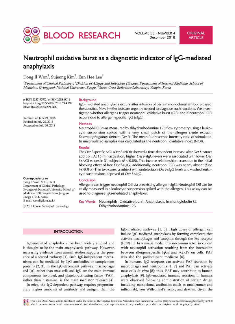
The mean fluorescence intensity (MFI) of rhodamine 123 and the mean FSC of neutrophils were determined (Fig. 1). Two regional gates were used for neutrophils: one in the FSC/SSC plot and the other in the rhodamine 123/SSC plot. The MFI value and mean FSC value were expressed as the geometric means of the main portion (M1 or M2) of the peak in the rhodamine 123 histogram and in the FSC histogram of gated neutrophils, respectively. The change in MFI for unstimulated and stimulated neutrophils was expressed as the neutrophil oxidative index (NOI), which was calculated using Equation 1:
NOI=MFI (stimulated)/MFI (unstimulated) ----------- (1).
The change in FSC (∆FSC) was calculated using Equation 2:
∆FSC=mean FSC (stimulated) - mean FSC (unstimulated) -- (2).
The cutoff value for distinguishing positive from negative results was 1.20 for NOI and 50 for ∆FSC.
CD63 assay: A BAT was performed by three-color flow cytometry [15]. For basophil priming, interleukin-3 (Sigma-Aldrich) was added to 100 L of whole blood and incubated for 10 min at 37oC. This primed whole blood was spiked with a patch of Der f extract, and the reaction mixture was incubated for 30 min at 37o in shaking water bath. Samples were marked with the fluorescently labeled antibodies CD123 phycoerythrin-cyanine 5, CD203c allo-phyocyanin and CD36 fluorescein isothiocyanate for 30 min at 4oC. Following red blood cell lysis with 3.0 mL lysing

bloodresearch.or.kr Blood Res 2018;53:299-306.
DHR assay for IgG-mediated anaphylaxis 301
Fig. 1. Flow cytometry data acquisition and analysis for oxidative burst in response to Der f extract. Calculations of NOI and ∆FSC are described in the Materials and Methods section. Abbreviations: Der f, Dermatophagoides farinae; FSC, forward scatter; MFI, mean fluorescence intensity; NOI, neutrophil oxidative index; SSC, sidescatter.
solution and a single washing step, the remaining pellet was resuspended in PBS for flow cytometry data acquisition (all from BD Biosciences). For the CD63+ proportion of baso-phils, the cutoff value for distinguishing positive from neg-ative results was 3.0%.
Determination of Der f-sIgE, Der f-sIgG (total), and Der f-sIgG4 levels
The level of Der f-specific (Der f-s) IgG (i.e., Der f-sIgG) antibodies (total) in the plasma samples was measured by enzyme-linked immunosorbent assay using RIDASCREEN
Specific IgG kits and discs of Der f (R-Biopharm AG, Darfmstadt, Germany), with a limit of detection (LOD) of 2.5 g/mL, and the levels of Der f-sIgE or Der f-sIgG4 anti-bodies were measured by a chemiluminescent immunoassay using ImmunoCAP Specific IgG (Thermo Fisher Scientific, Waltham, MA, USA), with an LOD of 0.1 g/mL.
Statistical analysesStatistical analyses were performed using SPSS Version
19.0 (SPSS, Inc., Chicago, IL, USA). The two groups were compared using regression analysis or Wilcoxon signed rank
Blood Res 2018;53:299-306. bloodresearch.or.kr
302 Dong Il Won, et al.
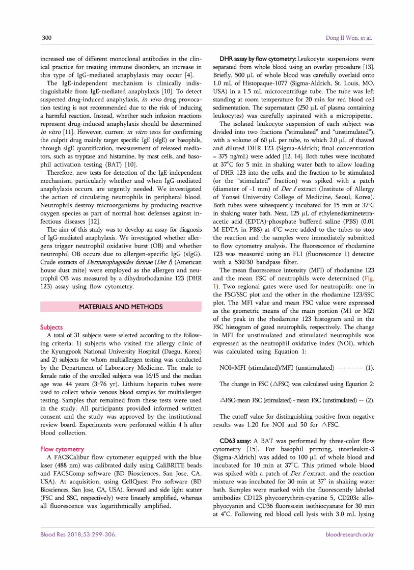
Fig. 2. Time course experiments showing effects of activation period on neutrophil reaction against Der f extract. Measurements were performed from 0 min (just prior to addition) to 40 min after adding Der f extract in 10 min intervals (total of five times). The typical results of four separate experiments are shown. (A) Raw data of FSC and MFI values. The difference between stimulated values and unstimulated values was significant (P<0.05) starting at 10 min activation for both FSC and MFI. Asterisks indicate significant stimulated values. (B) Calculated indices (∆FSC and NOI). The linearity of Der f-sNOI values according to the activation time was significant (P<0.01, asterisk), which was not observed for the ∆FSC values. Abbreviations: Der f, Dermatophagoides farinae; FSC, forward scatter; MFI, mean fluorescence intensity; NOI, neutrophil oxidative index; SSC, side scatter.
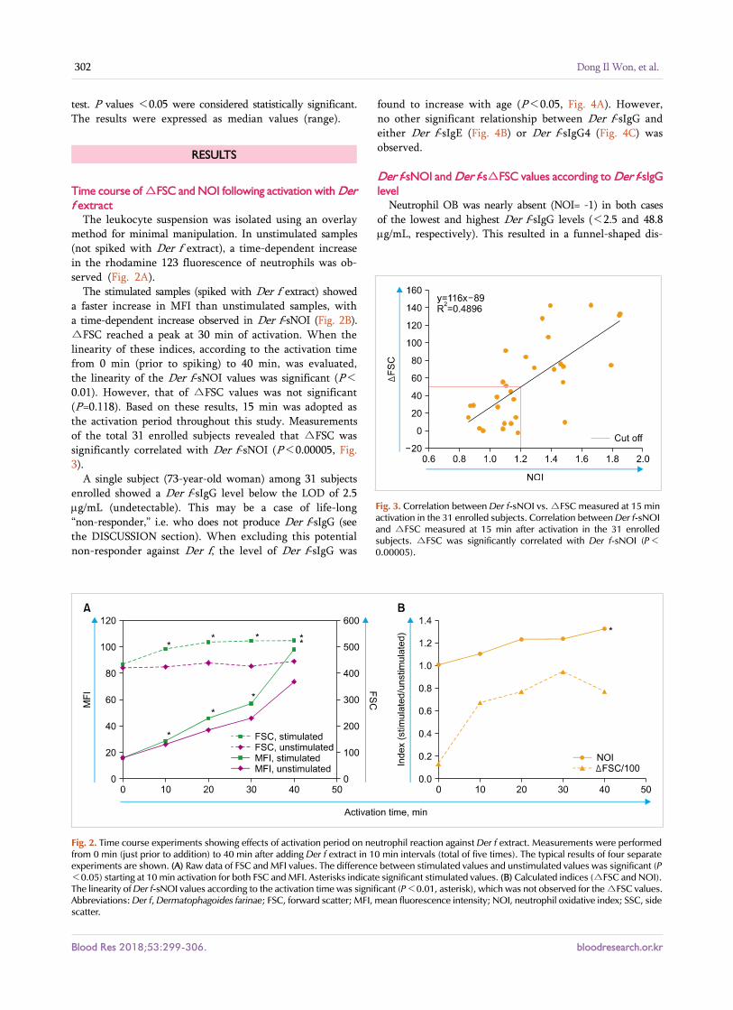
Fig. 3. Correlation between Der f-sNOI vs. ∆FSC measured at 15 min activation in the 31 enrolled subjects. Correlation between Der f-sNOI and ∆FSC measured at 15 min after activation in the 31 enrolled subjects. ∆FSC was significantly correlated with Der f-sNOI (P<0.00005).
test. P values <0.05 were considered statistically significant. The results were expressed as median values (range).
RESULTS
Time course of ∆FSC and NOI following activation with Der f extract
The leukocyte suspension was isolated using an overlay method for minimal manipulation. In unstimulated samples (not spiked with Der f extract), a time-dependent increase in the rhodamine 123 fluorescence of neutrophils was ob-served (Fig. 2A).
The stimulated samples (spiked with Der f extract) showed a faster increase in MFI than unstimulated samples, with a time-dependent increase observed in Der f-sNOI (Fig. 2B). ∆FSC reached a peak at 30 min of activation. When the linearity of these indices, according to the activation time from 0 min (prior to spiking) to 40 min, was evaluated, the linearity of the Der f-sNOI values was significant (P<
0.01). However, that of ∆FSC values was not significant (P=0.118). Based on these results, 15 min was adopted as the activation period throughout this study. Measurements of the total 31 enrolled subjects revealed that ∆FSC was significantly correlated with Der f-sNOI (P<0.00005, Fig. 3).
A single subject (73-year-old woman) among 31 subjects enrolled showed a Der f-sIgG level below the LOD of 2.5 g/mL (undetectable). This may be a case of life-long “non-responder,” i.e. who does not produce Der f-sIgG (see the DISCUSSION section). When excluding this potential non-responder against Der f, the level of Der f-sIgG was
found to increase with age (P<0.05, Fig. 4A). However, no other significant relationship between Der f-sIgG and either Der f-sIgE (Fig. 4B) or Der f-sIgG4 (Fig. 4C) was observed.
Der f-sNOI and Der f-s∆FSC values according to Der f-sIgG level
Neutrophil OB was nearly absent (NOI= -1) in both cases of the lowest and highest Der f-sIgG levels (<2.5 and 48.8 g/mL, respectively). This resulted in a funnel-shaped dis-

bloodresearch.or.kr Blood Res 2018;53:299-306.
DHR assay for IgG-mediated anaphylaxis 303
Fig. 4. Blood levels of Der f-sIgG (total) in 31 subjects and their correlation with (A) subject age (P<0.05); (B) Der f-sIgE level; and (C) Der f-sIgG4 level. In the panel C, IgG4 levels of five subjects with less than the limit of detection (0.1 g/mL) were assumed to be 0.05 g/mL for convenience. The red and blue arrows indicate extreme cases of Der f-sIgG levels (highest and lowest, respectively). In panel A, the case with the lowest (undetectable) Der f-sIgG levels (73-year-old woman, potential non-responder against Der f) was excluded from the regression analysis.
Fig. 5. Der f-sNOI and Der f-s∆FSC according to the Der f-sIgG level. The relationship between Der f-sIgG levels and (A) NOI; (B) 1/NOI (P<0.05); and (C) ∆FSC (P=0.201). The red and blue arrows indicate extreme cases of Der f-sIgG levels (highest and lowest, respectively). In panel B, the case with the lowest (undetectable) Der f-sIgG levels (73-year-old woman, potential non-responder against Der f) was excluded from the regression analysis.
tribution of the 31 subjects’ dots in the Der f-sIgG vs. Der f-sNOI plot (Fig. 5A). When the lowest level case (non-res-ponder against Der f) was excluded, the remaining subjects with detectable Der f-sIgG levels showed significant relation-ship between the two parameters (Fig. 5B); as the Der f-sIgG level increased, Der f-sNOI decreased (P<0.05). Der f-spe-cific ∆FSC (Der f-s∆FSC) appeared to be negatively but not significantly correlated with Der f-sIgG levels (Fig. 5C).
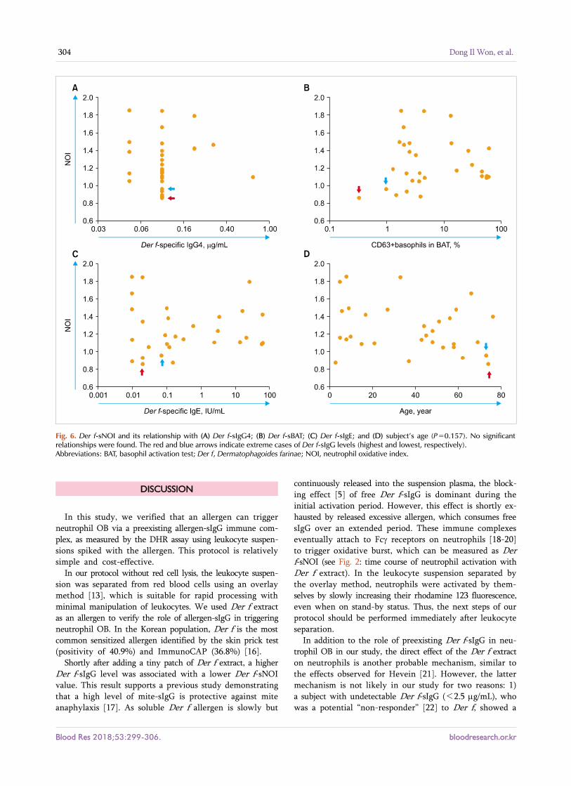
Relationship between Der f-sNOI and other parameters No significant relationship was found between Der f-sNOI
and Der f-sIgG4, Der f-specific BAT (Der f-sBAT, percentage of CD63+ basophils), or Der f-sIgE (Fig. 6A–D). A decreasing trend in Der f-sNOI was observed with increasing age, but the results were not significant (P=0.157).
Depriving leukocyte suspension of free preexisting Der f-sIgG We selected four subjects who were previously positive
for both Der f-s∆FSC and Der f-sNOI. Their leukocyte sus-pensions were washed three times with 3.0 mL of Roswell Park Memorial Institute medium to remove free preexisting Der f-sIgG. Neutrophil reactions were nearly absent in all the washed leukocyte suspensions, with a Der f-s∆FSC value (cutoff=50) of 2.0 (-6.0–15.0) and Der f-sNOI value (cutoff=1.20) of 1.05 (0.98–1.09). This suggests that neu-trophil OB following the addition of Der f extract occurred because of free preexisting Der f-sIgG in the suspension.
Blood Res 2018;53:299-306. bloodresearch.or.kr
304 Dong Il Won, et al.
Fig. 6. Der f-sNOI and its relationship with (A) Der f-sIgG4; (B) Der f-sBAT; (C) Der f-sIgE; and (D) subject’s age (P=0.157). No significant relationships were found. The red and blue arrows indicate extreme cases of Der f-sIgG levels (highest and lowest, respectively). Abbreviations: BAT, basophil activation test; Der f, Dermatophagoides farinae; NOI, neutrophil oxidative index.
DISCUSSION
In this study, we verified that an allergen can trigger neutrophil OB via a preexisting allergen-sIgG immune com-plex, as measured by the DHR assay using leukocyte suspen-sions spiked with the allergen. This protocol is relatively simple and cost-effective.
In our protocol without red cell lysis, the leukocyte suspen-sion was separated from red blood cells using an overlay method [13], which is suitable for rapid processing with minimal manipulation of leukocytes. We used Der f extract as an allergen to verify the role of allergen-sIgG in triggering neutrophil OB. In the Korean population, Der f is the most common sensitized allergen identified by the skin prick test (positivity of 40.9%) and ImmunoCAP (36.8%) [16].
Shortly after adding a tiny patch of Der f extract, a higher Der f-sIgG level was associated with a lower Der f-sNOI value. This result supports a previous study demonstrating that a high level of mite-sIgG is protective against mite anaphylaxis [17]. As soluble Der f allergen is slowly but
continuously released into the suspension plasma, the block-ing effect [5] of free Der f-sIgG is dominant during the initial activation period. However, this effect is shortly ex-hausted by released excessive allergen, which consumes free sIgG over an extended period. These immune complexes eventually attach to Fc receptors on neutrophils [18-20] to trigger oxidative burst, which can be measured as Der f-sNOI (see Fig. 2: time course of neutrophil activation with Der f extract). In the leukocyte suspension separated by the overlay method, neutrophils were activated by them-selves by slowly increasing their rhodamine 123 fluorescence, even when on stand-by status. Thus, the next steps of our protocol should be performed immediately after leukocyte separation.
In addition to the role of preexisting Der f-sIgG in neu-trophil OB in our study, the direct effect of the Der f extract on neutrophils is another probable mechanism, similar to the effects observed for Hevein [21]. However, the latter mechanism is not likely in our study for two reasons: 1) a subject with undetectable Der f-sIgG (<2.5 g/mL), who was a potential “non-responder” [22] to Der f, showed a
bloodresearch.or.kr Blood Res 2018;53:299-306.
DHR assay for IgG-mediated anaphylaxis 305
Der f-∆FSC of -0 and Der f-NOI of -1 (almost no neutrophil reaction); and 2) washed leukocyte suspensions from which Der f-sIgG was removed showed the same results.
We investigated the relationship between Der f-sNOI and parameters other than Der f-sIgG, Der f-sIgG4, Der f-sBAT, Der f-sIgE, and subjects’ age. No parameters were sig-nificantly correlated with Der f-sNOI. The insignificant but decreasing trend in Der f-sNOI values with age may be related to the increased Der f-sIgG levels during aging.
Upon in vivo basophil activation, granules fuse with the plasma membrane and their contents are released in the extracellular environment within minutes, which can induce a chain reaction of neutrophil OB [23-25]. We investigated whether this chain reaction occurs in our in vitro model using a leukocyte suspension and we did not find a significant correlation between Der f-sBAT and Der f-sNOI in the 31 subjects assessed (Fig. 6B). This finding suggests that neither in vitro basophil activation nor subsequent neutrophil OB occurs when our protocol is used.
Our protocol may be applicable for in vitro diagnosis of IgG-mediated anaphylaxis due to intravenously infused drugs or for detection of anti-drug antibodies that neutralize the biological activity of therapeutic proteins [26]. For this pur-pose, the amount of drug added should be sufficient to over-come the initial blocking effect of free sIgG. In contrast, when the allergen amount is optimized to reflect the blocking effect of free sIgG, this assay may be useful to assess the immunotherapy effectiveness.
We conclude that allergens can trigger neutrophil OB via preexisting allergen-sIgG and that neutrophil OB can be easily measured in a DHR assay using a leukocyte suspen-sion spiked with the allergen. These findings can be used for in vitro diagnosis of IgG-mediated anaphylaxis induced by intravenously infused drugs.
AuthorsÊ Disclosures of Potential Conflicts of Interest
No potential conflicts of interest relevant to this article were reported.
REFERENCES
1. Finkelman FD. Anaphylaxis: lessons from mouse models. J Allergy
Clin Immunol 2007;120:506-15.
2. Muñoz-Cano R, Picado C, Valero A, Bartra J. Mechanisms of
anaphylaxis beyond IgE. J Investig Allergol Clin Immunol
2016;26:73-82.
3. Finkelman FD, Khodoun MV, Strait R. Human IgE-independent
systemic anaphylaxis. J Allergy Clin Immunol 2016;137:1674-80.
4. Escribese MM, Rosace D, Chivato T, Fernández TD, Corbí AL,
Barber D. Alternative anaphylactic routes: the potential role of
macrophages. Front Immunol 2017;8:515.
5. Strait RT, Morris SC, Finkelman FD. IgG-blocking antibodies
inhibit IgE-mediated anaphylaxis in vivo through both antigen
interception and Fc gamma RIIb cross-linking. J Clin Invest
2006;116:833-41.
6. Jönsson F, Mancardi DA, Kita Y, et al. Mouse and human
neutrophils induce anaphylaxis. J Clin Invest 2011;121:1484-96.
7. Jönsson F, Mancardi DA, Zhao W, et al. Human FcRIIA induces
anaphylactic and allergic reactions. Blood 2012;119:2533-44.
8. Kajiwara N, Sasaki T, Bradding P, et al. Activation of human mast
cells through the platelet-activating factor receptor. J Allergy Clin
Immunol 2010;125:1137-45.
9. Vadas P, Gold M, Perelman B, et al. Platelet-activating factor, PAF
acetylhydrolase, and severe anaphylaxis. N Engl J Med 2008;
358:28-35.
10. Montañez MI, Mayorga C, Bogas G, et al. Epidemiology,
mechanisms, and diagnosis of drug-induced anaphylaxis. Front
Immunol 2017;8:614.
11. Khodoun MV, Strait R, Armstrong L, Yanase N, Finkelman FD.
Identification of markers that distinguish IgE- from IgG-mediated
anaphylaxis. Proc Natl Acad Sci U S A 2011;108:12413-8.
12. Walrand S, Valeix S, Rodriguez C, Ligot P, Chassagne J, Vasson
MP. Flow cytometry study of polymorphonuclear neutrophil
oxidative burst: a comparison of three fluorescent probes. Clin
Chim Acta 2003;331:103-10.
13. Robinson JP, Carter WO. Flow cytometric analysis of
granulocytes. In: Bauer KD, Duque RE, Shankey TV, eds. Clinical
flow cytometry: principles and application. 2nd ed. Baltimore,
MD: Williams & Wilkins, 1993:405-33.
14. Richardson MP, Ayliffe MJ, Helbert M, Davies EG. A simple flow
cytometry assay using dihydrorhodamine for the measurement of
the neutrophil respiratory burst in whole blood: comparison with
the quantitative nitrobluetetrazolium test. J Immunol Methods
1998;219:187-93.
15. Kim Z, Choi BS, Kim JK, Won DI. Basophil markers for
identification and activation in the indirect basophil activation
test by flow cytometry for diagnosis of autoimmune urticaria. Ann
Lab Med 2016;36:28-35.
16. Park HJ, Lee JH, Park KH, et al. A nationwide survey of inhalant
allergens sensitization and levels of indoor major allergens in
Korea. Allergy Asthma Immunol Res 2014;6:222-7.
17. Hirai T, Yoshioka Y, Takahashi H, et al. High-dose cutaneous
exposure to mite allergen induces IgG-mediated protection
against anaphylaxis. Clin Exp Allergy 2016;46:992-1003.
18. Fossati G, Bucknall RC, Edwards SW. Insoluble and soluble
immune complexes activate neutrophils by distinct activation
mechanisms: changes in functional responses induced by priming
with cytokines. Ann Rheum Dis 2002;61:13-9.
19. Coxon A, Cullere X, Knight S, et al. Fc gamma RIII mediates
neutrophil recruitment to immune complexes. a mechanism for
neutrophil accumulation in immune-mediated inflammation.
Immunity 2001;14:693-704.
20. Zhang W, Voice J, Lachmann PJ. A systematic study of neutrophil
degranulation and respiratory burst in vitro by defined immune
complexes. Clin Exp Immunol 1995;101:507-14.
21. Rojas E, Llinas P, Rodríguez-Romero A, et al. Hevein, an allergenic
lectin from rubber latex, activates human neutrophils' oxidative
burst. Glycoconj J 2001;18:339-45.
22. Shouval D, Roggendorf H, Roggendorf M. Enhanced immune
response to hepatitis B vaccination through immunization with
a Pre-S1/Pre-S2/S vaccine. Med Microbiol Immunol 2015;204:
57-68.

Blood Res 2018;53:299-306. bloodresearch.or.kr
306 Dong Il Won, et al.
23. Doener F, Michel A, Reuter S, et al. Mast cell-derived mediators
promote murine neutrophil effector functions. Int Immunol
2013;25:553-61.
24. Jönsson F, Mancardi DA, Albanesi M, Bruhns P. Neutrophils in
local and systemic antibody-dependent inflammatory and
anaphylactic reactions. J Leukoc Biol 2013;94:643-56.
25. Hart PH. Regulation of the inflammatory response in asthma by
mast cell products. Immunol Cell Biol 2001;79:149-53.
26. Shibata H, Nishimura K, Miyama C, et al. Comparison of different
immunoassay methods to detect human anti-drug antibody using
the WHO erythropoietin antibody reference panel for analytes.
J Immunol Methods 2018;452:73-7.


![arXiv:0803.2680v3 [astro-ph] 23 Mar 2008XMM-Newtonpn/rgsand 253ks of Suzaku xis/pindata. This is the first analysis of this full dataset. We inves tigated the spectral variability](https://img.pdfslide.us/doc/110x75/603680b4000e337f932e1cb3/arxiv08032680v3-astro-ph-23-mar-2008-xmm-newtonpnrgsand-253ks-of-suzaku-xispindata.jpg)


![Multi-soliton fusion phenomenon of Burgers equation and fission, … · 2019-11-19 · tween the wave vectors and velocities. Wazwaz [20–22] inves- tigated multiple soliton solutions](https://img.pdfslide.us/doc/110x75/5e78137a83639859e75dc2d8/multi-soliton-fusion-phenomenon-of-burgers-equation-and-fission-2019-11-19-tween.jpg)
![Characterisation of blast loading in complex, confined ... · design of weapons storage facilities. Wu et al. [11]inves-tigated confined explosions in a full-scale blast chamber,](https://img.pdfslide.us/doc/110x75/5f0794c67e708231d41db290/characterisation-of-blast-loading-in-complex-confined-design-of-weapons-storage.jpg)