Embed Size (px)
Citation preview
CASE REPORT Open Access
Blood pressure shifts resulting from aconcealed arteriovenous fistula associatedwith an iliac aneurysm: a case reportShintaro Doi1*, Yoshiaki Motoyama2 and Hiromi Ito2
Abstract
Background: A solitary iliac aneurysm (SIA) is more uncommon than an abdominal aortic aneurysm. The aneurysmis located in the deep pelvis and is diagnosed when it reaches a large size with symptoms of compression aroundadjacent structures and organs or when it ruptures. A definite diagnosis of an arteriovenous fistula (AVF) associatedwith a SIA is difficult preoperatively because there might not be enough symptoms and time for diagnosis. Here,we present a patient with asymptomatic rupture of SIA into the common iliac vein with characteristic bloodpressure shifts.
Case presentation: A 41-year-old man with a huge SIA underwent aortobifemoral graft replacement. Preoperatively,his blood pressure showed characteristic shifts for one or two heartbeats out of five beats, indicating that an AVF waspresent and that the shunt was about to having a high flow. During surgery, an AVF associated with the SIA was foundto be concealed owing to compression from the huge iliac artery aneurysm, and the shunt showed a highflow, resulting in shock during the surgery. No complications were noted after aortobifemoral graft replacement.Postoperatively, we noted an enhanced paravertebral vein on computed tomography (CT), which indicatedthe presence of an AVF.
Conclusions: Definite diagnosis of an AVF offers advantages in surgical and anesthetic management. Weemphasize that a large SIA can push the iliac vein and occlude an AVF laceration, concealing the enhancement of theveins in the arterial phase on CT. Blood pressure shifts might predict the existence of a concealed AVF that has a largeshunt. Even if the vena cava and the iliac veins are not enhanced on CT, anesthesiologists should carefully determinewhether their distal branches are enhanced.
Keywords: Abdominal aortic aneurysm, Arteriovenous fistula, Hemodynamics, Iliac artery
BackgroundA solitary iliac aneurysm (SIA), an aneurysm that locatesonly in the iliac artery, occurs in 0.6 % of the case of anabdominal aortic aneurysm (AAA) [1], and many of thepatients are free of symptoms. The aneurysm is locatedin the deep pelvis and is diagnosed when it reaches alarge size with symptoms of compression around adjacentstructures and organs or when it ruptures. An ilio-iliacarteriovenous fistula (AVF) occurs in less than 1 % of allcases of a common iliac artery aneurysm [2]. Becauseof its rarity and its various symptoms, the diagnosis
and treatment of an AVF associated with a SIA arechallenging to vascular surgeons and anesthesiologists.Here, we present a patient with asymptomatic ruptureof a SIA into the common iliac vein with characteris-tic blood pressure shifts.
Case presentationA 41-year-old man (height, 178 cm; weight, 58 kg) withno medical history was admitted to the emergency uniton foot complaining of severe right inguinal pain. Aright inguinal bulge was noted, and there was no lowerlimb edema. CT showed bilateral common iliac aneu-rysms (Fig. 1), and the internal iliac artery had a max-imum diameter of 8 cm (Fig. 2). An AVF was notdetected on CT, and chest radiography did not show
* Correspondence: [email protected] of Anesthesiology, Sanai Hospital, 4-35-17 Tajima, Sakura-ku,Saitama City, Saitama 338-0837, JapanFull list of author information is available at the end of the article
© The Author(s). 2016 Open Access This article is distributed under the terms of the Creative Commons Attribution 4.0International License (http://creativecommons.org/licenses/by/4.0/), which permits unrestricted use, distribution, andreproduction in any medium, provided you give appropriate credit to the original author(s) and the source, provide a link tothe Creative Commons license, and indicate if changes were made.
Doi et al. JA Clinical Reports (2016) 2:33 DOI 10.1186/s40981-016-0057-2
heart or lung disorders. Emergent laparotomy wasplanned for the aneurysm rupture.Preoperatively, his arterial pressure was stable with a
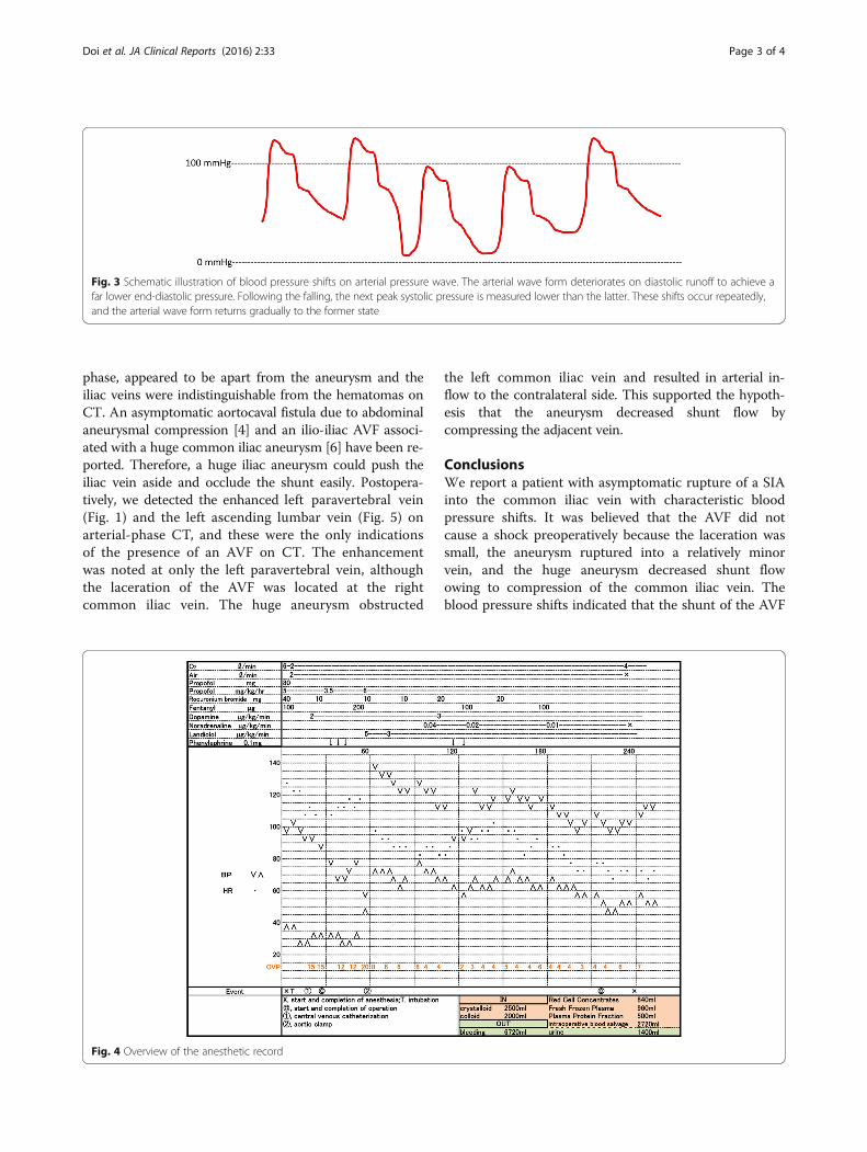
systolic pressure of approximately 100 mmHg; however,there was a characteristic hemodynamic change (Fig. 3),with pressure shifting between 95/48 mmHg and 85/25 mmHg for one or two heartbeats out of five beats,with normal sinus rhythm.Anesthetic induction was successful. The detection of
high central venous pressure (CVP) coincidently with re-duction of arterial pressure indicated the presence of anAVF. During the surgery, his systemic hemodynamiccondition worsened (Fig. 4). The CVP was initially
9 mmHg; however, it increased to 20 mmHg. His arterialpressure shifted frequently, and it reached a plateau of60/50 mmHg. Urgent laparotomy and impetuous aorticclamping resulted in quick hemodynamic recovery, andthe CVP reduced to approximately 6 mmHg. The rightcommon iliac artery aneurysm showed a communicationwith the right common iliac vein. Therefore, he underwentaortobifemoral graft replacement, and no complicationswere noted. Postoperatively, he was not diagnosed withany connective tissue disorders, such as Ehlers-Danlossyndrome and Marfan’s syndrome.
DiscussionWe presented a case of acute aneurysmal rapture intothe iliac vein. Sometimes, a definite diagnosis of anAVF associated with a SIA is difficult preoperativelybecause there might not be enough symptoms andtime for diagnosis [3]. An AVF associated with anAAA has the following triad of symptoms: congestiveheart failure, continuous abdominal bruit, and a pul-sating abdominal mass [1]; however, these symptomsare noted in only 20–50 % of reported cases [4]. AnAVF associated with a SIA might show lower limbedema as an additional feature; however, many casesdo not have hemodynamic symptoms [4–6]. Definitediagnosis of an AVF offers advantages for surgical andanesthetic management.Arterial pressure shifts rarely occur in a clinical situ-
ation. In the present case, these shifts indicated that anAVF was present and that the shunt was about to havinga high flow. These hemodynamic changes could be ex-plained by pooling of the transient increased shunt flowto a high-capacitance venous circuit and a decreased inpreload, which can produce low arterial pressure at thenext heartbeat. Simultaneously, an increase in venousreturn raised the blood pressure following a downwardshift in the blood pressure. We hypothesized that themechanical compression of a huge aneurysm should oc-clude the AVF and the fistula would appear by changingof lower limb posture, high blood pressure, or pulse ofthe aneurysm itself. The shift disappeared during theoperative preparation, indicating that shunt dilationdue to anesthetic agents and muscle relaxants decreasedperipheral resistance, including aneurysmal compressionat the vein.CT has been recommended to determine the sub-
types, sizes, and complications of aneurysms [1, 3–6].CT can contribute to the detection of an asymptom-atic AVF associated with a SIA [4–6]. Although ourcase had hemodynamic catastrophe, the CT findingsin our case were much fewer than the findings pre-sented in previous reports. We were unsure of thepresence of an AVF preoperatively because the dilatedvena cava, which was not enhanced in the arterial
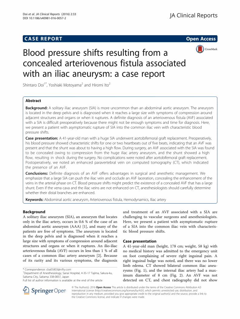
Fig. 1 Computed tomography showing bilateral common iliac arteryaneurysms and a dilated vena cava. Postoperatively, we could detectthe enhanced paravertebral vein (arrow). The non-enhanced vena cavaindicated an arteriovenous fistula located distal to the vena cava
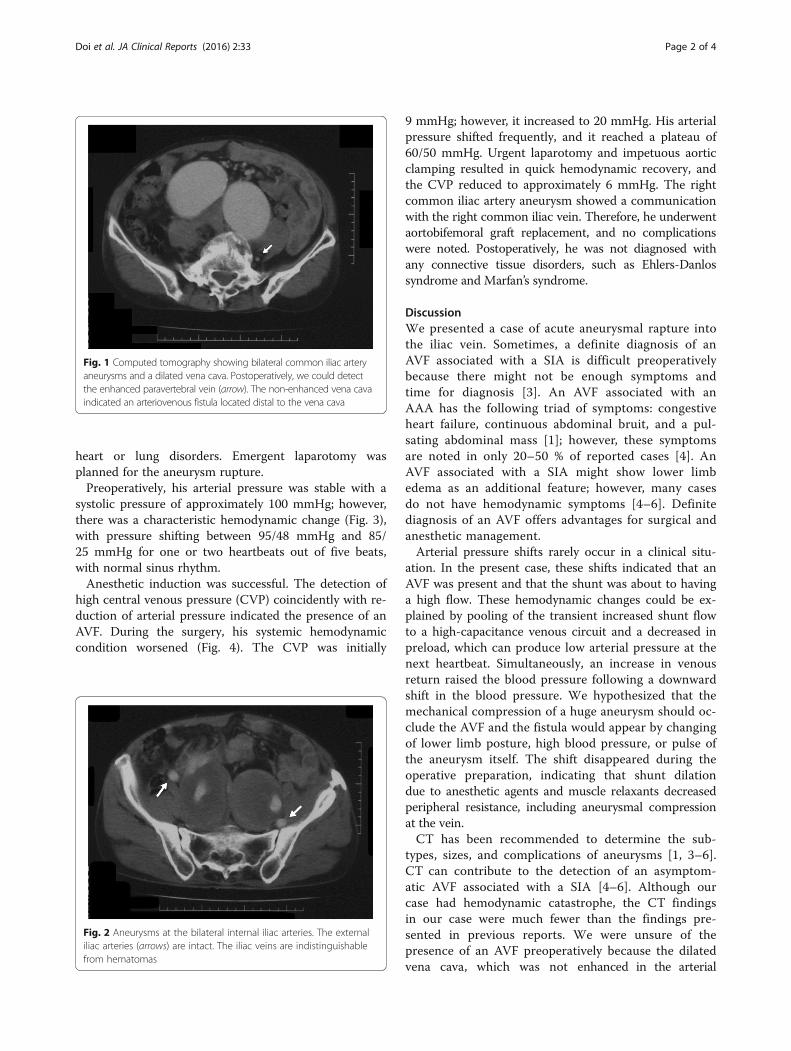
Fig. 2 Aneurysms at the bilateral internal iliac arteries. The externaliliac arteries (arrows) are intact. The iliac veins are indistinguishablefrom hematomas
Doi et al. JA Clinical Reports (2016) 2:33 Page 2 of 4
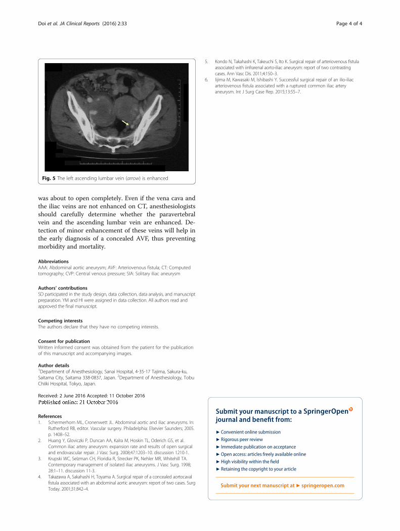
phase, appeared to be apart from the aneurysm and theiliac veins were indistinguishable from the hematomas onCT. An asymptomatic aortocaval fistula due to abdominalaneurysmal compression [4] and an ilio-iliac AVF associ-ated with a huge common iliac aneurysm [6] have been re-ported. Therefore, a huge iliac aneurysm could push theiliac vein aside and occlude the shunt easily. Postopera-tively, we detected the enhanced left paravertebral vein(Fig. 1) and the left ascending lumbar vein (Fig. 5) onarterial-phase CT, and these were the only indicationsof the presence of an AVF on CT. The enhancementwas noted at only the left paravertebral vein, althoughthe laceration of the AVF was located at the rightcommon iliac vein. The huge aneurysm obstructed
the left common iliac vein and resulted in arterial in-flow to the contralateral side. This supported the hypoth-esis that the aneurysm decreased shunt flow bycompressing the adjacent vein.
ConclusionsWe report a patient with asymptomatic rupture of a SIAinto the common iliac vein with characteristic bloodpressure shifts. It was believed that the AVF did notcause a shock preoperatively because the laceration wassmall, the aneurysm ruptured into a relatively minorvein, and the huge aneurysm decreased shunt flowowing to compression of the common iliac vein. Theblood pressure shifts indicated that the shunt of the AVF
Fig. 3 Schematic illustration of blood pressure shifts on arterial pressure wave. The arterial wave form deteriorates on diastolic runoff to achieve afar lower end-diastolic pressure. Following the falling, the next peak systolic pressure is measured lower than the latter. These shifts occur repeatedly,and the arterial wave form returns gradually to the former state
Fig. 4 Overview of the anesthetic record
Doi et al. JA Clinical Reports (2016) 2:33 Page 3 of 4
was about to open completely. Even if the vena cava andthe iliac veins are not enhanced on CT, anesthesiologistsshould carefully determine whether the paravertebralvein and the ascending lumbar vein are enhanced. De-tection of minor enhancement of these veins will help inthe early diagnosis of a concealed AVF, thus preventingmorbidity and mortality.
AbbreviationsAAA: Abdominal aortic aneurysm; AVF: Arteriovenous fistula; CT: Computedtomography; CVP: Central venous pressure; SIA: Solitary iliac aneurysm
Authors’ contributionsSD participated in the study design, data collection, data analysis, and manuscriptpreparation. YM and HI were assigned in data collection. All authors read andapproved the final manuscript.
Competing interestsThe authors declare that they have no competing interests.
Consent for publicationWritten informed consent was obtained from the patient for the publicationof this manuscript and accompanying images.
Author details1Department of Anesthesiology, Sanai Hospital, 4-35-17 Tajima, Sakura-ku,Saitama City, Saitama 338-0837, Japan. 2Department of Anesthesiology, TobuChiiki Hospital, Tokyo, Japan.
Received: 2 June 2016 Accepted: 11 October 2016
References1. Schermerhorn ML, Cronenwett JL. Abdominal aortic and iliac aneurysms. In:
Rutherford RB, editor. Vascular surgery. Philadelphia: Elsevier Saunders; 2005.p. 1408–52.
2. Huang Y, Gloviczki P, Duncan AA, Kalra M, Hoskin TL, Oderich GS, et al.Common iliac artery aneurysm: expansion rate and results of open surgicaland endovascular repair. J Vasc Surg. 2008;47:1203–10. discussion 1210-1.
3. Krupski WC, Selzman CH, Floridia R, Strecker PK, Nehler MR, Whitehill TA.Contemporary management of isolated iliac aneurysms. J Vasc Surg. 1998;28:1–11. discussion 11-3.
4. Takazawa A, Sakahashi H, Toyama A. Surgical repair of a concealed aortocavalfistula associated with an abdominal aortic aneurysm: report of two cases. SurgToday. 2001;31:842–4.
5. Kondo N, Takahashi K, Takeuchi S, Ito K. Surgical repair of arteriovenous fistulaassociated with iinfrarenal aorto-iliac aneurysm: report of two contrastingcases. Ann Vasc Dis. 2011;4:150–3.
6. Iijima M, Kawasaki M, Ishibashi Y. Successful surgical repair of an ilio-iliacarteriovenous fistula associated with a ruptured common iliac arteryaneurysm. Int J Surg Case Rep. 2015;13:55–7.
Submit your manuscript to a journal and benefi t from:
7 Convenient online submission
7 Rigorous peer review
7 Immediate publication on acceptance
7 Open access: articles freely available online
7 High visibility within the fi eld
7 Retaining the copyright to your article
Submit your next manuscript at 7 springeropen.com
Fig. 5 The left ascending lumbar vein (arrow) is enhanced
Doi et al. JA Clinical Reports (2016) 2:33 Page 4 of 4


![Piscivore-prey fish interactions - consequences of …lup.lub.lu.se/search/ws/files/5524614/2520970.pdfof trophic cascades and even resulting in ecosystem shifts [3] and weakening](https://img.pdfslide.us/doc/110x75/5f81a860ec52c522f3607656/piscivore-prey-fish-interactions-consequences-of-luplublusesearchwsfiles5524614.jpg)