Embed Size (px)
Citation preview
HAL Id: hal-00896591https://hal.archives-ouvertes.fr/hal-00896591
Submitted on 1 Jan 1970
HAL is a multi-disciplinary open accessarchive for the deposit and dissemination of sci-entific research documents, whether they are pub-lished or not. The documents may come fromteaching and research institutions in France orabroad, or from public or private research centers.
L’archive ouverte pluridisciplinaire HAL, estdestinée au dépôt et à la diffusion de documentsscientifiques de niveau recherche, publiés ou non,émanant des établissements d’enseignement et derecherche français ou étrangers, des laboratoirespublics ou privés.
BLOOD pH AND CATION LEVELS IN RELATIONTO EGG-SHELL FORMATION
R.-D. Hodges
To cite this version:R.-D. Hodges. BLOOD pH AND CATION LEVELS IN RELATION TO EGG-SHELL FORMATION.Annales de biologie animale, biochimie, biophysique, 1970, 10 (hs2), pp.199-213. <hal-00896591>
BLOOD pH AND CATION LEVELSIN RELATION TO EGG-SHELL FORMATION
R.-D. HODGES
Depavtment of Poultry Reseavch, Wye College(University of Lovcdon) near Ashfovd, Kent (Gveat Britain)
INTRODUCTION
The final processes of egg formation, the deposition of the albumen and shelaround the yolk, take place while the egg is passing down the oviduct. Normallythe time taken between the ovulation of the yolk and the oviposition of the finishedegg is about 26 hours and during the greater portion of this time, approximately20 hours, the egg remains in the shell gland. Two basic processes are carried outwhilst the egg is in the shell gland : firstly, the greater part of the plumping of thealbumen and secondly, the formation of the shell. These two processes require consi-derable quantities of ions, particularly the calcium which as calcium carbonate formsabout 95 p. 100 of the shell, but also other ions involved in the plumping processsuch as potassium and sodium. As the shell gland has little or no capacity for thestorage of these substances, most of the ions which are incorporated into the egghave to be derived from the shell gland blood supply when the gland is active. Theonly exceptions to this are the bicarbonate ions which go to form the shell carbonateand these are almost certainly derived mainly from the shell gland metabolic carbondioxide (HODGES and LoRCHr,R, Ig67).
The purpose of the present series of experiments was to obtain a comprehensivepicture of the fluctuations of several of the more important ions associated with plum-ping and shell formation both in systemic and shell gland blood and during the wholeof the shell formation cycle. Although a number of investigations of this nature havebeen performed in the past, most of them only measured calcium levels and thisonly on systemic blood (CHARr,!s and HOGI3!N, Ig33 ; POLIN and STURKIE, Ig57 1WINGET and SMITH, 1958 ; H!RTEI,!NDY and TAYI,OR, 1961). If shell gland bloodalso was sampled it was done only on a very limited scale (KNOwI,!s, HART and HAL-PIN, Ig35 ; WINGET, SMITH and HOOVER, Ig58 ; HUNSAKI;R and STURKIE, 1961).In this series the cations calcium, potassium and sodium have been measured andat the same time, in order to obtain a measure of the blood acid-base fluctuationswhich occur during shell formation, the blood pH was determined.
MATERIALS AND METHODS
The birds used in the experiments were actively-laying hybrid hens of the White Leghornstrain (Sterling White Links and Shavev 288’s). Each bird was anaesthetized with sodium penta-barbitone and ether and a plastic cannula was inserted into the posterior end of the inferior ovi-ducal vein (HODGES, 1965) using the technique described by HODGES (ig66). Cannulae were alsoinserted into the sciatic artery and vein in the upper left leg. From these three cannulae it waspossible to take samples of systemic arterial and venous blood and of shell gland venous blood.Since: the main artery supplying the shell gland, the hypogastric artery, branches off from thefemoral artery at the level of the left kidney (FREEDMAK and Sruxxm, 1963) the blood beingsampled from the sciatic artery was of similar composition to that entering the shell gland.
After the operation the birds were kept lightly anasthetized with ether and were heparinized.Hourly blood samples were taken throughout the period of each experiment and the timing ofsampling to coincide with regular hourly periods within the shell formation cycle was performedby timing the laying of the previous egg or by the oviposition or the entry of an egg into the shellgland during the experimental period. Blood samples for pH determination were taken into capil-lary tubes (80 [LI) and the pH values were measured with a Radiometer pH Micro-Electrode andpH-lTetcr z!. At the same time samples of 0.75 ml were taken into centrifuge tubes and the plasmawas used for the cation measurements. Plasma sodium and potassium were measured with anEEL flame photometer (BVOOTON, 1964) using 0.2 ml of plasma and plasma calcium was measuredby means of Trinder’s method (TRINDER, ig6o) using o, i ml of plasma.
The experimental series was controlled by using unan,csthctizcd, unrcstrained birds. Can-nulac were inserted into the wing veins of six birds and samples were taken every two hours overa period of approximately z4 hours.
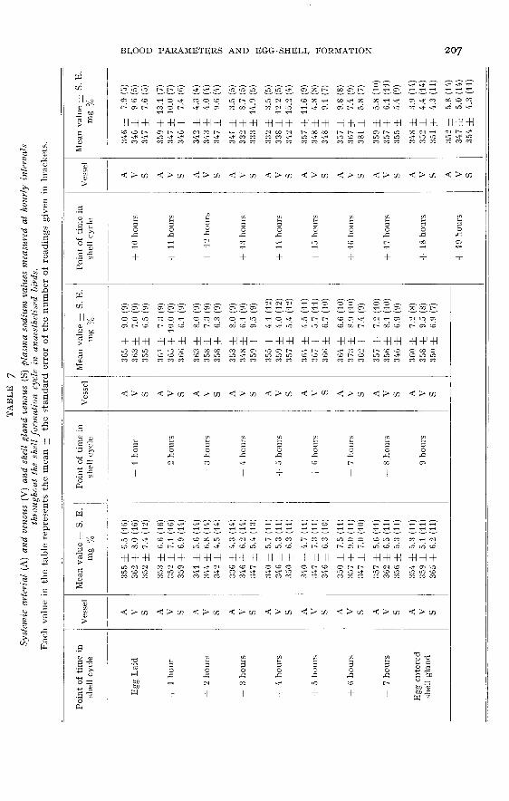
The time scale of the tables is divided into two parts. Firstly, there is the period of shell glandquiescence between the point of lay (Egg Laid) and the time when another egg entered the shellgland (Egg Entered Shell Gland). Secondly, there is the pcriod of shell formation lasting for20 hours.
RESULTS
pH. (Control values ― table I ; experimental values ― table 2)
The control pH values were rather variable, but did show a regular fluctuationthroughout the shell formation period, with a maximum level of about 7.38 at sixhours after the egg entered the shell gland and a minimum value of 7.32 at 16 hours
of shell formation. This pattern was somewhat obscured by a secondary fall in pHwhich occurred between four and six hours after lay.
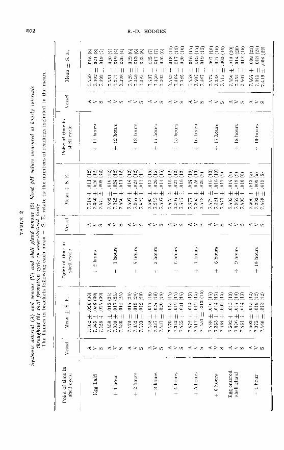
In the experimental birds the pattern of the arterial results was basically simi-lar to that of the controls, a maximum of 7.51 two hours after the egg entered theshell gland and a minimum of 7.q3 at thirteen hours of shell formation, but thesepoints occurred about three hours earlier in the experimental birds than in the con-trols.
The shell gland venous values, in general, closely followed the pattern of thearterial values at a rather more acid level. However, whereas this arterio-venousdifference was minimal during the period of shell gland quiescence (0.015 pH unitsfour hours after lay), during the following period of shell formation the differencegradually increased to reach a maximum of 0-079 pH units at the fifteenth hour ofshell formation (table 3). From then on the arterio-venous difference decreased untilthe egg was laid.
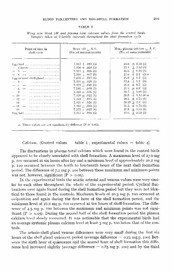
Calcium. (Control values ― table I ; experimental values ― table 4)
The fluctuations in plasma total calcium which were found in the control birdsappeared to be closely associated with shell formation. A maximum level of 27.6 mgp. 100 occurred at six hours after lay and a minimum level of approximately 20.2 mg
p. 100 occurred between the tenth to fourteenth hours of the next shell formation
period. The difference of 7.2 mg p. 100 between these maximum and minimum pointswas not, however, significant (P > 0.05).
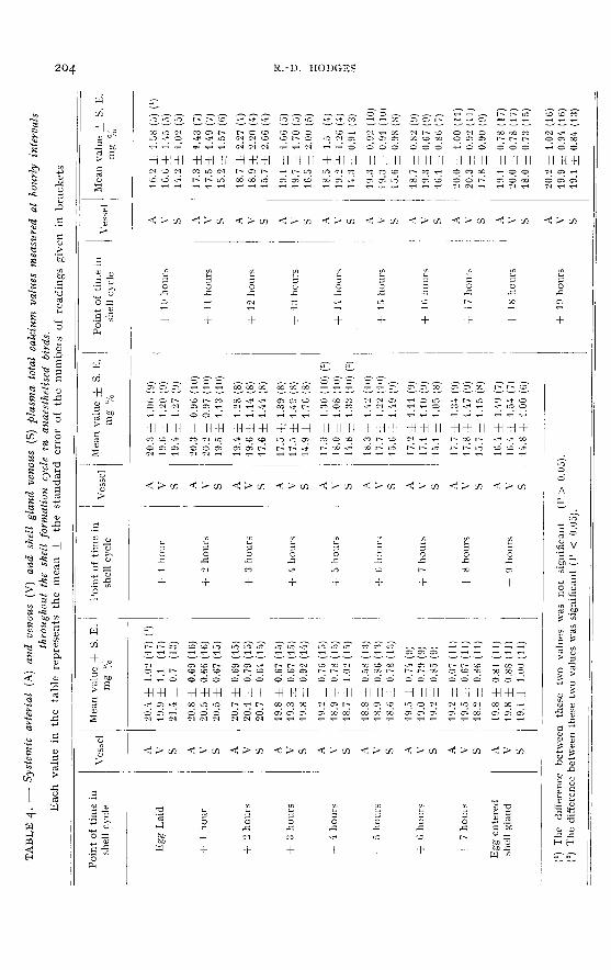
In the experimental birds the sciatic arterial and venous values were very simi-lar to each other throughout the whole of the experimental period. Cyclical fluc-tuations were again found during the shell formation period but they were not iden-tical to those found in the controls. Maximum levels of 20.3 mg p. 100 occurred at
oviposition and again during the first hour of the shell formation period, and theminimum level of 16.2 mg p. 100 occurred at ten hours of shell formation. The diffe-
rence of 4.5 mg p. 100 between the maximum and minimum points was not signi-ficant (P > 0.05). During the second half of the shell formation period the plasmacalcium level slowly recovered. It was noticeable that the experimental birds hadan average systemic plasma calcium level at least 3 mg p. 100 below that of the con-
trols.
The arterio-shell gland venous differences were very small during the first sixhours of the shell gland quiescent period (average difference = 0.21 mg p. 100). Bet-ween the sixth hour of quiescence and the second hour of shell formation this diffe-rence had increased slightly (average difference = 0.85 mg p. 100) and by the third
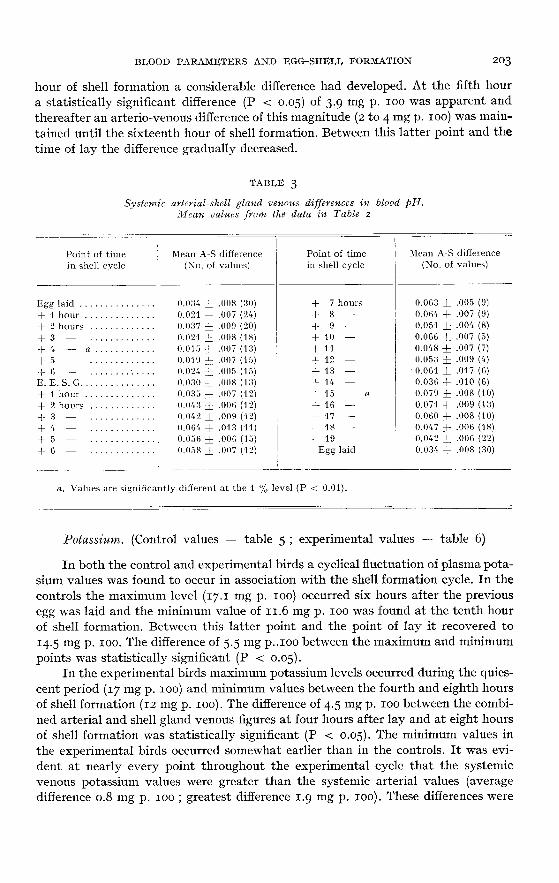
hour of shell formation a considerable difference had developed. At the fifth houra statistically significant difference (P < 0.05) of 3.9 mg p. 100 was apparent andthereafter an arterio-venous difference of this magnitude (2 to 4 mg p. 100) was main-tained until the sixteenth hour of shell formation. Between this latter point and thetime of lay the difference gradually decreased.
Potassium. (Control values ― table 5 ; experimental values ― table 6)
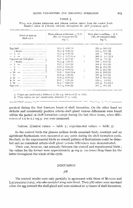
In both the control and experimental birds a cyclical fluctuation of plasma pota-sium values was found to occur in association with the shell formation cycle. In thecontrols the maximum level (17.1 mg p. 100) occurred six hours after the previousegg was laid and the minimum value of m.6 mg p. 100 was found at the tenth hour
of shell formation. Between this latter point and the point of lay it recovered to14.5 mg p. 100. The difference of 5.5 mg p..ioo between the maximum and minimum
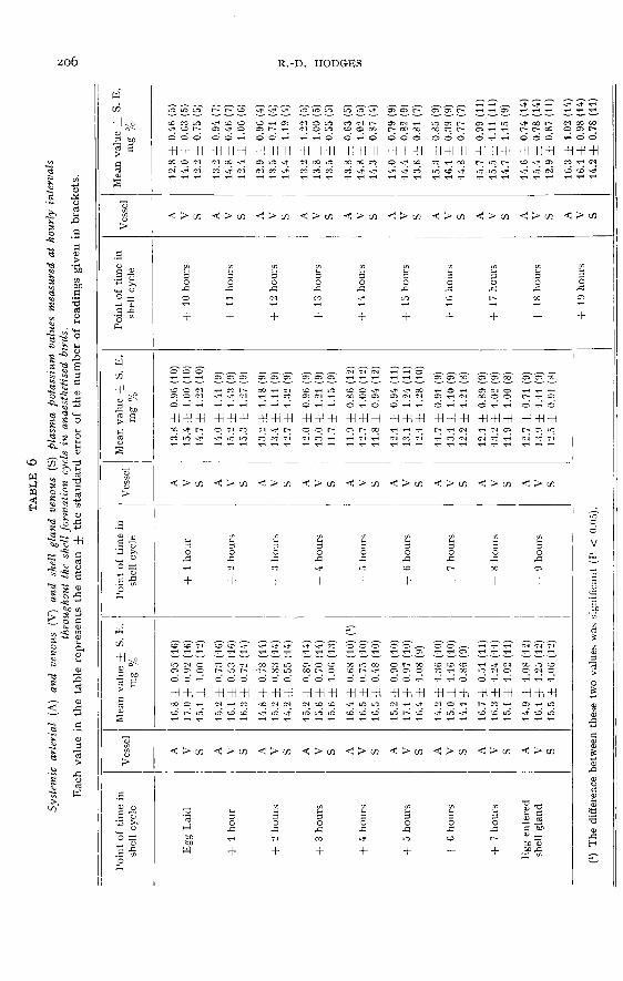
points was statistically significant (P < 0.05).In the experimental birds maximum potassium levels occurred during the quies-
cent period (17 mg p. 100) and minimum values between the fourth and eighth hoursof shell formation (12 mg p. ioo). The difference of 4.5 mg p. 100 between the combi-
ned arterial and shell gland venous figures at four hours after lay and at eight hoursof shell formation was statistically significant (P < 0.05). The minimum values inthe experimental birds occurred somewhat earlier than in the controls. It was evi-dent at nearly every point throughout the experimental cycle that the systemicvenous potassium values were greater than the systemic arterial values (averagedifference o.8 mg p. 100 ; greatest difference 1.9 mg p. ioo). These differences were
greatest during the first fourteen hours of shell formation. On the other hand nodefinite and consistently positive arterio-shell gland venous differences were foundwithin the period of shell formation except during the last three hours, when diffe-rences of i.o to 2.1 mg p. 100 were measured.
Sodium. (Control values ― table 5 ; experimental values ― table 7)
In the control birds the plasma sodium levels remained fairly constant and nosignificant fluctuations were measured at any point during the shell formation cycle.Similarly in the experimental birds no overall pattern of fluctuations could be detec-ted and no consistent arterio-shell gland venous differences were demonstrated.
There was, however, one anomaly between the control and experimental birds ;the values for the former were approximately 40 mg p. ioo lower than those for the
latter throughout the whole of the cycle.
DISCUSSION
!H
The control results were only partially in agreement with those of MoNGiN andLACASSAGNE (r964), who also sampled wing vein blood. Their pH values were maximalwhen the egg entered the shell gland and were minimal at y hours of shell formation.
The present results were overall at least o.oi pH units lower than those of DTovGrNand LncASSAGrr! and, in the case of the maximum point, approximately 5 hourslater in the shell formation cycle. However, it was not easy to relate their time scaleto the present one. Another point of difference was that whereas during the shellgland quiescent period the present results were in the process of recovering from theminimal values which had occurred before the oviposition of the previous egg, 3IONGINand I,ncnssAGrr!’s results were constant and maximal.
In discussing the experimental results it is necessary to acknowledge that thepH values would be adversely affected by the anaesthetic. Although the averagearterial pH in these experiments (7-474) was slightly higher than that found forunanaesthetised birds of a similar strain (7.410), and thus respiratory depressiondue to the anaesthetic would not seem to have occured, other experiments haveshown that birds with a similar arterial pH may have an unusually high PC02 and
bicarbonate, showing that some respiratory disturbance had occured. This was mini-mised by keeping the birds only lightly anaesthetised after the actual operation.
The sciatic venous values were very irregular and did not follow the trendsoccurring in the sciatic arterial and wing vein blood. This may have been due tothe fact that the venous cannulae were inserted well into the vein, thus drawing offblood from the region of the junction of the sciatic vein with the renal portal vein.It is possible that blood from the renal portal vein is not truly representative ofsystemic venous blood.
The arterial and shell gland venous results possessed maximum and minimumvalues which coincided closely with those of MoNGiN and I,acassnGn! (1964), butthe actual cyclical pattern of these results corresponded more closely with the presentcontrols than with the values of MonGrN and I,ncnsseGrr!. After oviposition pHvalues slowly increased to a maximum just after the next egg entered the shell glandand, as the next phase of shell formation was initiated, the pH gradually declinedto the minimum at 13 hours of shell formation. Thereafter it recovered to about
midway between minimum and maximum points at the time of lay.Although the shell gland venous pH values in general followed the trends of
the arterial values, the arterio-venous pH difference varied according to the phase ofshell formation and thus appears to be closely associated with the process of egg shellformation. The difference was minimal during quiescence and at the beginning ofshell formation, but steadily increased from then on until the fifteenth hour of shellformation (there was some irregularity in the results between 9 and 14 hours). Afterthe fifteenth hour the difference decreased rapidly until the point of lay, indicatingthat the rate of shell formation may be decreasing during this period .
It was originally considered that the egg shell carbonate was derived from bicar-bonate ions removed from the blood stream by the shell gland (GuTOwsmA andMITCHELL, r945), and the removal of such bicarbonate ions could account for changesin the pH of the shell gland venous blood. GUTOWSKA and l!ItTCa!!,!,’s theory receivedsupport from the fluctuations of systemic bicarbonate found by MONGIN and LACAS-SAGNE (r96q). More recently, however, HODGES and LbRClIrR (1967) have demons-trated that the egg shell carbonate is derived from the metabolic carbon dioxide ofthe shell gland and that during this process considerable amounts of hydrogen ionsare produced. The development of the large A-V. difference would therefore appearto be mainly associated with the production of hydrogen ions by the shell gland and
the changes that occur in the systemic pH, pC02 and bicarbonate levels concurrentlywith this A-V. difference would appear to be due to the introduction of large amountsof hydrogen ions into the circulation and to the compensatory efforts of the birdsregulatory mechanisms. In the present experimental data the systemic pH began agradual recovery after the thirteenth hour of shell formation even though the processof shell formation was still actively occurring, thus indicating the initiation of acompensatory mechanism. From the data of PTOVGIN and I,ACASSAGN! (1964) it canbe seen that a slight respiratory alkalosis develops during shell formation as a partialcompensation for the increasing metabolic acidosis. However, this acidosis developssteadily until about 17 hours of shell formation (three hours before lay), and then theprocess is reversed along the line of a metabolic alkalosis indicating active compen-sation by the kidneys. On the other hand, ANUERSON (r967) has shown, by measuringthe pH of the urine, that secretion of acid by the kidneys begins soon after the ini-tiation of active shell formation (z5-i6 hours before the egg is laid), indicating thatcompensation by the kidneys has begun long before the reversal of the metabolicacidosis. This suggests that by 17 hours of shell formation shell secretion is decreasingand that the kidney, which before was unable to compensate for the production ofhydrogen ions, can now reverse the metabolic acidosis. However, in the present datathe reversal apparently began earlier, at the thirteenth hour of shell formation, sugges-ting that the kidneys of these birds were able to reverse the metabolic acidosis at anearlier stage. Without further acid-base data this interpretation can only be a sugges-tion.
Calcium
The plasma calcium results are in general agreement with previous work. WIXGETand SMITH (1958) demonstrated similar fluctuations in total plasma calcium duringthe shell formation cycle and HERTEEENDY and TAm,oR (ig6i) showed a similaroverall drop in plasma calcium between shell gland quiescence and the period ofactive shell formation. However, the drop in systemic plasma calcium which wasfound in both the control and experimental birds in the present experiments was notfound to be statistically significant. This may have been due to the very large varia-tion that can occur between birds (HER’rW ,G?!Dy and T.NYLOR, ig6i).
This fall in systemic plasma calcium, in the present experimental birds, continueduntil the tenth hour of shell formation. Thereafter the trend was reversed and levels
returned to normal just after oviposition, in spite of the fact that shell calcificationappeared to continue at a steady rate across most of the period of shell formation.This reversal in the fall of plasma calcium may be associated with a rapid mobilisationof calcium from the medullary bone. FUSSEL (1966) has demonstrated that there isfrequently a considerable increase in urinary phosphorus output during the last eightor ten hours of shell formation, indicating a rapid breakdown of bone mineral. Theautoradiographic studies of TYLER (y54) have also shown that the calcium laiddown during the last few hours of shell formation is mainly of skeletal origin. In thepresent series not all of the birds were early layers and thus the whole of the rise inplasma calcium cannot be ascribed to bone mobilisation ; part of this rise, at leastduring the last four or five hours before lay, must be attributed to renewed absorptionof calcium from the intestine.
The present data is also in general agreement with previous work on the rates
of calcification of the egg shell. The work of BURMESTER, SCOTT and CARD (1939),BuRnzESTEx (1940) and BRADFIELD (1951) have indicated that during the first fourhours that the egg spends in the shell gland the rate of calcification gradually increasesand thereafter a rapid and constant rate of calcification occurs throughout the remai-ning sixteen hours before lay. In the present results a large arterio-venous differencehad developed by the fourth hour after entry into the shell gland, and this continuedfor about twelve hours. However, after the sixteenth to seventeenth hours the diffe-rence rapidly decreased, indicating a gradual decrease in the rate of shell formation.
Potassium
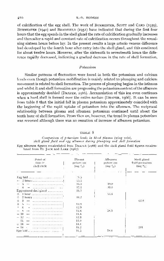
Similar patterns of fluctuation were found in both the potassium and calciumlevels even though potassium mobilisation is mainly related to plumping and calciummovement is related to shell formation. The process of plumping begins in the isthmusand whilst it and shell formation are progressing the potassium content of the albumenis approximately doubled (DRAPER, ig66). Accumulation of this ion even continueswhen a hard shell is formed over the entire surface (DRAPER, zg66). It can be seenfrom table 8 that the initial fall in plasma potassium approximately coincided withthe beginning of the rapid uptake of potassium into the albumen. The reciprocalrelationship between plasma and albumen potassium continued until about thetenth hour of shell formation. From then on, however, the trend in plasma potassiumwas reversed although there was no cessation of increase of albumen potassium.
No arterio-shell gland venous potassium differences were found throughoutalmost the whole of the plumping/shell formation period and this was to be expectedas, on the basis of the amount of potassium transferred into the albumen over theperiod of twenty hours, a rough calculation only gives an average A-V. differenceof o.2 mg p. 100, which would not become evident in the results. However, a diffe-rence of about 1-2 mg p. 100 was measured during the last three hours of shell forma-tion. This can probably be explained by some results of EL JACK and LAKE (1967)(table 8). They measured the ionic constituents of the shell gland fluid at two points,during plumping and shortly before the egg was laid. They found that the potassiumlevel of plumping fluid was only 16 mmol Jl. whilst that taken towards the end of shellformation was 75 mmol/1. If such an unusually high potassium level could be shownto occur only during the last three hours of shell formation, then this would accountfor the arterio-venous difference during this period.
Sodium
Whilst plumping is occurring the total amount of sodium in the albumenincreases by 50 p. 100, from approximately no mmol. to approximately 160 mmol.
(DRAPER, i966). However, owing to the very large uptake of water, the concentrationin the albumen drops from 127 mmol/1. in the magnum to about 60 mmol/1. whenlaid. These comparatively small amounts of sodium were apparently not large enoughto show up as either an overall plasma variation or as an A-V. difference.
There was one rather anomalous factor present in the plasma sodium results.The control results were consistently about qo mg p. 100 lower than the experimentalresults. There does not seem to be, at present, any satisfactory explanation for this.
In the cases of the pH, the calcium and the potassium results it was noted thata maximum level was found at about the time of entry of an egg into the shell glandand a minimum level occurred at, or just after, the mid-point of the period of shellformation. In all three cases the levels returned gradually towards normal after theminimum point was passed and thus, although shell formation continued steadilyfor several more hours, the trend in all these three parameters was reversed. Becauseof this close similarity in time and trend it seemed possible that there was somecommon cause or factor underlying all three. This common factor may be the activity(excretory or resorbtive) of the kidney, but it will not be possible to demonstratethis until the complete pattern of excretion during shell formation is known.
ACKNOWLEDGEMENTS
The author wishes to express his thanks to Miss C. FREEMAN for technical assistance.The Radiometer blood acid-base equipment was purchased with a grant from the Central
Research Fund of the University of London.
SUMMARY
r. Cannulations of the inferior oviducal vein, the sciatic artery and the sciatic vein wereperformed upon actively-laying, anaesthetised hens. A control series of unansesthetized birdswere cannulated only in the wing vein. All cannulac were sampled at hourly intervals during theshell formation cycle and blood pH, plasma total calcium, potassium and sodium were measured.
2. pH. Arterial pH was at a maximum of 7.31 two hours after the egg entered the shell glandand at a minimum of 7.43 at thirteen hours of shell formation. The shell gland venous blood ingeneral followed the arterial trends at a more acid level but, during shell formation, an increasing:1-!’. difference developed, reaching a maximum of 0.079 pH units at IS hours of shell formation.
3. Calcium. Cyclical fluctuations of plasma total calcium were found with a maximum level(20 mg p. 100) during the quiescent period and a minimum level (16 mg p. ioo) at 10 hours ofshell formation. During shell formation an A-V. difference of 2-4 mg p. ioo developed.
4. Potassium. A cyclical pattern of plasma potassium associated with the shell formationcycle was again found. Maximum levels (17 mg p. 100) occurred during the quiescent period andminimum values (12 mg p. ioo) at 4 to 8 hours of shell formation. An A-V. potassium difference(r-2 mg p. 100) was only found during the last three hours before lay.
5. Sodium. No cyclical pattern of sodium values or consistent A-V. differences were foundwhich could be associated with the shell formation cycle.
6. The significance of the results was discussed in relation to the processes of plumping andshell formation.
RÉSUMÉ
VARIATIONS DU pH ET DES CATIONS DU SANGLORS DE LA FORMATION DE LA COQUILLE DE L’œUF CHEZ LA POULE
i. Des canules ont été insérées dans l’artère et la veine sciatique et dans la veine inférieurede l’oviducte de poules anesthésiées. Des poules témoins non anesthésiées furent seulement canu-lées dans la veine alaire. Des prélèvements de sang ont été effectués chaque heure pendant la for-tion de la coquille et le pH du sang ainsi que la teneur en calcium total, potassium et sodium duplasma ont été mesurés.
2. pH. Les valeurs artérielles présentent un maximum de 7,51 deux heures après l’entréedans la glande coquillière et un minimum de 7,45 treize heures après le début de la formation dela coquille. Le sang veineux de la glande coquillière tend en général à suivre les valeurs artériellesà un niveau plus acide mais, pendant la formation de la coquille, une différence artério-veineusecroissante se développe qui atteint un maximum dc 0,079 unités pH au stade rs heures de forma-tion de la coquille.
3. Calcium. La teneur en calcium total du plasma présente des variations cycliques, le niveaumaximal (20 mg p. 100) intervenant pendant la période de repos de la glande coquillière et leniveau minimal (16 mg p. 100) après 10 heures de formation de la coquille. Au cours du dépôtde la coquille, une différence artério-veineuse de 2 à mg p. 100 se développe au niveau de laglande coquillière.
4. Potassium. Il apparaît également des fluctuations cycliques de la kaliémie liées à la for-mation de la coquille. Les valeurs maximales (17 mg p. 100) sont trouvées durant la période de reposet les valeurs minimales (12 mg p. ioo) entre et 8 heures de formation de la coquille. Il n’existeaucune différence entre les teneurs des artères et des veines de la glande coquillère sauf pendantles trois dernières heures précédant l’oviposition où un écart de i à 2 mg p. 100 est enregistré.
5. Sodium. Aucun comportement cyclique de la natrémie ni de différence artério-veineuseutérine ne sont trouvés qui puissent être associés au cycle de formation de la coquille.
6. La signification de ces résultats est discutée en rapport avec le !< plumping o et la formationde la coquille de l’oeuf.

REFERENCES
Arrnsasov R. S., 1967. Acid-base changes in the excreta of the laying hen. Vet. Rec., 80, 314-5.BRADFIELD J. R. G., i95i. Radiographic studies on the formation of the hen’s egg shell. J. Exp. Biol.28, izs-4o.
Buavtrarra B. I2., 1940. A study of the physical and chemical changes of the egg during its passagethrough the isthmus and uterus of the hen’s oviduct. J. Exp. Zool., 84, 445-Soo.BUR&dquo;ESTER B. R., SCOTT H. 1B1., CARD L. H., 1939. Rate of egg shell formation in the hen. Proc.
Vllth it’orld’s Poultry Seience Gongress, Cleveland, Ohio., pp. 9g-ror.CHARLES E., 140CBl!-N L., 1933. The serum calcium and magnesium level in the ovarian cycle of the
laying hen. Quart.]. Exp. Physiol., 23, 343-9.DRAPER !B1. H., i966. The transport of minerals to the white of the hen’s Pro,. ):IlItli ll’oild’s
Poultry Science congress, Kiev, pp. 333-6.EL JACK :B1. H., LAKE I’. E., 1967. The content of the principal inorganic ions and carbon dioxide in
uterine fluids of the domestic hen. J. Reprod. Pertil,. 13, 127-32.FpEEDMAN S. L., SruahiE P. D., 1963. Blood vessels of the chicl<en’s- uterus (shell gland). Amer. J.
Anat., 113, 1-7.FussEL M. IL, 1966. Observations on the intestinal absorption and urinary excretion of calcium andphosphorus in the hen. Proc. XIIith ¡Forld’s Poultry Science Congress, l!iev., pp. 3og-i3.GUTOWSK.a M. S., NIITCHELL C. A., 1945. Carbonic anhydrase in the calcification of the egg shell.
Poultry Sci., 24, 159-67.HERTELENUV F., TA VLOR T. G., 1961. Changes in blood calcium associated with egg shell calcification
in the domestic fowl. I. Changes in total calcium. Poultry .Sci., 40, io8-I4.HODGES R. D., 1965. The blood supply to the avian oviduct, with special reference to the shell gland.
J. Anat., 99, 485-506.Honcrs R. D., i966. Changes in blood pO. and pH during the formation of the egg shell in laying hens.P!oc..Y//7! 117orld’s Poultry Science Congre,.s, Kiev, pp. 3r4-3i9.IfoDCCS R. D., LORCHER K., 1967. Possible sources of the carbonate fraction of egg shell calcium car-
bonate. Nature, 216, 6og-io.HUNSAKER W. G., STURKIE P. D., 1961. Removal of calcium from uterine blood during shell forma-
tion in the chicken. 1’oultry Sci., 40, 1348-52.KNOWLES H. R., HART E. B., Hwcrr:v J. G., r93!. The variation in the calcium level of the blood of
the domestic fowl. Poultry Sci., 14, 83-g.MONGIN P., Lwcwssncve L., 1964. The physiology of the formation of the shell of the hen’s egg and
the acid-base equilibrium of the blood (in French). C. R. Acad. Sci., Paris, 258, 3093-4.POLIN 17., STURKIE P. D., 1957. The influence of the parathyroid on blood calcium levels and shell
deposition in laying hens. Endocrixology, 60, 778-784.Tamur P., 1960. Colorimetric micro-determination of calcium in serum. Analyst, 85, 889-94.TYLER C., r9;¢. Studies on egg shells. IV. The site of deposition of radio-active calcium and phosphorus.J. Sci. Food Agric., 5, 335-9.IVINGET C. M., SMITH A. H., 1958. Changes in the plasma calcium concentr ti r during egg formation.
Poultry .Sci., 37, go9-m.B’l’mcaT C. 31., SMITH A. H., HoovER G. N., 1958. Arterio-vcnons differences in plasma calcium
concentration in the shell gland of the laying hen during ;hell formation. Poatltry Sci., 37, 1325 8.lvooToN I. D. P., 1964. Micro-analysis in rnedical biochemistry. London, J. and A. Churchill Ltd. p. 71


![Synthesis of Hydroxyapatite from egg shell and preparation ... · Ahmed M. Saeed [6] have worked on synthesis of calcium hydroxyapatite powder from hen’s eggshell and orthophosphoric](https://img.pdfslide.us/doc/110x75/603a19cdd92f8913c757fb39/synthesis-of-hydroxyapatite-from-egg-shell-and-preparation-ahmed-m-saeed-6.jpg)