Embed Size (px)
Citation preview
Biology 224Human Anatomy and Physiology IIWeek 4; Lecture 1; MondayDr. Stuart S. Sumida
Changes in the Circulatory and Respiratory Systems at
Birth
FETAL RESPIRATORY & CIRCULATORY CONDITIONS
•Lungs small and collapsed.•Lungs filled with amniotic fluid.•Lung volume too small to allow complete output of right atrium through lung circuit.•(Another way of saying this: Back-pressure of lungs too great to allow much blood flow through lungs.)•There is enough blood flow to allow growth and development, but not gas transfer.•No need for gas transfer in lungs while in utero.
So...
Before birth, it would be desirable for blood leaving the right atrium to – for the most part – bypass the lung. There are two ways:
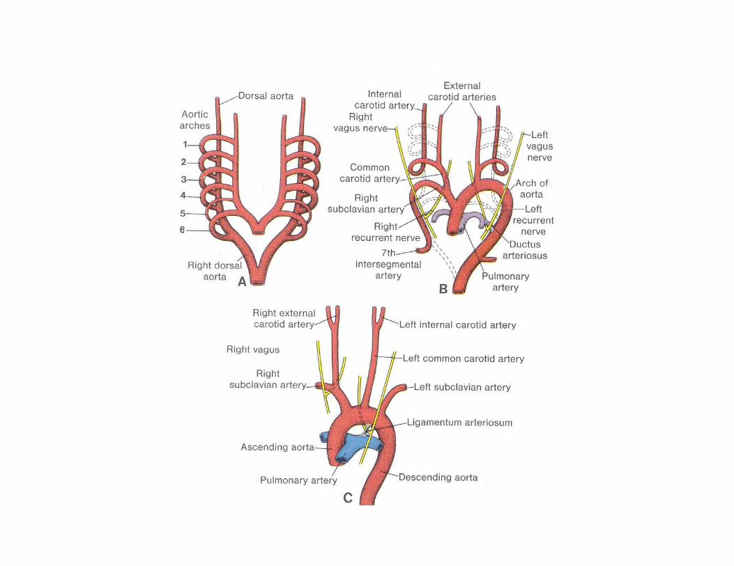
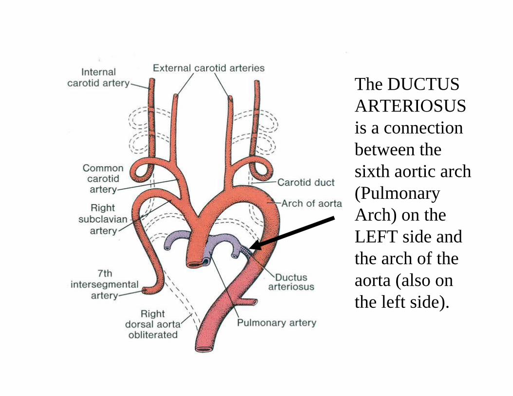
1st: This is done via a structure called the DUCTUS ARTERIOSUS. The DUCTUS ARTERIOSUS is a connection between the sixth aortic arch (Pulmonary Arch) on the LEFT side and the arch of the aorta (also on the left side).
2nd: The FORAMEN OVALE is a hole between the right and left atria, allowing blood to pass from the right side to the left and not ever get pumped to the lungs.
The DUCTUS ARTERIOSUS is a connection between the sixth aortic arch (Pulmonary Arch) on the LEFT side and the arch of the aorta (also on the left side).
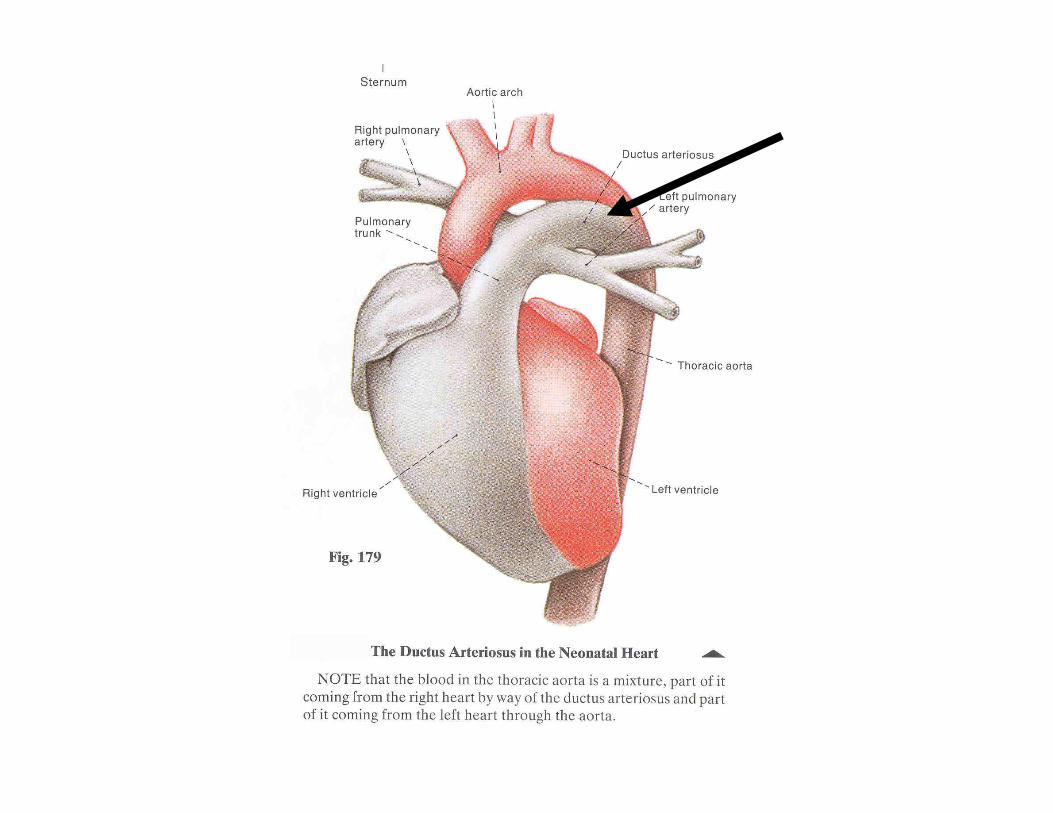
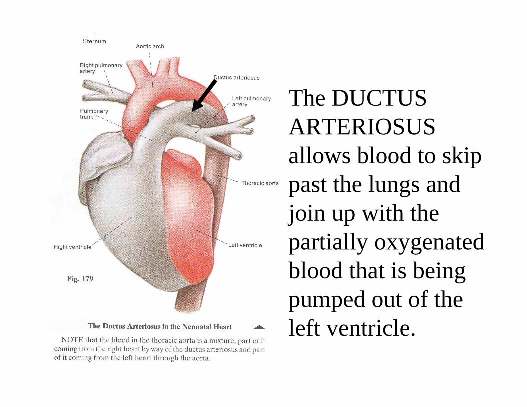
The DUCTUS ARTERIOSUS allows blood to skip past the lungs and join up with the partially oxygenated blood that is being pumped out of the left ventricle.
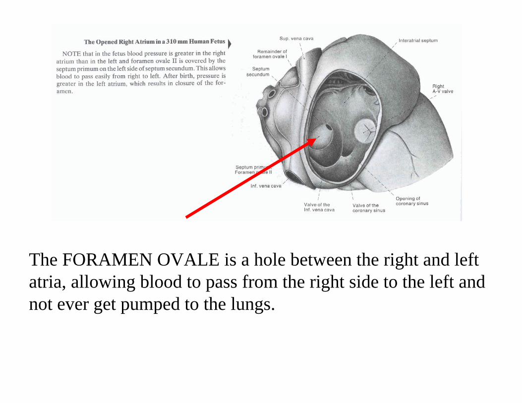
The FORAMEN OVALE is a hole between the right and left atria, allowing blood to pass from the right side to the left and not ever get pumped to the lungs.
So......where does spent blood from the fetus go?
Well, mom of course. It goes to mom via the UMBILICAL VEIN.
Remember that the distal end of the internal iliac vein is the old connection of the UMBILICAL VEIN which goes toward the placenta.
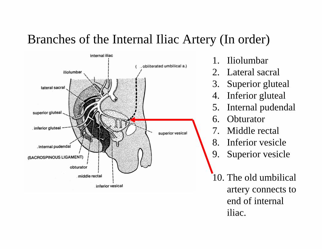
Branches of the Internal Iliac Artery (In order)1. Iliolumbar2. Lateral sacral3. Superior gluteal4. Inferior gluteal5. Internal pudendal6. Obturator7. Middle rectal8. Inferior vesicle9. Superior vesicle
10. The old umbilical artery connects to end of internal iliac.

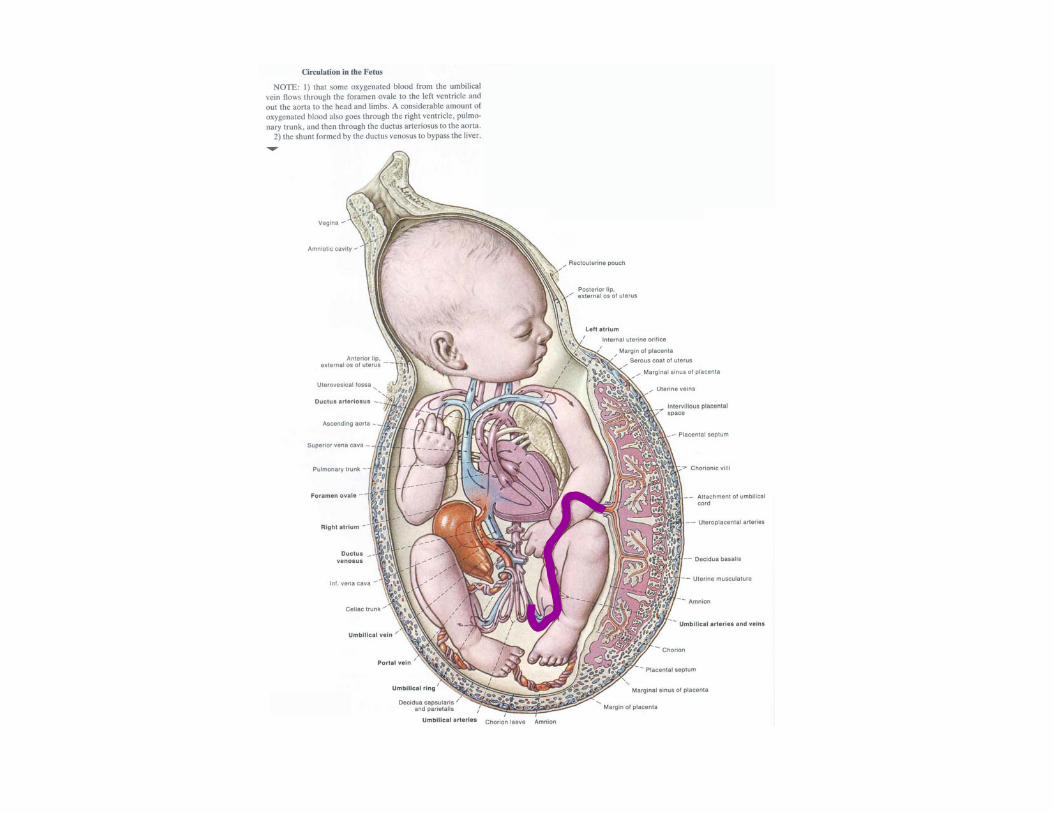
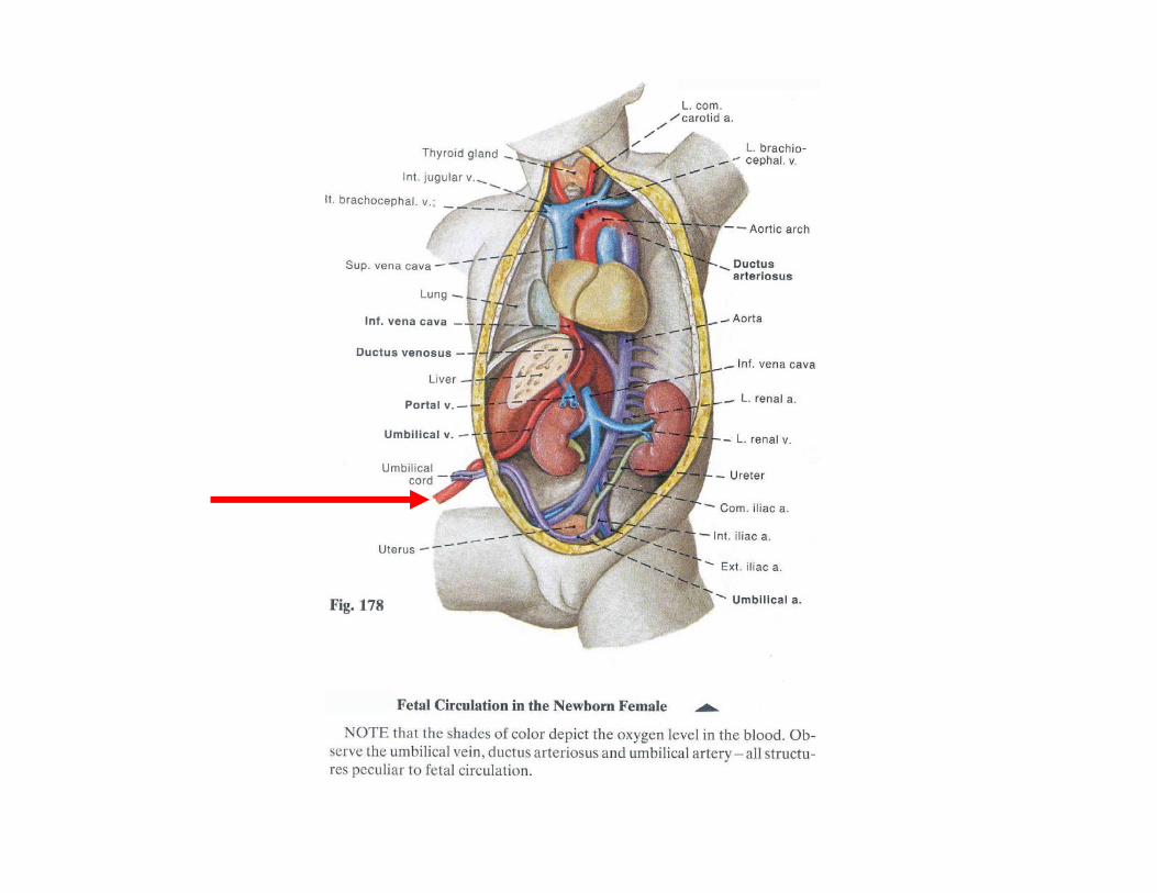
Arterial blood from mother & placenta – laden with nutrients and oxygen – come to fetus via (now) the umbilical vein hooked up to the inferior vena cava.
Thus, the blood returning to the fetal heart is actually, partially oxygen and nutrient rich.
This sets up the potential problem of it mixing in the right atrium with deoxygenated blood returning from the superior vena cava.
HOWEVER: Due to the angle of entry of the blood from the inferior vena cava and superior vena cava, their streams do not mix much.
MORE OXYGEN-RICH BLOOD FROM INFERIOR VENA CAVA PASS THROUGH FORAMEN OVALE OVER TO THE LEFT SIDE.
From left atrium, moderately oxygen-rich blood passes to left ventricle and then out arch of aorta.
Blood richest in oxygen & nutrients goes to heart wall, head (brain), neck, and arms.
Somewhat more oxygen/nutrient depleted blood from superior vena cava – of course – enters right atrium, then right ventricle, then pulmonary artery.
MOST of the right ventricular blood exits via the left pulmonaryarterial branch – then to the DUCTUS ARTERIOSUS.
This means that it eventually mixes with somewhat richer blood in the descending aorta, but ONLY AFTER the aorta has delivered blood to head, neck, and arms.
MORE ON GETTING FRESH BLOOD IN...
Remember, UMBILICAL VEINS enter the inferior vena cava. But soon, they hook up with the hepatic portal system.
The right UMBILICAL VEIN eventually degenerates, leaving only the left.
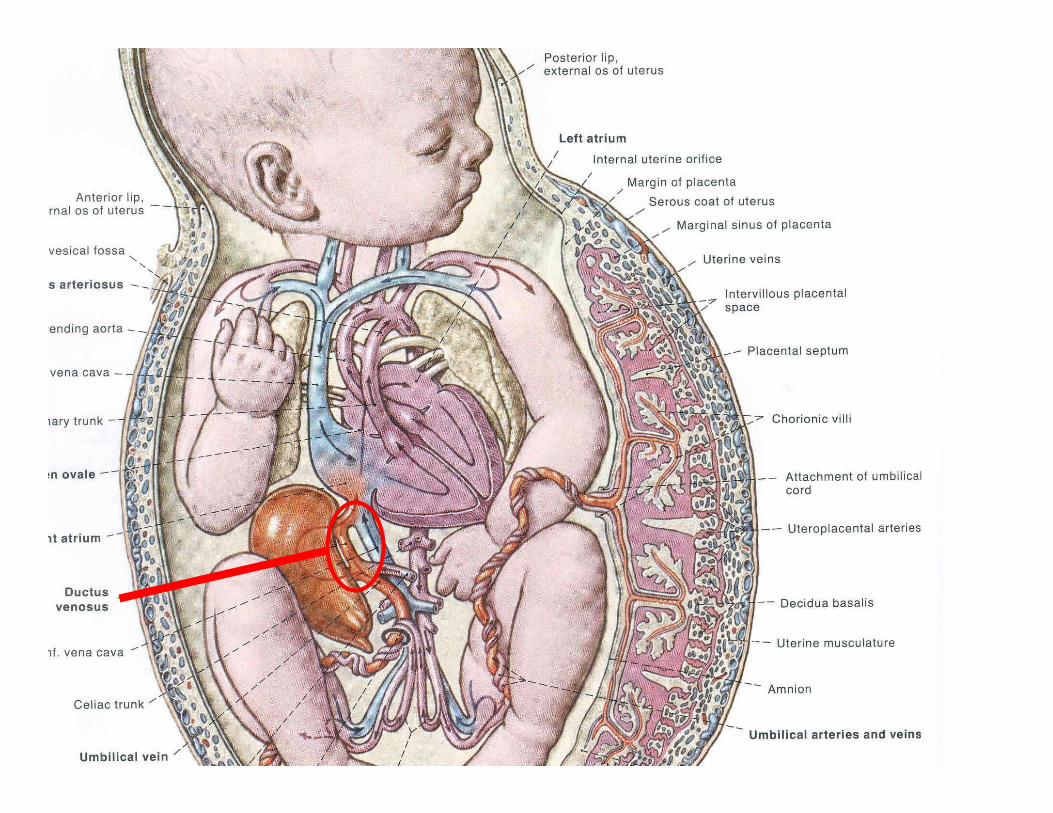
It would not be desirable to mix oxygenated blood (from mom, umbilical vein) with deoxygenated in liver. So – a new pathway from the placenta to the upper region of the inferior vena cava bypasses the liver. It is called the DUCTUS VENOSUS. It runs straight from the portal vein, bypassing the liver, to the inferior vena cava.
CIRCULATORY CHANGES AT BIRTH – I
Things that need to be changed:
1. If fetal circulatory pattern were to persist after birth, right atrialblood would continuously spill to the left side via the foramen ovale.
2. Blood of the pulmonary arch would miss the lung and go to the aorta, mixing there.
3. This would result in hypoxia – lack of adequate oxygen in arterial blood.
4. Lungs would be bypassed continuously, deteriorating quickly.
To prevent this, foramen ovale and ductus arteriosusMUST CLOSE AT THE MOMENT OF BIRTH.
CIRCULATORY CHANGES AT BIRTH – II
1. In the fetus, foramen ovale is covered by an interatrial flap valvethat allows blood to pass from right to left atrium, but not in opposite direction.
2. After child is delivered, fetal blood vessels in placenta contract, returning blood they contain (about 100 milliliters) to the infant.
3. Child takes first breath. Lung capillaries fill with blood. The first pulse of oxygenated blood returns from lungs to left atrium with next heartbeat.
4. As a result of suddenly higher flood of blood INTO let atrium, PRESSURE IN THE LEFT ATRIUM IS NOW HIGHER THAN THAT IN THE RIGHT. The increased pressure holds the interatrial flap valve shut – thus CLOSING FORAMEN OVALE.
CIRCULATORY CHANGES AT BIRTH – III
5. At moment of first breath, a powerful VASOCONSTRICTION closes the ductus arteriosus. (In a few days fibrous tissue begins to occlude it; in adult, it is left as a ligament, the LIGAMENTUM ARTERIOSUM.)
6. When blood stops flowing in from placenta via umbilical vein, ductus venosus closes off.
7. In a few days fibrous tissue begins to occlude it; in adult, it is left as a ligament, the LIGAMENTUM VENOSUM. The umbilical vein become the ROUND LIGAMENT OF THE LIVER (also known as “ligamentum teres”).
8. Together, ligamentum venosum and round ligament tie postnatal portal vein to navel ventrally and inferior vena cava dorsally.
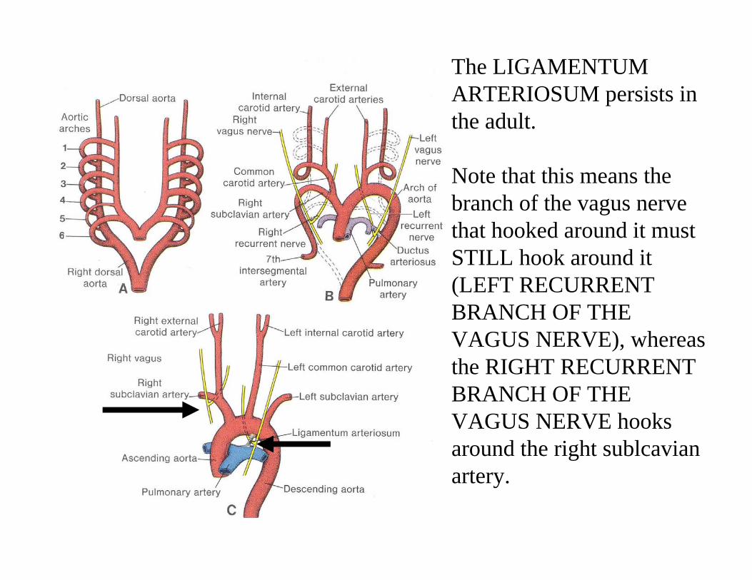
The LIGAMENTUM ARTERIOSUM persists in the adult.
Note that this means the branch of the vagus nerve that hooked around it must STILL hook around it (LEFT RECURRENT BRANCH OF THE VAGUS NERVE), whereas the RIGHT RECURRENT BRANCH OF THE VAGUS NERVE hooks around the right sublcavianartery.
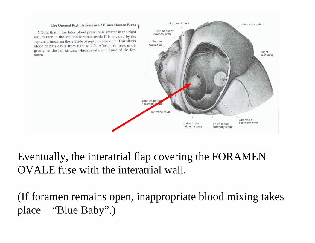
Eventually, the interatrial flap covering the FORAMEN OVALE fuse with the interatrial wall.
(If foramen remains open, inappropriate blood mixing takes place – “Blue Baby”.)
OTHER CHANGES AT BIRTH – HEMOGLOBIN FUNCTION
Due to fetal mixing of blood, FETAL HEMOGLOBIN is adapted specially.
FETAL HEMOGLOBIN can combine with oxygen even more easily than ADULT HEMOGLOBIN – so it can pick oxygen OFF of the adult hemoglobin.
This special property is lost within a few days after birth.
CHANGES NEAR BIRTH – LIVER FUNCTION
At approximately week 27-30, enzyme function in the fetal liver changes to promote storage of GLYCOGEN.
This stores up glycogen as a food source in case of temporary starvation between birth and mother’s first ability to produce milk for nursing.