Embed Size (px)
Citation preview
Blood and Bone Marrow
Histology SSNNovember 18, 2004
Presented by:Nadia Goodwin & Missy Walker
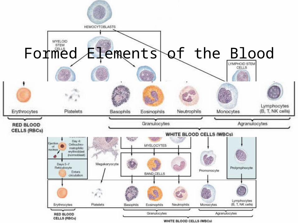
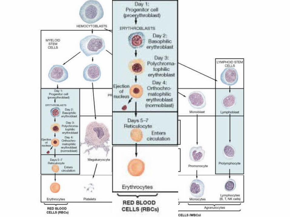
Formed Elements of the Blood
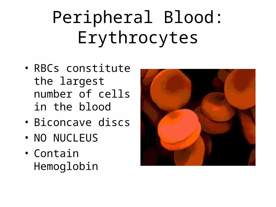
Peripheral Blood: Erythrocytes
• RBCs constitute the largest number of cells in the blood
• Biconcave discs• NO NUCLEUS• Contain Hemoglobin
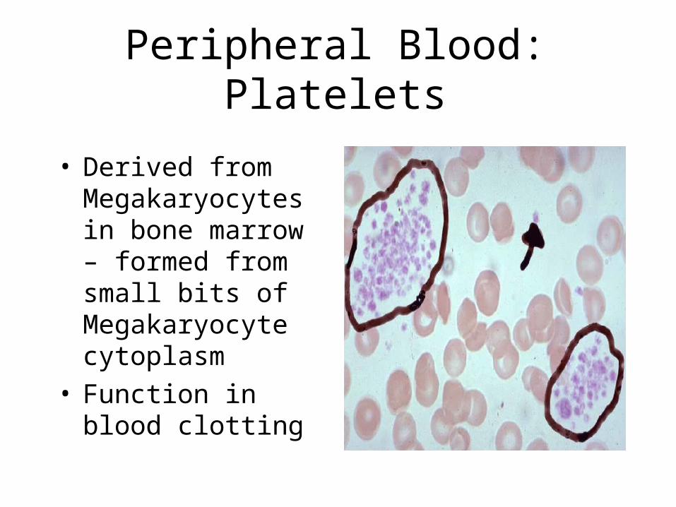
Peripheral Blood: Platelets
• Derived from Megakaryocytes in bone marrow – formed from small bits of Megakaryocyte cytoplasm
• Function in blood clotting
Peripheral Blood: Leukocytes
• GRANULOCYTES– Neutrophils– Basophils– Eosinophils
• AGRANULOCYTES– Lymphocytes (T and B cells)– Monocytes (Macrophages)
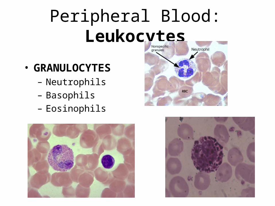
Peripheral Blood: Leukocytes
• GRANULOCYTES– Neutrophils
– Basophils
– Eosinophils
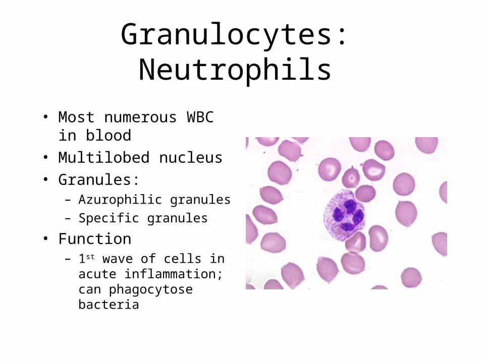
Granulocytes: Neutrophils
• Most numerous WBC in blood
• Multilobed nucleus
• Granules:– Azurophilic granules
– Specific granules
• Function– 1st wave of cells in acute
inflammation; can phagocytose bacteria
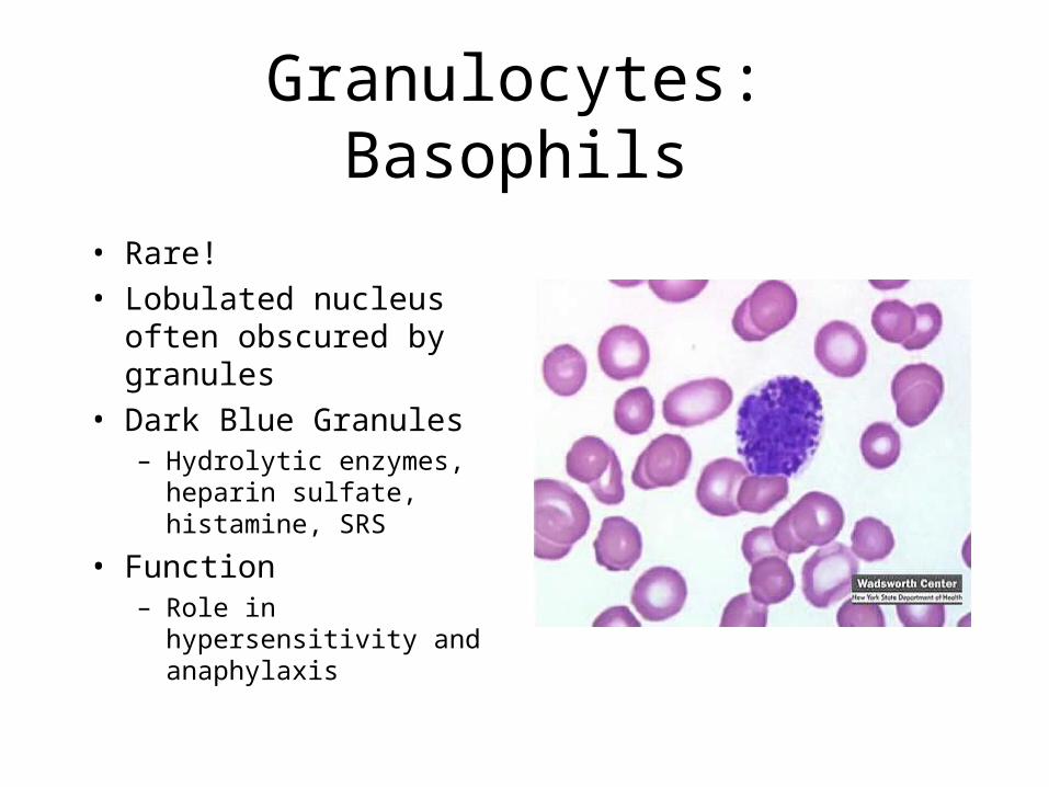
Granulocytes: Basophils
• Rare!
• Lobulated nucleus often obscured by granules
• Dark Blue Granules– Hydrolytic enzymes,
heparin sulfate, histamine, SRS
• Function– Role in hypersensitivity and
anaphylaxis
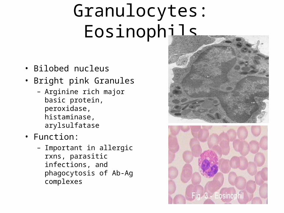
Granulocytes: Eosinophils
• Bilobed nucleus
• Bright pink Granules– Arginine rich major basic
protein, peroxidase, histaminase, arylsulfatase
• Function: – Important in allergic rxns,
parasitic infections, and phagocytosis of Ab-Ag complexes
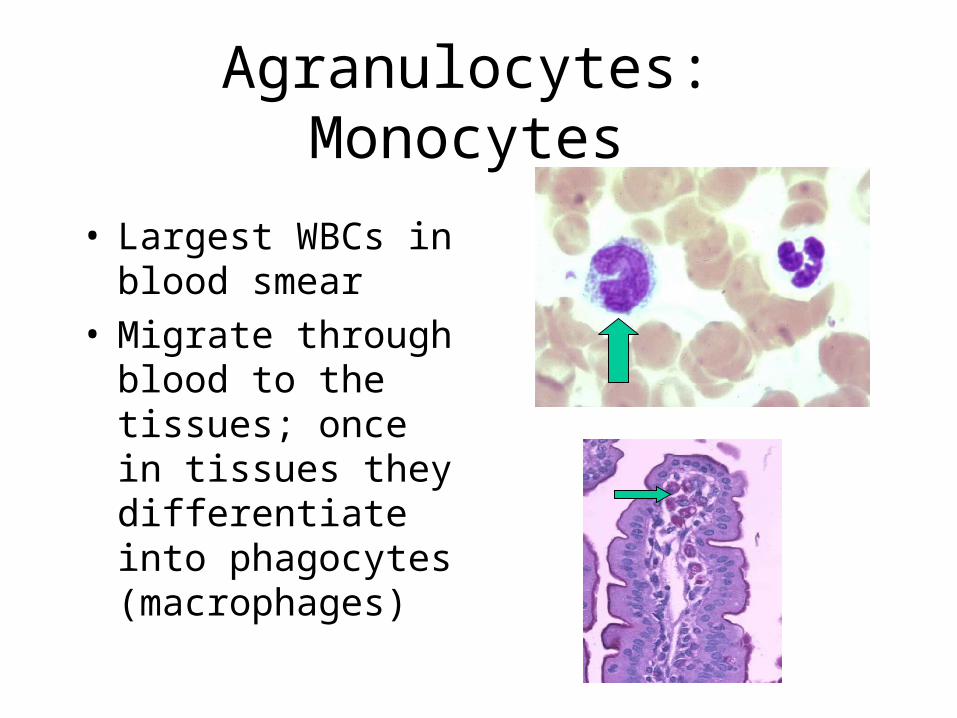
Agranulocytes: Monocytes
• Largest WBCs in blood smear
• Migrate through blood to the tissues; once in tissues they differentiate into phagocytes (macrophages)
Agranulocytes: Lymphocytes
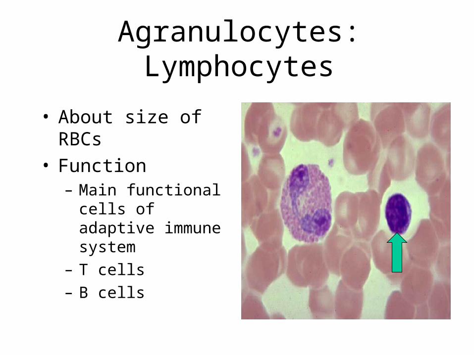
• About size of RBCs• Function
– Main functional cells of adaptive immune system
– T cells
– B cells
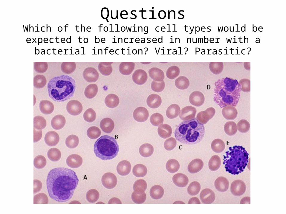
QuestionsWhich of the following cell types would be expected to be increased in
number with a bacterial infection? Viral? Parasitic?
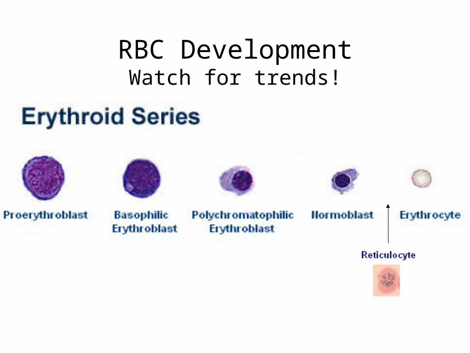
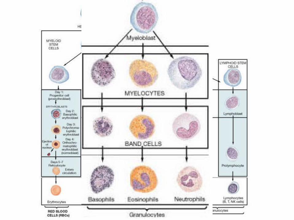
RBC DevelopmentWatch for trends!
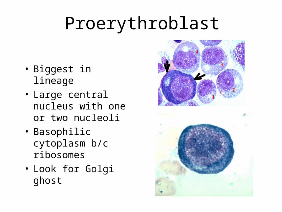
Proerythroblast
• Biggest in lineage• Large central nucleus
with one or two nucleoli
• Basophilic cytoplasm b/c ribosomes
• Look for Golgi ghost
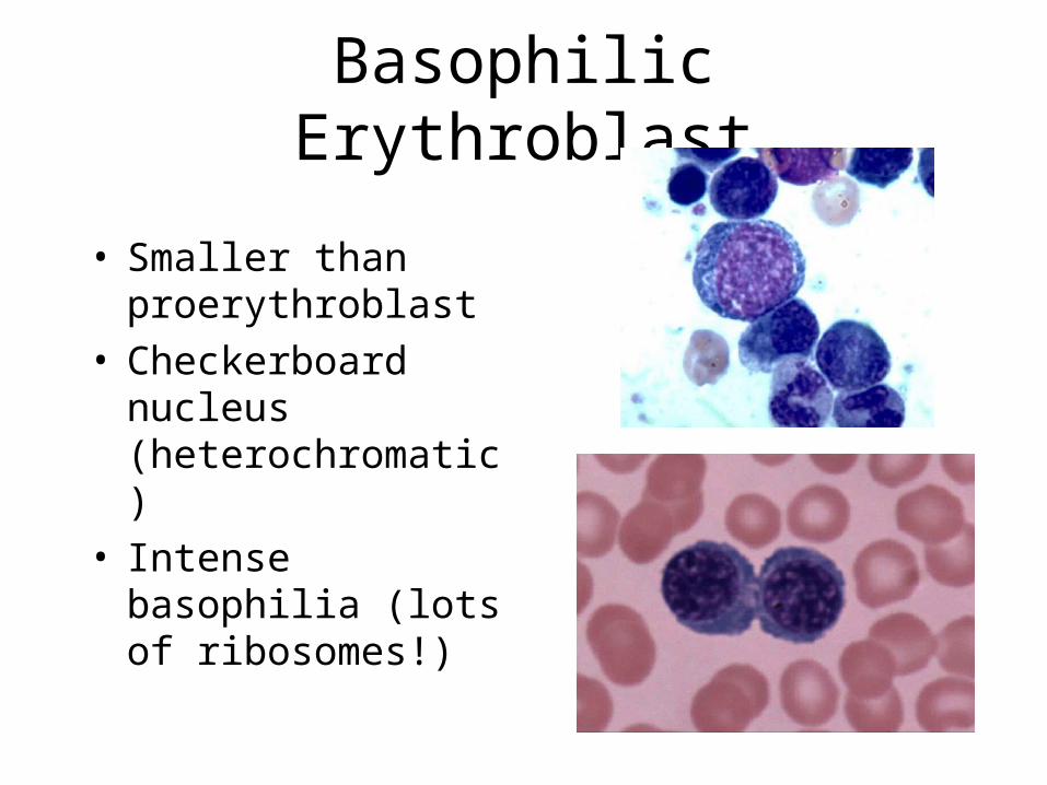
Basophilic Erythroblast
• Smaller than proerythroblast
• Checkerboard nucleus (heterochromatic)
• Intense basophilia (lots of ribosomes!)
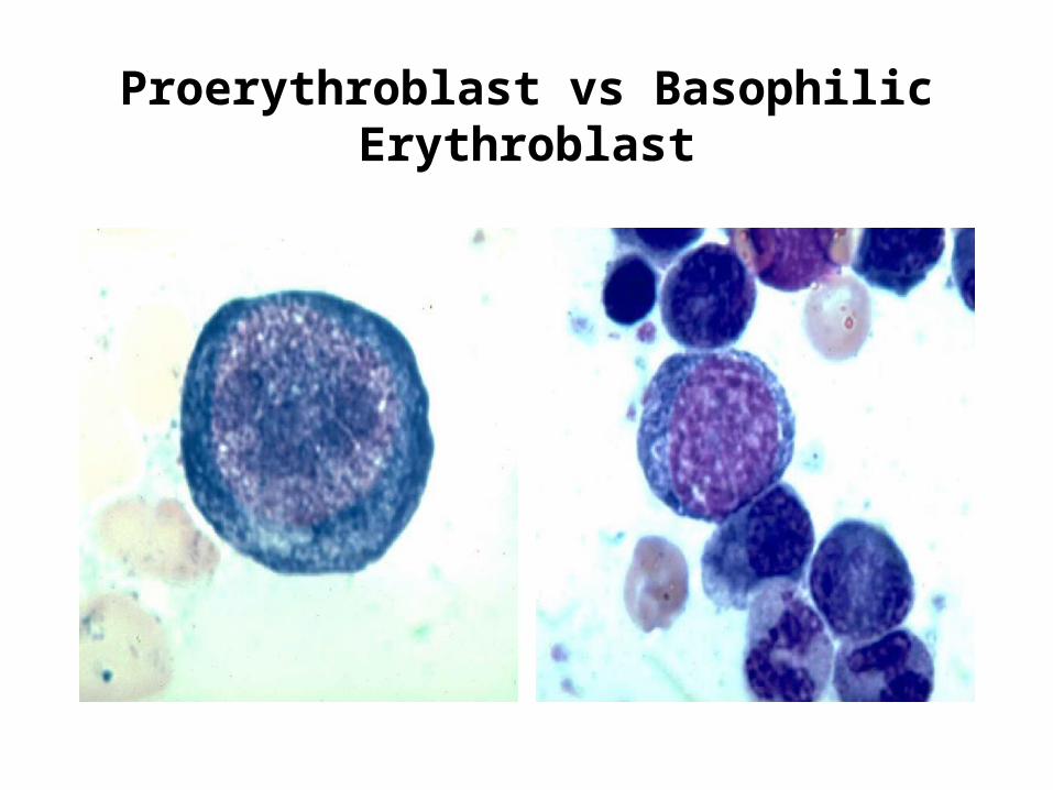
Proerythroblast vs Basophilic Erythroblast
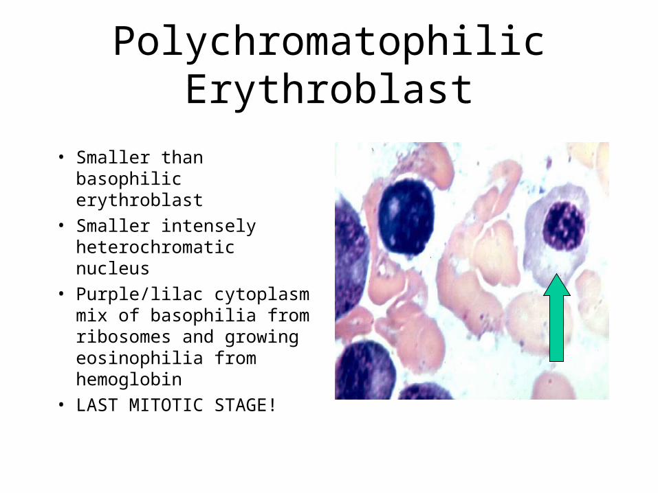
Polychromatophilic Erythroblast
• Smaller than basophilic erythroblast
• Smaller intensely heterochromatic nucleus
• Purple/lilac cytoplasm mix of basophilia from ribosomes and growing eosinophilia from hemoglobin
• LAST MITOTIC STAGE!
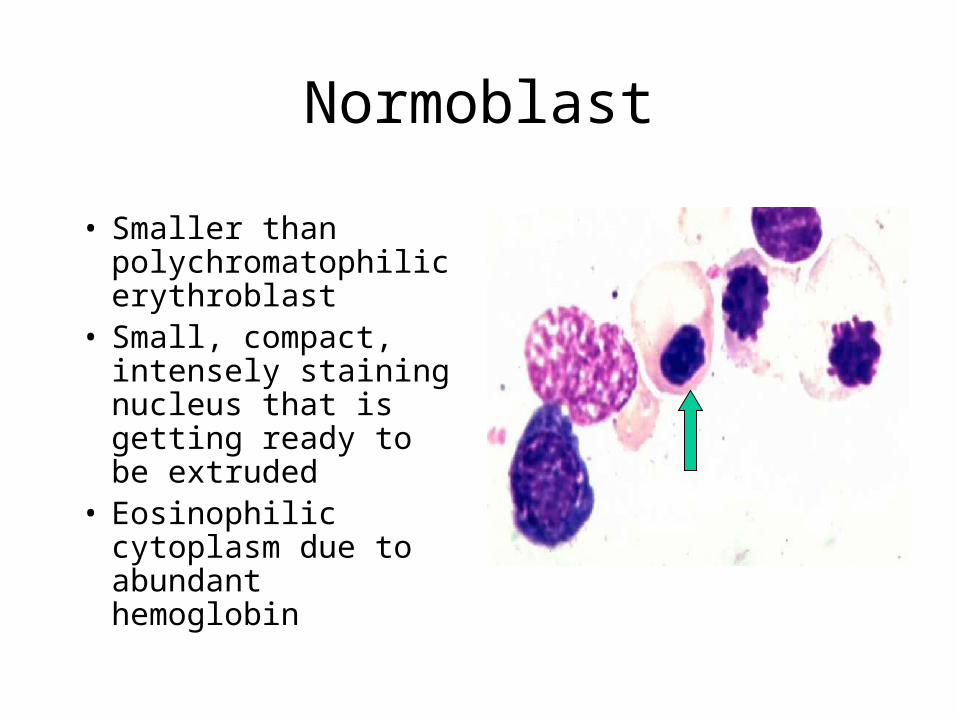
Normoblast
• Smaller than polychromatophilic erythroblast
• Small, compact, intensely staining nucleus that is getting ready to be extruded
• Eosinophilic cytoplasm due to abundant hemoglobin
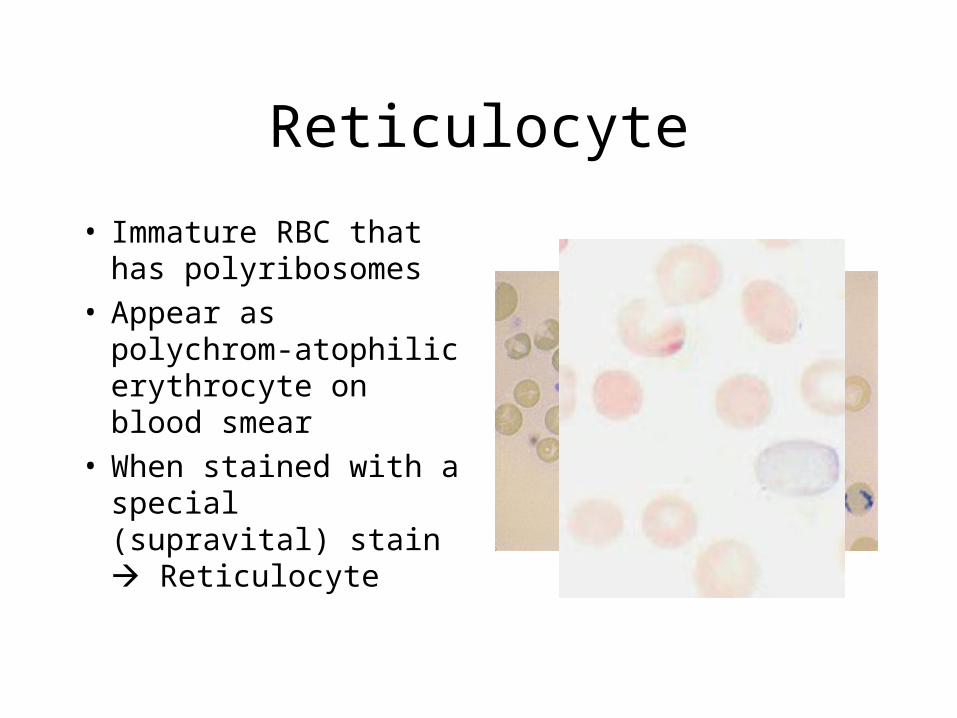
Reticulocyte
• Immature RBC that has polyribosomes
• Appear as polychrom-atophilic erythrocyte on blood smear
• When stained with a special (supravital) stain Reticulocyte
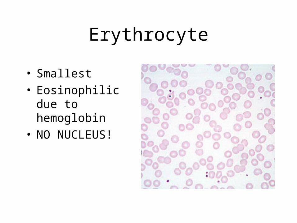
Erythrocyte
• Smallest• Eosinophilic due to
hemoglobin• NO NUCLEUS!
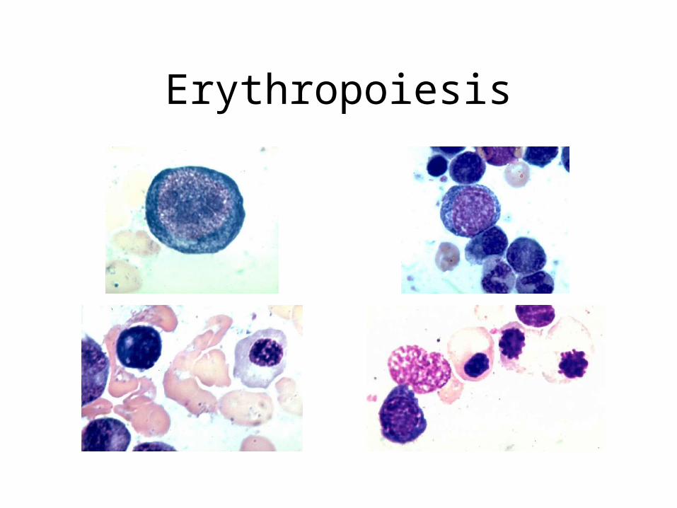
Erythropoiesis
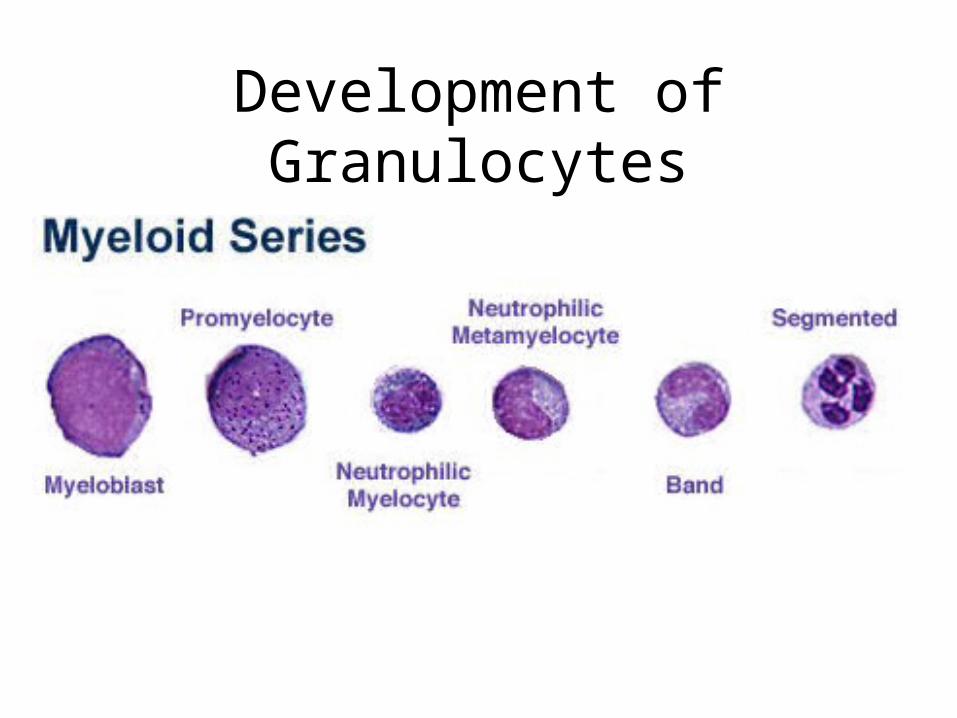
Development of Granulocytes
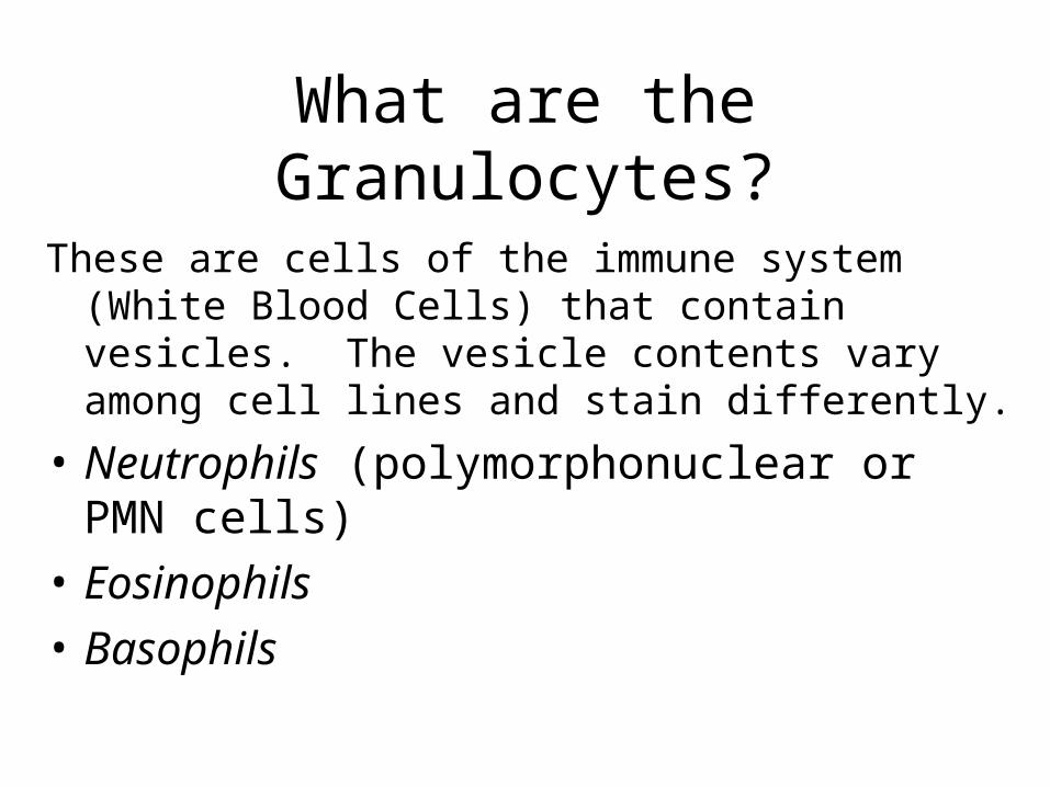
What are the Granulocytes?
These are cells of the immune system (White Blood Cells) that contain vesicles. The vesicle contents vary among cell lines and stain differently.
• Neutrophils (polymorphonuclear or PMN cells)
• Eosinophils
• Basophils
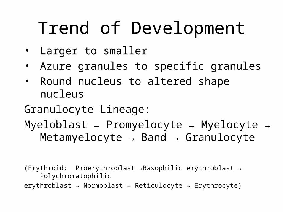
Trend of Development• Larger to smaller
• Azure granules to specific granules
• Round nucleus to altered shape nucleus
Granulocyte Lineage:
Myeloblast → Promyelocyte → Myelocyte → Metamyelocyte → Band → Granulocyte
(Erythroid: Proerythroblast →Basophilic erythroblast → Polychromatophilic
erythroblast → Normoblast → Reticulocyte → Erythrocyte)
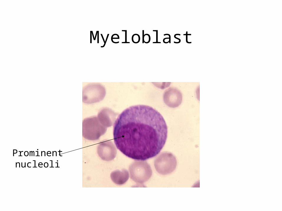
Myeloblast
Prominent nucleoli
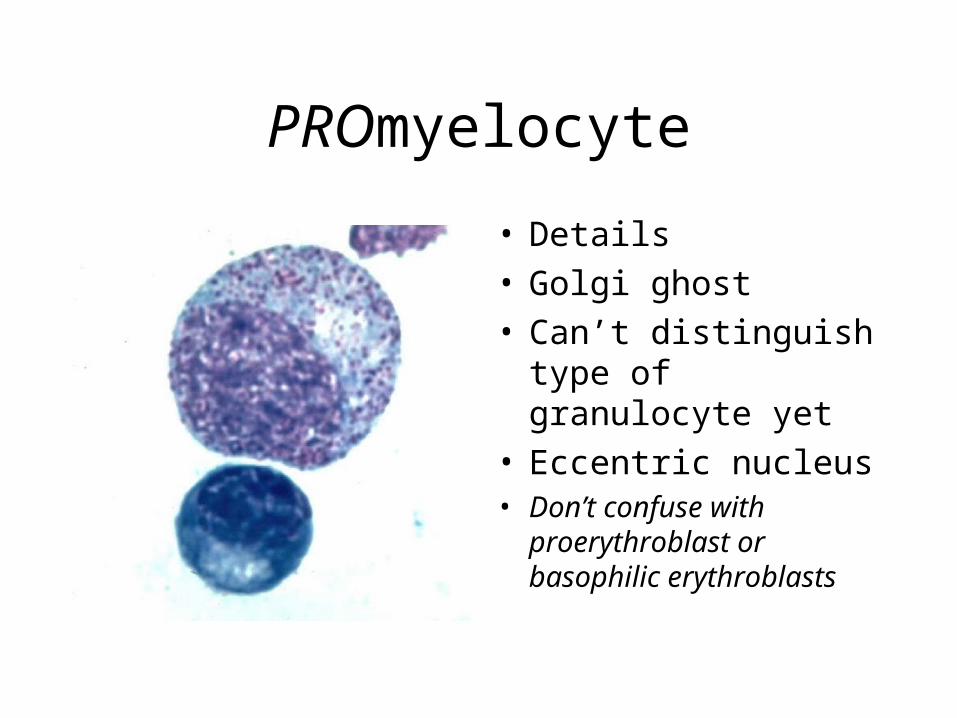
PROmyelocyte
• Details• Golgi ghost• Can’t distinguish type
of granulocyte yet• Eccentric nucleus• Don’t confuse with
proerythroblast or basophilic erythroblasts
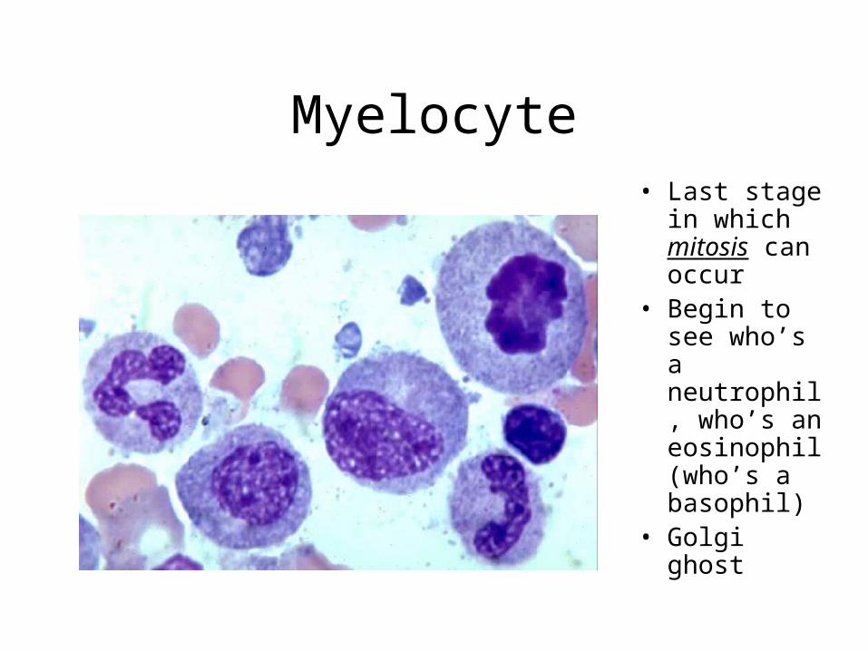
Myelocyte• Last stage in
which mitosis can occur
• Begin to see who’s a neutrophil, who’s an eosinophil (who’s a basophil)
• Golgi ghost
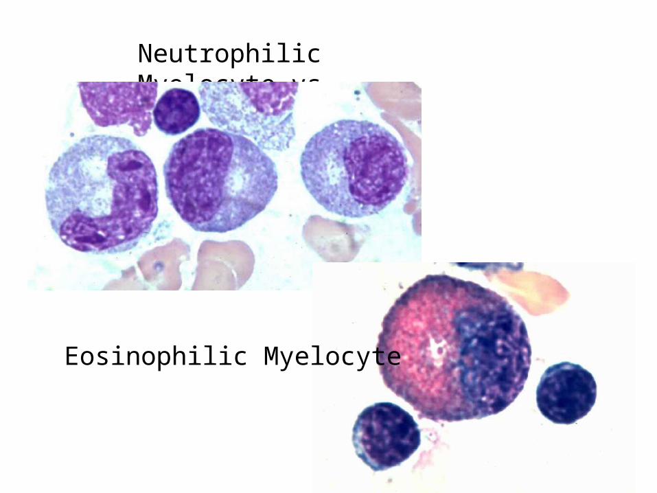
Neutrophilic Myelocyte vs
Eosinophilic Myelocyte
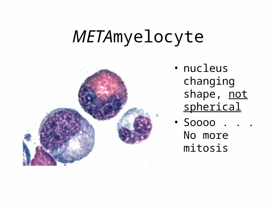
METAmyelocyte
• nucleus changing shape, not spherical
• Soooo . . . No more mitosis
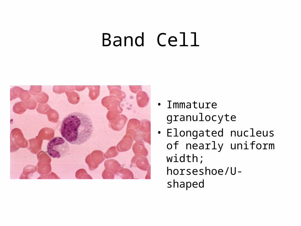
Band Cell
• Immature granulocyte• Elongated nucleus of
nearly uniform width; horseshoe/U-shaped
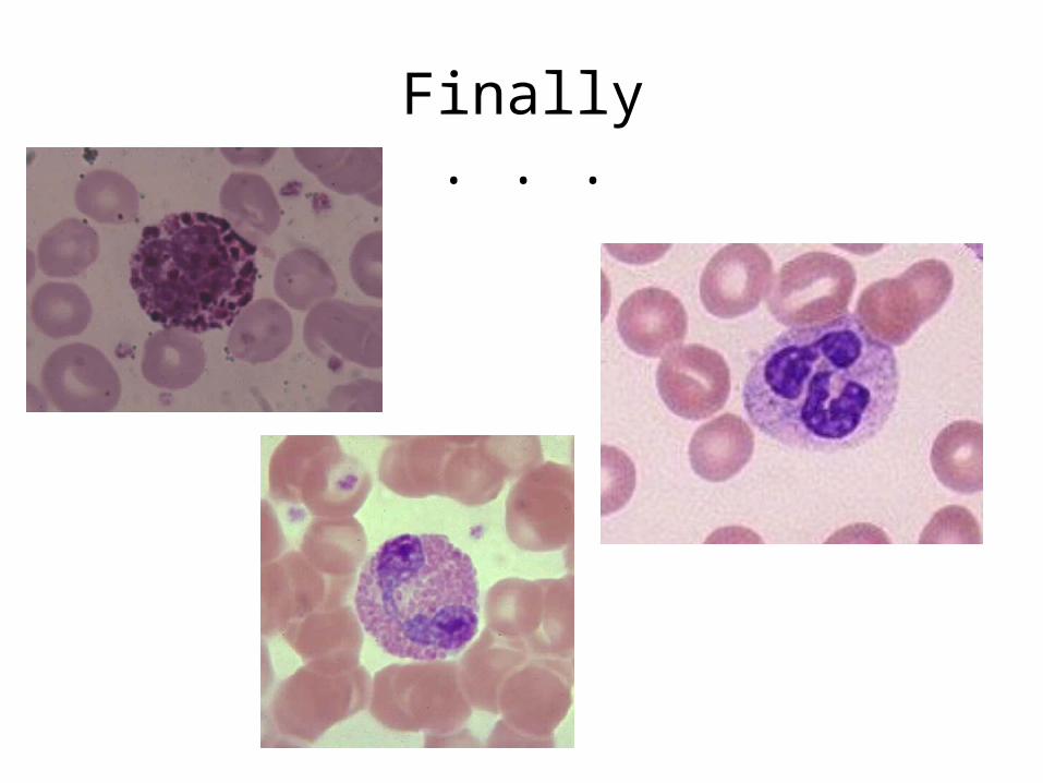
Finally . . .
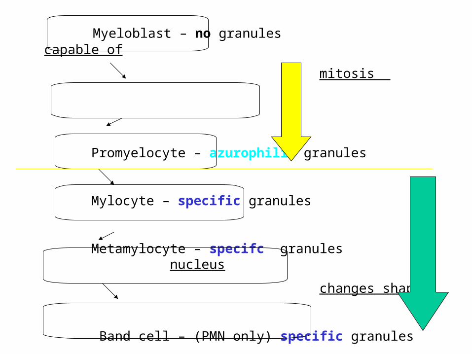
Myeloblast – no granules capable of
mitosis
Promyelocyte – azurophilic granules
Mylocyte – specific granules
Metamylocyte – specifc granules nucleus
changes shape
Band cell – (PMN only) specific granules
Mature Granulocyte – specific granules
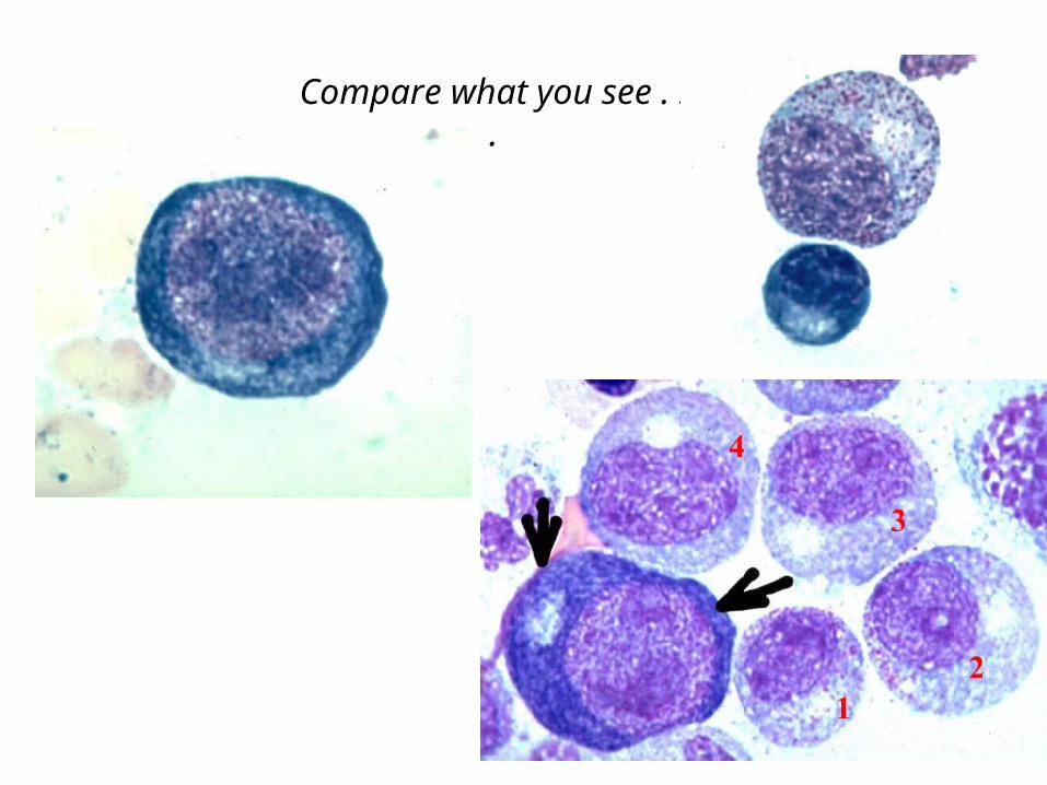
Compare what you see . . .
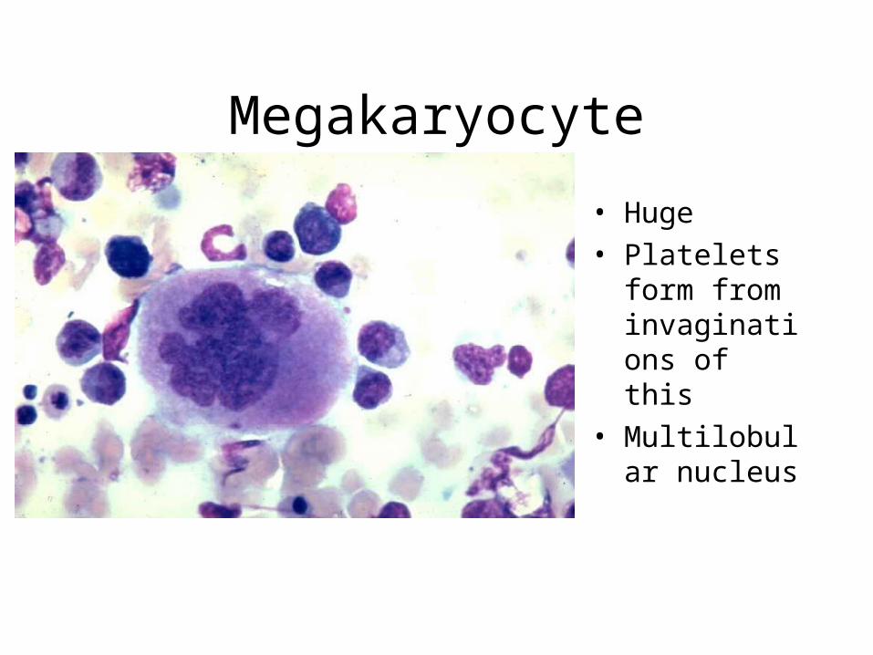
Megakaryocyte
• Huge
• Platelets form from invaginations of this
• Multilobular nucleus
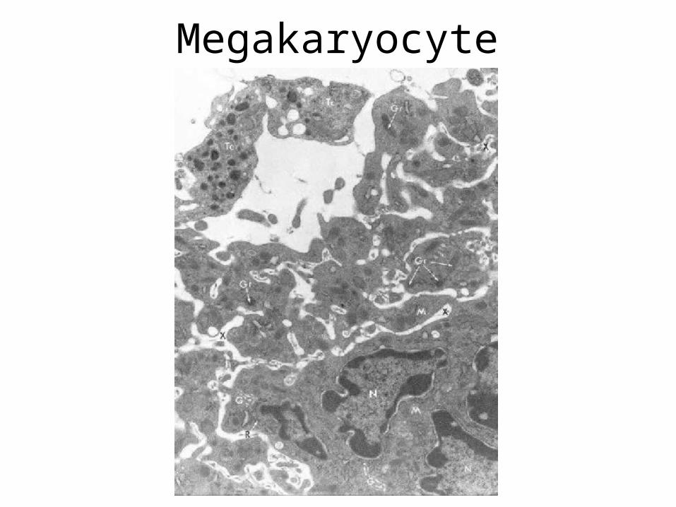
Megakaryocyte
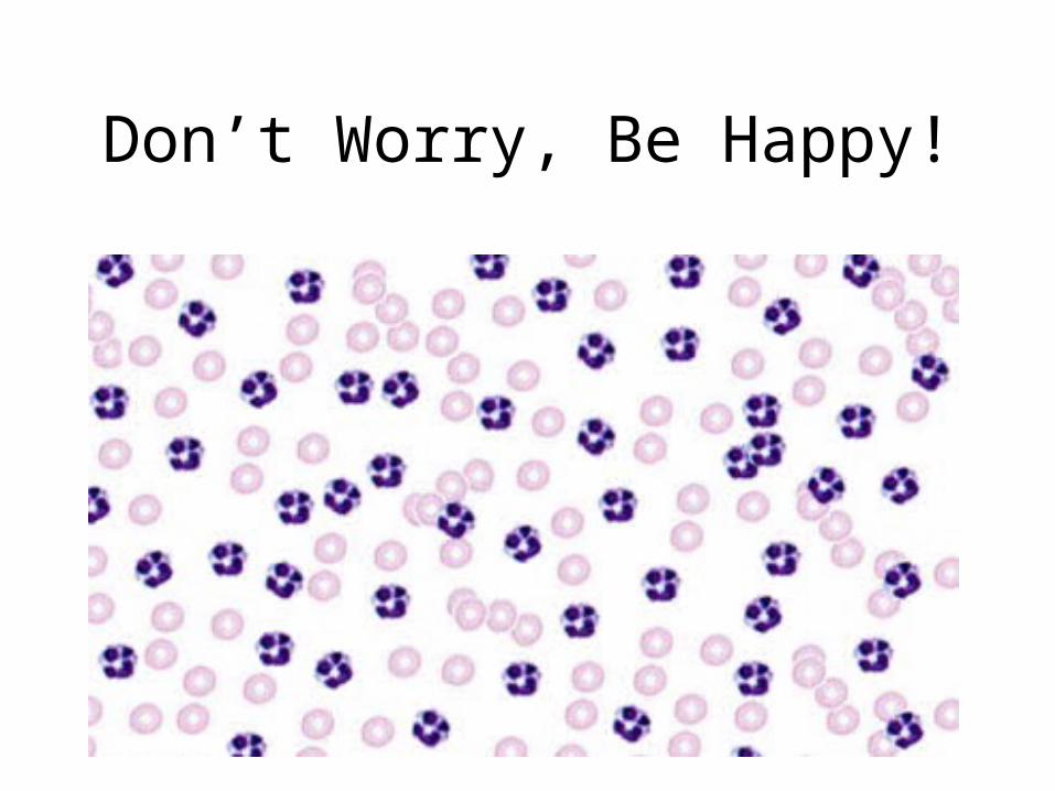
Don’t Worry, Be Happy!