Embed Size (px)
Citation preview

Blobs and Color Vision
DAVID H. HUBEL
Department of Neurobiology, Harvard Medical School, Boston, ~VIA 02115
Two main topics will thread their way through this paper: The struc- ture of the monkey striate cortex, including its inputs and outputs, and the physiological basis of color vision. When Margaret Livingstone and I began this work four years ago we did not set out to study color, but the structures in the cortex that we have been looking at turn out to be strongly involved in that sense. The work to be described here was done entirely in macaque and squirrel monkeys.
First, let me remind you of the structure and connections of the monkey striate cortex. In the classical visual pathway in mammals, infor- mation is transmitted from the eyes to the lateral geniculate body and from there to the primary visual cortex. Each geniculate layer receives its input from one eye only, and a cell in the lateral geniculate is conse- quently supplied by a single eye, the left or right, depending on the layer in which that cell is situated (Fig. 1). The magnocellular geniculate layers project to layer 4Co~ and the parvocellular to 4C6, with small contribu- tions to 4A and 6 (Fig. 2). The output of the cortex is from pyramidal cells of the upper (2 and 3) and the lowermost (5 and 6) layers; layer 4 cells do not project out of the cortex. Layers 2 and 3 send their axons mainly to other cortical regions, and it is these pathways that are presumably in- volved in perception. Layers 5 and 6 project mainly to subcortical areas, especially to the superior colliculus and, in a recurrent path, back to the lateral geniculate.
When responses were studied at different levels in the path from ret- ina to cortex, it was found that up to and including layer 4C cells re- sponded best to spots of light, and usually had concentric, center- surround receptive fields, whereas most or all cells in the upper (2 and 3) and lower (5 and 6) layers responded best to specifically oriented slits, bars, or edges of light (1). Taken literally, this would mean that the entire output of the striate cortex consisted of orientation-specific cells, and, in particular, that the entire projection of the upper layers to other cortical regions was in the form of orientation-specific cells. As we shall see, these generalizations turn out to require some revision.
91

92 Hubel
~" r
~.o
r~a~a
0 ~ -. 0 ,.C~ ~ "~ ~-
" ~ ' ~ 0
~ 5
~ . . -='S ~- -#= .~ " ~ b O ~
~0.~
< ~- ~ ~
. ~ o ~" o
. . ~ ~

Blobs and Color Vision 93
I =========================== I : : : : : : : : : : : : : : : : : : : : : : : : : : : : - i ==========================
!iiiiii:iil lliiii i iil E!ii !i t . . . ~ ~:i~:~i~:] li~.:~iiiiiiiiiii:~i~iiiii i~ 1 , l:i~.
iiiii �9 ,
ill',: ,3
OCULAR DOMINANCE COLUMNS
[•CONTRALATERAL EYE
IPSILATERAL EYE
6 c
Fig. 2. Diagram of geniculate input to striate cortex [From Hubel, D. H., and Wiesel, T. N. (1972), ]. Comp. Neurol. 146, 441-450, Fig. 18].
For years one has known that cortical cells are functionally segre- gated in two different ways. A fiber from a single layer of the lateral ge- niculate body, and hence from one eye, breaks up as it approaches layer 4 of the cortex into patches of terminals with terminal-free gaps between them (2). All the fibers belonging to that eye supply these same patches, and fibers from the other eye supply the gaps. This is the basis of the partial eye segregation in the form of eye-dominance columns (3). Such a segregation is absent or poorly developed in the squirrel monkey (4). The second system concerns receptive-field orientation. As one threads an electrode along the cortex parallel to the surface, the optimum stimulus orientation changes in small steps in a very regular way--about 10~ microns (5). If, instead, one goes downward through the cortex, orienta- tion stays constant. It follows from this orderliness that cells of similar

94 Hubel
orientation specificity are grouped in narrow vertical slabs arranged per- pendicular to the the cortical surface (5,6).
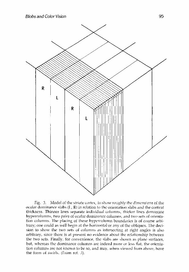
These two systems are shown in what we now term the Ice-Cube Model, Mark I (3) (Fig. 3). Classical anatomical staining methods, like the Golgi, or stains for Nissl substance or myelin, give no hint of these subdi- visions. A Nissl stain of a tangential section through layers 2 and 3 shows no subdivision of any kind. The ocular dominance columns have been demonstrated anatomically by a number of more modern methods, for example, by making geniculate lesions in single layers and using the Nauta technique for staining degenerating fiber terminals or by putting radioactive proline in one eye and letting it be transported by way of the geniculate to the cortex, where it can then be seen in autoradiographs in layer 4. So far the only way of seeing orientation columns has been through the use of the 2-deoxyglucose method (7). Figure 3 is necessarily a caricature: The two sets of columns are not known to be or thogonal-- in fact, they probably do not have any constant relat ionship--and the ori- entation columns are almost certainly not straight for any great distance.
Around 1977 there appeared reports of another kind of periodicity. Weber et al. (8) and, independently, Hendrickson et al. (9) and Hubel and Wiesel (4) all observed faint puffs of label in layer 3 of squirrel mon- key cortex after injection of proline into the eye or geniculate; one won- dered (wrongly) if these might in some way be concerned with ocular dominance. In 1978 Margaret Wong-Riley stained sections of cortex for the mitochondrial enzyme cytochrome oxidase and observed similar la- beled aggregations in layers 2 and 3 and also, faintly, in 5 and 6 (personal communication). She commented on the similarity of these structures to the proline puffs. It was not until 1980 that the cytochrome oxidase tech- nique was used to stain the upper layers of striate cortex in tangential sections (10,11). Astonishingly, the densities were then seen to form a quasiregular polka-dot pattern of patches, which we have designated, by the legitimate English term, "blobs" (12). These are shown in Fig. 4, a flat mounted section through layer 3 of macaque striate cortex, stained for cytochrome oxidase. The blobs are roughly 200 microns in diameter and are spaced at 0.5 mm intervals. (Area 18 also shows a series of coarser periodicities that we shall say more about later.) In macaques, they line up in parallel rows that intersect the 17-18 border at 90 ~ , suggesting some relationship to ocular dominance columns, and it has in fact been estab- lished by several independent methods that the blobs are aligned along the centers of ocular dominance columns (13,14). No such rows appear in the squirrel monkey, in which ocular dominance columns are lacking (Fig. 5a). In both species the blobs extend through layers 2 and 3, are not seen in 4, but reappear faintly in 5 and 6, where they lie in register with the upper layer blobs. In Fig. 5b, a deeper section through the same block of cortex, one can make out the blobs in layers 5 and 6, lying directly be- low those in 2 and 3. Layers 4A and 4C stain uniformly and densely. Fig- ure 5, a and b, also shows a set of regular, much coarser, periodicities in area 18; these have been of great interest to us in recent months.
Blobs and Color Vision 95
\
J
J
R
L
\
\
\
J
J
Fig. 3. Model of the striate cortex, to show roughly the dimensions of the ocular dominance slabs (L, R) in relation to the orientation slabs and the cortical thickness. Thinner lines separate individual columns, thicker lines demarcate hypercolumns, two pairs of ocular dominance columns, and two sets of orienta- tion columns. The placing of these hypercolumn boundaries is of course arbi- trary; one could as well begin at the horizontal or any of the obliques. The deci- sion to show the two sets of columns as intersecting at right angles is also arbitrary, since there is at present no evidence about the relationship between the two sets. Finally, for convenience, the slabs are shown as plane surfaces, but, whereas the dominance columns are indeed more or less flat, the orienta- tion columns are not known to be so, and may, when viewed from above, have the form of swirls. (From ref. 3).

96 Hubel
Fig. 4. This macaque visual cortex was stained for cytochrome oxidase after it had been sliced tangentially. The section passes through layers 2 and 3.
We naturally wondered whether ceils in the blobs might have physi- ological properties different from the cells between blobs, and two years ago we set out to correlate physiology with histology by recording from upper-layer cells in tangential penetrations. We very soon observed that cells between blobs were sharply tuned for orientation, whereas cells in- side blobs showed a complete lack of orientation selectivity (15). Cells on the fringes of blobs showed a slight, but clear, orientation preference. We were at first skeptical, but have penetrated through and recorded from some 78 mm of cortex, traversed 72 blobs, and have seen nonoriented cells in all but nine of these (16).
Figure 6, for example, shows a section of cortex through layer 3, stained for cytochrome oxidase, and indicates the positions of two paral- lel, closely spaced electrode tracks. Polar histograms of response vs slit movement direction are shown for cells recorded at various points along these tracks. Polar histograms are simply plots in polar coordinates of av- erage response vs direction of movement of a slit oriented at 90 ~ to this direction. An elongated graph represents a cell sharply tuned for orienta- tion, whereas a circle represents a cell with no orientation specificity. Be- tween blobs the cells were well-oriented; in blobs, all of the cells we re- corded showed no hint of orientation preference, responding actively as a line swept across the receptive field, whatever its orientation. We thus

Fig. 5. Tangential sections (cytochrome oxidase stain) through the visual cortex of the squirrel monkey. The sections pass through the 17-18 border, which runs obliquely in the figure, with area 17 below and to the right and 18 above and to the left. (a) The section passes through layer 3, and the blobs can be seen easily in area 17. (b) The section is tangential to layer 5, where the blobs can again be seen, though faintly; these lie in register with the upper-layer blobs (From ref. 1).

98 Hubel
Fig. 6. Tangential section of macaque striate cortex showing two elec- trode tracks and polar-coordinate histograms for some of the cells studied in the penetration.
have little doubt that all of the cells in any blob are insensitive to line ori- entation.
We have already said that, as one moves across the cortex, the optimal orientation changes in regular steps. We wondered whether the iso-orientation lines (e.g., the close-spaced lines in the Ice-Cube Model of Fig. 3) are thrust aside by the blobs, or are unper turbed by them. Figure 7 helps answer this. Here, as the electrode traversed some 5 m m of cortex, we plotted orientation shifts (y-axis) through five entire cycles of 180 ~ .

9O + 0-
9C
-4- 0-
121
.~- O" c 0
4,-- l...
6 o
0
OOB
90"
+
N
90"
4-
00
�9
00
00
Depth in mm 2
s
D
: ' ~ . �9
� 9
go �9
Pen 1 Sept. 10/82
3 4
s
�9 000 9 0 ~
000 �9
Fig. 7. Plot of preferred orientation against distance along electrode track for a single 5-ram penetration through the upper layers of macaque striate cor- tex. The rectangles indicate blobs, within which the cells showed no preferred orientation. Note that the linearity of the plot is unperturbed by the presence of the first blob.
99

1 O0 Hubel
The rectangle indicates a place where a blob, was encountered, and of course no orientations could be plotted for about 200 t~m, but on leaving the blob, the sequence resumed as though nothing had happened. The orientation columns thus seem to be unperturbed by the presence of the blobs.
These results can be summarized by showing our modified Mark II Ice-Cube Model (Fig. 8). The blobs spanning the layers, interrupted only by layer 4, are thrust into the columnar systems as though by a twist drill. So, ironically, one ends up, for historical reasons, calling the ocular dom- inance and orientation groupings "columns," though they are really slabs, whereas the blobs really are columns, in the Greek sense.
A lack of orientation in blob cells could have one of two interpreta- tions: The cells might receive convergent input from neighboring cells of all orientations, and hence be higher-order cells, though lacking orienta- tion tuning; or they could be simpler, perhaps receiving direct input from layer 4C or from the lateral geniculate body. When we looked more closely at the receptive fields of these cells, it was immediately clear that they are simpler, and that most have concentric center-surround fields. They have much in common with geniculate cells and layer 4 cells, al- though they do show some striking differences.
Over the past year, the main thrust of our research has been in fur- ther exploring the physiological properties of blob cells and their connec- tions. Both sets of results are fascinating, but at the moment it is too early to describe them, except in the most tentative fashion. Unlike the rest of the cells in the upper layers, blob cells do seem to receive input from the lateral geniculate body directly rather than by way of layer 4C--several anatomical experiments point strongly in this direction, although none so far provide definitive proof (16-18).
Blob cells and nonblob cells seem to have different outputs, the blob cells projecting selectively to a set of much coarser blobs in area 18, and the nonblob cells to the nonblob parts of 18 (personal observation). This only serves to emphasize the extreme specificity with which the nervous system is wired, with 17 projecting to 18, not only in topographic cor- respondence, but also with correspondence of the finer-grained blob--nonblob subdivisions.
Perhaps most exciting to us, at present, is the increasingly strong im- pression we are gaining that a high proportion (but perhaps not all) of the blob cells are specifically concerned with color. Color-specific cells have been studied for some years at more peripheral levels in the visual system--in the retina and especially in the geniculate--and the behavior of these cells has helped explain the physiological basis for the complex and often counterintuitive laws of color mixing, that blue light added to yellow produces white, for example. The cortical blob cells seem to take the process a step further; they begin to explain the physiology behind the psychophysics explored by Edwin Land (t9,20) and Jameson and Hurvich (21) over the past twenty years, especially the fact that colors in

Blobs and Color Vision 101
\
\
\
\
R
\
N
< >
/ \1
2 & 3
4B
I ',,' "1',' I \ \ , L ! i 4C
/ /
/ /
5 & 6
Fig. 8. Our revised model of the striate cortex.
a scene are so incredibly constant despite marked changes in the spectral content of the light source (19-21). This is something our visual system does that a photographic film cannot do: An outdoor film used inside with tungsten light gives the all-too-familiar pinks or oranges instead of whites. A knowledge of color photography may indeed explain why Land recognized what color vision experts had either missed or not era-

102 Hubel
phasized for many centuries. The blob cells behave in a way, in response to color stimuli, that suggests that they may form the building blocks for this system.
As is typical in much research, we thus find ourselves p lunged into a field that we originally had no definite plans to study. We began with an interest in blobs, and that led us to color because color is what the blobs seem to be largely concerned with. Perhaps the most exciting thing about s tudying the brain is the feeling of unexpectedness, the inability to pre- dict exactly what one may be doing in even a few months time. This makes it hard to apply for research money- -g ran t ing agencies and one's fellow scientists expect to be presented with concise, long-range p lans - - but it does make for variety and a feeling of exhilaration.
REFERENCES
1. Hubel, D. H. (1982), (Nobel Lecture) Nature 299, 515. 2. Ferster, D., and LeVay, S. (1978), ]. Comp. Neurol. 182, 923. 3. Hubel, D. H., and Wiesel, T. N. (1977), (Ferrier Lecture) Proc. R. Soc. Lond.
B. 278, 377. 4. Hubel, D. H., and Wiesel, T. N. (1978), Soc. Neurosci. Abst. 4, 632. 5. Hubel, D. H., and Wiesel, T. N. (1974), J. Comp. Neurol. 158, 295. 6. Hubel, D. H., and Wiesel, T. N. (1968), J. Physiol. 195, 215. 7. Hubel, D. H., Wiesel, T. N., and Stryker, M. P. (1977), Nature 269, 328. 8. Weber, J. T., Kaas, J. H., Huerta, M. F., and Harting, J. K. (1977), Soc.
Neu~'osci. Abst. 3, 580. 9. Hendrickson, A. E., Wilson, J. R., and Ogren, M. P. (1978), J. Comp. Neurol.
182, 123. 10. Horton, J. C., and Hubel, D. H. (1980), Soc. Neurosci. Abst. 6, 315. 11. Humphrey, A. L., and Hendrickson, A. E. (1980), Soc. Neurosci. Abst. 6, 315. 12. The Oxford English Dictionary (1978), (Murray, J. A. H., Bradley, H., Craigie,
W. A., and Onions, C. T., eds.), Oxford University, Oxford, vol. 1, p. 926. 13. Horton, J. C., and Hubel, D. H. (1981), Nature 292, 762. 14. Hendrickson, A. E., Hunt, S. P., and Wu, J. -Y. (1981) Nature 292, 605. 15. Hubel, D. H., and Livingstone, M. S. (1982), Soc. Neurosci. Abst. 8, 706. 16. Livingstone, M. S., and Hubel, D. H. (1982), Proc. Natl. Acad. Sci. USA
7%6098. 17. Fitzpatrick, D., Itoh, K., and Diamond, I. T. (1983), ]. Neurosci. 3, 673-702. 18. Weber, J. T., Huerta, M. F., Kaas, J. H., and Harting, J. K. (1983), ].
Comp.Neurol. 213, 135. 19. Land, E. H. (1959), Parts I and II Proc. Natl. Acad. Sci. USA 45, 115 and 636. 20. Land, E. H. (1964) Am. Scientist 52, 247. 21. Jameson, D., and Hurvich, L. M. (1961), ]. Opt. Soc. Am. 51, 46.


![ALLISON HUBEL, PH.D. Education · 2020-03-11 · Allison Hubel 3 [12] Hubel A, Stroncek D, Whitley CB, Pan D, and McCullough J. “Mobilization and transduction of peripheral blood](https://img.pdfslide.us/doc/110x75/5f82edf2a3c93513b0643e56/allison-hubel-phd-education-2020-03-11-allison-hubel-3-12-hubel-a-stroncek.jpg)