Embed Size (px)
Citation preview
Received 02/05/2018 Review began 02/13/2018 Review ended 02/20/2018 Published 02/26/2018
© Copyright 2018Gebreselassie et al. This is an openaccess article distributed under theterms of the Creative CommonsAttribution License CC-BY 3.0.,which permits unrestricted use,distribution, and reproduction in anymedium, provided the originalauthor and source are credited.
Superior Vena Cava Obstruction: A RareCause of Recurrent Esophageal VaricealBleedingAgazi Gebreselassie , Ahmad Awan , Hamid Yaqoob , Adeyinka Laiyemo
1. Gastroenterology, Howard University Hospital 2. Department of Internal Medicine, Howard UniversityHospital 3. Department of Medicine, Howard University Hospital
Corresponding author: Agazi Gebreselassie, [email protected] Disclosures can be found in Additional Information at the end of the article
Abstract“Downhill” esophageal varices are formed in upper two-thirds of the esophagus as aconsequence of a superior vena cava obstruction. We present a case of 55-year-old African-American female with a medical history of multiple comorbidities, including end-stage renaldisease, who presented with an upper gastrointestinal bleed and was found to have distendedneck veins on physical examination. She gave a history of the insertion of an intravenouscentral line in her neck area for hemodialysis purposes about six years previously. Anendoscopy showed the presence of esophageal varices and computed tomography (CT) of theabdomen showed the presence of a superior vena cava (SVC) obstruction. The patient wasmanaged supportively. This case represents a rare cause of acute upper gastrointestinalbleeding in an individual with a central line for dialysis leading to SVC thrombosis.
Categories: Internal Medicine, Gastroenterology, OncologyKeywords: esophageal varices, downhill varices, superior vena cava obstruction
IntroductionThe most common cause of esophageal varices is portal hypertension. These varices are alsoknown as ‘uphill esophageal varices’, as they are found upstream to the venous flow. Thesevarices are usually found in the lower third of the esophagus. Conversely, ‘downhill’ esophagealvarices are formed in the upper two-thirds of the esophagus as a consequence of superior venacava (SVC) obstruction and are considered to be among the rare causes of gastrointestinalbleeding [1-2]. Non-bleeding varices may be found in patients with extrinsic or intrinsic SVCobstruction undergoing a screening upper endoscopy [1]. We present a rare case of recurrentupper gastrointestinal bleeding from a chronic SVC thrombosis in a patient with end-stagerenal disease on hemodialysis.
Case PresentationA 55-year-old African-American female with a medical history significant for hypertension,end-stage renal disease, hypothyroidism, and history of coronary artery disease with coronarystent placement presented to the emergency department with abdominal pain and persistentvomiting of ingested material for one day. While in the emergency department, she was notedto have three episodes of painless vomiting of bright red blood. The amount of bleeding was400 milliliters (ml), 700 ml, and 500 ml, respectively. She remembered that she had similarepisodes of vomiting of blood in the past and she was told she had a bleeding vessel in her ‘food
1 2 2 3
Open Access CaseReport DOI: 10.7759/cureus.2226
How to cite this articleGebreselassie A, Awan A, Yaqoob H, et al. (February 26, 2018) Superior Vena Cava Obstruction: A RareCause of Recurrent Esophageal Variceal Bleeding. Cureus 10(2): e2226. DOI 10.7759/cureus.2226
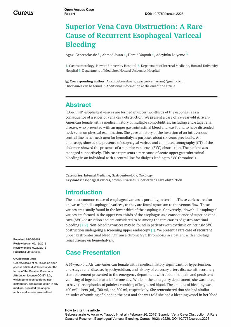
pipe’. She did not remember if any interventions were done to treat it. She gave a history of theinsertion of an intravenous central line in her neck area for hemodialysis purposes about sixyears previously. The line was removed later on because it formed a clot. She had no history ofliver disease, abdominal distension, or jaundice. On examination, she was not in acute distress.There was no conjunctival pallor and no scleral icterus. Physical examination was notable forengorged veins of the anterior chest (Figure 1). She had no ascites or other stigmata of chronicliver disease. Laboratory investigation revealed hemoglobin of 12.9 g/dL with a platelet countof 206,000 per microliter. Liver enzymes were within normal limit and liver synthetic functionwas preserved.
FIGURE 1: Markedly engorged neck veins (arrows)
2018 Gebreselassie et al. Cureus 10(2): e2226. DOI 10.7759/cureus.2226 2 of 6
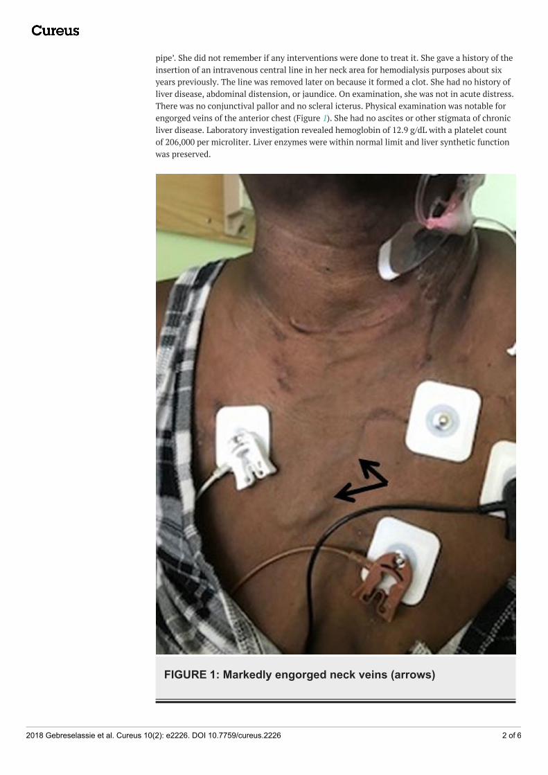
Intravenous lines were secured and the patient was started on intravenous proton pumpinhibitors, an octreotide drip, and intravenous ceftriaxone for possible esophageal bleedingfrom cirrhosis in the emergency department. The patient was admitted to the intensive careunit for close monitoring. The patient underwent esophagogastroscopy after resuscitation,which revealed esophageal varices in the mid-esophageal area but no active bleeding wasfound (Figure 2). The varices were of moderate size with no red wale signs. Variceal bandligation was not performed.
FIGURE 2: Esophagoscopy showing moderately sized mid-esophageal varices (arrows)
An abdominal ultrasound did not show features of cirrhosis or portal hypertension. Computedtomography (CT) of the chest with intravenous contrast showed significant superior vena cavanarrowing at the right atrial junction with large collateral venous channels, including aprominent right internal mammary vein and a large azygous vein (Figures 3, 4). Collateralvessels in the posterior left and anterior chest walls were also appreciated.
2018 Gebreselassie et al. Cureus 10(2): e2226. DOI 10.7759/cureus.2226 3 of 6
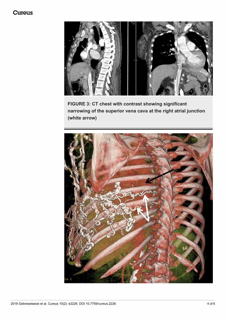
FIGURE 3: CT chest with contrast showing significantnarrowing of the superior vena cava at the right atrial junction(white arrow)
2018 Gebreselassie et al. Cureus 10(2): e2226. DOI 10.7759/cureus.2226 4 of 6
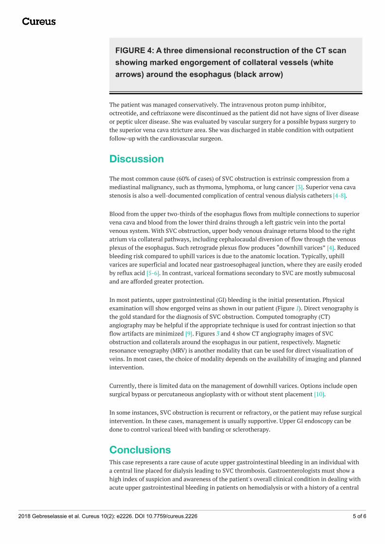
FIGURE 4: A three dimensional reconstruction of the CT scanshowing marked engorgement of collateral vessels (whitearrows) around the esophagus (black arrow)
The patient was managed conservatively. The intravenous proton pump inhibitor,octreotide, and ceftriaxone were discontinued as the patient did not have signs of liver diseaseor peptic ulcer disease. She was evaluated by vascular surgery for a possible bypass surgery tothe superior vena cava stricture area. She was discharged in stable condition with outpatientfollow-up with the cardiovascular surgeon.
DiscussionThe most common cause (60% of cases) of SVC obstruction is extrinsic compression from amediastinal malignancy, such as thymoma, lymphoma, or lung cancer [3]. Superior vena cavastenosis is also a well-documented complication of central venous dialysis catheters [4-8].
Blood from the upper two-thirds of the esophagus flows from multiple connections to superiorvena cava and blood from the lower third drains through a left gastric vein into the portalvenous system. With SVC obstruction, upper body venous drainage returns blood to the rightatrium via collateral pathways, including cephalocaudal diversion of flow through the venousplexus of the esophagus. Such retrograde plexus flow produces “downhill varices” [4]. Reducedbleeding risk compared to uphill varices is due to the anatomic location. Typically, uphillvarices are superficial and located near gastroesophageal junction, where they are easily erodedby reflux acid [5-6]. In contrast, variceal formations secondary to SVC are mostly submucosaland are afforded greater protection.
In most patients, upper gastrointestinal (GI) bleeding is the initial presentation. Physicalexamination will show engorged veins as shown in our patient (Figure 1). Direct venography isthe gold standard for the diagnosis of SVC obstruction. Computed tomography (CT)angiography may be helpful if the appropriate technique is used for contrast injection so thatflow artifacts are minimized [9]. Figures 3 and 4 show CT angiography images of SVCobstruction and collaterals around the esophagus in our patient, respectively. Magneticresonance venography (MRV) is another modality that can be used for direct visualization ofveins. In most cases, the choice of modality depends on the availability of imaging and plannedintervention.
Currently, there is limited data on the management of downhill varices. Options include opensurgical bypass or percutaneous angioplasty with or without stent placement [10].
In some instances, SVC obstruction is recurrent or refractory, or the patient may refuse surgicalintervention. In these cases, management is usually supportive. Upper GI endoscopy can bedone to control variceal bleed with banding or sclerotherapy.
ConclusionsThis case represents a rare cause of acute upper gastrointestinal bleeding in an individual witha central line placed for dialysis leading to SVC thrombosis. Gastroenterologists must show ahigh index of suspicion and awareness of the patient's overall clinical condition in dealing withacute upper gastrointestinal bleeding in patients on hemodialysis or with a history of a central
2018 Gebreselassie et al. Cureus 10(2): e2226. DOI 10.7759/cureus.2226 5 of 6
catheter, especially in patients with no stigmata of chronic liver disease. There are no standardguidelines to manage acute bleeding associated with ‘downhill’ varices; we suggest thatimproved knowledge, prompt diagnosis, and management on a case-by-case basis. Usingavailable endoscopic, radiological, and surgical interventions may lead to successful outcomes.
Additional InformationDisclosuresHuman subjects: Consent was obtained by all participants in this study. Conflicts of interest:In compliance with the ICMJE uniform disclosure form, all authors declare the following:Payment/services info: All authors have declared that no financial support was received fromany organization for the submitted work. Financial relationships: All authors have declaredthat they have no financial relationships at present or within the previous three years with anyorganizations that might have an interest in the submitted work. Other relationships: Allauthors have declared that there are no other relationships or activities that could appear tohave influenced the submitted work.
References1. Siegel Y, Schallert E, Kuker R: Downhill esophageal varices: a prevalent complication of
superior vena cava obstruction from benign and malignant causes. J Comput Assist Tomogr.2015, 39:149–52. 10.1097/RCT.0000000000000183
2. Areia M, Romãozinho JM, Ferreira M, et al.: “Downhill” varices. A rare cause of esophagealhemorrhage. . Rev Esp Enferm Dig. 2006, 98:359–61.
3. Rice TW, Rodriguez RM, Light RW: The superior vena cava syndrome: clinical characteristicsand evolving etiology. Medicine (Baltimore). 2006, 85:37–42.10.1097/01.md.0000198474.99876.f0
4. Hussein FA, Mawla N, Befeler AS, et al.: Formation of downhill esophageal varices as a rarebut serious complication of hemodialysis access: a case report and comprehensive literaturereview. Clin Exp Nephrol. 2008, 12:407–15. 10.1007/s10157-008-0055-4
5. Okamoto E, Amano Y, Fukuhara H, et al.: Does gastreoesophageal reflux have an influence onbleeding from esophageal varices?. J Gastroenterol. 2008, 43:803-808. 10.1007/s00535-008-2232-3
6. Greenwell M, Basye S, Dhawan S, et al.: Dialysis catheter-induced superior vena cavasyndrome and downhill esophageal varices. Clin Nephrol. 2007, 67:325–30.
7. Blam ME, Kobrin S, Siegelman ES, Scotiniotis IA: “Downhill esophageal varices as aniatrogenic complication of upper extremity hemodialysis access. Am J Gastroenterol. 2002,97:216–18. 10.1111/j.1572-0241.2002.05417.x
8. Pop A, Cutler AF: Bleeding downhill esophageal varices: a complication of upper extremityhemodialysis access. Gastrointest Endosc. 1998, 47:299–303. 10.1016/S0016-5107(98)70331-1
9. Qanadli SD, Hajjam M, Bruckert F, et al.: Helical CT phlebography of the superior vena cava:diagnosis and evaluation of venous obstruction. AJR Am J Roentgenol. 1999, 172:1327–33.10.2214/ajr.172.5.10227511
10. Kalra M, Gloviczki P, Andrews JC, et al.: Open surgical and endovascular treatment of superiorvena cava syndrome caused by nonmalignant disease. J Vasc Surg. 2003, 38:215–23.10.1016/S0741-5214(03)00331-8
2018 Gebreselassie et al. Cureus 10(2): e2226. DOI 10.7759/cureus.2226 6 of 6

![A rare cause of dysphagia due to esophageal intramural ......disease, GERD, and corrosive esophageal injury [4, 11– 14]. Abnormalities in esophageal motility, including, un-coordinated](https://img.pdfslide.us/doc/110x75/60b5b1e8c993b14a95327914/a-rare-cause-of-dysphagia-due-to-esophageal-intramural-disease-gerd-and.jpg)