Embed Size (px)
Citation preview
919 Fraser Drive Unit 11 Burlington ON L7L 4X8 Tel: 866-344-3954 Fax: 289-288-0122
www.cytodiagnostics.com
Biotin Labeling Kit - Manual
PRODUCT DESCRIPTION
Cytodiagnostics Biotin Labeling Kit provides all the reagents needed to label antibodies, proteins and other macromolecules with biotin through the modification of primary amines. This product contains an NHS-activated form of biotin that contains a unique chromophore in the linker region. The kit also contains organic solvent, modification buffer, and spin filters for purifying the biotin conjugates.
BACKGROUND
Biotin binds to streptavidin/avidin conjugates with high affinity. Antibodies or protein conjugated with several biotin molecules can amplify signal through streptavidin-conjugated reporter enzyme, thereby increasing the sensitivity of immunoassays. Biotinylated antibodies or proteins can be used in a variety of applications including ELISA, blotting, and immunohistochemistry applications.
It is important to know the extent of biotin modification for effective assay development. While there are many reagents available for biotinylating proteins, determining the amount of biotin incorporated has historically depended on the use of the 4’-hydroxyazobenzene-2-carboxylic acid (HABA) reagent. HABA binds weakly to avidin and absorbs at 500 nm. Biotin competes with HABA for binding to avidin, thereby lowering the absorbance at 500 nm. This method consumes precious sample and is only useful for concentrated proteins because the difference in absorbance due to biotin displacement of HABA from avidin is very small and difficult to accurately measure at low concentrations.
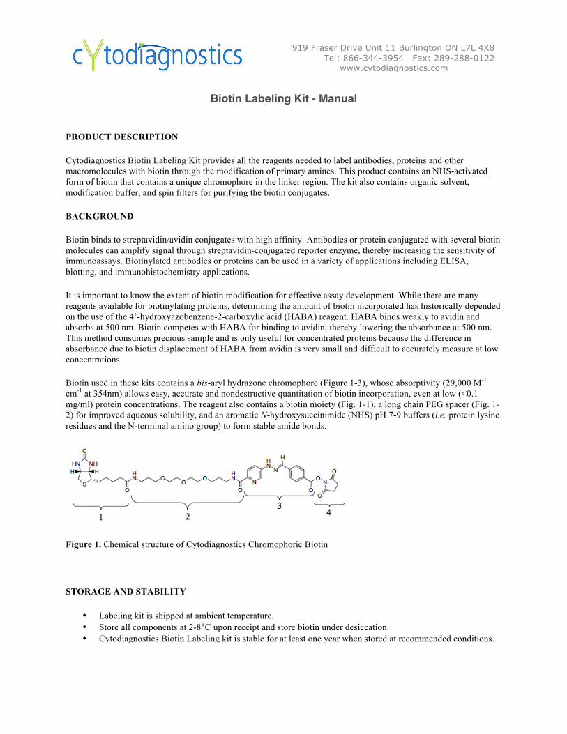
Biotin used in these kits contains a bis-aryl hydrazone chromophore (Figure 1-3), whose absorptivity (29,000 M-1 cm-1 at 354nm) allows easy, accurate and nondestructive quantitation of biotin incorporation, even at low (<0.1 mg/ml) protein concentrations. The reagent also contains a biotin moiety (Fig. 1-1), a long chain PEG spacer (Fig. 1-2) for improved aqueous solubility, and an aromatic N-hydroxysuccinimide (NHS) pH 7-9 buffers (i.e. protein lysine residues and the N-terminal amino group) to form stable amide bonds.
Figure 1. Chemical structure of Cytodiagnostics Chromophoric Biotin
STORAGE AND STABILITY
• Labeling kit is shipped at ambient temperature. • Store all components at 2-8°C upon receipt and store biotin under desiccation. • Cytodiagnostics Biotin Labeling kit is stable for at least one year when stored at recommended conditions.
919 Fraser Drive Unit 11 Burlington ON L7L 4X8 Tel: 866-344-3954 Fax: 289-288-0122
www.cytodiagnostics.com
BEFORE YOU BEGIN
SAFETY AND HANDLING
• Wear appropriate personal protective equipment when handling reagents.
OTHER REQUIRED SUPPLIES AND EQUIPMENT
o Antibody or protein, free of salts or contaminants. See Troubleshooting section for removing salts or other contaminants. Additional Modification Buffer (100 mM phosphate, 150 mM NaCl, pH 7.2-7.4) may be required for buffer exchange.
o Molecular biology grade water o Shaker capable of gentle agitation o Vortex o Microcentrifuge
REAGENT PREPARATION
• Dissolve SureLINKTM Chromophoric Biotin in DMF immediately prior to use. Tap down and equilibrate the biotin vial to room temperature prior to opening to avoid moisture condensation. The NHS-ester moiety of Cytodiagnostics chromophoric biotin is hydrolyzed when exposed to water.
Extended storage of the biotin/DMF solution is not recommended. Aliquots may be stored at -20°C under desiccation for short periods of time.

919 Fraser Drive Unit 11 Burlington ON L7L 4X8 Tel: 866-344-3954 Fax: 289-288-0122
www.cytodiagnostics.com
QUICK REFERENCE PROTOCOL
Rehydrate or transfer antibody/protein in an appropriate buffer
Dissolve Cytodiagnostics Chromophoric Biotin in DMF at 20 mg/mL
Add biotin to protein solution
Incubate the reaction mixture at room temperature for 2 hours with gentle agitation
Remove unconjugated biotin from protein using spin filter or by dialysis
Measure degree of biotin incorporation with spectrophotometer
Elapsed time: approximately 3 hr
Hands-on time: approximately 30 min
919 Fraser Drive Unit 11 Burlington ON L7L 4X8 Tel: 866-344-3954 Fax: 289-288-0122
www.cytodiagnostics.com
BIOTIN CONJUGATION PROTOCOL
This protocol describes the conjugation reaction for 1 mL of IgG at 0.2 to 5.0 mg/mL. Other volumes and concentrations can be used (see Calculations section). Other proteins can also be labeled using this protocol.
1. Rehydrate or transfer the antibody into 1 mL Modification Buffer (100 mM phosphate, 150 mM NaCl, pH 7.2-7.4) at 0.2-5.0 mg/mL.
Buffers containing Tris, imidazole, glycine or primary amines should not be used due to competition with the conjugation reaction. If the sample is stored in one of these buffers, remove by buffer exchange against the Modification Buffer. PBS (10 mM phosphate, 150 mM NaCl, pH 7.2-7.4) is not recommended due to poor buffering capacity.
2. Immediately prior to use, prepare a 20 mg/ml (25 nmole/µ l) stock solution of Cytodiagnostics Chromophoric Biotin in anhydrous DMF.
Tap down and equilibrate biotin vial to room temperature prior to opening to avoid the condensation of moisture. The NHS-ester moiety of chromophoric biotin is hydrolyzed when exposed to water.
If high biotin molar excess is chosen for a highly concentrated protein solution, the concentration of biotin stock solution can be increased to 40 mg/mL (see Calculation section for more details).
3. Add an appropriate volume of 20 mg/ml Biotin to the antibody or protein solution.
See the Calculation section Part 2 to determine the appropriate volume of Biotin. The solution may become cloudy, which does not affect the labeling reaction. The percentage of biotin solution added should be less than 5% of the total reaction volume to minimize protein precipitation by DMF.
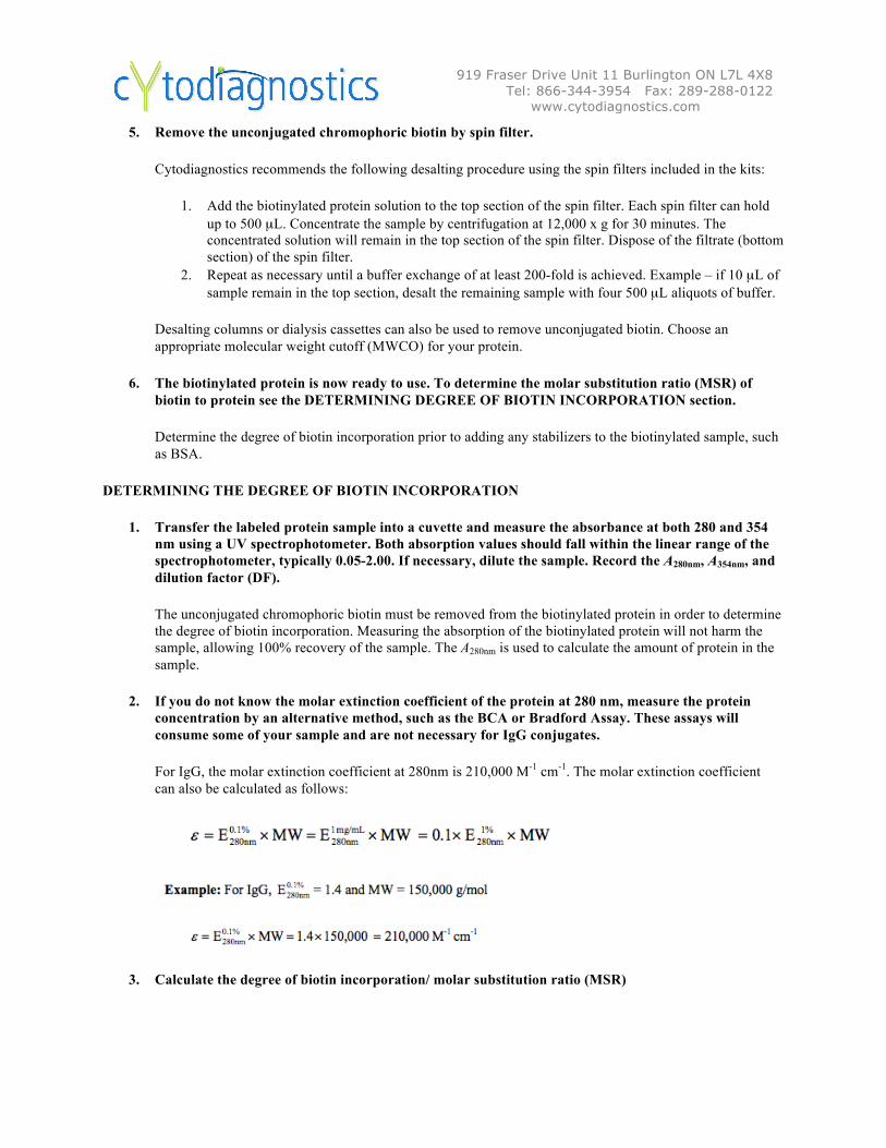
Examples for Conjugation Reactions: When antibodies at 1 mg/mL are used for labeling, a 40-fold molar excess of biotin is recommended for ELISA and a 10-fold molar excess of biotin is recommended for Western blotting.
4. Incubate at room temperature for two hours with gentle agitation.
Longer incubation times, up to overnight, may be used. However, microbial growth and protein degradation may be a concern.
919 Fraser Drive Unit 11 Burlington ON L7L 4X8 Tel: 866-344-3954 Fax: 289-288-0122
www.cytodiagnostics.com
5. Remove the unconjugated chromophoric biotin by spin filter.
Cytodiagnostics recommends the following desalting procedure using the spin filters included in the kits:
1. Add the biotinylated protein solution to the top section of the spin filter. Each spin filter can hold up to 500 µL. Concentrate the sample by centrifugation at 12,000 x g for 30 minutes. The concentrated solution will remain in the top section of the spin filter. Dispose of the filtrate (bottom section) of the spin filter.
2. Repeat as necessary until a buffer exchange of at least 200-fold is achieved. Example – if 10 µL of sample remain in the top section, desalt the remaining sample with four 500 µL aliquots of buffer.
Desalting columns or dialysis cassettes can also be used to remove unconjugated biotin. Choose an appropriate molecular weight cutoff (MWCO) for your protein.
6. The biotinylated protein is now ready to use. To determine the molar substitution ratio (MSR) of biotin to protein see the DETERMINING DEGREE OF BIOTIN INCORPORATION section.
Determine the degree of biotin incorporation prior to adding any stabilizers to the biotinylated sample, such as BSA.
DETERMINING THE DEGREE OF BIOTIN INCORPORATION
1. Transfer the labeled protein sample into a cuvette and measure the absorbance at both 280 and 354 nm using a UV spectrophotometer. Both absorption values should fall within the linear range of the spectrophotometer, typically 0.05-2.00. If necessary, dilute the sample. Record the A280nm, A354nm, and dilution factor (DF).
The unconjugated chromophoric biotin must be removed from the biotinylated protein in order to determine the degree of biotin incorporation. Measuring the absorption of the biotinylated protein will not harm the sample, allowing 100% recovery of the sample. The A280nm is used to calculate the amount of protein in the sample.
2. If you do not know the molar extinction coefficient of the protein at 280 nm, measure the protein concentration by an alternative method, such as the BCA or Bradford Assay. These assays will consume some of your sample and are not necessary for IgG conjugates.
For IgG, the molar extinction coefficient at 280nm is 210,000 M-1 cm-1. The molar extinction coefficient can also be calculated as follows:
3. Calculate the degree of biotin incorporation/ molar substitution ratio (MSR)
919 Fraser Drive Unit 11 Burlington ON L7L 4X8 Tel: 866-344-3954 Fax: 289-288-0122
www.cytodiagnostics.com
In the following example, A280nm = 1.209, A354nm = 1.160, and DF = 1
A. Molar concentration of biotin
Cytodiagnostics Chromophoric Biotin contains a bis-aryl hydrazone with a molar extinction coefficient of 29,000 M-
1 cm-1 at 354nm. The molar concentration of biotin in the labeled protein is calculated as follows:
B. Molar concentration of protein
The concentration of the biotinylated protein is easily determined from the A280nm and molecular weight if the molar extinction coefficient of the protein is known. Cytodiagnostics Chromophoric Biotin contributes to the absorbance of the protein at 280 nm, so a correction must be made to the A280nm. Alternatively, the protein concentration can be calculated using total protein assays such as BCA (Pierce) or Bradford (Bio-Rad).
919 Fraser Drive Unit 11 Burlington ON L7L 4X8 Tel: 866-344-3954 Fax: 289-288-0122
www.cytodiagnostics.com
C. Degree of biotin incorporation/ molar substitution ratio (MSR)
The degree of biotin incorporation is given as a ratio of the molar concentration of biotin to the molar concentration of protein. This dimensionless number is termed the molar substitution ratio, abbreviated MSR.
CALCULATIONS
Part 1: Determining the desired biotin:antibody (or protein) molar ratio for the conjugation reaction.
When labeling antibodies at a concentration of 1 mg/mL, Cytodiagnostics recommends a 40-fold molar excess of biotin for conjugates that will be used in ELISA and a 10-fold molar excess of biotin for conjugates that will be used in Western blotting. See “Use of biotin conjugates in various assays” section for more information.
Various biotin:antibody molar ratios can be used with the Biotin Labeling kit. In general, the degree of biotin incorporated depends on the following:
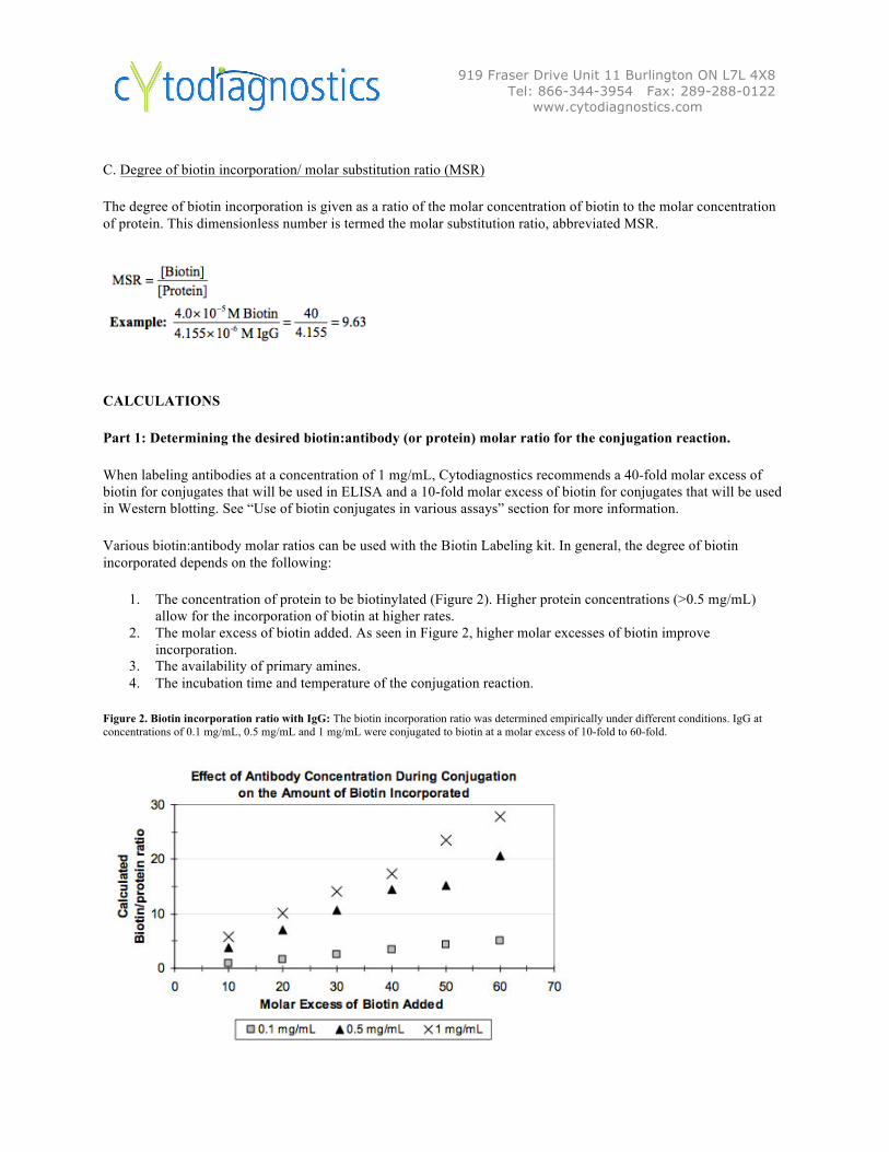
1. The concentration of protein to be biotinylated (Figure 2). Higher protein concentrations (>0.5 mg/mL) allow for the incorporation of biotin at higher rates.
2. The molar excess of biotin added. As seen in Figure 2, higher molar excesses of biotin improve incorporation.
3. The availability of primary amines. 4. The incubation time and temperature of the conjugation reaction.
Figure 2. Biotin incorporation ratio with IgG: The biotin incorporation ratio was determined empirically under different conditions. IgG at concentrations of 0.1 mg/mL, 0.5 mg/mL and 1 mg/mL were conjugated to biotin at a molar excess of 10-fold to 60-fold.
919 Fraser Drive Unit 11 Burlington ON L7L 4X8 Tel: 866-344-3954 Fax: 289-288-0122
www.cytodiagnostics.com
Part 2: Determining the amount of Cytodiagnostics Chromophoric Biotin for the conjugation reaction. The volume of chromophoric biotin required for the conjugation reaction can be determined once the desired biotin molar excess ratio (See Part 1.) has been determined.
Part 3. Calculating the percentage of DMF in the conjugation reaction:
DMF exposure may cause irreversible conformational changes to antibodies and other proteins. These conformational changes may have impact on long-term stability and antigen-binding activity. It is preferred to limit the percentage of DMF in conjugation reactions to less than 5% (Melnikova et al.).
If a large biotin molar excess is chosen for a highly concentrated protein solution, the DMF percentages may exceed 5% when a 20 mg/mL biotin stock solution is used. In these situations, the concentration of biotin stock solution can be increased to 40 mg/mL (50 nmol/µL) to reduce the concentration of DMF.
Calculate the percentage of DMF using the following equation:
% DMF = Vol. of biotin stock ÷ Vol. of protein to be biotinylated
Example: using the example in Part 2, 10.7 µL of 20 mg/mL biotin stock is added to 1000 µL of 1 mg/mL IgG.
%DMF =10.7 µL÷1000 µL=1.1% DMF
919 Fraser Drive Unit 11 Burlington ON L7L 4X8 Tel: 866-344-3954 Fax: 289-288-0122
www.cytodiagnostics.com
RECOMMENDED USE OF CONJUGATES
Cytodiagnostics Biotin Labeling kit can be used to label IgG or other proteins for a variety of immunoassay methods. The conjugate concentration that will provide the best signal to background in your specific assay may vary and should be determined for each conjugate. Include positive and negative controls in each immunoassay for proper review of experimental results and successful troubleshooting.
Use of biotin conjugates in various assays.
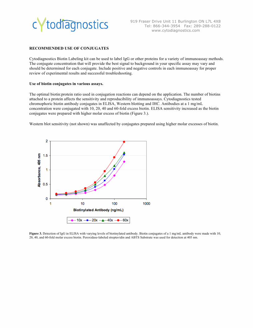
The optimal biotin:protein ratio used in conjugation reactions can depend on the application. The number of biotins attached to a protein affects the sensitivity and reproducibility of immunoassays. Cytodiagnostics tested chromophoric biotin antibody conjugates in ELISA, Western blotting and IHC. Antibodies at a 1 mg/mL concentration were conjugated with 10, 20, 40 and 60-fold excess biotin. ELISA sensitivity increased as the biotin conjugates were prepared with higher molar excess of biotin (Figure 3.).
Western blot sensitivity (not shown) was unaffected by conjugates prepared using higher molar excesses of biotin.
Figure 3. Detection of IgG in ELISA with varying levels of biotinylated antibody. Biotin conjugates of a 1 mg/mL antibody were made with 10, 20, 40, and 60-fold molar excess biotin. Peroxidase-labeled streptavidin and ABTS Substrate was used for detection at 405 nm.
919 Fraser Drive Unit 11 Burlington ON L7L 4X8 Tel: 866-344-3954 Fax: 289-288-0122
www.cytodiagnostics.com
FREQUENTLY ASKED QUESTIONS
Q: What biomolecules can be conjugated with the Biotin Labeling Kit?
A: Proteins, peptides, and oligonucleotides can be conjugated using this technology. Proteins and peptides can be modified using primary amino groups on lysine residues and the N-terminus. Modified oligonucleotides with primary amino groups are suitable for conjugation.
Q: What determine the degree of biotin incorporation?
A: The degree of biotin incorporation depends on the concentration of protein, the molar excess of biotin added, the availability of primary amines in protein, and the incubation temperature and time of conjugation reaction.
Q: Is free chromophoric biotin a concern following the conjugation protocol?
A: In immunoassays such as ELISA and Western blotting, the effect of free chromophoric biotin is minimal as most of the unconjugated biotin is removed in the washing steps. However, free biotin must be removed in order to quantitate the amount of biotin incorporation.
Q: How do I know how many primary amines my protein has?
A: Without knowledge of the protein sequence, the number of primary amines can be estimated by multiplying the MW of the protein by 0.0006. This formula estimates the number of lysine residues given an average weight of 110 Daltons for an amino acid, and an average lysine content of 6.6% in a protein (Dayhoff, M.O., 1978). Due to steric or functional constraints, some lysine residues may not be available for conjugation.
Q: How much biotin should I use in the conjugation reaction?
A: In general, adding between 10 and 60-fold molar excess of Chromophoric Biotin is sufficient for most applications. The optimal amount of biotin added will depend on the number of available primary amines, the assay conditions, the protein concentration, the incubation temperature, and the incubation time. A two hour, room temperature antibody conjugation reaction at a 5 mg/mL concentration with 40-fold molar excess of biotin works well in ELISA. We recommend preparing multiple conjugates with various molar ratios of biotin. Over-modification of the protein with biotin may inhibit protein function.
Q: Unlabeled protein samples are stored in buffer with Tris, sodium azide, or glycine. Can I still conjugate the proteins to Chromophoric Biotin? A: Tris, sodium azide and glycine will react with the Chromophoric Biotin in the conjugation reaction. Remove them by buffer exchange against 0.1 M sodium phosphate, 0.15 M NaCl, pH 7.2-7.4.
Q: Do I have to dissolve Chromophoric Biotin in DMF?
A: Cytodiagnostics Chromophoric Biotin is soluble in either DMSO or DMF to ~50 mg/mL. As with all NHS esters, exposure to aqueous buffers should be limited to prevent inactivation of the NHS ester by hydrolysis.
Q: How long can you store rehydrated Cytodiagnostics Chromophoric Biotin?
A: It is not recommended that you store Cytodiagnostics Chromophoric Biotin solution for future use. The 0.5 and 1.0 mg sizes of Cytodiagnostics Chromophoric Biotin are designed for single use, although multiple conjugates can be made.
Q: Why do I have to store Cytodiagnostics Chromophoric Biotin under desiccation?
A: The desiccator helps to remove moisture to prevent inactivation of the NHS ester by hydrolysis.
Q: My protein normally has an absorbance at 354 nm. Can I still quantitate the amount of biotin? A: If your unlabeled protein has an absorbance at 354 nm, you may subtract the portion of the to the unlabeled protein if you know the extinction coefficient at 354 nm. Otherwise, use the standard HABA assay to determine biotin incorporation.
Q: My unlabeled protein contains a protein-based stabilizer such as BSA. How do I prepare my sample for conjugation? A: BSA may be removed by commercially available albumin removal kits, such as Bio-Rad Affi-gel blue. If the unlabeled protein and contaminating protein are sufficiently different in size, an appropriate gel filtration column can be used to purify the sample. The contaminating protein can also be removed by immunoprecipitation, if you have an antibody to recognize the contaminating protein.
919 Fraser Drive Unit 11 Burlington ON L7L 4X8 Tel: 866-344-3954 Fax: 289-288-0122
www.cytodiagnostics.com
Q: My antibody concentration is less than 0.2 mg/mL. Can I still label my antibody with Cytodiagnostics Chromophoric Biotin? A: Yes, antibodies at a concentration of less than 0.2 mg/mL can still be labeled, but the biotin incorporation rate is low. For best results, concentrate the sample to 0.2 – 5.0 mg/mL using the included spin filters.
Q: Adding more Chromophoric Biotin brings the percentage of DMF or DMSO in the conjugation reaction above 5%. Can I still use this molar ratio of biotin? A: Yes. Instead of dissolving chromophoric biotin in DMF at 20 mg/mL, dissolve it at 40 mg/mL in order to bring the percentage of DMF in the final reaction to less than 5%.
Q: Do I have to purify my sample using the spin filters included in the Cytodiagnostics Biotin Labeling Kit? A: No, you do not have to use the included 10,000 MWCO spin filters. The excess biotin can be removed with a variety of desalting columns or dialysis cassettes. For conjugation to antibodies, 50,000 MWCO Vivaspin 6 centrifugal concentrators also work well.
Q: How much mass will the incorporated biotin add to my protein?
A: Each mole of Chromophoric Biotin will add 695.83 Daltons to the molecular weight of the biotinylated protein.
919 Fraser Drive Unit 11 Burlington ON L7L 4X8 Tel: 866-344-3954 Fax: 289-288-0122
www.cytodiagnostics.com
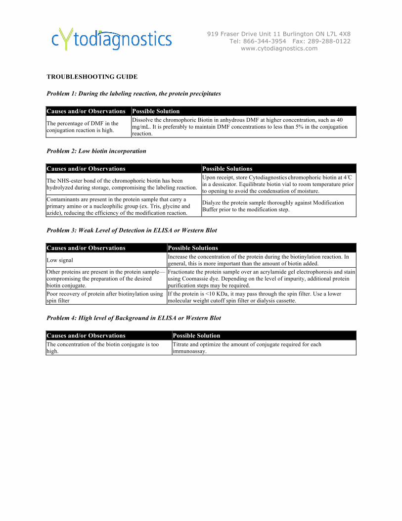
TROUBLESHOOTING GUIDE
Problem 1: During the labeling reaction, the protein precipitates
Causes and/or Observations Possible Solution
The percentage of DMF in the conjugation reaction is high.
Dissolve the chromophoric Biotin in anhydrous DMF at higher concentration, such as 40 mg/mL. It is preferably to maintain DMF concentrations to less than 5% in the conjugation reaction.
Problem 2: Low biotin incorporation
Causes and/or Observations Possible Solutions
The NHS-ester bond of the chromophoric biotin has been hydrolyzed during storage, compromising the labeling reaction.
Upon receipt, store Cytodiagnostics
chromophoric biotin at 4 ̊C in a dessicator. Equilibrate biotin vial to room temperature prior to opening to avoid the condensation of moisture.
Contaminants are present in the protein sample that carry a primary amino or a nucleophilic group (ex. Tris, glycine and azide), reducing the efficiency of the modification reaction.
Dialyze the protein sample thoroughly against Modification Buffer prior to the modification step.
Problem 3: Weak Level of Detection in ELISA or Western Blot
Causes and/or Observations Possible Solutions
Low signal Increase the concentration of the protein during the biotinylation reaction. In general, this is more important than the amount of biotin added.
Other proteins are present in the protein sample—compromising the preparation of the desired biotin conjugate.
Fractionate the protein sample over an acrylamide gel electrophoresis and stain using Coomassie dye. Depending on the level of impurity, additional protein purification steps may be required.
Poor recovery of protein after biotinylation using spin filter
If the protein is <10 KDa, it may pass through the spin filter. Use a lower molecular weight cutoff spin filter or dialysis cassette.
Problem 4: High level of Background in ELISA or Western Blot
Causes and/or Observations Possible Solution The concentration of the biotin conjugate is too high.
Titrate and optimize the amount of conjugate required for each immunoassay.
919 Fraser Drive Unit 11 Burlington ON L7L 4X8 Tel: 866-344-3954 Fax: 289-288-0122
www.cytodiagnostics.com
REFERENCES
Melnikova, Y. et al., (2000) Antigen-Binding Activity of Monoclonal Antibodies after Incubation with Organic Solvents. Biochemistry (Moscow) 65 (11), 1256-1265
Dayhoff, M.O. (1978) Atlas of Protein Sequence and Structure, Suppl. 2, National Biomedical Research Foundation, Washington.
For Research Use Only
The products listed herein are for research use only and are not intended for use in human or clinical diagnosis. Nothing disclosed herein is to be construed as a recommendation to use these products in violation of any patents. The information presented above is believed to be accurate. However, said information and product are offered without warranty or guarantee since the ultimate conditions of use and the variability of the materials treated are beyond our control. We cannot be responsible for patent infringements or other violations that may occur with the use of these products. No claims beyond replacement of unacceptable material or refund of purchase price shall be allowed. All claims regarding product performance must be made within 30 days following date of delivery.
Disclaimer
The recommendations of this bulletin are provided solely for the benefit of users who need practical guidance on immunoassay procedures. Because experimental conditions for the use of the suggested products are beyond the control of Cytodiagnostics Inc, it is impossible for Cytodiagnostics Inc. to implicitly guarantee the performance of the mentioned products for any and all assay procedures.