Embed Size (px)
Citation preview

FACOLTÀ DI SCIENZE AGRARIE E ALIMENTARI Department of Food, Environmental and Nutritional Sciences (DeFENS)
Graduate School in Molecular Sciences and Plant, Food and
Environmental Biotechnology
PhD programme in Food Science, Technology and Biotechnology
XXV cycle
Biotechnological and Spectroscopical Evaluation of Selected
Lactobacillus plantarum Strains with Probiotic and
Nutraceutical Potentialities
Scientific field AGR/16
MARIA CHIARA REMAGNI
Tutor: Prof. Diego Mora
Co-tutors: Dott.ssa Tiziana M. P. Cattaneo
Dott. Domenico Carminati
PhD Coordinator: Prof. Maria Grazia Fortina
2011/2012

ABSTRACT: Probiotics are non-pathogenic microorganisms that, when ingested in adequate
amounts, exert a positive influence on their host‘s health. A variety of microorganisms,
typically food grade lactic acid bacteria (LAB), have been evaluated for their probiotic potential
and are applied as adjunct cultures in various types of food products or in therapeutic
preparations. Within the genus Lactobacillus, Lactobacillus plantarum is a member of the
facultatively heterofermentative group of lactobacilli. It is a heterogeneous and versatile species
that is encountered in a variety of environmental niches, including dairy, meat, fish, and many
vegetable or plant fermentations. L. plantarum strains have also been found in many cheese
varieties. Moreover, strains of L. plantarum have proven ability to survive gastric transit and
colonize the intestinal tract of humans and other mammals. Thus, the aim of this Ph.D. thesis
was to select and characterize strains belonging to Lactobacillus plantarum species for their
probiotic and nutraceutical potentialities which could be added to other foods for exploitation as
carrier of functional supplements.
Eighty Lactobacillus plantarum strains isolated from different matrixes were evaluated for their
probiotic and nutraceutical potential. After a preliminary subtractive screening based on the
presence of msa and bsh genes, the strains which showed the presence of these two genes, were
characterized for their nutraceutical features dealing with their ability to produce riboflavin and
to remove cholesterol from growth medium.
At the same time, a spectroscopical characterization of these bacteria was carried out. Strains
were tested for their ability to grow in milk whey and the fermentation parameters, such as
microbial growth, lactose decrease and lactic acid increase, were monitored by a NIR optic
probe. Results obtained show how this non-destructive technique could be a viable alternative
to traditional methods. In fact, the NIR proposed regression models were very satisfactory for
all of the three considered analytes showing an high correlation coefficient coupled with low
errors both in calibration and prediction.
Thanks to this technique, it was verified whether strains with different ability to remove
cholesterol can show changes in the cell wall. Preliminary data obtained by PCA analysis
showed the suitability of these techniques for the study of the bacterial cell walls: changes in the
spectra seemed to highlight the possibility to distinguish not only different strains but also
different effects produced by the addiction of cholesterol in the medium.
The potential of NIR coupled with Aquaphotomics and multivariate techniques to identify
species has been investigated for evaluating its feasibility in microbiology. Spectral data
analysis revealed very promising results for discrimination and classification of samples,
demonstrating how this technique successfully distinguished between different bacteria.
The research of lactic acid bacteria with probiotic and nutraceutical potentialities offer the
opportunity to improve the nutritional quality of many fermented foods by increasing their
added value. The development of new functional foods by fermentation may contribute to a
further expansion of the market for this class of products.
These new functional foods could pave the way for the development of food products
"functional" for specific consumer groups with special needs such as the elderly, adolescents,
pregnant women, children, athletes and vegetarians.
RIASSUNTO: I probiotici sono microrganismi non patogeni che, se ingeriti in quantità
adeguate, esercitano un influsso positivo sulla salute dell‘ospite. All'interno del genere
Lactobacillus, Lactobacillus plantarum è un membro del gruppo degli eterofermentanti
facoltativi. Si tratta di una specie eterogenee e versatile che è presente in una varietà di nicchie
ambientali, tra cui latticini, carne, pesce, verdure e molti o fermentazioni vegetali. Inoltre, ceppi
di L. plantarum hanno mostrato la capacità di sopravvivere di transito gastrico e colonizzare il
tratto intestinale sia di esseri umani che di altri mammiferi. Pertanto, l'obiettivo della presente
tesi di dottorato è stato quello di selezionare e caratterizzare i ceppi appartenenti alla specie

Lactobacillus plantarum per le loro potenzialità probiotiche e nutraceutiche al fine di poter
utilizzare questi ceppi come carrier di integratori funzionali.
Ottanta ceppi di Lactobacillus plantarum isolati da diverse matrici sono stati valutati per il loro
potenziale probiotico e nutraceutico. Dopo uno screening preliminare basato sulla presenza dei
geni msa e bsh, i ceppi che mostravano la presenza di questi due geni, sono stati caratterizzati
per le loro caratteristiche nutraceutiche riguardanti la capacità di produrre riboflavina e di
rimuovere il colesterolo dal mezzo di crescita.
Allo stesso tempo è stata effettuata una caratterizzazione spettroscopica. I ceppi sono stati
testati per la loro capacità di crescere in siero del latte e i parametri di fermentazione, come la
crescita microbica, la diminuzione lattosio e l‘aumento dell'acido lattico sono stati monitorati da
una sonda ottica NIR. I risultati ottenuti mostrano come questa tecnica non distruttiva possa
essere una valida alternativa ai metodi tradizionali. Infatti, i modelli di regressione proposti
sono molto soddisfacenti per tutti i tre analiti considerati mostrando un alto coefficiente di
correlazione accoppiato con bassi errori sia in calibrazione che in predizione.
Grazie a questa tecnica, si è verificato se i ceppi con differente capacità di rimuovere il
colesterolo possano mostrare cambiamenti nella parete cellulare. Dati preliminari ottenuti
dall‘analisi PCA hanno evidenziato l'idoneità di tali tecniche per lo studio delle pareti cellulari
batteriche: le variazioni negli spettri sembravano evidenziare la possibilità di distinguere non
solo ceppi diversi, ma anche diversi effetti prodotti dalla dipendenza di colesterolo nel mezzo.
Inoltre è stata studiata la capacità della tecnica NIR accoppiata ad Aquaphotomics e alla
statistica multivariata di identificare specie batteriche.
L‘analisi dei dati spettrali ha rivelato risultati molto promettenti per la discriminazione e
classificazione dei ceppi dimostrando come questa tecnica sia in grado di distinguere batteri
diversi.
La ricerca di batteri lattici con potenzialità probiotiche e nutraceutiche offre l'opportunità di
migliorare la qualità nutrizionale di molti alimenti fermentati, aumentandone il valore aggiunto.
Lo sviluppo di nuovi alimenti funzionali mediante fermentazione può contribuire ad una
ulteriore espansione del mercato per questa classe di prodotti.
Questi nuovi alimenti potrebbero aprire la strada per lo sviluppo di prodotti alimentari
"funzionali" per gruppi specifici di consumatori con esigenze speciali, come gli anziani,
adolescenti, donne incinte, bambini, atleti e vegetariani

INDEX 1. Introduction
1.1 Lactic acid bacteria
1.1.1 General characteristics and technological applications
1.1.2 LAB and metabolism
1.2 Probiotics
1.2.1 functional aspects
1.3 Lactobacillus plantarum
1.3.1 General aspects
1.3.2 Probiotic aspects
1.3.3 Technolgical aspects
1.4 Infrared spectroscopy
1.4.1 Theoretical princeps of infrared spectroscopy
1.4.2 FT-NIR instrumentation
1.4.3 NIR sample presentation system
1.4.4 NIR detectors
1.4.5 NIR advantages
1.5 Reference
2. Preliminary selection of Lactobacillus plantarum strains with probiotic potentialities
2.1 Introduction
2.1.2 Probiotics: beneficial effects on human health
2.2 Materials and Methods
2.2.1 Strains selection
2.2.2 Molecular typing
2.2.3 Research for msa and bhs genes
2.2.4 BSH activity
2.2 Results and Discussion
2.3 Conclusions
2.4 References
3. Evaluation of riboflavin production in Lactobacillus plantarum strains
3.1 Introduction
3.2 Materials and Methods
3.2.1 Strains selections
3.2.2 Detection of rib operon
3.2.3 Evaluation of rib operon by Real- Time PCR
3.2.4 Detection of riboflavin production
3.3 results and Discussion
3.4 Conclusions
3.5 References
4. Lactic acid bacteria cholesterol removal capability and related cell membrane fatty acid
modifications
4.1 Introduction
4.2 Materials and Methods
4.2.1 Bacterial strains and colture conditions
4.2.2 Evaluation of cholesterol removal from the growth media
4.2.3 Detection of bile salt hydrolase (bsh) gene
4.2.4 Phenotypic assays

4.2.5 Cellular fatty acids composition
4.2.6 Statistical analysis
4.3 Results and Discussion
4.3.1 cholesterol removal cabability of LAB
4.3.2 Influence of the grow phase on cholesterol removal
4.3.3 Bile resitance and BSH activity
4.3.4 Effect on cholesterol on cellular fatty acids
4.4 Conclusions
4.5 References
5. The use of NIR spectroscopy for monitoring milk whey biotransformation process
using L.plantarum
5.1 Introduction
5.2 Materials and Methods
5.2.1 Inoculum preparation
5.2.2 Substrate preparation
5.2.3 Fermentation tests
5.2.4 Determination of lactose and lactic acid content by HPLC
5.2.5 Near Infrared Spectroscopy
5.2.6 Data processing
5.3 Results and Discussion
5.4 Conclusions
5.5 References
6. NIR and MIR spectroscopy in the study of cholesterol removal ability of L. plantarum
6.1 Introduction
6.2 Materials and Methods
6.2.1 Bacterial strains and colture conditions
6.2.2 MIR spectroscopy
6.2.3 NIR spectroscopy
6.2.4 Data processing
6.3 Results and discussions
6.4 Conclusions
6.5 References
7. Near Infrared spectroscopy and Aquaphotomics as a tool for bacterial classification
7.1 Introduction
7.2 Materials and Methods
7.2.1 Strain selection
7.2.2 Molecular typing
7.2.3 Sprectroscopical typing
7.3 Results and Discussion
7.3.1 molecular typing
7.3.2 Sprectroscopical typing
7.4 Conclusions
7.5 References

1. INTRODUCTION

1
1.1 LACTIC ACID BACTERIA
1.1.1 General characteristics and technological applications
Lactic acid bacteria (LAB) are a heterogeneous group of microorganisms, whose species may
possess different morphology, metabolism, adaptability and growth characteristics. They are
Gram +, ubiquitous, rods or cocci shaped, non-motile, non-sporeforming, facultative or obligate
anaerobic, heterotrophic, fermenting sugars according to different metabolic processes,
however, mainly producing lactic acid.
They are divided into different families and in particular:
Family Lactobacillaceae: genus Lactobacillus and Pediococcus;
Family Streptococcaceae: genus Streptococcus and Lactococcus;
Family Leuconostoceae: genus Leuconostoc, Oenococcus and Weissella
The optimum growth temperature is variable between different species and according to this
classification, LAB are divided in mesophilic or thermophilic bacteria.
In relation to their ability to ferment sugars, they are classified into:
Homofermentative: ferment only sugars to produce lactic acid as a primary metabolite;
Heterofermentative: ferment sugars to produce, in addition to lactic acid, acetic acid,
ethyl alcohol and CO2 as primary metabolites.
The microorganisms belonging to this group are, in general, very demanding from the
nutritional point of view and require complex substrates for their growth. For this reason,
despite their ubiquity, LAB tend to colonize only those matrices which possess a composition
rich in constituents and suitable for their multiplication.
The metabolites associated with the multiplication of LAB are the basis of fermentation
processes of different fermented foods such as dairy products (cheese, cream and yoghurt),
salami and sausages, vegetables (sauerkraut), sourdoughs and wine.
The production of lactic acid and other primary metabolites may induce changes in the structure
of proteins, in food rheology and inhibits pathogenic and alterative microorganisms.
Furthermore, it extends the shelf life of products.
Concerning lactic acid bacteria, in order of their use in food technology, it is useful to deepen
the study of different phenotypic traits.
To evaluate the technological functionality of LAB, it is necessary to study their phenotypic
traits such as:
- the ability to ferment lactose and galactose or the ability to use other carbon substrates (citric
acid, carbohydrate fractions associated with casein);
- the possibility to use different nitrogen fractions (peptides and amino acids);
- resistance to osmotic stresses;
- the ability to grow in different conditions of temperature characteristics of a particular process;
- the ability to interact positively with other microorganisms or to inhibit them;
- the proteolytic activity,
- the phage resistance;
- the autolytic ability;
- the capability to produce extracellular polysaccharides .
The development of phenotypic and genotypic techniques has favored the possibility of
recognizing the presence of more biotypes within the same species. It has been frequently
highlighted as strains belonging to the same species, characterized by significant genetic
polymorphisms, may have different metabolic attitudes. This assumes, therefore, a
technological relief: it is possible to use strains with different characteristics useful for the
transformation or to prepare mixtures of different biotypes able to interact and cooperate during
the fermentation process. In this sense, the biodiversity observed in certain species of

2
microorganism can be considered an opportunity of practical interest (Mucchetti et al., 2006).
1.1.2 Lactic acid bacteria and metabolism
LAB have a simple metabolism and even though they require complex precursors present in the
growth medium to supply their nutritional requirements. The nutritional requirements of LAB
usually include, in addition to carbon sources, amino acids, vitamins, nucleic acids and salts.
Environments designed to provide microorganisms this pool of substances available in free
form are not many, and despite the ubiquity of LAB, only a relatively small group of natural
substrates (mainly milk, vegetables and meat) is designed to promote a massive growth.
1.1.2.1 Carbohydrates fermentation
An essential aspect of the metabolism of LAB is carbohydrates fermentation in order to obtain
the needed energy for cells biosynthetic processes. LAB are able to use different
monosaccharides and disaccharides. This attitude varies in relation to the enzyme equipment of
each species and it presents different efficiency in relation to the physiological characteristics of
the biotype and environmental conditions.
The fermentation process that derives from the development of the LAB lactic microflora
involves the total or partial utilization of fermentable sugars present, that are subtracted to the
potential use by germs spoilage, and induces the modification of the chemical-physical
characteristics of the matrix resulting in the accumulation of metabolites primary such as lactic
acid, ethanol and CO2 (Figure 1.1).
Figure 1.1: Lactic acid bacteria homolactic fermentation pattern
LAB can be divided into two groups: homofermentative and heterofermentative depending on
which of the two main fermentative pattern they use. In the first case, from a molecule of a
monosaccharide, such as glucose, it is possible to obtain a theoretical yield of two molecules of
pyruvate which, in normal conditions (presence of sugars and limited presence of oxygen), are
reduced to lactic acid (Figure 1.2).

3
Figure 1.2: Homolactic fermentation: Embden-Meyerhof-Parnas pattern
Different monosaccharides (galactose, fructose, mannose etc..) may be fermented by LAB
through the way of glucose-6-phosphate pathway or alternatively, through the way of the
tagatose-6-phosphate although less widespread among LAB (Figure 1.3).
4
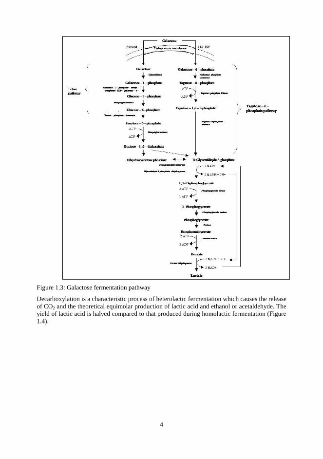
Figure 1.3: Galactose fermentation pathway
Decarboxylation is a characteristic process of heterolactic fermentation which causes the release
of CO2 and the theoretical equimolar production of lactic acid and ethanol or acetaldehyde. The
yield of lactic acid is halved compared to that produced during homolactic fermentation (Figure
1.4).

5
Figure 1.4: Heterolactic fermentation: phosphoketolase pathway
Many species of LAB, not only heterofermentative, are able to also use the pentose sugars. In
general, specific permease located in cytoplasmic membrane are involved in the transportation
of these sugars inside the cell. In the cytoplasm, pentoses are phosphorylated and converted by
specific epimerase or isomerase to ribulose-5-phosphate.
Citrate is an important carbon source, among all carbon sources present in the matrix, for LAB
and different species are able to transform citrate in aromatic compounds. It is not directly used
as electrons acceptor but it is converted, by citrate lyase, in acetate and oxalacetate from whose
decarboxylation, piruvate is obtained (Figure 1.5).

6
Figure 1.5: Citric acid utilization from LAB
1.1.2.2 Proteolysis
LAB are particularly demanding in amino acids. This nutritional need is variable among
different species and, in some cases, is linked to specific characteristics of the single strain.
Milk proteins, in particular casein, are too large to be able to permeate through the membrane
into the cytoplasm of the bacterial cell. At the same time, the nitrogenous non-proteic fraction,
with a lower molecular weight, composed by peptides with different sizes, free amino acids,
urea and nitrogen bases, is not always sufficient to allow an adequate bacterial growth.
LAB, therefore, utilize endo and exopeptidases to hydrolyze casein and whey proteins, which
allow the production of peptides with compatible size with transport across the cell membrane.
Then, the peptides will transform in simple compounds in the cytoplasm. Amino acids and
oligopeptides can be used by the cell for biosynthetic finality or alternatively, demolished to
acids or amines and/or processed through other metabolic pathways, if they are in excess of the
cellular needs. The catabolism of amino acids is considered one of the metabolisms of major
importance for the formation of flavor and aroma in cheese.
1.1.2.3 Lipolysis and polysaccharides production
The attitude of LAB to degrade the lipidic fraction of milk is less deepened because it is
generally believed that the lactic bacteria are not able to hydrolyse triglycerides characteristic of
the fat fraction of milk even though it is recognized an esterase activity against diglycerides and
monoglycerides.

7
Many strains of LAB are also capable of producing different exopolysaccharides, which may
get stuck on the cell wall to form a sort of capsule or released in the medium. The role of these
substances is of considerable interest, in particular in relation to the possibility to obtain yoghurt
and/or fermented milk-based beverages with different rheological characteristics and viscosity.
1.1.2.4 Bacteriocins production
Bacteriocins are secondary metabolites, which are produced in larger amounts after the
exponential phase of bacterial growth. It is a heterogeneous group of molecules characterized
by different spectrum of inhibition, structure, molecular weight, conditions of activity and
stability (Mucchetti et al., 2006).
8
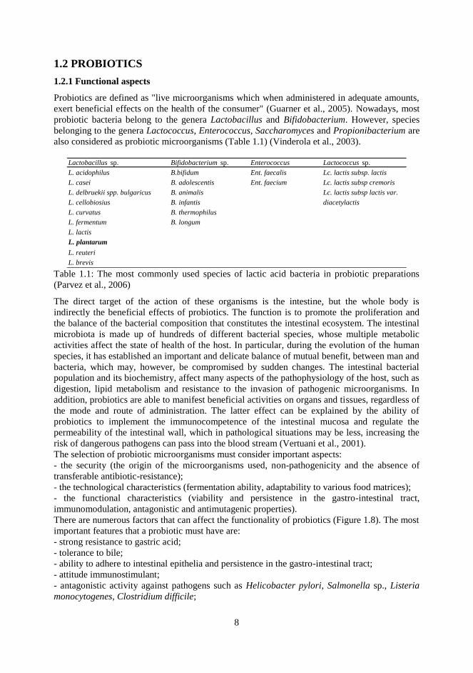
1.2 PROBIOTICS
1.2.1 Functional aspects
Probiotics are defined as "live microorganisms which when administered in adequate amounts,
exert beneficial effects on the health of the consumer" (Guarner et al., 2005). Nowadays, most
probiotic bacteria belong to the genera Lactobacillus and Bifidobacterium. However, species
belonging to the genera Lactococcus, Enterococcus, Saccharomyces and Propionibacterium are
also considered as probiotic microorganisms (Table 1.1) (Vinderola et al., 2003).
Lactobacillus sp. Bifidobacterium sp. Enterococcus Lactococcus sp.
L. acidophilus B.bifidum Ent. faecalis Lc. lactis subsp. lactis
L. casei B. adolescentis Ent. faecium Lc. lactis subsp cremoris
L. delbruekii spp. bulgaricus B. animalis Lc. lactis subsp lactis var.
L. cellobiosius B. infantis diacetylactis
L. curvatus B. thermophilus
L. fermentum B. longum
L. lactis
L. plantarum
L. reuteri
L. brevis Table 1.1: The most commonly used species of lactic acid bacteria in probiotic preparations
(Parvez et al., 2006)
The direct target of the action of these organisms is the intestine, but the whole body is
indirectly the beneficial effects of probiotics. The function is to promote the proliferation and
the balance of the bacterial composition that constitutes the intestinal ecosystem. The intestinal
microbiota is made up of hundreds of different bacterial species, whose multiple metabolic
activities affect the state of health of the host. In particular, during the evolution of the human
species, it has established an important and delicate balance of mutual benefit, between man and
bacteria, which may, however, be compromised by sudden changes. The intestinal bacterial
population and its biochemistry, affect many aspects of the pathophysiology of the host, such as
digestion, lipid metabolism and resistance to the invasion of pathogenic microorganisms. In
addition, probiotics are able to manifest beneficial activities on organs and tissues, regardless of
the mode and route of administration. The latter effect can be explained by the ability of
probiotics to implement the immunocompetence of the intestinal mucosa and regulate the
permeability of the intestinal wall, which in pathological situations may be less, increasing the
risk of dangerous pathogens can pass into the blood stream (Vertuani et al., 2001).
The selection of probiotic microorganisms must consider important aspects:
- the security (the origin of the microorganisms used, non-pathogenicity and the absence of
transferable antibiotic-resistance);
- the technological characteristics (fermentation ability, adaptability to various food matrices);
- the functional characteristics (viability and persistence in the gastro-intestinal tract,
immunomodulation, antagonistic and antimutagenic properties).
There are numerous factors that can affect the functionality of probiotics (Figure 1.8). The most
important features that a probiotic must have are:
- strong resistance to gastric acid;
- tolerance to bile;
- ability to adhere to intestinal epithelia and persistence in the gastro-intestinal tract;
- attitude immunostimulant;
- antagonistic activity against pathogens such as Helicobacter pylori, Salmonella sp., Listeria
monocytogenes, Clostridium difficile;

9
- resistance to phages;
- high vitality and stability during the preparation and storage of food in which they are
conveyed (Mattila-Sandholm et al., 2002).
Figure 1.8: Main technological factors that influence the functionality of probiotics according to
Mattila-Sandholm et al., 2002.
Many probiotic bacteria are of human origin. This choice is based on the belief that these
strains, when ingested with foods fermented by them, are better adapted to colonize the human
intestine. Currently there is little scientific evidence to support this observation, and recent
studies have shown that the character of a probiotic strain is not dependent on its origin but it is
a species-and strain-specific characteristic (Bude Ugarte et al., 2006; Schillinger 2005; Minelli
et al., 2004).

10
1.3 LACTOBACILLUS PLANTARUM
1.3.1 General aspects
The genus Lactobacillus comprises more than 50 species, many of which are often used as
starter cultures for the production of fermented foods as they contribute to the conservation, to
the flavor and the to the rheology of the products. In addition, many species of this genus,
including Lactobacillus crispatus, Lactobacillus gasseri and Lactobacillus plantarum, are able
to colonize the human and animal gut.
Lactobacillus plantarum belongs to the phylogenetic group of Lactobacillus casei -
Pediococcus of the genus Lactobacillus and in particular to the phylogenetic subgroup of L.
plantarum (Bringel et al., 2005).
It is a Gram-positive bacterium, facultative heterofermentative, non-mobile, non-spore-forming,
rod-shaped with rounded ends, usually from 0.9 to 1.2 µm at 3-8 µm, facultative anaerobic, able
to grow at 15°C but not at 45°C (de Vries et al., 2006). Some strains, in certain conditions, are
capable of nitrate reduction.
The fermentation profile is very wide: this species is, in fact, able to ferment many
carbohydrates, except for rhamnose. Lactic acid is produced both in the D and L forms (Bishop
et al., 1993).
Lactobacillus plantarum is one of the most frequently microorganisms isolated from
sourdoughs, and in a wide range of environmental niches, including dairy, meat products such
as salami, and many fermented vegetables (olives and sauerkraut).
At this microorganism is recognized an important role in the process of fermentation due to its
ability to rapidly acidify the medium and the ability to produce bacteriocins, preventing the
growth of pathogenic microorganisms and spoilage, improving the hygienic safety and food
preservation (de Vries et al., 2006).
1.3.2 Probiotic aspects
Lactobacillus plantarum is a microorganism that grows abundantly in many environmental
niches. Many clinical tests were conducted to evaluate the effects due to the ingestion of
Lactobacillus plantarum by subjects suffering from various diseases. Recent studies have
demonstrated that the recruitment of adequate quantities of Lactobacillus plantarum 299v on
the part of subjects characterized by high levels of cholesterol in the blood, leads to a significant
reduction of LDL and fibrinogen, which are risk factors for the development of heart disease
(Parvez et al., 2006).
A considerable attention has been paid to the ability of probiotics to inhibit intestinal infections
caused by pathogens. Lactobacillus plantarum is able, among the constituents of the natural
bacterial flora of the whole intestinal tract, to bind to the intestinal villi and to prevent access of
pathogenic bacteria to the intestinal wall, keeping the balance of the endogenous flora and
modulating the immune system associated with it (GALT: gut-associated lymphoyd tissue or
gut-associated lymphoid tissue) (de Vries et al., 2006).
Some strains of Lactobacillus plantarum are capable of interacting with the specific receptor of
mannose present on the surface of the intestinal cells (erythrocytes). This bond promotes
adhesion to the intestinal epithelium and its colonization by Lactobacillus plantarum, and
causes an inhibitory effect against pathogens causing intestinal infections, especially those
caused by Escherichia coli, due to competition for binding to mannose receptors present on
erythrocytes (Pretzer et al., 2005).
Lactobacillus plantarum has demonstrated therapeutic efficacy in infections associated with
Clostridium difficile, and its administration (in association with other lactobacilli) in capsules or
in functional foods (yoghurt) has been proposed in association with antibiotic treatment for

11
preventive purposes (de Vries et al ., 2006).
Lactobacillus plantarum in fact, is able to produce different substances capable of inhibiting the
development of pathogenic bacteria. This effect is due to the production of organic acids which
reduce the pH of the medium, and the production of anti-microbial metabolites such as
hydrogen peroxide and bacteriocins (Bishop et al., 1993).
1.3.3 Technological aspects
Probiotics most widely used in food belong to the genera Lactobacillus and Bifidobacterium. In
particular for the genus Lactobacillus species most commercially used are L. casei/paracasei, L.
acidophilus and L. rhamnosus (Stanton et al., 2003).
The dairy industry has used probiotic cultures as a tool for the development of new products:
some dairy products, in fact, have proved good vehicles of probiotics for human consumption
(Champagne & Garner, 2005), although some of the probiotic species mentioned above do not
develop well in milk or remain viable during the life of the product, limiting its industrial
application.
This justifies the need for a continuous search for new strains with probiotic potential, able to
diversify the range of dairy products available on the market (Farmworth, 2003).
L. plantarum has the ability to change cyclically habitat (from the gastro-intestinal human to
fermented foods, and vice versa) demonstrating its adaptability and competitiveness, which in
turn justified by the plasticity of its genome recently sequenced. The large number of membrane
proteins suggests that L. plantarum has the ability to adhere to different surfaces and to
potential substrates for growth. Moreover, the high number of genes encoding the regulatory
functions, indicates a marked ability to adapt to many and different environmental conditions.
All this explains the ability of L. plantarum to grow in wide range of food ecosystems (de Vries
et al., 2006).
These characteristics make Lactobacillus plantarum potential and interesting candidate for
possible applications in the field of fermented milks and justify their interest for further study
on its probiotic properties.

12
1.4 INFRARED SPECTROSCOPY
1.4.1 Theoretical principles of infrared spectroscopy (Workman & Weyer, 2008; Burns &
Ciurczak, 2001; Siesler, 2002)
Infrared spectroscopy can be defined as the analysis of materials regarding their tendency to
absorb light in a certain area of the electromagnetic radiation. In particular, it is used to indicate
the separation, detection and recording of changes in energy (resonance peaks) involving nuclei,
atoms or entire molecules. These energetic variations are due to the interaction between
radiation and matter, specifically the emission, absorption or diffusion of electromagnetic
radiation or particles. Infrared spectroscopy is applied for quantitative and qualitative analysis.
Its most important and characteristic application field is the identification of organic compounds
that give rise, especially in the mid-infrared region, to generally complex spectra with several
maxima and minima absorption peaks. In many cases, in fact, the infrared spectrum of an
organic compound provides a unique fingerprint that is easily distinguishable from other
compounds.
The high selectivity of the methodology often allows the quantitative determination of an
analyte in a complex mixture without prior separation. The theoretical basis of the interaction
between matter and radiation is the quantum nature of energy transfer from the radiation to
matter and vice versa. In fact both the matter and the electromagnetic field have a ―dual nature‖,
i.e. the ability to behave both as waves and as particles. Electromagnetic radiations, the best
known of which is light, are nothing but a form of transport of energy electromagnetic thought
space. According to studies of James Clerck Maxwell, the movement of electrical charges can
generate waves of radiant energy in space. They are the result of the superposition of an electric
field and a magnetic field orthogonal mutually coupled: each of them is the source of the other
and propagates with a sinusoidal movement in both space and time (Figure 1.10).
Figure 1.10: Representative model of the electromagnetic radiation.
The directions of oscillation in space of electric and magnetic fields are perpendicular to the
direction of propagation which has a wave nature. The wave character of the electromagnetic
radiation is commonly described by its wavelength (, measured in nanometers (nm = 10-9
m),
the wave number (v), which represents the number of waves present in a unit length, measured
in reciprocal centimeters (cm-1
), the speed (V) with the wave advance, and the number of
wavelengths that pass in a given point per unit of time, frequency (, measured in hertz ( Hz =
s-1
). The relationship between these quantities is given by the expression formula

13
v=l V
=
where V is the velocity of the electromagnetic wave in vacuum, i.e. the rate of radiation
diffusion. Maxwell discovered that the propagation speed was constant for all the
electromagnetic waves in vacuum, and it was equal to 2.998x1010 cm s-1
, i.e. the speed of light.
Thus, being the speed propagation constant, the frequency can be deduced from the wavelength
and vice versa. The entire electromagnetic spectrum is composed of several areas defined by
specific wavelengths as shown in Figure 1.11. This division gives rise to five major groups: the
visible region, the ultraviolet and ionizing radiation, characterized by high frequencies and short
wavelengths, and the infrared and radio waves, characterized by low frequency and high
wavelengths. The infrared region of the spectrum comprises radiation with wave numbers
ranging from about 12500 to 10 cm-1
. It‘s usually divided into three regions: the higher energy
near-IR (NIR), (4000-10000 cm−1
) exciting overtone or harmonic vibrations; the mid-infrared
(IR), (4000–400 cm−1
) used to study fundamental vibrations and associated rotational-
vibrational structure; the far-infrared (FIR) (400–10 cm−1
) used for rotational spectroscopy.
Figure 1.11: Electromagnetic spectrum.
The radiation shows its particle nature when interacts with matter. It does not transmit a
continuous quantity of energy, as in classical physics, but ―packets‖ of quantized energy. It can
therefore be considered as a stream of particles called photons. The interaction between
radiation and matter happens when the quantum energy transfer occurs between the
electromagnetic wave and the energy states of matter and vice versa. By considering that the
radiation consists of photons and the energy transmitted by a photon is proportional to the
frequency of the electromagnetic wave, the amount of energy that a photon of a certain wave
transmits to the matter can be calculated through the Einstein -Planck relation:
hc
E = hv = = hcv

14
where E is the energy in Joules, h is Planck‘s constant (6.62x10⁻³⁴ J / s) and v is the frequency
of the radiation in Hertz.
This function shows that the energy of a photon or of a monochromatic radiation (single
frequency) depends on its wavelength () or by its frequency (). A radiation beam can have an
intensity more or less strong depending on the amount of photons per time unit and area unit,
but the quantum energy (E) is always the same for a given frequency of radiation. The
electromagnetic spectrum is the radiation set consisting in a series of photons or
electromagnetic waves at increasing energy and it can therefore be divided into regions,
corresponding to well-defined fields of energy. Thus, the electromagnetic radiation it is not
distributed in a continuous way but in a quantized way and consequently also the energetic
events occurring at the atomic or molecular level. From these considerations, Bohr (Burns &
Ciurczak, 2001) in 1914 laid the foundation for a correct interpretation of the spectra of atoms
and molecules with the following postulates:
1. The atomic systems exist in stable states, without emitting electromagnetic energy.
2. The absorption or the emission of electromagnetic energy occurs when an atomic system
changes from one energy state to another.
3. The process of absorption or emission corresponds to a photon of radiant energy
hv = E‘-E’’, where E’ –E’’ is the difference in energy between two states of an atomic system.
So, according to quantum physics, a molecule can not rotate or vibrate freely with any value of
energy, but it is subjected to what are called quantum restrictions. So when the energy of a
radiation goes through the energy of a molecule that is vibrating, there is a transfer of energy
that can be measured and graphically represented as a variation of energy (in the ordinate) and
wavelength (in the abscissa) as a spectrum. According to the third postulate of Bohr (Burns &
Ciurczak, 2001), the passage of energy from a photon to a molecule can take place only if the
photon has a frequency, and therefore energy, equal to that is necessary to move the molecule
from the ground to the excited state.

15
Figure 1.12: Atomic quantum jumps
The three groups of lines, represented in Figure 1.12, correspond to three different arrangements
of electrons. The lowest energy corresponds to the most stable configuration, called basic
configuration. The next level corresponds to the first excited level. If a photon, with an energy
equal to the difference between the two considered configurations, strikes the molecule, an
electron in the basic state has a certain probability to move to the next level. Thus, the photon is
absorbed by the molecule. After some time, typically 10-8
seconds, the electron returns to its
basic state with the emission of a photon of energy equal to the jump in energy between the two
levels. Higher energy photons can lead the electron to a second level or to subsequent levels of
excitement. High energy photons in the ultraviolet region can also split the electron from the
atom which remains positively charged (ionized). In the infrared region, with low energy,
photons are not able to excite the molecule, but they may induce vibrational motions of
electrons. Even in this case energies associated with various modes of vibration are quantized.
Energy of the ground state and excited states are flanked by vibrational states. The system of
the possible levels jumping greatly increases and gives rise to very complicated emission and
absorption spectra. Electromagnetic radiations in the microwave, even less energy, are not able
to induce vibrations but only the rotation of the molecule. So the effects of radiation on matter
vary depending on the frequency of the radiation, as represented in Figure 1.13.

16
Figure 1.13: Molecular effects of UV, VIS, IR and microwave radiations.
For these reasons the absorption or the emission of energy by matter is one of the most
important identification marks provided by nature. When a beam of radiation is passed through
an absorbent material, the intensity of the incident radiation (I) will be greater than that emitted
(I0). So it is possible to go back to the frequency of the radiation that was absorbed and thus to
the jump of energy of the molecule. Jumps with a given energy level may be restricted to
certain molecules; thus it‘s possible to understand what molecules make up the matter. The total
energy of a molecule can be considered as the sum of the contributions of the electronic,
rotational and vibrational energies: Etot = Eel + Erot + Evib
In the atomic spectra, electronic interactions regarding the electrons in the valence shell are the
only possible; regarding molecules, for each electronic state, usually several vibrational and
rotational states are possible. In the case of NIR, even combinations of these and the presence of
overtones occur.
A photon, that has an amount of energy that is two or three times the energy required to bring a
molecule to a higher energy level, will produce changes in the second or third level, thus
forming the second or third overtone.
Consequently, the number of possible energy levels for a molecule is much larger than that for
an atomic particle. That is why the atomic spectra appear as lines, while those molecular consist
of hundreds or thousands of absorption lines so closed together that they appear as bands of
absorption.
In the area of the electromagnetic spectrum defined as near-infrared, the energies involved seem
to result in a change in the vibrational motion of molecules and in particular of the links they
contain. In fact, absorptions of the ground states usually fall in the region between 2500 and
15000 nm (4000-660 cm-1
) defined as mid-infrared (MIR), while absorptions of states with
multiple frequencies to those of the ground state, called overtones, are characteristic of the area
of NIR.
A molecule absorbs infrared radiation when it vibrates in such a way that its electric dipole
moment changes during vibration. The electric dipole moment μ is a vector quantity μ = qd,
where q is the electric charge and d is the vectorial distance of charge q from a defined origin
point of coordinates for the molecule. When the molecule vibrates, its charge distribution, with
respect to this origin, may change or remain unchanged, depending on the structure of the
molecule. Not all the vibrations of a particular molecular structure necessarily absorb infrared
radiation, but only those vibrations that are changing the electric dipole moment of the
molecule. Models to explain the vibrations are based on the concept of ―harmonic oscillator‖,

17
which consists of two masses connected by a spring (Figure 1.14):
Figure 1.14: Harmonic oscillator
When set in motion, the system will oscillate or vibrate back and forward along the axis
determined by the spring, at a certain frequency, depending on the masses of the spheres and the
stiffness of the spring. A sphere with small mass is lighter and easier to move than one with a
large mass. So the smaller masses oscillate at higher frequencies than large masses. A very stiff
spring is difficult to deform and quickly returns to its original shape when the force of
deformation is removed. On the other hand, a weak spring is easily deformed; in addition, a
stiffer spring will oscillate at frequencies higher than a weak spring. A generic chemical bond
between two atoms can be considered as a simple harmonic oscillator. The link is the spring,
and the two atoms or groups of atoms, held together by the binding, are the masses. Each atom
has a different mass, and single, double and triple bonds have different degrees of stiffness, so
that each combination of atoms and bonds has its particular harmonic frequency.
Mathematically, the system behavior is described by Hooke (Burns & Ciurczak, 2001):
=1
2pc
k (m1+m2)
m1m2 where c is the speed of light, k is the spring constant (dyne * 5 * 105 cm
-1) and m1 and m2 are
the masses of the involved atoms.
At any temperature above absolute zero, all the small and simple harmonic oscillators that make
up any molecule vibrate intensely. The frequency of vibration of the molecules matches the
frequencies that characterize the infrared radiation. If a vibrating molecule is hit with IR light,
the molecule could absorb energy delivered by radiation, if this exactly combines with the
frequencies of the different harmonic oscillators that make up the molecule. When light is
absorbed in the small molecule, oscillators continue to vibrate at the same frequency, but since
they have absorbed the energy of light, have greater amplitude, resulting in a lengthening of the
―spring‖. The absorption intensity is also influenced by the polarity of the bond on which the
radiation affects: the more polarity of a bond, the greater is its absorption. This model
represents, with a good approximation, only the symmetric diatomic molecules. Although the
harmonic model is often used to explain the vibrational spectroscopy, it has some limitations
because it fails to describe the possible energetic transitions that can occur in a molecule that
has a large number of atoms and especially not symmetrically arranged, as in most of organic
molecules in food.
The infrared radiation is absorbed by a molecule when the radiation has enough energy to
induce vibrational transitions on the molecule itself. The basic types of vibration caused by the
incidence of IR radiation are divided into two categories: stretching and bending, as shown in
18
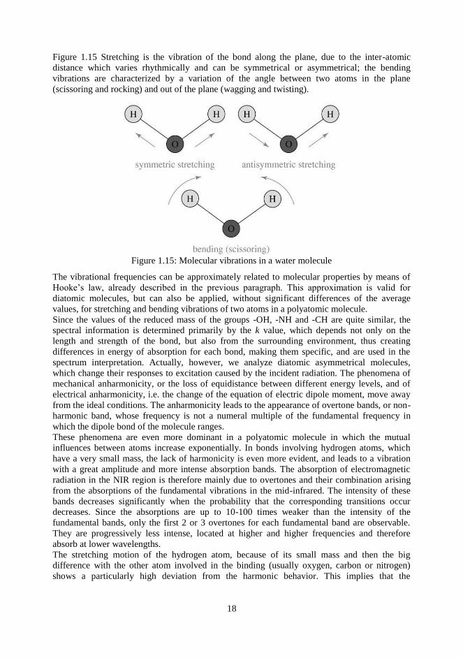
Figure 1.15 Stretching is the vibration of the bond along the plane, due to the inter-atomic
distance which varies rhythmically and can be symmetrical or asymmetrical; the bending
vibrations are characterized by a variation of the angle between two atoms in the plane
(scissoring and rocking) and out of the plane (wagging and twisting).
Figure 1.15: Molecular vibrations in a water molecule
The vibrational frequencies can be approximately related to molecular properties by means of
Hooke‘s law, already described in the previous paragraph. This approximation is valid for
diatomic molecules, but can also be applied, without significant differences of the average
values, for stretching and bending vibrations of two atoms in a polyatomic molecule.
Since the values of the reduced mass of the groups -OH, -NH and -CH are quite similar, the
spectral information is determined primarily by the k value, which depends not only on the
length and strength of the bond, but also from the surrounding environment, thus creating
differences in energy of absorption for each bond, making them specific, and are used in the
spectrum interpretation. Actually, however, we analyze diatomic asymmetrical molecules,
which change their responses to excitation caused by the incident radiation. The phenomena of
mechanical anharmonicity, or the loss of equidistance between different energy levels, and of
electrical anharmonicity, i.e. the change of the equation of electric dipole moment, move away
from the ideal conditions. The anharmonicity leads to the appearance of overtone bands, or non-
harmonic band, whose frequency is not a numeral multiple of the fundamental frequency in
which the dipole bond of the molecule ranges.
These phenomena are even more dominant in a polyatomic molecule in which the mutual
influences between atoms increase exponentially. In bonds involving hydrogen atoms, which
have a very small mass, the lack of harmonicity is even more evident, and leads to a vibration
with a great amplitude and more intense absorption bands. The absorption of electromagnetic
radiation in the NIR region is therefore mainly due to overtones and their combination arising
from the absorptions of the fundamental vibrations in the mid-infrared. The intensity of these
bands decreases significantly when the probability that the corresponding transitions occur
decreases. Since the absorptions are up to 10-100 times weaker than the intensity of the
fundamental bands, only the first 2 or 3 overtones for each fundamental band are observable.
They are progressively less intense, located at higher and higher frequencies and therefore
absorb at lower wavelengths.
The stretching motion of the hydrogen atom, because of its small mass and then the big
difference with the other atom involved in the binding (usually oxygen, carbon or nitrogen)
shows a particularly high deviation from the harmonic behavior. This implies that the

19
fundamental stretching bands located in the mid-infrared between 3000 and 2400 cm-1
, at the
limit with the NIR region, induce overtones and combination bands in the NIR region, thus
making the absorption related to the secondary vibrational modes of hydrogen, the main feature
of a near infrared spectrum. In fact most of the absorptions in this region are derived from the
first, second and third overtones, corresponding to the fundamental vibrations of the bonds -CH,
-NH, -OH, -SH, and their combination bands. The interactions due to the presence of hydrogen
bonds between molecules of the sample are particularly noticeable, since they cause
enlargements of the bands and shifts to lower frequencies. Very weak bands are related to the
vibrations of C-C, C-F and C-Cl bonds.
The low-intensity of absorption in the NIR region may at first seem to be a limit, since it seems
to decrease the sensitivity of the technique. Actually, at a practical level, this is a big advantage
because it allows the direct analysis of a sample, without diluting or dispersing it into inert
matrices as normally happens in traditional spectroscopic techniques, and also to obtain
representative spectra of the whole sample, since the optical paths used are very long.
Moreover, even if NIR bands are larger and liable to overlap more than in other spectral
regions, chemometric techniques available today are able to extract a lot of information even
from complex spectra such as NIR spectra.
IR measurements can be performed both in transmission and in reflectance mode; in the case of
transmission mode, the intensity of light transmitted through the sample compared to the
intensity of incident light is measured:
T = I/I0
Lambert and Beer (Burns & Ciurczak, 2001) observed that the amount of radiation absorbed or
transmitted from a solution or a medium was the exponential function of the concentration of
the adsorbent and the radiation path length through the sample:
A = log 1/T = ka cl
where ka is the molar extinction coefficient, c is the concentration and l is the optical path length
of incident radiation through the sample. In the case of acquisition of reflectance data, the
intensity of the reflected light compared to the intensity of incident light is measured:
R = Irifl/I0
According to the Kubelka-Munk law (Burns & Ciurczak, 2001), reflectance depends on the
coefficient of absorption kr and the coefficient of dispersion of a sample s:
f(R∞) = kr/s
where R∞ is the absolute reflectance.
Experimentally the relative reflectance, i.e. the intensity of light that is reflected from the
sample compared to the intensity of the reflected light in a reference material with a high and
constant absolute reflectance, will be measured; examples of used materials are Teflon, MgO,
discs of high purity ceramic materials. In practice, the relative reflectance is often converted
into apparent absorbance A’, using an empirical relationship between analyte concentration and
reflectance, similar to the Lambert and Beer‘s law:
A’ = log 1/R = a’c
where c is the concentration and a’ is a constant of proportionality.
However, if the matrix is highly absorbent or the analyte shows intense absorption bands, the
linear relationship between absorbance and concentration fails. Both for the transmittance and
the reflectance mode, the proposed equations are obtained from ideal situations, and are
applicable only when the absorptions are weak or the product between concentration and molar
extension coefficient is small. In the case of NIR spectroscopy, the matrix, which cannot be

20
separated from the analyte, has the major absorption and can absorb at the same wavelengths of
the analyte.
Often, the spectroscopic measurement is affected by scattering phenomena, or light diffusion on
the surface, especially in the case of the acquisition of solid samples: in fact, the more the
incident radiation is scattered, the less the beam penetrates deep into and therefore the lower
will be the absorbance (apparent or real). The scattering phenomena depend primarily on the
physical properties of the sample (particle size, crystal environment) and can cause shifts in the
baseline of the spectrum and lead to phenomena of collinearity at different wavelengths.
The signal dependence of the signal from the physical properties of the sample is a significant
disadvantage when NIR is used for qualitative determinations such as product identification or
monitoring of chemical parameters during a process (humidity, homogeneity), and quantitative
analysis of one or more components. To avoid this, some mathematical spectra pre-treatments
have been developed to be applied before the data processing.
1.4.2 FT-NIR instrumentation (Settle, 1997; Da-Wen Sun, 2008)
A genericFT-NIR instrument is formed by a number of basic components: the source of
radiation, a wavelength selector, a system of sample exposure to radiation and a detector.
Radiation sources
For FTIR instrumentation nichrome coil source is commonly used. Helium neon laser source is
used for timing operations in an FTIR.
The NIR radiation sources are mainly incandescent bulbs or emitting diodes (LEDs). Each
source has a specified emission range of wavelengths; for example, incandescence sources are
effective for visible radiation while LEDs are limited to specific wavelengths depending on the
material used. For each of these sources, especially with incandescent lamps, filters must be still
used to eliminate the portion of radiation irrelevant for the analysis purposes and which can lead
to excessive sample heating . For some very specific applications the use of lasers is emerging.
Wavelength selectors
The selection refers to the method used to separate specific wavelengths, in order to obtain the
best resolution. The most common method for selecting wavelengths is the use of filters, made
by layers of clear or colored glass and covered with aluminum, in order to pass only specific
wavelengths or groups of them. More filters can be put together to make more accurate
selections. In modern systems, the diffraction grating is used: this is a surface that reflects
infrared radiation and is engraved with a number of parallel lines, which leads, for the
diffraction, the division of the incident radiation into separate wavelengths. The selection of
wavelengths to be addressed to the sample or to the detector occurs by rotating the grating and
thus changing the incident angle of the radiation source. The critical point of the system is just
the rotation mechanism, which must be extremely precise.
Another category of monochromators, which today is widely used for being fast and precise,
and which characterizes all the instruments based on the Fourier transform (FT), is the
interferometers. The traditional model of spectrometer is modified by replacing the
monochromator with the interferometer discovered by Michelson in 1891, which is still widely
used in most of the NIR instruments (Figure 1.16)

21
Figura 1.16: Michelson inteferometer.
It is based on the principle that two different waves can add up and then combined
constructively each other in a peak with maximum intensity when they are involved, or they can
combine annulling each other when are out of phase (Figure 1.17). To do so, the radius from the
source is split into two parts so that they have the same propagation conditions. The first part
will be reflected on a fixed mirror, while the second on a moving mirror. The two mirrors are
positioned at right angles to each other and the two parts of the radius from the source are
orthogonally directed and separated with a semi-transparent mirror (beamsplitter) with
reflectivity equal to 50%. Once reflected, the two parts are recombined, but with different
phases, since the displacement of the moving mirror causes a delay which in turn induces the
out of phase of the fixed mirror. After recombination, only a certain wavelength will be
enhanced with a peak, while the other will be deleted (Figure 1.17).
Fixed mirror
Translating
mirror
Fixed mirror
Translating
mirror
Figure 1.17: Generation of interference through the interferometer.
Therefore, the moving mirror is able to select all the wavelengths in a fixed range. The
transmitted, diffused or reflected light reaches the detector, which sends a signal to the analog-
digital converter that converts the signal into digital data which are then analyzed by a software
that, applying the Fourier transform, translates the data into a spectral interferogram.
FT spectroscopy has the advantage to simultaneously analyze all frequencies; conversely,

22
traditional spectroscopy, using the grating monochromator, sends to the detector a single
wavelength at each time.
1.4.3 NIR Sample presentation systems (Burns & Ciurczak, 2001)
When a sample is exposed to radiation, interaction with matter can be occur in several ways: the
light can be absorbed by the sample, reflected, and in part or completely transmitted through the
sample (Figure 1.18).
Figure1.18: Light and matter interaction modes
The mode and the degree to which these effects occur depends on the physical state of the
sample and the reading system used. The radiation transmission systems are mainly used for
liquid samples or for thin layers solids, while the reflection mode is most useful for solid
samples. If the sample does not reflect or transmit radiation well enough, the transflectance can
be used as a measuring parameter. In this mode, the radiation penetrates the sample, part is
absorbed, the rest is then reflected on a non-absorbent surface on the bottom of the cell and re-
transmitted through the sample to the detector. Several types of presentation systems are
currently available, strictly dependent on the construction technology.
Fiber-optics
NIR instrumentation is a growing field, thanks to the development of optical fibers that allow
the direct and simple acquisition of spectra by placing the tip of the fiber on the surface and / or
inside the sample. The fibers can have two different optical geometries: diffuse reflectance
fibers (for solid matrices) and transmission fibers (for liquid matrices). The operating principle
of diffuse reflectance fibers is illustrated in Figure 1.19: the radiation beam from the NIR source
strikes the sample, the fraction not absorbed by the sample is reflected and reaches the detector.

23
IR source
Radiation
Input radiation
Output radiation
Sample
Reflected energy
Figure 1.19: Operating principle of diffuse reflectance fiber optic.
Using transmission fibers (Figure 1.20), the light beam strikes the sample, passes through it and
it is collected by the detector. In this optical geometry, the light passes through the sample once.
Figure 1.20: Operating principle of transmission optical fiber.
Optical fibers withstand stressful environmental conditions, are easily integrated into machines
and allow conveying the signal unchanged for tens of meters, permitting the centralization of
the measurement devices into a single structure. Moreover, the use of optical fiber, positioned
directly on the sample surface, allows non-invasive, non destructive and in line measurements.
Integrating sphere
For measurements on heterogeneous solid samples, the most suitable sampling system is the
integrating sphere, whose operating principle is illustrated in Figure 1.21.

24
Figure 1.21: Operating principle of the integrating sphere
The radiation strikes a mirror outside the sphere which in turn directs the radiation to the
sample. The part of radiation not absorbed by the sample is reflected back to the inner surface
of the sphere to be collected by the detector. The sample is usually collected in a container with
a bottom transparent to radiation, and equipped with a geometry that allows the rotation of the
sample, which is necessary when working with non-homogeneous samples.
Scanning transmission system for solids
Although transmission systems are mainly used for liquid samples or for thin layers of solids, it
is possible to scan solid samples in transmission mode. In this case, the light beam strikes the
sample, passes through it and it is collected by the detector. This sampling technique is used to
measure the whole mass of the sample, especially to determine hardness or composition.
Table 1.2 shows the advantages and disadvantages between diffuse reflectance and transmission
measurements on solid samples. As the table shows, diffuse reflectance measurements allow
the analysis of the whole spectral range and therefore the collecxtion of information on
combination bands and overtone spectral region, while transmission measurements,
characterized by a low energy, allow the acquisition of spectra in a smaller range of
wavelengths.
Diffuse reflectance measurements Ttransmission measurements
High energy Low energy due to the high sample
absorption
The analysis requires sample homogeneity
(the surface should be representative of the
whole mass of the sample)
The whole mass of the sample is measured
Analysis lasting from 1 to 30 seconds Analysis lasting from 10 seconds to 2
minutes
Whole spectral range: from 12500 to 3600
cm-1
(800-2780 nm)
Reduced spectral range: from 12500 to 7000
cm-1
(800-1400 nm)
Combination bands and overtone working
zone
Overtone bands working zone
Usually applied to samples surface
(coatings etc.)
Usually applied to the sample core (hardness
or composition etc.)
Table 1.2: Comparison between diffuse reflectance and transmission measurements.

25
1.4.4 NIR detectors (Burns & Ciurczak, 2001)
The most characteristic element for the instrument is the detector. Its task is to receive the
radiation from the sample and turn it into an electrical signal. The detector must be sensitive to
the wavelength of interest: generally sulphide or lead selenide detector are used for the spectral
region between 1100 and 2500 nm and silicon detectors for the NIR region at short length
(SWNIR) between 400 and 1100 nm. Most applications of the NIR technique are intended to
obtain the spectrum of a sample, i.e. the graphical representation of absorbance or transmittance
as a function of wavelength or wavenumber. An additional request is the ability of the tool to
acquire a white or a background (baseline) to subtract from the sample spectrum. This can be
done before the scan or, in some cases, i.e. in dual-beam instruments, continuously and
simultaneously with the reading.
1.4.5 NIR advantages (Burns & Ciurczak, 2001)
The practical advantages offered by FT-NIR are:
• Improved signal to noise ratio. The high value of this ratio allows the well resolved spectra
with fast scans in few seconds
• Less energy loss and hence a greater energy to the detector. The optics of the FT-NIR in fact
allows the availability of a passing energy greater than that of dispersive instruments, where the
available energy is limited by the need of the use of splits.
• Improved accuracy and precision in the wavelengths discrimination.
• Increased speed of spectra collection.
1.5 REFERENCES
Bringel F et al., 2005, Lactobacillus plantarum subsp. argentoratensis subsp. nov., isolated
from vegetable matrices. International Journal of Systematic and Evolutionary Microbiology,
55: 1629-1634.
Bude Ugarte M et al., 2006, Nonstarter lactobacilli isolated from soft and semihard Argentinean
cheeses: genetic characterization and resistance to biological barrier. J. Food Prot. 69: 2983-
2991.
Burns DA, Ciurczak EW Handbook of near-infrared analysis. Burns DA, Ciurczak EW eds,
CRC Press, Marcel Dekker Inc, New York, 2001.
Champagne C, Garner N, 2005, Challenger in the addition of probiotic culture sto foods.
Critical Reviews in Food Science and Nutrition, 45: 61-84.
Da-Wen Sun Infrared Spectroscopy for Food Quality Analysis and Control. Da-Wen Sun ed,
Academic Press, Elsevier Inc, USA, 2008.
de Vries M et al., 2006, Lactobacillus plantarum-survival, functional and potential probiotic
properties in the human intestinal tract. International Dairy Journal, 16: 1018-1028.
Farmworth E. The future for fremented food. In E. R. Farworth (Ed.), Handbook of fermented
functional foods (pp. 27-58). Boca Raton, Florida, USA: CRC Press.
Guarner F et al., 2005, Should yogurt cultures be considered probiotic? British J. of Nutrition
93: 783-786.
Mattila-Sandholm T et al., 2002, Technological challenger for future probiotic foods. Int. Dairy
J. 12:173-182.
Minelli E et al., 2004, Assessment of novel probiotic Lactobacillus casei strain for the
production of functional dairy foods. Int Dairy J. 14: 723-736.
Mucchetti G, Neviani E (2006). Microbiologia e tecnologia lattiero-casearia (pp. 145-160, 758-
763). Ed. tecniche nuove. Milano.
Parvez, S et al., 2006, Probiotics and their fermented food products are beneficial for health. J.
Appl. Microbiol. 100: 1171-1185.

26
Pretzer G et al., 2005, Biodiversity-based identification and functional characterization of the
mannose-specific adhesin of Lactobacillus plantarum. J. Bacteriol. 187: 6128-6136.
Schillinger U et al.,2005, ―In vitro‖-adherence and other properties of lactobacilli used in
probiotic yoghurt-like products. Int. Dairy J. 12. 1289-1297.
Schmidt EJ et al., 2001, Activities of cholic and acid-derived antimicrobial agents against
multidrug-resistant bacteria. J. Antimicrob. Chemoth 47: 671-674.
Siesler HW Near-infrared spectroscopy: principles, instruments, applications. Siesler HW,
Ozaki Y, Kawata S, Heise HM eds, John Wiley & Sons, Weinheim, Germany, 2002.
Stanton C et al., 2003, Challenges facing development of probiotic-containing functional foods.
In E. R. Farnworth (Ed.), Handbook of fermented functional foods (pp. 27-58). Boca Raton,
Florida, USA: CRC Press.
Vertuani S et al., 2001 Probiotici e prebiotici-impieghi attuali e prospettive future. Integratore
Nutrizionale 26: 26-32.
Vinderola CG, Reinheimer JA, 2003, Lactic acid starter and probiotic bacteria: a comparative
―in vitro‖ study of probiotic characteristics and biological barrier resistance. Food Res Int, 36:
895-904.
Workman J, Weyer L Practical guide to interpretive near-infrared spectroscopy CRC Press,
Taylor & Francis Group, New York, 2008.

2. PRELIMINARY SELECTION OF LACTOBACILLUS
PLANTARUM STRAINS WITH PROBIOTIC
POTENTIALITIES

28
2.1 INTRODUCTION
2.1.1Probiotics: benficial effects on human health
The beneficial effects of probiotics on the human organism are numerous. Several studies have
shown that probiotic bacteria may decrease the incidence, duration and severity of some gastro-
intestinal diseases (Parvez et al., 2006).
The considerable interest in this subject is confirmed by the scientific literature, resulting from
exploration of the therapeutic potential of probiotics (Figure 2.2) in various contexts, which will
be hereafter considered.
Figure 2.1. Main beneficial effects due to the consumption of probiotics (Parvez et al., 2006)
1. Detoxification properties. The intestine plays a central role in the process of detoxification
of the body through its function as double barrier, both mechanical and immunological.
Probiotics are considered detoxifying agents, able to counteract the generation of harmful
metabolites through direct activity (degradation of toxic agents) and indirect (decreased
activity of enzymes pro-toxin).
2. Reduction of lactose intolerance. Approximately 70% of the world population is intolerant
to lactose because of a low β-galactosidase activity, responsible for the conversion of
lactose into glucose and galactose (Vertuani et al., 2001). The beneficial effects due to the
assumption of probiotics results from their ability to ferment lactose, resulting in higher
tolerance in intolerant people (Parvez et al., 2006).
3. Immunomodulation. Probiotics are able to influence various mechanisms of the immune
response, such as humoral or cell-mediated immunity. Regarding the humoral response,
numerous scientific studies show that a treatment with probiotics belonging to
Lactobacillus casei and Lactobacillus acidophilus determines a rise in the production of
IgA, which improves the function of the gut as a barrier. An interesting aspect of the

29
modulation of the immune system by probiotics is their ability to influence the mediated
response by T cells in the intestinal epithelium, through the production of cytokines. It has
also been shown that lactobacilli are able to stimulate the activity of macrophages against
different species of bacteria. Probably this effect is determined by absorption through the
intestinal walls of a soluble antigen or translocation of lactobacilli in the bloodstream. The
immunomodulating action of probiotics also explains the decrease in the symptoms
associated with food allergies, in which there is an increase in intestinal permeability to
allergens, caused by chronic inflammation of the mucosa.
4. Rheumatoid arthritis. It is an auto-immune disease that affects joints and other internal
organs. The etiology is not entirely clear, but there are studies that indicate this disease as a
consequence of intestinal infection or a food allergens action. In these circumstances, the
intestinal mucosa is damaged, resulting in increased permeability to antibodies, which
attack the body's structures and trigger an inflammatory process. The consumption of
probiotics by patients suffering from this disease, showed an improvement of their clinical
situation (Parvez et al., 2006).
5. Anti-infective activities. An important application of probiotics is the prevention of
opportunistic infections resulting from antibiotic treatment. A considerable part of the
intestinal microflora is destroyed by antibiotics, thus promoting the development of
pathogenic organisms such as Candida albicans and Clostridium difficile, which can lead
to infection, sepsis, colitis and diarrhea. Numerous studies confirm that the intake of
probiotics, concomitantly with antibiotic therapy, is able to reduce the incidence of
opportunistic infections and to restore, more quickly, the physiological structure of the
intestinal microflora.
6. Intestinal disorders. The use of probiotics has a positive influence on inflammatory and
infectious diseases of the intestine. It has been hypothesized that the mechanism involved
include the reduction of intestinal pH through the stimulation of lactic acid production by
the intestinal microflora, the direct effects of antagonism of pathogenic microorganisms
and the process of immunostimulation.
7. Chemo-protective activities. The scientific data available concern the reduction of the risk
of developing colon cancer, which is one of the most important causes of mortality. The
experiments, conducted on animal treated with probiotic lactobacilli and bifidobacteria,
after treatment with chemical carcinogens, showed the decrease of specific tumor markers.
An hypothetic explanation of this effects on the development of tumors is given by the
ability of lactobacilli to suppress the growth of bacterial species which convert pro-
carcinogens into carcinogens. This property determines the reduction of the concentration
of carcinogenic substances in the gut. Moreover, lactobacilli subtract mutagenic
compounds at the intestinal level, thus preventing them from being absorbed. There is no
direct experimental evidence on men yet, but the increase of consideration of probiotics as
potentially useful in the prevention of neoplastic diseases encourage further research
(Vertuani et al., 2001).
8. Hypertension. Preliminary studies indicate that probiotics or fermented products may affect
the control of blood pressure. In this regard, clinical studies have been conducted. These
studies allowed us to document the anti-hypertensive effect caused by the ingestion of
probiotics.
9. Control of cholesterol. Cholesterol is essential for many body functions. It acts as a
precursor of various hormones and vitamins and is a component of biological membranes.
High levels of cholesterol or other fats in the blood, however, are considered risk factors
for the development of heart disease. The cholesterol-lowering activity of probiotics is
controversial (Parvez et al., 2006), it may be due to a direct action of cellular uptake of
cholesterol or indirectly, by hydrolysis of bile salts.

30
The functional requirements of probiotics should be established by using in vitro methods and
the results of these studies should be reflected in controlled human studies.
Among the most important features that a probiotic must have, the ability to adhere to intestinal
epithelia and the tolerance to bile were in depth investigated.
Adhesion of probiotic strains to the intestinal surface and the subsequent colonization of the
human GI-tract has been suggested as an important prerequisite for probiotic action. Adherent
strains of probiotic bacteria are likely to persist longer in the intestinal tract and thus have better
possibilities of showing metabolic and immunomodulatory effects than non-adhering strains
(Saarela et al., 2000).
Adhesion provides an interaction with the mucosal surface facilitating the contact with gut
associated lymphoid tissue mediating local and systemic immune effects. Thus, only adherent
probiotics have been thought to effectively induce immune effects and to stabilize the intestinal
mucosal barrier (Salminen et al., 1998).
Lactobacillus plantarum is encountered inhabitant of the human intestinal tract, and some
strains are marketed as probiotics. Their ability to adhere to mannose residues is a potentially
interesting characteristic with regard to proposed probiotic features such as colonization of the
intestinal surface and competitive exclusion of pathogens (Pretzer et al., 2005).
The capacity to recognize the same receptor sites has been proposed to enable probiotic
microorganisms to inhibit colonization of pathogens by competitive exclusion, which might
prevent infections in the small intestine (Reid & Burton, 2002). Studies reported how the
adherent bacteria can inhibit the intestinal infections by enterotoxigenic Escherichia coli
(ETEC), which causes travelers‘ diarrhea by the recognition of the same adherence sites on the
intestinal epithelial surface (Pretzer et al., 2005).
Recently, the potentially ‗probiotic gene‘(msa) encoding the mannose-specific adhesin (MSA)
of L. plantarum has been identified, using a biodiversity-based approach including phenotype-
genotype correlation and mutation analysis in L. plantarum strain WCFS1 (Pretzer et al., 2005).
In recent years, the ability of probiotic strains to produce bile salt hydrolase (BSH) has become
the focus of attention on account of its influence on cholesterol metabolism and hence BSH
activity can be explored as a functional probiotic biomarker for the selection of the probiotic
strains (Kumar et al., 2012).
Bile salt hydrolase (BSH) is an enzyme produced by several bacterial species in the human or
animal gastrointestinal tract that catalyzes the glycine- or taurine-linked bile salt deconjugation
reaction (Patel et al., 2009).
The ability of probiotic strains to hydrolyze bile salts has often been included among the criteria
for probiotic strain selection, and a number of bile salt hydrolases (BSHs) have been identified
and characterized.
BSHs are very specific for certain bile types, and their duration of contact to bile ensures the
bacterial survival into varying bile environments. Studies carried out in Lactobacillus
plantarum WCFS1, which had four bsh genes, and L. acidophilus NCFM, which had two bsh
genes, supported this assumption (Patel et al., 2009).
BSH activity involves deconjugation of bile salts to primary bile salts: primary bile acids are
synthesized in the liver from cholesterol and are conjugated with either glycine or taurine before
their secretion. The carboxyl group of the bile acid and the amino group of the amino acid are
linked by an amide bond. (Figure 2.2a)

31
Figure 2.2 (A) Chemical structure of bile acids. (B) Reaction catalyzed by BSH enzymes. BSHs
cleave the peptide linkage of bile acids, which results in removal of the amino acid group from
the steroid core. The resulting unconjugated bile acids precipitate at low pH. (C) Detection of
BSH activity. The white precipitates around colonies and the clearing of the medium are
indicative of BSH activity (Begley et al., 2006).
This activity is strictly related to the ability of strains to remove cholesterol. Many authors
reported how the cholesterol removal ability is linked to bile salt deconjugation and co-
precipitation (Liong et al., 2006).
Moreover, BSH activity protects bacteria from the toxicity of conjugated bile salts (they repress
bacterial growth), being considered a detoxification mechanism of vital importance to bacterial
communities (such as lactobacilli), which are typically associated to the human GIT (Zago et
al., 2011).
Thus, the aim of this study was to investigate the presence of msa and bsh genes in L.plantarum
strains involving in MSA and BSH activity that are considered to be associated with probiotic
features. Moreover, positive strains were tested for BSH activity using a phenotypic assay.
2.2 MATERIALS AND METHODS
2.2.1 Strain selection
A total of 80 strains were used in this study. Seventy-nine strains of Lactobacillus plantarum,
belonging to the collection of the CRA-FLC (Fodder and Dairy Productions Research Centre,
Lodi), were isolated from different matrixes (23 strains from dairy products, 38 strains from
olive brines and 18 strains from grape marcs); the type strain ATCC 14917T,
obtained from the
American Type Culture Collection (Rockville, MD) was used as reference strain. Strains were
kept as frozen stocks at -80 °C in MRS broth (Merck, Darmstadt, Germany) added with 20%
(v/v) glycerol. Frozen stock cultures were thawed and subcultured twice (2% inoculum) in MRS
at 30 °C overnight before use.
2.2.2 Molecular typing
Bacterial DNA was extracted using a Chelex®
100 method described by Walsh et al. 1991.
Two-hundred μl of culture were washed twice with 500 μl di TE0.1 pH 8.0 (TE0.1: Tris-HCl 10
mM, EDTA 0.1 mM) and the pellet was harvested and resuspended in 245 μl of the same buffer
and 5 μl of lysozyme (stock solution 50 mg/ml). After that, the strains were incubated at 56°C
for 45 min.
To each eppendorf were added 218.7 μl of TE0.1 pH 8.0, 5 μl of DTT 1 M, 10 μl of EDTA 0.5
M pH 8.0, 12.5 μl of 20% SDS and finally 3.8 μl of proteinase K (stock solution 20 mg/ml).
The strains were incubated for 1 hour at 37°C and than 500 μl of a 5% Chelex® 100 (Sigma –

32
Aldrich) solution were added. This ionic resin allows the DNA purification by precipitation of
all impurities generated by bacterial lysis. The extracted DNA was diluted 1:10 and used as a
template for PCR reactions.
RAPD-PCR was performed using M13 primer (5‘ – GAGGGTGGCGGTTCT – 3‘) as
described by Rossetti and Giraffa (2005). Amplification reactions were carried out in 25 μl
volume containing 2 mM of M13 primer, 2.5 units/100 μl of AmpliTaq DNA polymerase
(Applied Biosystems, Monza, Italy), 3 mM MgCl2, 2.5 ng/μl of total DNA, and 0.2 mM of each
dNTP. PCR amplifications were performed in a Perkin–Elmer thermal cycler (model 9700
Applied Biosystem). The cycling program consisted of an initial denaturation step at 94 °C for
120 s and then 40 cycles of: 94 °C for 60 s, 45 °C for 20 s, and 72 °C for 120 s. The final
elongation was performed at 72 °C for 10 min.
PCR products were analyzed by electrophoresis through 1.5% agarose gels at 26 V for 12 h in
1× Tris-acetate EDTA (TAE) buffer (1 × TAE: 40 mmol/l Tris acetate, 1 mmol/l EDTA, pH
8.0) stained with ethidium bromide, and visualized under UV light.
The fingerprinting profiles were processed using Bionumerics software (5.1 Applied Maths,
Belgium). Similarity among different profiles was evaluated by applying the Pearson‘s
correlation coefficient. Strains representative of different genotypic profiles were selected and
their taxonomic affiliation was confirmed by PCR according to Torriani et al. (2001).
2.2.3 Research for msa and bsh genes
Total DNAs were also used to detect the presence of bsh (bile salt hydrolase) and msa (mannose
specific adhesing) genes, encoding for the bile salt hydrolase (BSH) and the mannose-specific
adhesin (MSA), respectively (Zago et al., 2011). PCR primers were designed on the basis of the
gene sequences of bsh (GenBank accession number AL935262.1) and msa (GenBank accession
number AL935255.1) of L. plantarum WCFS1. The primers had the following sequences: 5‘-
CGTATCCAAGTGCTCATGGTTTAA-3‘ (bsh for, nucleotide position 150568 to 150593 of
the bsh gene), 5‘-ATGTGTACTGCCATAACTTATCAATCTT-3‘ (bsh rev, nucleotide position
151487 to 151460 of the bsh gene), 5‘-GCTATTATGGGGATTACGTTG-3‘ (msa for,
nucleotide position 133061 to 133081 of the msa gene) and 5‘-
CTGTCTTGACAATAGCCATATA-3‘ (msa rev, nucleotide position 134801 to 134781). The
primers were designed to obtain PCR product lengths of 919 bp (for bsh) and 1740 bp (for
msa). The strain LMG 9211 was used as positive control for both PCR amplifications.
PCR amplifications were performed in 20 µl volumes with 0.5 mM of each primer (Biotez,
Berlin, Germany), 2.5 units/100 μl of AmpliTaq DNA polymerase (Applied Biosystems,
Monza, Italy), 1.5 mM MgCl2, 20 ng of total DNA, and 200 mM of each dNTP. DNA
amplifications were performed by a Perkin Elmer thermal cycler (mod. 9700, Applied
Biosystems) and were set as follows: 4 min at 94°C; 30 cycles of 30 s at 94°C, 30 s at 64°C
(bsh for/rev) or 52°C (msa for/rev), and 1 min at 72°C; the final extension step consisted of 7
min at 72°C. The PCR products were separated on a 1% agarose gel in Tris-Acetate EDTA
buffer, prepared as previous described.
2.2.4 BSH activity
BSH activity of the strains was detected using a plate screening procedure according to Zago et
al. (2011). Briefly, overnight cultures were spotted on MRS agar plates containing 0.37 g/l of
CaCl2 and 0.5% sodium salt of taurodeoxycholic acid (TDCA) or 0.5% sodium salt of
glycodeoxycholic acid (GDCA) (Sigma-Aldrich). Plates were anaerobically incubated at 37°C
for 72 h. The presence of halos around colonies or white opaque colonies indicated BSH
activity. Enterococcus faecium LMG 16170 was used as BSH-positive strain. The inoculums of
each strain in MRS without supplementation was included as negative control.

33
2.3 RESULTS AND DISCUSSION
Several functional attributes have already been demonstrated for L. plantarum strains isolated
from fermented foods (Adlerberth et al., 1996; Nguyen et al., 2007; Georgieva et al.,2008;
Belviso et al., 2009).
Considering the role that L. plantarum plays in food systems, the aim of this work was to select
potentially probiotic L. plantarum strains from a wide collection of strains isolated from
different matrixes..
In the present study 80 L.plantarum strains were investigated for their probiotic properties.
These strains, identified on the basis of species-specific PCR for L. plantarum group, with
different M13 PCR fingerprinting profiles and coming from different matrixes, were screened
for the presence of msa and bsh genes, that are considered to be associated with probiotic
features. The msa gene was selected to indicate the mannose-specific adhesion of L. plantarum,
being one of the first identified genes associated with a particular probiotic effect (Pretzer et al.,
2005). The bsh gene, coding for the BSH activity, was selected for its importance in bile salt
hydrolysis which, ultimately, leads to lower blood serum cholesterol levels (Begley et al.,
2006).
Thirty-four (Table 2.1) out of the 80 strains showed the expected amplicons for both genes.
These 33 strains and the L. plantarum type strain ATCC14917T were selected for further
characterization.
Strains Origin Strains Origin Strains Origin
Lp751 Pecorino Pisano Cheese 50339 Olive Brine 50357 Olive Brine
Lp752 Pecorino Pisano Cheese 50341 Olive Brine 50358 Olive Brine
Lp755 Pecorino Pisano Cheese 50343 Olive Brine 50360 Olive Brine
Lp793 Canestrato Pugliese Cheese 50346 Olive Brine 50361 Olive Brine
Lp794 Monte Veronese Cheese 50347 Olive Brine 50362 Olive Brine
Lp797 Morlacco Cheese 50348 Olive Brine 50363 Olive Brine
Lp799 Monte Veronese Cheese 50350 Olive Brine 50366 Olive Brine
Lp804 Monte Veronese Cheese 50351 Olive Brine 50368 Olive Brine
Lp813 Spressa Cheese 50352 Olive Brine 50369 Olive Brine
Lp994 Argentinian Cheese 50353 Olive Brine 50371 Olive Brine
50337 Olive Brine 50356 Olive Brine 50372 Olive Brine
ATCC 14917 Type Strain
Table 2.1: Lactobacillus plantarum strains used for characterisation
The ability to adhere to mannose residues is a potentially interesting characteristic with regard
to colonization of the intestinal surface and competitive exclusion of pathogens. The product of
the msa gene, identified as a mannose-specific adhesin, was found to mediate adhesion to
human colonic cell line HT-29 (Adlerberth et al., 1996), and to agglutinate S. cerevisiae cells
(Pretzer et al., 2005). All the strains carried the msa gene.
A relevant property of candidate probiotics is BSH activity, which involves deconjugation of
bile salts to primary bile salts: all 34 strains, which possessed bsh gene, demonstrated the ability

34
to hydrolyze both sodium glycodeoxycholate and sodium taurodeoxycholate, as shown by halos
around colonies after growth in MRS-GDCA and by the appearance of white opaque colonies
after growth in MRS-TDCA, respectively.
The deconjugation activity would play a role in maintaining the equilibrium of the gut
microflora. In fact, the inhibition of common intestinal bacteria has been related to the presence
of free (deconjugated) bile acids rather than the conjugated ones It has been also suggested that
bile salts hydrolase enzyme might be a detergent shock protein that enables lactobacilli to
survive the intestinal bile stress (Vinderola & Reinheimer, 2003).
Another phenomenon related to the presence of the deconjugation activity is the reduction of
serum cholesterol. In this sense, bile salt deconjugation is a desirable property when selecting
strains to be used as a dietary adjunct (Liong et al., 2006).
As shown above, all 34 L. plantarum strains possessed the bsh gene and expressed BSH
activity. Schmidt et al. (2001) also showed that, at least in lactobacilli, bile salt resistance could
not be related to the presence of BSH. BSH activity, however, is a widespread trait in L.
plantarum (Zago et al., 2011).
2.4 CONCLUSIONS
This study showed that potentially probiotic L. plantarum strains could be isolated from dairy
products and that strains of human origin are not necessarily the most suitable for probiotic
application. This seems corroborated by the wide finding on genotypic traits (such as adhesion
ability and capacity to hydrolase bile salts) which are considered to be typical of probiotic
bacteria, within the L. plantarum dairy isolates studied here.
Further phenotypic analysis to confirm expression of the studied genes and in vitro (such as
adhesion ability, antibiotic resistance, and cell hydrophobicity) and in vivo studies are required
to better detect really L. plantarum candidates to be used in functional foods.
2.5 REFERENCES
Adlerberth I et al., 1996, A mannose specific adherence mechanism in Lactobacillus plantarum
conferring binding to the human colonic cell line HT-29. Appl. Environ. Microbiol. 62: 2244-
2251.
Begley M et al., 2006, Bile salt hydrolase activity in probiotics. Appl. Environ. Microbiol. 72:
1729-1738.
Belviso S et al., 2009, In vitro cholesterol-lowering activity of Lactobacillus plantarum and
Lactobacillus paracasei strains isolated from the Italian Castelmagno PDO cheese. Dairy Sci.
Technol 89: 169-176.
Bude Ugarte M et al., 2006, Nonstarter lactobacilli isolated from soft and semihard Argentinean
cheeses: genetic characterization and resistance to biological barrier. J. Food Prot. 69: 2983-
2991.
Georgieva R et al., 2008, Identification and vitro characterization of Lactobacillus plantarum
strains from artisanal Bulgarian white brined cheeses. J. Basic Microb. 48: 234-244.
Guarner F et al., 2005, Should yogurt cultures be considered probiotic? British J. of Nutrition
93: 783-786.
Kumar R et al., 2012, Bile salt hydrolase (bsh) activity screening of Lactobacilli: in vitro
selection of indogenous Lactobacillus strains with potential bile salt hydrolyzing and
cholesterol lowering ability. Probiotics and Antimicrobial Proteins 4: 162-172
Liong MT, Shah NP, 2005, Bile salt deconjugation ability, bile salt hydrolase activity and
cholesterol co-precipitation ability of lactobacilli strains. Int. Dairy J. 15:391–398.
Mattila-Sandholm T et al., 2002, Technological challenger for future probiotic foods. Int. Dairy
J. 12:173-182.
Minelli E et al., 2004, Assessment of novel probiotic Lactobacillus casei strain for the

35
production of functional dairy foods. Int Dairy J. 14: 723-736.
Nguyen TDT et al., 2007, Characterization of Lactobacillus plantarum PH04, a potential
probiotic bacterium with cholesterol-lowering effects. Int. J. Food Microbiol. 113, 358-361.
Parvez, S et al., 2006, Probiotics and their fermented food products are beneficial for health. J.
Appl. Microbiol. 100: 1171-1185.
Patel AK et al., 2009, Probiotic bile salt hydrolase: current developments and perspectives.
Appl. Biochem. Biotechnol. 162: 166-180.
Pretzer G et al., 2005, Biodiversity-based identification and functional characterization of the
mannose-specific adhesin of Lactobacillus plantarum. J. Bacteriol. 187: 6128-6136.
Reid G, Burton J, 2002. Use of Lactobacillus to prevent infection by pathogenic bacteria.
Microbes Infect. 4:319–324.
Rossetti L, Giraffa G 2005 Rapid identification of dairy lactic acid bacteria by M13-generated,
RAPD-PCR fingerprint databases. J. Microbiol. Meth. 63: 135-144.
Saarela MG et al., 2000, Probiotic bacteria: safety, functional and technological properties. J.
Biotechnol. 84:197–215.
Salminen SJ et al., 1998, Adhesion of some probiotic and dairy Lactobacillus strains to Caco-2
cell cultures. Int. J.Food Microbiol. 41: 45-51
Schillinger U et al.,2005, ―In vitro‖-adherence and other properties of lactobacilli used in
probiotic yoghurt-like products. Int. Dairy J. 12. 1289-1297.
Schmidt EJ et al., 2001, Activities of cholic and acid-derived antimicrobial agents against
multidrug-resistant bacteria. J. Antimicrob. Chemoth 47: 671-674.
Torriani S et al., 2001, Differentiation of Lactobacillus plantarum, L. pentosus, and L.
paraplantarum by recA gene sequence analysis and multiplex PCR assay with recA gene-
derived primers. Appl. Environ. Microbiol. 67: 3450-3454.
Vertuani S et al., 2001 Probiotici e prebiotici-impieghi attuali e prospettive future. Integratore
Nutrizionale 26: 26-32.
Vinderola CG, Reinheimer JA, 2003, Lactic acid starter and probiotic bacteria: a comparative
―in vitro‖ study of probiotic characteristics and biological barrier resistance. Food Res Int, 36:
895-904.
Walsh PS 1991, Chelex® 100 as a medium for simple extraction of DNA for PCR- based typing
from forensic material. Biotechniques 10: 506–513.
Zago M et al., 2011, Characterization and probiotic potential of Lactobacillus plantarum strains
isolated from cheeses. Food Microbiol 28: 1033-1040.

3 EVALUATION OF RIBOFLAVIN PRODUCTION IN
LACTOBACILLUS PLANTARUM STRAINS

37
3.1 INTRODUCTION
The relationship between nutrition and health is constantly developing and linking the concepts
of "functional food" (where functional refers to the nutraceutical and healthy aspects of food) to
the new trend of achieving wellness consuming foods with high added value. Food is
considered "functional" if, in addition to a high nutritional value, proves to have beneficial
effects on the body, to preserve the health of the individual, to mitigate the risk of the onset of
disease. Foods enriched in probiotics are certainly the model of functional food.
Besides probiotic LAB, certain strains of LAB are able to produce/release and/or increase
specific beneficial compounds in foods. These functional ingredients are sometimes referred to
as nutraceuticals, a term that describes ―a food (or part of a food) that provides medical or
health benefits, including the prevention and/or treatment of a disease‖. These ingredients can
be macronutrients, micronutrients (such as vitamins) or non-nutritive compounds and can be
naturally present in certain foods or added during processing (Le Blanc et al., 2011).
Vitamins are micronutrients that are essential for the metabolism of all living organisms. They
are found as precursors of intracellular coenzymes that are necessary to regulate vital
biochemical reactions in the cell. Humans are incapable of synthesizing most vitamins, and they
consequently have to be obtained exogenously (i.e. from their diet). Although most vitamins are
present in a variety of foods, vitamin deficiencies still exist in many countries including highly
industrialized nations mainly because of malnutrition, not only as a result of insufficient food
intake but also because of unbalanced diets (Le Blanc et al., 2011).
Vitamins are generally classified into two groups, the fat-soluble vitamins, which include
vitamins A, D, E and K, and the water-soluble vitamins, which include vitamin C, biotin
(vitamin H or B7) and a series of B vitamins — thiamin (B1), riboflavin (B2), niacin (B3),
pantothenic acid (B5), pyridoxine (B6), folic acid (B9) and cobalamin (B12). The fat-soluble
vitamins appear to function as integral parts of cell membranes while, the water-soluble
vitamins act as coenzymes, often as carriers of a particular chemical group (Burgess et al.,
2009).
The ability of lactic acid bacteria (Lactococcus lactis, Streptococcus thermophilus, Leuconostoc
spp. and Lactobacillus plantarum are the most studied species) to produce spontaneously
vitamins can be commercially exploited in view of the high cost of chemical synthesis or
extraction from the raw materials of these important molecules. The aim of the development of
the research of strains able to produce vitamins is to use the fermented food, obtained with
strains selected for the ability to produce these compounds, as carrier of these natural
substances with biological value reducing the costs of artificial fortification with synthetic
vitamins.
The B vitamins produced by microorganisms are riboflavin (vitamin B2), folic acid and folate
(vitamin B9) and cyanocobalamin (vitamin B12).
Riboflavin (vitamin B2) is an essential component of basic cellular metabolism since it is the
precursor of the coenzymes flavin mononucleotide (FMN) and flavin adenine dinucleotide
(FAD). The latter two biomolecules play a central role in metabolism acting as hydrogen
carriers in biological redox reactions involving enzymes such as NADH dehydrogenase
(Burgess et al., 2004).
Many microorganisms, plants, and fungi possess the biosynthetic ability to produce riboflavin.
However, vertebrates, including humans, lack this ability and must therefore obtain this vitamin
from their diet. Dietary, riboflavin is present in liver, egg yolk, milk, and meat, whereas the
vitamin is commercially synthesized for nutritional use in the fortification of various food
products such as bread and breakfast cereals.
Riboflavin is synthesized by many bacteria and its biosynthetic pathway has been studied
extensively in Bacillus subtilis and Escherichia coli.

38
Riboflavin biosynthesis (Figure 3.1) requires the precursors guanosine 5'-triphosphate (GTP)
and ribulose 5-phosphate. The first step of the GTP-dependent branch of the biosynthetic
pathway is encoded by ribA in E. coli. In B. subtilis it is also encoded by ribA but in this case
ribA is a bifunctional enzyme which also catalyses the formation of 3,4-dihydroxy-2-butanone
4-phosphate from ribulose 5-phosphate. RibA overexpression in B. subtilis increases riboflavin
yield up to 25%, suggesting that this enzyme is rate-limiting in riboflavin biosynthesis (Burgess
et al., 2009).
However, in Lactococcus lactis, overexpression of ribA alone does not lead to increased
riboflavin production (Burgess et al., 2004).
To be biologically active, riboflavin must be converted to its coenzyme forms, FMN and FAD.
This is accomplished by a bifunctional flavokinase/FAD synthetase, which is encoded in B.
subtilis by ribC or ribF in E. coli. Certain mutations in the B. subtilis ribC gene have been
found to result in riboflavin overproduction (Coquard et al., 1997; Kreneva and Perumov,
1990). B. subtilis also encodes a separate monofunctional riboflavin kinase encoding gene, ribR,
which can suppress the effect of ribC mutations, restoring normal riboflavin production
(Burgess et al., 2009).
Figure 3.1: Riboflavin biosynthesis (Burgess et al., 2009).
Some bacteria and fungi are capable of riboflavin overproduction. In bacteria this trait can be
achieved either by metabolic engineering or by exposure to purine analogues and/or the toxic
riboflavin analogue roseoflavin. Particularly, in Lactococcus lactis both of these approaches
have been used with success (Burgess et al., 2004) Recent studies have reported on the selection
of riboflavin overproducing strains for potential food applications, for example, the
manufacture of vitamin B2-enriched dairy products, which were found to improve the
riboflavin status of deficient rats (Capozzi et al., 2011).
Thus, the present study reports on the selection of food-grade, riboflavin-producing LAB
isolated from different matrixes.
3.2 MATERIALS AND METHODS
3.2.1 Strains selection
A total of 34 strains were used in this study: 33 strains of Lactobacillus plantarum, belonging to
the collection of the CRA-FLC (Fodder and Dairy Productions Research Centre, Lodi), were
isolated from different matrixes (10 strains from dairy products and 23 strains from olive

39
brines); the type strain ATCC 14917T, obtained from the American Type Culture Collection
(Rockville, MD) was used as reference strain.
Strains were kept as frozen stocks at -80 °C in MRS broth (Merck, Darmstadt, Germany) added
with 20% (v/v) glycerol. Frozen stock cultures were thawed and subcultured twice (2%
inoculum) in MRS at 30 °C overnight before use (Table 3.1).
Strains Origin Strains Origin Strains Origin
Lp751 Pecorino Pisano Cheese 50339 Olive Brine 50357 Olive Brine
Lp752 Pecorino Pisano Cheese 50341 Olive Brine 50358 Olive Brine
Lp755 Pecorino Pisano Cheese 50343 Olive Brine 50360 Olive Brine
Lp793 Canestrato Pugliese Cheese 50346 Olive Brine 50361 Olive Brine
Lp794 Monte Veronese Cheese 50347 Olive Brine 50362 Olive Brine
Lp797 Morlacco Cheese 50348 Olive Brine 50363 Olive Brine
Lp799 Monte Veronese Cheese 50350 Olive Brine 50366 Olive Brine
Lp804 Monte Veronese Cheese 50351 Olive Brine 50368 Olive Brine
Lp813 Spressa Cheese 50352 Olive Brine 50369 Olive Brine
Lp994 Argentinian Cheese 50353 Olive Brine 50371 Olive Brine
50337 Olive Brine 50356 Olive Brine 50372 Olive Brine
ATCC 14917 Type Strain
Table 3.1: Lactobacillus plantarum strains used for the characterization
3.2.2 Detection of the rib operon
Strains were tested by PCR to search for the presence of genes of the rib operon encoding for
the production of riboflavin (vitamin B2).
Bacterial DNA was extracted using a Chelex®
100 method described by Walsh et al. 1991.
Two-hundred μl of culture were washed twice with 500 μl di TE0.1 pH 8.0 (TE0.1: Tris-HCl 10
mM, EDTA 0.1 mM) and the pellet was harvested and resuspended in 245 μl of the same buffer
and 5 μl of lysozyme (stock solution 50 mg/ml). After that, the strains were incubated at 56°C
for 45 min.
To each eppendorf were added 218.7 μl of TE0.1 pH 8.0, 5 μl of DTT 1M, 10 μl of EDTA 0.5 M
pH 8.0, 12.5 μl of 20% SDS and finally 3.8 μl of proteinase K (stock solution 20 mg/ml). The
strains were incubated for 1 hour at 37°C and than 500 μl of a 5% Chelex® 100 (Sigma –
Aldrich) solution were added. This ionic resin allows the DNA purification by precipitation of
all impurities generated by bacterial lysis. The extracted DNA was diluted 1:10 and used as a
template for PCR reactions.
Total DNAs were used to detect the presence of rib operon which encode for the production of
riboflavin.
PCR primers were designed on the basis of the alignment of operon sequences of L. plantarum
WCFS1 (GenBank Accession number: AL 935263.1) and JDM1 (GenBank Accession number:
CP001617.1). The primers had the following sequences: 5‘ – GGGTGGCGAGCTACCAAAAT
– 3‘ (rib for) and 3‘ – CGTGGTTAAAACGCCGAACATTA – 5‘ (rib rev). The primers were
designed to obtain a PCR product length of 3190 bp. L. plantarum WCFS1 which contains an

40
incomplete rib operon, in which the entire ribG and part of ribB are absent from the genome,
was used as control strain.
PCR amplifications were performed in 20 µl volumes with 0.5 mM of each primer, 2.5
units/100 μl of AmpliTaq DNA polymerase (Applied Biosystems, Monza, Italy), 1.5 mM
MgCl2, 20 ng of total DNA, and 200 mM of each dNTP. DNA amplifications were performed
by a Perkin Elmer thermal cycler (mod. 9700, Applied Biosystems) and were set as follows: 3
min at 94°C; 30 cycles of 30 s at 94°C, 30 s at 58°C and 30 s at 72°C; the final extension step
consisted of 7 min at 72°C. The PCR products were separated on a 1% agarose gel in Tris-
Acetate EDTA buffer (1 × TAE: 40 mmol/l Tris acetate, 1 mmol/l EDTA, pH 8.0) stained with
ethidium bromide, and visualized under UV light.
3.2.3: Evaluation of rib operon expression by real time PCR
3.2.3.1 Reverse PCR
Total RNA was isolated from 1.5 ml of an overnight culture, by using TRIzol reagent (1 ml)
(Invitrogen, New Zeland). TRIzol is a mono-phasic solution, phenol-based which maintains,
during the sample homogenization, the integrity of the RNA, while disrupting cells and
dissolving cell components. Addiction of chloroform (200 μl) and the subsequent centrifugation
(12000 rpm x 15 min at 4°C) allow the separation of the solution into an aqueous phase and
organic phase. RNA remains exclusively in the aqueous phase. After transfer the aqueous
phase, the RNA is recovered by precipitation with isopropyl alcohol (500 μl), it is free of
proteins and DNA contamination and stored at -80°C. RNA concentration was measured using
a spectrophotometer (Jasco, Japan).
One μg/100 μl of total RNA were transcribed into cDNA using High Capacity cDNA reverse
Trascription Kit (Life Technologies, Italy). Retrotrascription was performed in 20 µl volumes
with 1X of random primer, 50 units/μl of Reverse Transcriptase, 20 units/μl of RNAse inhibitor,
and 1X of each dNTP. The amplification was performed by a Perkin Elmer thermal cycler
(mod. 9700, Applied Biosystems) and were set as follows: 25°C for 10 min, 37°C for 120 min
and 85°C for 5 min.
3.2.3.2 Real - time PCR
PCR primers to detect the ribG gene, the endogenous reference (glyceraldehydes - 3- phosphate
gene) and the internal Taqman Minor Groove Binder (MGB) probe were designed with Primer
Express version 3.0 (Life Technologies, Italy). The probe was labelled at the 5‘ end with a the
reporter dye FAM.
Forward and reverse primers for the rib gene detection had the following sequences
respectively: primer for: 5‘ – GGAATTCGGCAAGTCGTGATTG – 3‘, rev: 5‘ –
CAAAAGATACTGACGTCCCTTACCA – 3‘.The probe sequence was 5‘ – (FAM)
TCCCCATCCAATCGTG – 3‘. RT-PCR amplified an internal fragment of the ribG gene with
size of 75 bp.
Forward and reverse primers for the endogenous gene detection had the following sequence,
respectively: for: 5‘ – CCCTGTACGTGGTGGTAACTT – 3‘, rev: 5‘ –
CACCAGTTGAATGAGGAATA – 3‘ The probe sequence was 5‘ – (FAM)
CTGCCGGTGTAAACAC – 3‘. RT-PCR amplified an internal fragment of the endogenous
gene with the size of 71 bp.
Real time PCR was carried out in a 25 μL volume and contained TaqMan universal PCR master
mix, No AmpErase UNG (Life Technologies, Italy), Gene Expression Assay Mix (final
concentration 1X) and 2.5 μL of template cDNA. Thermal cycling conditions were the
following: 95 °C for 10 min, 45 cycles of 95 °C for 15 s and 60 °C for 1 min. Thermal cycling,
data collection and data analysis were carried out with the ABI 7300 Real time PCR System

41
(Life Technologies, Italy). The amplification were normalized to the constantly expressed
housekeeping gene and relative transcript levels were calculated applying the equation 2-∆∆Ct
.
3.2.4 Detection of riboflavin production
The evaluation of riboflavin production was carried out with strains which expressed the entire
rib operon by a microbiological assay using the Lactobacillus rhamnosus ATCC 7469 as test
strain because it needs riboflavin to grow. Riboflavin Assay Medium (RAM; Difco, USA) free
from riboflavin but containing all other nutrients and vitamins essential for the growth of L.
rhamnosus ATCC 7469 was used (Salvetti et al., 2003). The inoculum for assay was prepared
in MRS for 16-24 h at 35-37 °C.
The cells were recovered by centrifugation at 9000 g for 10 min, washed three times and
resuspended in 10 ml of sterile 0.85% saline solution. The cell suspension was diluted to a
turbidity of 35-40% of transmittance, corresponding to 0.45±0.05 of absorbance at 660 nm.
Fifty μL of this suspension were used to inoculate tubes containing RAM. A standard curve was
constructed before performing the assay according to a reference procedure. The standard curve
was obtained by using different concentration of riboflavin (Sigma, Italy), ranging from 0 to 30
ng/ml. Tubes were incubated at 37 °C overnight, and after growth the absorbance was measured
to evaluate the linearity of the curve.
To detect the riboflavin production, the L. plantarum strains were cultivated in RAM at 37 °C
overnight, cells eliminated by centrifugation, and spent broths filter sterilized. Different
concentrations (2%, 5%, 10%) of spent broths were added to RAM inoculated with the test
strain to detect the presence of riboflavin and to determine the amount by interpolation from the
standard curve.
3.3 RESULTS AND DISCUSSION
In order to investigate the potentiality of the 34 L. plantarum strains to produce riboflavin, a
PCR based method was applied. Among all the strains analysed, only 10, from dairy matrix,
and type strain ATCC 14917 highlighted the presence of complete rib operon and these strains
were subject of further studies of gene expression. Transcription analysis of rib operon revealed
that the strain ATCC14917 showed the high level of expression followed by Lp794. Lp 813 was
used as calibrator to normalize the expression level (Figure 3.1). Statistical analysis (one way
ANOVA and Duncan‘s test) confirmed the obtained results highlighting a significance for
P<0.05.

42
Figure 3.1: Expression level of tested strains
These strains were first tested to verify their ability to grow in RAM medium which not contain
riboflavin. All strains grew, and the amount of the riboflavin produced and released into RAM
were detected by a microbiological assay using a strain auxotroph for riboflavin. The standard
curve carried out with L rhamnosus ATCC 7469 on the basis of OD660 values obtained with
increasing amounts of riboflavin in RAM medium gave a linear response (R2
= 0.9719). The
interpolation of OD660 values, obtained by adding 2%, 5% or 10% of L. plantarum spent broths,
to RAM on the standard curve allowed to determine the amount of riboflavin produced by each
strains.
Figure 3.2 shows that L. plantarum ATCC 14917 is the highest producer of free riboflavin,
whereas Lp 813 was the lowest. The amount of riboflavin produced is in accordance with the
relative level of gene expression determined by RT-qPCR.
Figure 3.2: Riboflavin content for the tested strains
Results obtained were confirmed by the evaluation of the kinetic of production during 24 hours
of growth for the stains ATCC 14917 and Lp813 (Figure 3.3).

43
Figure 3.3: Kinetic of riboflavin production
The results obtained are in accordance with those reported in literature. Many authors reported
how LAB are able to produce riboflavin for food enrichment. Le Blanc et al. (2001) reported
how riboflavin concentrations can sometimes vary in certain dairy products because of
processing technologies and through the action of micro-organisms utilized during food
processing. This author reported the case of buttermilk and yogurt where riboflavin levels
increased significantly (1.7 and 2.0 mg/l) compared with unfermented milk (1.2 mg/l). It has
been shown that most yogurt starter cultures decreased riboflavin concentrations whereas others
can increase the levels of this essential vitamin up to 60% of the initial concentration present in
unfermented milk (Le Blanc et al., 2011).
The study of genes which regulate the biosynthesis of this vitamin carried out in Lactococcus
lactis highlights that the presence of the biosynthetic genes and thus biosynthetic ability is not
conserved within LAB. The presence or absence of the rib biosynthetic genes does not appear
to be linked to whether the LAB in question is a hetero- or homofermentative species, whether
they are pathogenic species or phylogenetically closely related (Burgess et al., 2004).
3.4 CONCLUSIONS
The research of lactic acid bacteria capable of producing vitamins offers the opportunity to
improve the nutritional quality of many fermented foods by increasing their added value. The
development of new functional foods naturally rich in vitamins by fermentation may contribute
to a further expansion of the market for this class of products.
The availability of complete sequences of microbial genomes offers an overview of possibilities
to fast select microorganisms of biotechnological interest. Through genetic selection strategies
is now possible to screen the genomes of entire microbial collections and identify, by searching
gene expression of genes involved in the biosynthesis of vitamins and functional genomics
studies, strains capable of producing these and other compounds of nutritional interest.
The use of lactic acid bacteria to produce fermented foods are characterized by a high content of
vitamins of the group B may restrict or prevent the use of a fortification of these products with
synthetic compounds. These new functional foods could help people with illnesses from
vitamin deficiency, but could also pave the way for the development of food products
"functional" for specific consumer groups with special needs such as the elderly, adolescents,
pregnant women, children, athletes and vegetarians.

44
3.5 REFERENCES
Burgess C et al., 2004, Riboflavin production in Lactococcus lactis: potential for in situ
production of vitamin-enriched foods. Appl Environ Microbiol 70: 5769–5777.
Burgess, C et al., 2009, Bacterial vitamin B2, B11 and B12 overproduction: an overview. Int J
Food Microbiol 133: 1–7.
Capozzi V et al., 2011, Biotechnological production of vitamin B2-enriched bread and pasta. J
Agric Food Chem 59: 8013–8020.
Coquard D et al., 1997, Molecular cloning and characterization of the ribC gene from Bacillus
subtilis: a point mutation in ribC results in riboflavin overproduction. Mol. Gen. Genet. 254:
81–84.
Kreneva RA, DA Perumov, 1990. Genetic mapping of regulatory mutations of Bacillus subtilis
riboflavin operon. Mol. Gen. Genet. 222: 467– 469.
Salvetti et al., 2003 Rapid determination of vitamin B2 secretion by bacteria growing on solid
media. J. Appl. Microbiol 95: 1255-1260
Walsh PS et al., 1991, Chelex ® 100 as a medium for simple extraction of DNA for PCR- based
typing from forensic material. Biotechniques 10: 506–513.

4 LACTIC ACID BACTERIA CHOLESTEROL
REMOVAL CAPABILITY AND RELATED CELL
MEMBRANE FATTY ACID MODIFICATIONS

46
4.1 INTRODUCTION
Cholesterol is a polycyclic alcohol having a secondary -OH group at position 3‘. It plays an
important role as a component of cell membranes in vertebrates and, in many of the normal
metabolic pathways as a precursor of steroid hormones, bile acids and vitamin D. An intricate
balance is physiologically maintained between the biosynthesis, utilization and transport of
cholesterol in a healthy organism (Yazid et al.1999). When the endogenous cholesterol
produced by the liver and the cholesterol dietary intake exceeds the amount required by the
body, serum cholesterol concentration increases and may represent a risk factor for
cardiovascular diseases. The growing awareness of consumers about eating food with an
excessive ratio of cholesterol and saturated fatty acids leads to the consumption of food with
low cholesterol content (Parodi 2009). Milk and dairy products also represent a source of lipids
and cholesterol, even if they play an important role in a healthy diet, because of their high
nutritional value (Belviso et al. 2009). Several treatments both physical and chemical (Aloğlu et
al. 2006; Kim et al. 2004), have been developed to reduce the cholesterol level in milk and dairy
products, but these methods resulted expensive and often involved changes in the sensory
characteristics of the products. On the other hand some reports indicate that fermented dairy
products, like kefir, possess hypocholesterolaemic properties. (Guzel-Seydim et al. 2011). This
property is due to the presence of certain strains of lactic acid bacteria (LAB) that determine a
reduction of bile salts levels in the intestine, acting directly on bile acids in the gastrointestinal
tract thanks to the bile salt hydrolase (bsh) activity. Therefore, new bile is synthesized in the
liver from serum cholesterol, causing the hypocholesterolemic effect (Mann and Spoerry 1974,
Rossi et al. 2000; Nguyen et al. 2007; Zhang et al. 2008). These findings have aroused great
interest in the understanding of the mechanism by which these products were able to exert
cholesterol-lowering action and in particular the role played by LAB. The exact mechanism by
which certain strains of LAB are able to reduce cholesterol is not completely clear. Different
hypotheses have been suggested to explain the hypocholesterolemic properties, including
assimilation by growing cells, binding to bacterial cellular surface, deconjugation of bile acids
catalyzed by the bsh activity, disruption of cholesterol micelle, co-precipitation of cholesterol
with deconjugated bile salts, and conversion of cholesterol removed ( Pereira et al. 2002;
Lambert et al. 2008; Liong et al. 2006; Lye et al. 2010a; Lye et al. 2010b; Johnsson et al. 1995).
Concerning this hypotesis, several reports have been published on cholesterol removal from
laboratory media containing cholesterol. Kimoto et al. (2002) investigated the ability to remove
cholesterol from laboratory media of growing cells, resting cells and heat-killed cells. The
author observed that bacterial growth stimulated the removing of cholesterol, which also
occurred in the presence of the heat-killed cells. Moreover the same authors detected
differences in the fatty acid distribution pattern for cells grown with and without cholesterol, suggesting that cholesterol removed was incorporated into the cellular membrane. Recently, the
cholesterol attachment to bacterial cells studied by electron microscopy, showed that cholesterol
was adhered to the surface of lactobacilli cells upon fermentation and it was removed together
with the cells from the media upon centrifugation (Lye et al. 2010a). More information is thus
needed to strengthen the hypotheses raised. Therefore, the objective of this study was to test and
compare the cholesterol removing abilities of different LAB species, some of which have never
been studied for this property before, and to investigate the capacity of this molecule to change
the cellular fatty acids composition of microorganisms to which it adheres.

47
4.2. MATERIALS AND METHODS
4.2.1. Bacterial strains and culture conditions
Fifty–eight strains were used in this study (Table 4.1). Forty-three strains, belonging to the
collection of the CRA-FLC, were isolated from different dairy products. Fourteen strains were
obtained from culture collections and used as reference strains. One strain, Lactobacillus
acidophilus LA-5®, a commercial starter preparation (Chr. Hansen, Hørsholm, Denmark), was
used as a positive control (Belviso et al. 2009). Strains were kept as frozen stocks at -80 °C in
MRS broth (Merck, Darmstadt, Germany) for lactobacilli or in M17 broth (Merck) for
streptococci and enterococci with added 20% (v/v) glycerol. Frozen stock cultures were thawed
and subcultured twice (2% inoculum) in MRS or M17 at 37 °C overnight before use.
Cholesterol reduction (%)
Species ≤ 15 > 15 ≤ 20 > 20
Lactobacillus plantarum 803, 995, 997,
ATCC14917
752 885
Lactobacillus delbr. ssp.
bulgaricus
ATCC11842 - V15
Lactobacillus acidophilus - - LA-5®
Lactobacillus helveticus 9, 46, 1030,
ATCC15009
75, 153 -
Lactobacillus paracasei 955, 1230, 1235,
1236, DSMZ5622
913 -
Lactobacillus delbr. ssp.
lactis
A21, 223, 255, 544,
ATCC12315
- -
Streptococcus
macedonicus
- 631, 661 627, 638,
656, LMG18488
Streptococcus
thermophilus
374 402, 408, 478, 563,
ATCC19258
-
Enterococcus gilvus DSMZ 15689 993, LMG 13601
Enterococcus italicus 985, 991, LMG
22039
987, 988 989
Enterococcus faecium 677, 1275, 3F7, F14,
ATCC 19434
710 -
Enterococcus mundtii - LMG 10748 -
Enterococcus
casseliflavus
2364, 2370,
LMG14406,
LMG10745
- -
Table 4.1: Cholesterol reduction ability of the LAB strains studied. Strains were divided into
three classes according to the percentage of cholesterol removed from the culture media.
4.2.2 Evaluation of cholesterol removal from the growth media
4.2.2.1 Preliminary screening
A stock solution of cholesterol (1 g/l) was prepared by dissolving cholesterol at 99% purity
(Sigma-Aldrich, Milan, Italy) in an aqueous solution containing 12% of Tween 80 and 26.4% of
ethanol (Sigma-Aldrich) (Belviso et al. 2009). The stock solution was filter sterilized and added
to MRS or M17 broth to obtain a final concentration of 100 mg/l (MRS-Ch, M17-Ch). Each
strain was inoculated at 2% in duplicate into MRS-Ch or M17-Ch broths and incubated at 37 °C
for 24 h. After centrifugation at 5000 x g for 10 min, the supernatants were collected and stored

48
at 4 °C for the following cholesterol determination.
4.2.2.2 Influence of the growth phase on cholesterol removal
Strains showing the best cholesterol lowering capability were inoculated (2%) in MRS-Ch or
M17-Ch broths, incubated at 37 °C and sampled at 0, 10, 16, 24 and 48 h. At the same time,
viable cells were enumerated by plate count on MRS or M17 agar media at 37 °C for 24 h.
Results were expressed as CFU/ml. At each sampling time, cultures were centrifuged at 5000 x
g for 10 min, and the supernatants were collected and stored at 4 °C for cholesterol
determination with the GC.
4.2.2.3 Cholesterol determination
Cholesterol levels in supernatant were determined by the method of Fletouris et al. (1998) with
modifications. An aliquot (0.4 ml) of the supernatant was mixed with 4 mL of methanolic KOH
0.5 mol/l, vortexed for 15 s, heated at 80 °C for 15 min and cooled. After cooling, 1 ml of
sterile distilled water and 4 ml of the internal standard solution of 5-α-cholestane (0.02 mg 5-α-
cholestane/ml hexane) were added. The mixture was vortexed for 1 min and centrifuged at 2000
x g for 1 min. The hexane layer was separated, dried under nitrogen, diluted with 1 ml of
hexane and injected into the GC. The GC analysis was carried out by a Trace GC
(ThermoFisher, Rodano, Italy) provided with the Ultrafast module (UFM) and an automatic
sampler. A 5% phenyl, 95% dimethylpolysiloxane column, 2.5 m in length and with an internal
diameter of 0.32 mm and film thickness of 0.05 µm, was used. One µL was injected in the
Programmed Temperature Vaporisation (PTV) split injector (split ratio 1:20). The following
PTV temperature program was adopted: 60 °C, for 0.1 min, rate at 12 °C/s up to 320 °C, held
for 4 min. Oven conditions were: 80 °C for 1 min, rate 60 °C/min up to 240 °C, held for 1 min.
The carrier gas was hydrogen (2.4 ml/min, constant flow) and the detector was a FAST flame
ionisation detector (FID) kept at 320 °C. Data processing was performed by the Chrom-Card
software (ThermoElectron, Milan, Italy). The concentration (mg/l) of cholesterol was calculated
using the internal standard method and applying the correction factor cholesterol/5-α-
cholestane, previously calculated by analysing solutions of known concentration of both
constituents. All assays were repeated in duplicate. The amounts of cholesterol removed by the
test cultures were expressed as percentages of cholesterol reduction compared to the cholesterol
content determined in the uninoculated control broth and calculated as follows: (100 x B)/A,
where B = average of cholesterol (mg/l) in culture supernatant; A = average cholesterol (mg/l)
in uninoculated control broth.
4.2.3 Detection of the bile salt hydrolase (bsh) gene
Strains showing high ability of cholesterol reduction were tested for the presence of the bsh
gene by PCR amplification. PCR primers were designed on the basis of the gene sequences of
bsh (GenBank accession number AL935262.1) of L. plantarum WCFS1. The primers had the
following sequences: 5‘-CGTATCCAAGTGCTCATGGTTTAA-3‘ (bsh for, nucleotide
position 150568 to 150593 of the bsh gene) and 5‘-
ATGTGTACTGCCATAACTTATCAATCTT-3‘ (bsh rev, nucleotide position 151487 to
151460 of the bsh gene). The strain LMG 9211 was used as positive control for both PCR
amplifications.
PCR amplifications were performed in 20 µl volumes with 0.5 mM of each primer (Biotez,
Berlin, Germany), 2.5 units/100μl of AmpliTaq DNA polymerase (Applied Biosystems, Monza,
Italy), 1.5 mM MgCl2, 20 ng of total DNA, and 200 mM of each dNTP. DNA amplifications
were performed by a Perkin Elmer thermal cycler (mod. 9700, Applied Biosystems) and were
set as follows: 4 min at 94°C; 30 cycles of 30 s at 94°C, 30 s at 64°C (bsh for/rev) or 52°C (msa
for/rev), and 1 min at 72°C; the final extension step consisted of 7 min at 72°C. The PCR

49
products were separated on a 1% agarose gel in Tris-Acetate EDTA buffer.
4.2.4 Phenotypic assays
4.2.4.1 BSH activity
BSH activity of the strains was detected using a plate screening procedure according to Zago et
al. (2011). Briefly, overnight cultures were spotted on MRS agar plates containing 0.37g/l of
CaCl2 and 0.5% sodium salt of taurodeoxycholic acid (TDCA) or 0.5% sodium salt of
glycodeoxycholic acid (GDCA) (Sigma-Aldrich). Plates were anaerobically incubated at 37°C
for 72 h. The presence of halos around colonies or white opaque colonies indicated BSH
activity. Enterococcus faecium LMG 16170 was used as BSH-positive strain. The inoculums of
each strain in MRS without supplementation was included as negative control.
4.2.4.2 Bile resistance
The ability of strains to grow in the presence of 0.3%, 0.5% and 1.0% of bile (Sigma-Aldrich)
was determined according to the method of Vinderola & Reihneimer (2003). Each strain was
inoculated (2% v/v) into MRS broth on M17 broth with 0.3, 0.5 or 1% (w/v) of bile (Sigma
Chemical Co., St. Louis, MO USA). Cultures were incubated at 37°C (25°C for lactococci) and,
after 24 h, A560 nm was measured and compared to a control culture (without bile salts).
The results were expressed as the percentage of growth (A560 nm) in the presence of bile salts
compared to the control.
4.2.5 Cellular fatty acid composition
Cellular fatty acid composition was determined on colonies harvested from a culture plate at the
stationary growth phase, as described by Morandi et al (2012). About 40 mg bacterial cells were
saponified with 1 ml basic methanol (45 g NaOH dissolved in 300 ml deionized
water:methanol, 1/1, v/v); tubes containing the saponified mixture were vortexed for 5–10 s and
kept in a boiling water bath for 30 min. After cooling to room temperature, 2 ml 6 M
HCl/methanol (325:275, v/v) was added and the sample was heated in an 80 uC water bath for
10 min. The sample was cooled rapidly, 1.25 ml methyl tertbutyl ether/hexane (1:1, v/v) was
added and the tube was turned end over end for about 10 min. The lower aqueous phase was
discarded by pipetting, 3 ml 0.3 M NaOH was added and the tube was turned end over end for 5
min. Then, the organic phase was transferred into a 2 ml glass vial. Fatty acids methyl esters
were injected into a gas chromatograph (Thermo Fischer) equipped with a flame ionisation
detector and a high polar fused silica capillary column (SP 2560 Supelco, Bellefonte, PA, USA)
of 100-m length, with an internal diameter of 0.25 mm and a film thickness of 0.20 µm. The
temperature program was as follows: 45 °C for 8 min, 12 °C/min up to 173 °C, 47 min holding,
4 °C/min up to 220 °C, 20 min holding time. The split injector and the FID were held at 250 °C.
Hydrogen was used as the carrier gas at a flow rate of 1 mL/min and the split ratio was 1:80.
The identification of the fatty acid methyl esters in the samples was performed using the BAME
Mix standard (Sigma-Aldrich) and FAME mix standard cod. 47885-U (Supelco). Data
processing was carried out by the Chrom-Card software (ThermoElectron).
4.2.6 Statistical analysis
Data analysis was carried out with the SPSS software (version 17.0; SPSS Inc., Chicago, IL).
One-way ANOVA analysis of variance was used to test the effect of the different variables on
the measured factors. Duncan's test was used to perform multiple comparisons between the
means. All data are presented as mean ± standard deviation.
50
4.3 RESULTS AND DISCUSSION
4.3.1 Cholesterol removal capability of LAB
The 58 strains showed different levels of abilities in removing the cholesterol added to the
growth media. After 24 h of growth, a reduction of the cholesterol in the cells free culture
supernatants was determined. The amount of cholesterol removed from the substrate ranged
from a minimum of 0% to a maximum of 33.31%. The cholesterol reduction was strain-specific
and four strains of different species (i.e., Lact. plantarum 885, Lact. acidophilus LA-5®
, Lact.
delbrueckii ssp. bulgaricus V15 and Ent. italicus 989) showed the highest percentage of the
cholesterol removal capability (Table 4.1). Several Streptococcus and Enterococcus strains also
caused a decrease in cholesterol, whereas Lact. delbrueckii ssp. lactis and Ent. casseliflavus
strains showed the lowest cholesterol removal ability.
4.3.2. Influence of the growth phase on cholesterol removal
The four strains that showed the highest activity on cholesterol were selected to study the trend
of cholesterol removal during a complete cycle of growth (Table 4.2). The strains cultivated in
growth media in the presence of cholesterol showed the highest increase in cell population after
16 h (Lact. acidophilus LA-5®
and Ent. italicus 989) or 24 h (Lact. plantarum 885 and Lact.
delbrueckii ssp. bulgaricus V15). The number of viable cells then decreased until the end of the
48-h incubation period. The amount of cholesterol found in the cell-free culture supernatants at
each time was related to growth and viability of the bacterial cells.
The strains also showed an increased ability to remove cholesterol during the bacterial
exponential growth phase. The level of cholesterol in the medium increased with a decreasing
number of viable and cultivable cells, resulting in lower removal ability. This behaviour was
recorded for Lact. plantarum 885, Ent. italicus 989 and Lact. acidophilus LA-5®. For Lact.
delbrueckii ssp. bulgaricus V15, the amount of cholesterol removed did not change when the
cells entered the mortality of growth phase. The significance of these results were confirmed by
statistical analysis, which showed the influence of the time of growth on the percentage of
cholesterol capture. Lact. plantarum 885 and Lact delbrueckii subsp. bulgaricus V15 showed
the highest ability to remove cholesterol from the medium after 24 hours, whereas Lact.
acidophilus LA-5®
and Ent. Italicus 989 exhibited their highest ability after 16 h (Table 4.2).
To verify the possible presence of products of cholesterol conversion, a GC/MS analysis of the
cell-free culture supernatant was performed. No compounds identifiable as cholesterol
derivatives were detected (data not shown).

51
Strains
Time (h) Total amount
of cholesterol
removed (%)
Sig 10 16 24 48
Removal log
CFU/ml Removal
log
CFU/ml Removal
log
CFU/ml Removal
log
CFU/ml
Lp 885 0.61±0.86c 0.87±0.07 21.12±6.12
b 1±0.03 33.31±0.22
a 1.28±0.02 20.67±0.33
b 0.38±0.01 75,71 **
La LA-5®
18.97±0.88b 1.65±0.04 24.09±0.73
a 2.04±0.01 21.03±1.38
b 1.09±0.01 10.69±0.03
c 0.38±0.02 74,78 **
Ldb V15 7.24±2.23b 1.16±0.25 15.60±2.57
ab 1.42±0.11 19.29±1.07
a 1.62±0.02 18.80±4.87
a 0.76±0.03 60,93 *
Eit 989 7.59±3.89b 0.73±0.05 26.45±2.17
a 1.33±0.03 13.90±2.96
b 1.06±0.01 10.59±1.47
b 0.63±0.02 58,53 **
Table 4.2: Comparison of cholesterol removal capability and growth of the tested strains related to the time of growth. Values expressed as
percentage of reduction for cholesterol removal and as log CFU/ml for bacterial growth. Data are showed as means and standard deviations. Data
followed by different letters are significantly different. *significance at P < 0.05; **significance at P < 0.01.
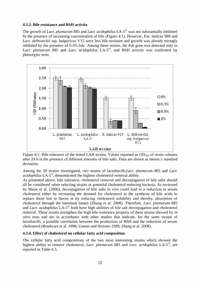
52
4.3.3. Bile resistance and BSH activity
The growth of Lact. plantarum 885 and Lact. acidophilus LA-5®
was not substantially inhibited
by the presence of increasing concentration of bile (Figure 4.1). However, Ent. italicus 989 and
Lact. delbrueckii ssp. bulgaricus V15 were less bile-resistant and growth was already strongly
inhibited by the presence of 0.3% bile. Among these strains, the bsh gene was detected only in
Lact. plantarum 885 and Lact. acidophilus LA-5®, and BSH activity was confirmed by
phenotypic tests.
Figure 4.1: Bile tolerance of the tested LAB strains. Values reported as OD560 of strain cultures
after 24 h in the presence of different amounts of bile salts. Data are shown as means ± standard
deviation.
Among the 58 strains investigated, two strains of lactobacilli,Lact. plantarum 885 and Lact.
acidophilus LA-5®, demonstrated the highest cholesterol removal ability.
As presented above, bile tolerance, cholesterol removal and deconjugation of bile salts should
all be considered when selecting strains as potential cholesterol-reducing bacteria. As reviewed
by Maire et al. (2006), deconjugation of bile salts in vivo could lead to a reduction in serum
cholesterol either by increasing the demand for cholesterol in the synthesis of bile acids to
replace those lost in faeces or by reducing cholesterol solubility and thereby, absorption of
cholesterol through the intestinal lumen (Zhang et al. 2008). Therefore, Lact. plantarum 885
and Lact. acidophilus LA-5® both have high abilities of bile salt deconjugation and cholesterol
removal. These results strengthen the high bile resistance property of these strains showed by in
vitro tests and are in accordance with other studies that indicate, for the same strains of
lactobacilli, a possible correlation between the production of BSH and the reduction of serum
cholesterol (Brashcars et al. 1998; Usman and Hozono 1999; Zhang et al. 2008).
4.3.4. Effect of cholesterol on cellular fatty acid composition
The cellular fatty acid compositions of the two most interesting strains which showed the
highest ability to remove cholesterol, Lact. plantarum 885 and Lact. acidophilus LA-5®, are
reported in Table 4.3.

53
Fatty acids Lact. plantarum 885 Lact. acidophilus LA-5®
- cholesterol + cholesterol - cholesterol + cholesterol
C12 2.49±0.128 * 1.92±0.056
3.84±0.054 ** 4.39±0.043
C14 18.12±0.555 * 15.64±0.220
8.47±0.124 ** 7.19±0.055
C14:1 0.15±0.042 ns 0.24±0.013
0.51±0.042 * 0.31±0.017
C15 0.36±0.040 ns 0.49±0.007
0.77±0.103 ns 0.50±0.025
C16 19.99±0.475 * 18.09±0.147
20.83±0.114 ** 17.86±0.224
C16:1cis9 2.71±0.022 ns 2.78±0.030
2.51±0.082 ns 2.81±0.093
C18 0.57±0.003 ns 0.54±0.009
1.78±0.063 ** 0.66±0.026
C18:1trans 1.21±0.193 ns 1.34±0.028
1.27±0.039 ns 1.50±0.069
C18:1cis9 24.46±0.495 ** 35.44±0.091
36.42±0.017 ns 35.13±0.185
C18:1cis11 2.76±0.099 ns 2.46±0.063
1.61±0.052 ns 1.57±0.155
C18:2 0.58±0.056 * 0.35±0.007
0.91±0.132 ns 0.79±0.020
C19 (dihydrosterculic) 20.91±0.039 ** 16.81±0.057
17.39±0.071 ** 23.11±0.137
Others 5.68±0.076 * 3.91±0.130
3.71±0.137 ns 4.18±0.084
Saturated 62.45±0.206 ** 53.49±0.349
53.07±0.056 ns 53.71±0.408
Unsaturated 31.87±0.281 ** 42.61±0.219
43.22±0.082 ns 42.10±0.499
Table 4.3: Cellular fatty acids composition of LAB strains grown in medium with or without
cholesterol. Values expressed as percentage of total fatty acids.
The two strains showed differences independently of the presence of cholesterol. Lact.
plantarum 885 grown in the absence of cholesterol was characterized by a higher percentage of
saturated fatty acids (SFA) than Lact. acidophilus LA-5®
(+9.38%), mainly due to the high
amount of C14. As for the other SFA, C16 was present in a rather similar amount in the two
strains, whereas C18 was higher in Lact. acidophilus LA-5®. Moreover, the latter strain showed
the highest amount of unsaturated fatty acids (+11.35%), especially C18:1 c9.
The presence of cholesterol in the medium of Lact. plantarum 885 elicited a highly significant
(P < 0.01) decrease in the overall percentage of SFA and a corresponding increase in the
unsaturated ones. The main contribution to this result was due to the variation of C19 cyc and
C18:1 cis9, respectively (Table 4.3).
According to Kimoto et al. (2002), who suggested that cholesterol incorporation into the
cellular membrane alter the fatty acid profiles, changes in hexadecanoic, octadecanoic and total
saturated and unsaturated acids were observed.
Saturated fatty acids confer rigidity to the lipids in the membranes, whereas the double bonds in
the unsaturated fatty acids are responsible for the increase in this physical property. The balance
between saturated and unsaturated fatty acids is important in maintaining the optimum degree
of membrane fluidity and in bacterial membranes, branched-chain and cyclopropane fatty acids
behave like unsaturated ones (Dowhan et al. 2008). Cholesterol plays an important role in
biological membranes since it is believed to modulate their fluidity by interacting with their
complex lipid components, like phospholipids (Dowhan et al. 2008). A membrane composed

54
only of unsaturated lipids is in a fluid state, characterized by a degree of disorder, whereas a
membrane composed of more saturated lipids is in a more solid gel state. Cholesterol increases
the degree of order in the former and renders the latter more fluid (Dowhan et al. 2008).
The changes in lipid composition enable the microorganisms to maintain the membrane
characteristics and functions in response to environmental conditions (Aricha et al. 2004). The
incorporation of cholesterol to the bacterial membrane induces a change in the balance between
saturated and unsaturated fatty acids. This modification was more evident in Lact. plantarum
885, in which the original fatty acid composition was richer in saturated acids than that of Lact.
acidophilus LA-5®. This result leads to the hypothesis that the incorporation of cholesterol
increases the amount of unsaturated fatty acids to maintain membrane fluidity. On the contrary,
the high percentage of unsaturated fatty acids in Lact. acidophilus LA-5® appeared to be able to
maintain the membrane‘s physical characteristics, even in the presence of cholesterol.
4.4 CONCLUSION
Some LAB strains confirmed the ability to remove cholesterol from culture media. The amount
of cholesterol decreased during growth phase and increased during the resting or mortality
phase. The release of cholesterol when the cells reached the resting or mortality phase
suggested that the cholesterol was reversibly incorporated within the bacterial cell membranes.
Regarding the changes in fatty acid composition, as affected by the presence of cholesterol in
the culture media, the two strains with the highest cholesterol removal ability, Lact. plantarum
885 and Lact. acidophilus LA-5® showed a different behaviour that appeared to be related to the
needs of the cells to maintain membrane fluidity, while depending on the original fatty acid
composition.
These preliminary findings will lead to further studies aimed at better characterizing LAB
strains for use in developing fermented dairy products with reduced cholesterol content or able
to induce hypocholesterolemic effects in vivo.
4.5 REFERENCES
Aricha B et al., 2004, Differences in membrane fluidity and fatty acid composition between
phenotypic variants of Streptococcus pneumoniae. J Bacteriol 186: 4638-4644.
Belviso S et al., 2009, In vitro cholesterol-lowering activity of Lactobacillus plantarum and
Lactobacillus paracasei from the Italian Castelmagno PDO cheese. Dairy Sci Technol 89: 169-
176.
Brashcars MM et al., 1998, Bile salt deconjugation and cholesterol removal from media by
Lactobacillus casei. J Dairy Sci 81: 2103-2110.
Dowhan W et al., 2008, Functional roles of lipids in membranes. In: Biochemistry of Lipids,
Lipoproteins and Membranes (5th Edition), pp. 1-37 (edited by D.E. Vance and J.E. Vance,
Elsevier Science).
Fletouris DJ et al., 1998, Rapid determination of cholesterol in milk and milk products by direct
saponification and capillary gas chromatography. J Dairy Sci 81: 2833- 2840.
Gilliland SE et al.,1984, Assimilation of cholesterol of Lactobacillus acidophilus. App. Environ
Microbiol 49: 377-381.
Guzel- Seydim ZB et al., 2011 Review: functional properties of kefir. Crit Rev Food Sci Nutr
51: 261- 268.
Johnsson T et al., 1995, Cellular fatty acid profiles of Lactobacillus and Lactococcus strains in
relation to the oleic acid content of the cultivation medium. Appl and Environ Microbiol 61:
4497-4499.
Kimoto H et al., 2002, Cholesterol temoval from media by Lactococci. J Dairy Sci 85: 3182-
3188.
Lambert JM et al., 2008, Functional analysis of four bile salt hydrolase and penicillin acylase

55
family members in Lactobacillus plantarum WCFS1. Appl and Env Microbiol; 74: 4719-4726.
Li SJ & Cronan JE Jr., 1993, Growth rate regulation of Escherichia coli acetyl coenzyme A
carboxylase, which catalyzes the first step of lipid biosynthesis. J. Bacteriol 175: 332–340.
Liong MT & Shah NP, 2006, Effects of a Lactobacillus casei synbiotic on serum lipoprotein,
intestinal microflora, and organic acids in rats. J Dairy Sci 89: 1390-1399.
Lye HS et al.,2010a, Mechanisms of cholesterol removal by Lactoballi under conditions that
mimic the human gastrointestinal tract. Int Dairy J 20: 169-175.
Lye HS et al., 2010b, Removal of cholesterol by Lactobacilli via incorporation of and
conversion to coprostanol. J Dairy Sci 93, 1383-1392.
Maire B et al., 2006, Bile salt hydrolase activity in probiotics. Appl Environ Microbiol 72:
1729–1738.
Mann GV & Spoerry A, 1974, Studies of a surfactant and cholesteremia in the Maasai. The
American J of Clinical Nutrition 27: 464-469.
Miller L & Berger T, 1985, Bacteria identification by gas chromatography of whole cell fatty
acid. Hewlett Packard Application note 228-41, 1-8.
Morandi S et al., 2012 Enterococcus lactis sp. nov. from Italian raw milk cheeses. Int. J Syst
Evol Microbiol 62: 1992-1996
Nguyen TDT et al., 2007, Characterization of Lactobacillus plantarum PH04, a potential
probiotic bacterium with cholesterol-lowering effects. Int J Food Microbiol 113: 358-361.
Noh DO et al., 1997 Incorporation of cholesterol into the cellular membrane of Lactobacillus
acidophilus ATCC 43121. J Dairy Sci 80: 3107–3113.
Parodi PW, 2009, Has the association between saturated fatty acid, serum cholesterol and
coronary heart desease been over emphasized? Int Dairy J 19: 345-361.
Pereira DIA & Gibson GR, 2002, Cholesterol assimilation by lactic acid bacteria and
Bifidobacteria isolated from human gut. Applied and Env Microbiol 68: 4689-4693.
Rossi EA et al., 2007 Effect of a novel fermented soy product on the serum lipids of
hypercholesterolemic rabbits Arq Bras Cardiol 74: 213-216.
Usman & Hosono A, 1999 Bile tolerance, taurocholate deconjugation and bonding of
cholesterol by Lactobacillus gasseri strain. J Dairy Sci 82: 243-248.
Vinderola CG & Reinheimer JA, 2003, Lactic acid starter and probiotic bacteria, a comparative
―in vitro‖ study of probiotic characteristics and biological barrier resistance. Food Res Int 36:
895-904.
Zago M et al., 2011,. Characterization and probiotic potential of Lactobacillus plantarum
strains isolated from cheeses. Food Microbiol 28: 1033-1040.
Zhang M et al.,2008, Characterization and selection of Lactobacillus strains for their effect on
bile tolerance, taurocholate deconjugation and cholesterol removal. World J Microbiol
Biotechnol 24: 7-14.

56
5. THE USE OF NIR SPECTROSCOPY FOR
MONITORING MILK - WHEY
BIOTRANSFORMATION PROCESSES USING
LACTOBACILLUS PLANTARUM

57
5.1 INTRODUCTION
The dairy industry generates significant liquid waste, whose disposal requires a large amount of
capital investment. The majority of total milk used for manufacturing cheese is discarded as
whey, which retains a lot of nutrients (Jalen, 2002). The most abundant nutrients are lactose,
soluble proteins, organic acids and salts. The disposal of whey as waste can have a high
environmental impact. To overcome this problem, a better alternative could be to process whey
in order to obtain new added value products. Whey contains approximately 4.5% lactose, 0.8%
proteins, 1% salts and 0.1-0.8% lactic acid (Wenge et al., 1999) Availability of a high amount
of lactose in whey and the presence of other essential nutrients for Lactic Acid Bacteria (LAB)
growth make whey a potential raw material for the production of different bio-products using
biotechnological means, such as fermentation processes.
LAB are Gram positive micro-organisms, non-spore forming, catalase negative, non-motile,
microaerophilic, rod or cocci that ferment sugars to mainly produce lactic acid (John et
al.,2007).
Among LAB, selected species of the Lactobacillus genus are widely used as probiotics in dairy
products. Probiotics are defined as active microorganisms that show health benefits for the host
by improving the properties of indigenous microbial community when consumed in adequate
amounts. They should have technological characteristics to allow their production on a large
scale and their incorporation into food products without losing viability and functionality but, at
the same time, without creating unpleasant flavours (Guarner et al., 2005).
One of these species, Lactobacillus plantarum, is industrially important and is involved in many
vegetable fermentations as well as being a frequent inhabitant of the human intestinal tract.
Some Lactobacillus plantarum strains are already on the market as probiotics. High acid
tolerance is an important feature that prevents contamination during fermentation processes. L.
plantarum is a fair lactic acid producer.
Bioprocess optimization is often limited by the inability to measure biomass, nutrients and
products concentration in a time frame that allows process adjustment.
For accurate control of fermentation processes, it is very important to monitor accurately and
rapidly substrate and product concentrations during incubation. However, the analysis of these
compounds requires sample preparations, and in some cases several steps of purification are
needed, which delays obtaining the final results from analyses.
Near-infrared transmittance spectroscopy (NIRS) offers the advantage of rapid sample
processing with results available within 5 min. It is a nondestructive technique that requires no
sample preparation and can measure several broth constituents simultaneously. Furthermore,
authentic standards are not necessary after the prediction equation is developed. With the use of
NIRS as a routine analysis method, opportunities for on-line control processes and fault analysis
are much greater than can presently be achieved with existing measurements, such as HPLC
(Guarner et al., 2005; Vaccari et al., 1994). Nevertheless, the NIRS method also has
disadvantages, such as interference caused by unexpected components in the sample, with
overlapping spectral features, which can introduce errors in the analysis. However, the main
advantage in using NIRS is its ability to monitor in real time the progress of fermentation. This
allows the optimization of the harvesting time during batch culture, the selection of feeding
times with growth-limiting substrate during feed-batch fermentation, and the detection of steady
state for continuous fermentation. In all cases NIRS could save medium and operation time .
NIRS has mainly been used for the evaluation of nutritional components in animal feed.
Recently it has been used for determining natural products in plants, monitoring cell and
biomass concentrations, characterizing polymers, enhancing production of lactic acid in a fully
automated plant-controlled process, and also for analyzing dairy foods (Macedo et al., 2002).
The main advantage in using NIRS is its ability to monitor in real time the progress of
58
fermentation allowing the optimization of the starter culture during processes. Thus, this study
aimed to develop a control system for monitoring fermentation using NIR techniques
recognized as rapid and accurate methods to also correct as quickly as possible anomalies
during the milk fermentation processes. This can give way to new added value products starting
from milk whey recovery (Bouzas et al., 1991).
5.2 MATERIALS AND METHODS
5.2.1 Inoculum preparation
Tests were performed using strains of Lactobacillus plantarum belonging to the internal
collection of CRA-FLC (Lodi, Italy). The strains were reactivated from frozen stock at –80°C,
revitalized in MRS broth (Merk, Germany) and incubated at 30°C±1°C overnight. Culture was
centrifuged for 10 min at 7000 rpm, at 4°C±1°C. The supernatant was removed, and the cellular
pellet suspended in equal volume of Ringer solution (Oxoid, Basingstoke, England). Microbial
count was determined for direct counts with optical microscope (Leitz, Wetzlar, Germany). The
optimal value of microbial count, established in 9.0±0.2 log CFU/ml, was normalized by
diluting or concentrating cellular pellet. The standardized cell suspension was added at a rate of
1% to reconstituted whey, used as substrate of microbial growth.
5.2.2 Substrate preparation
Whey was prepared in accordance with the method proposed by Parnesar et al. (2007) with
some modifications. Whey powder was reconstituted at 7% with deionized water and subjected
to agitation for dissolution. After preliminary tests, the percentage of recovery was established
when the final percentage of lactose equal to 4.5±0.1 was reached. After heat treatment with
flowing steam for 10 minutes to allow the precipitation of proteins, and spinning at 8000 rpm
for 10 minutes at 4°C±1°C, 0.75% of yeast extract (Merck, Germany) and 1% of calcium
carbonate (Carlo Erba, Italy) were added to the supernatant whey (clarified whey). Then, the
substrate was pasteurized in flowing steam for 30 min and cooled to 4°C+1°C.
5.2.3 Fermentation tests
Pasteurized whey was inoculated and incubated in thermostatically-controlled water bath at
30°C±1°C for 48 hours. Aliquots of 2 ml were taken at fixed intervals (0, 6, 8: 11, 15, 18, 24,
28, 32 and 48 hours) and microbial count was determined using MRS agar, after the plates
incubation for 48 hours at 30°C ±1°C in anaerobic conditions.
5.2.4 Determination of lactose and lactic acid content by HPLC
One gram of fermented whey was retrieved with sulphuric acid 0,009 N, centrifuged, filtered
and injected into an ion exchanger column (Aminex HPX 87H, 300 mm x 7.8 mm, 9 mm
diameter average Biorad, U.S.A) in isocratic conditions (65°C±0.1) with HPLC system
(Kontron "instrument", Switzerland). Components were eluted with an aqueous solution of
sulphuric acid 0.009 N at a speed of 0.6 ml/min (Bouzas et al., 1991). Refractometer
REFRACTIVE INDEX 475 (Kontron "instrument", Switzerland) was used as detector. Data
processing was made using Kromasystem 2000 software (Kontron "instrument", Switzerland)
and quantification was made by comparison with an external standard consisting of a solution
of acids and sugars at known concentrations.7
5.2.5 Near infrared Spectroscopy
FT-NIR spectra were acquired in trasflectance mode using a FT-NIR® - NIRFlex N-500
(BUCHI Assago, Milan, Italy) spectrometer. Analysis were conducted using an optic fiber with
an optical path of 0.08 mm, submerged directly in the whey samples at established intervals, at
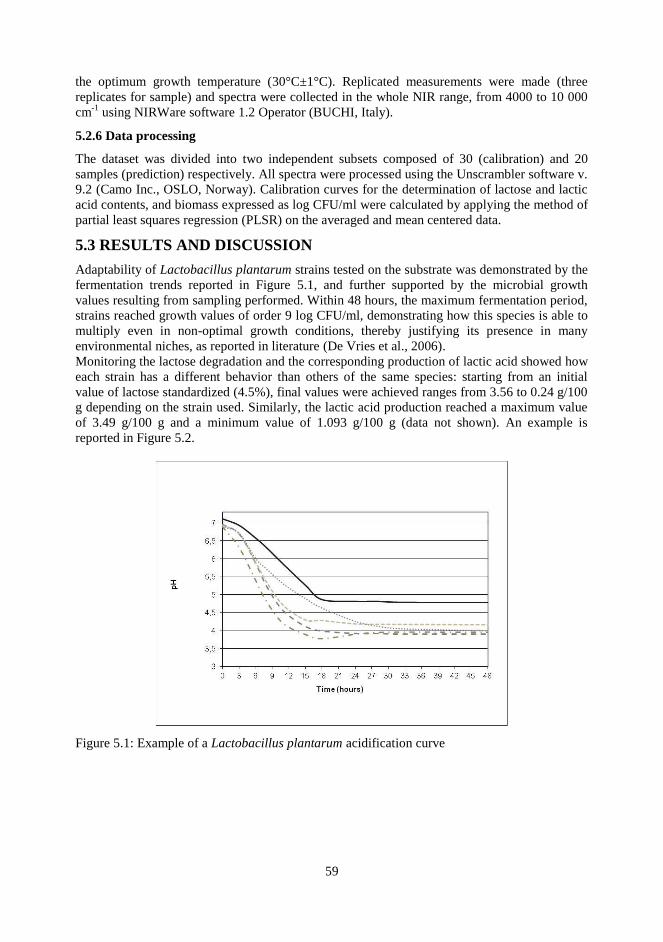
59
the optimum growth temperature (30°C±1°C). Replicated measurements were made (three
replicates for sample) and spectra were collected in the whole NIR range, from 4000 to 10 000
cm-1
using NIRWare software 1.2 Operator (BUCHI, Italy).
5.2.6 Data processing
The dataset was divided into two independent subsets composed of 30 (calibration) and 20
samples (prediction) respectively. All spectra were processed using the Unscrambler software v.
9.2 (Camo Inc., OSLO, Norway). Calibration curves for the determination of lactose and lactic
acid contents, and biomass expressed as log CFU/ml were calculated by applying the method of
partial least squares regression (PLSR) on the averaged and mean centered data.
5.3 RESULTS AND DISCUSSION
Adaptability of Lactobacillus plantarum strains tested on the substrate was demonstrated by the
fermentation trends reported in Figure 5.1, and further supported by the microbial growth
values resulting from sampling performed. Within 48 hours, the maximum fermentation period,
strains reached growth values of order 9 log CFU/ml, demonstrating how this species is able to
multiply even in non-optimal growth conditions, thereby justifying its presence in many
environmental niches, as reported in literature (De Vries et al., 2006).
Monitoring the lactose degradation and the corresponding production of lactic acid showed how
each strain has a different behavior than others of the same species: starting from an initial
value of lactose standardized (4.5%), final values were achieved ranges from 3.56 to 0.24 g/100
g depending on the strain used. Similarly, the lactic acid production reached a maximum value
of 3.49 g/100 g and a minimum value of 1.093 g/100 g (data not shown). An example is
reported in Figure 5.2.
Figure 5.1: Example of a Lactobacillus plantarum acidification curve
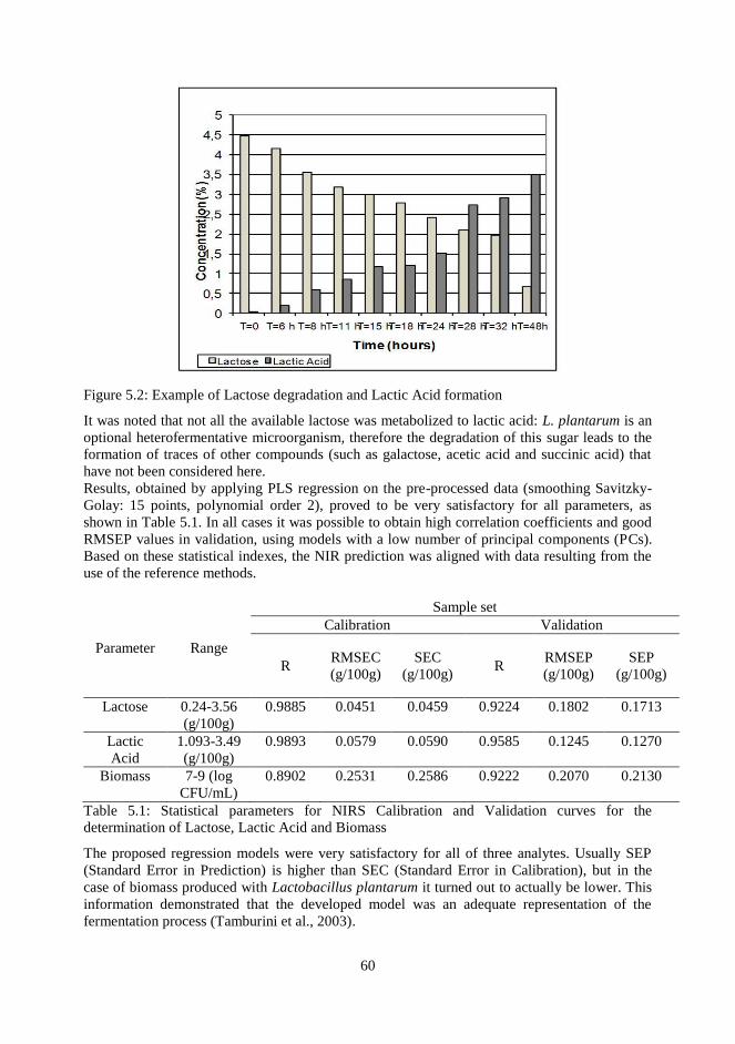
60
Figure 5.2: Example of Lactose degradation and Lactic Acid formation
It was noted that not all the available lactose was metabolized to lactic acid: L. plantarum is an
optional heterofermentative microorganism, therefore the degradation of this sugar leads to the
formation of traces of other compounds (such as galactose, acetic acid and succinic acid) that
have not been considered here.
Results, obtained by applying PLS regression on the pre-processed data (smoothing Savitzky-
Golay: 15 points, polynomial order 2), proved to be very satisfactory for all parameters, as
shown in Table 5.1. In all cases it was possible to obtain high correlation coefficients and good
RMSEP values in validation, using models with a low number of principal components (PCs).
Based on these statistical indexes, the NIR prediction was aligned with data resulting from the
use of the reference methods.
Parameter Range
Sample set
Calibration Validation
R RMSEC
(g/100g)
SEC
(g/100g) R
RMSEP
(g/100g)
SEP
(g/100g)
Lactose 0.24-3.56
(g/100g)
0.9885 0.0451 0.0459 0.9224 0.1802 0.1713
Lactic
Acid
1.093-3.49
(g/100g)
0.9893 0.0579 0.0590 0.9585 0.1245 0.1270
Biomass 7-9 (log
CFU/mL)
0.8902 0.2531 0.2586 0.9222 0.2070 0.2130
Table 5.1: Statistical parameters for NIRS Calibration and Validation curves for the
determination of Lactose, Lactic Acid and Biomass
The proposed regression models were very satisfactory for all of three analytes. Usually SEP
(Standard Error in Prediction) is higher than SEC (Standard Error in Calibration), but in the
case of biomass produced with Lactobacillus plantarum it turned out to actually be lower. This
information demonstrated that the developed model was an adequate representation of the
fermentation process (Tamburini et al., 2003).

61
To identify the specific absorption bands, related to the determination of lactic acid and lactose,
and the evaluation of biomass content, the second derivative of NIR spectra was computed: an
example is reported in Figure 5.3.
Figure 5.3: Example of NIR second derivative spectra obtained from a microbial starter in whey
as a function of time.
The spectral region between 6500 and 7500 cm-1
appeared to be super-imposable to all tested
times. Indeed, in this area it was possible to identify the absorption bands related to the
stretching and bending of C–H bond (6960 and 7084 cm-1
), and the absorption band of the first
overtone of O-H stretching, positioned at 6840 cm-1
. Another common area was identified
around 4400 cm-1
, ascribable to the stretching of O-H and C-H bonds; furthermore, the second
overtone of C–H stretching was identified around 5115 cm-1
(Williams 2001).
In particular, as reported in Figure 5.4, a relationship between bands intensity and concentration
was noticed: the NIR bands that increased with the incubation time were located in the same
spectral region for all the calibrated parameters (5000 – 5200 cm-1
). The evidence found by the
correspondance with the main whey constituents demonstrated how, even in complex matrices,
quantitative answers relating to a single constituent, can be optimized.

62
Figure 5.4: A more in depth portion of the NIR second derivative spectra obtained from a
microbial starter in whey as a function of time.
5.4 CONCLUSIONS
On the basis of the obtained statistical parameters for each studied index determined by
both reference methods of analysis and FT-NIR technology, it was possible to confirm
the applicability of NIRS for the direct determination of lactose, lactic acid, and
microbial count in milk whey. The use of an optic probe proved to be a viable
alternative to existing methods of fermentation monitoring, particularly with respect to
biomass determination. A complex analytical matrix, such as a culture medium with
suspended cells, at varying concentration wasn‘t an impediment for a rapid, reliable,
and accurate determination of the studied components.
5.5 REFERENCES
de Vries M et al., 2006, Lactobacillus plantarum—survival, functional and potential probiotic
properties in the human intestinal tract International Dairy Journal, 16: 1018-1028.
Guarner F et al., 2005, Should yoghurt cultures be considered probiotic? British Journal of
Nutrition, 93: 783-786.
J. Bouzas J et al., 1991, Sugars and organic acids in raw and pasteurized milk Manchego
cheeses with different degrees of late blowing defect. Journal of Food Science, 56: 276-278.
Jelen P, 2002, ―Whey processing‖ in Encyclopedia of dairy science, Ed by H. Roginski, J. N.
Fuquay and P. F. Fox, Academic press, San Diego, CA, p 2739-2745.
John RP et al.,2007, Fermentative production of lactic acid from biomass: an overview on
process developments and future perspectives. Applied Microbiolgy and Biotechnology, 74:
524-534.
P.C. Williams and K. Norris, Near infrared technology in the agricultural and food industries,
Ed by P. Williams and K. Norris American Association of Cereal Chemists, St. Paul, MN, USA
(2001)
Panesar PS et al., 2007, Bioutilization of whey for lactic acid production Food Chemistry, 105:
1-14.
Tamburini E et al., 2003, Near-Infrared Spectroscopy: a tool for Monitoring submerged

63
Fermentatioin Process using an Immersion Optical-Fiber Probe. Applied spectroscopy, 57: 132-
138.
Wenge F et al., 1999, Lactic acid production from lactose by Lactobacillus plantarum: kinetic
model and effects of pH, substrate, and oxygen Biochemical Engineering Journal, 3: 163-170.
6. NIR AND MIR SPECTROSCOPY IN THE STUDY OF
CHOLESTEROL REMOVAL ABILITY OF
LACTOBACILLUS PLANTARUM STRAINS

65
6.1 INTRODUCTION
Cholesterol is a polycyclic alcohol having a secondary -OH group at position 3‘. It plays an
important role as a component of cell membranes in vertebrates and, in many of the normal
metabolic pathways as a precursor of steroid hormones, bile acids and vitamin D.
The consumption of food rich in lipids, especially saturated fatty acids and cholesterol, has a
predominant role in the rise in the heart disease. Thus, the consumer demand on food with
lower amount of these components and with additional healthy properties is increasing (Belviso
et al., 2009).
Dairy products play an important role in a healthy diet because of their high nutritional value,
but since in some cases they also represent a source of lipids, many attempts were carried out to
reduce their fat concentration. As the consumption of fermented milk containing bacterial
strains provides beneficial effects, such as the reduction of serum cholesterol levels on human
health (Gilliland et al., 1985), several researches on bacterial strains with potential
hypocholesterolaemic properties were then carried out.
In particular, the consumption of dairy products containing probiotic bacteria has been proposed
as a means to lower cholesterol in serum (Ling & Shah, 2005).
Probiotics are defined as a ―live microbial supplement that beneficially affects the host by
improving its intestinal microbial balance‖. Many authors have shown how these bacteria are
able to decrease blood cholesterol content, proposing several in vitro studies about the
mechanism of cholesterol-lowering action of probiotic bacteria even if the exact mechanism(s)
remain unclear (Liong & Shah, 2005; Gilliland et al., 1985; Tahri et al., 1996). Recent papers
tried to show how strains isolated from dairy matrixes have the ability to remove cholesterol
during their growth. In particular, Belviso et al. (2009) tried to do this with Lactobacillus
plantarum hypothesizing the co-precipitation of choelesterol with bile salts.
Within the genus Lactobacillus, L. plantarum is a member of the facultative heterofermentative
group of lactobacilli and it is present in a variety of environmental niches, including dairy,
meat, fish, and many vegetable or plant fermentations. Moreover, strains of L. plantarum have
proven ability to survive gastric transit and colonize the intestinal tract of humans and other
mammals (Zago et al., 2011).
Methods have been proposed to investigate whether cholesterol was really assimilated by
strains or whether it remains free in culture medium. Many authors used the method of Rudel &
Morris (1973): cholesterol can be estimated by visible spectroscopy using o-phthalaldehyde
reagent after extraction of unsaponified matter (cholesterol) with an organic solvent such as
hexane. The application of this method requires a long time and so spectroscopic methods have
been proposed using MIR and NIR techniques7-9
.
Infrared spectroscopic methods are simple, cost-effective, rapid, nondestructive and have the
potential for routine analysis if proper calibration and validation procedures with data
acquisition protocols could be established. Budinova et al. (1997) have used the FTIR spectral
region between 950 and 1100 cm for the detection of cholesterol in blood and serum and Shaw
& Mantsch (2000) used MIR attenuated total reflectance (ATR) technique (in the spectral
region between 2800 and 3000 cm) to develop a statistical model using eight partial least square
(PLS) factors. This study is one of the first attempts to use FTIR-ATR spectroscopy to estimate
cholesterol in milk products.
The application of chemometrics to analyse the spectra is an important step in spectroscopy.
The most commonly used multivariate calibration methods is principal component analysis
(PCA). Discriminant analysis is commonly used in classification to group objects or data of
similar characteristics into clusters based on statistical measure. The success of these methods
depends upon the choice of proper spectral data and the number of variables employed in the
calibration model.
66
The aim of this study was to verify, by the use of NIR and MIR spectroscopic techniques,
whether strains with different ability to remove cholesterol can show changes in the cell wall
and if it was possible to determine the amount of the cholesterol removed.
6.2 MATERIALS AND METHODS
6.2.1. Bacterial strains and culture conditions
Four L. plantarum strain (Lp752, Lp885, Lp995, Lp997) belonging to the collection of CRA-
FLC (Lodi, Italy) and isolated from Italian dairy products, and the type strain ATCC14917T,
stored as frozen stock at –80°C, were cultivated overnight in MRS broth (Merck, Germany) at
30°C±1°C. After growth, 2% (v/v) of fresh culture was transferred in MRS and MRS broth
supplemented with a stock solution of cholesterol (10% w/v) to obtain a final concentration of
0.1 g/l.
Cholesterol stock solution (1 g/l) was prepared by dissolving 100 mg of cholesterol (Sigma-
Aldrich, Milan) in an aqueous solution of 26,4% ethanol and 12% Tween 80 (polyoxyethylene
sorbitan monooleate) (Sigma-Aldrich, Milan). After growth for 24 h at 30°C±1°C, the
following samples were analyzed: A) cells harvested (7000 rpm for 10 min at 4°C) by
centrifugation and washed twice with ultrapure water B) culture supernatant. The pH measured
on samples B was 3.9 ± 0.2.
6.2.2 MIR spectroscopy
One gram of sample A was distributed on ZnSe tablets ( 13mm, h 2mm, PIKE Technologies,
USA) to collect, at room temperature, ATR-FTIR spectra (128 scans, resolution 4 cm-1
) using a
FT-IR 420 spectrometer (JASCO Europe, Cremella, Como, Italy), in the range between 900 and
1750 cm-1
.
6.2.3 NIR spectroscopy
FT-NIR spectra were collected on samples B in transmission mode (pathlength 0.2 mm, 32
scans, resolution 8 cm-1, 6 replicates) with FT-NIR® NIRFlex N-500 spectrometer (BUCHI,
Italy) in the whole NIR range (4000-10000 cm-1) with 1.2 NIRWare Operator (BUCHI, Italy)
at controlled temperature (40°C 1°C). Data were processed with Unscrambler v. 9.2 (Camo
Inc., OSLO, Norway).
6.2.4 Data processing
MIR average spectra of sample A (5 replicates), after subtraction of an average spectrum of
water or supernatant, depending on the matrix, were pretreated with smoothing (Savitzky
Golay, 7 points, polynomial order 0). NIR data were processed by using Unscrambler software
v. 9.2 (Camo Inc., OSLO, Norway) in order to evaluate qualitative differences between culture
supernatants (in absence and in presence of cholesterol). PCA analyses were performed by
pretreating spectral data with SNV (Standard Normal Variate), Savitzky – Golay second
derivative (8 points, polynomial order 2) and mean centering.
6.3 RESULTS AND DISCUSSION
Lactic acid bacteria (LAB) are Gram+ microorganisms and their cell wall is primarily made up
of peptidoglycan (ca. 40-80% of the dry weight of the wall), which is a polymer of N-
acetylglucosamine (NAG) and Nacetylmuramic acid (NAM). Other important constituents are:
teichoic acid, a polymer of glycopyranosyl glycerol phosphate, teichuronic acid, which is
similar to teichoic acid, but replaces the phosphate functional groups with carboxyls, and
surface proteins6. Therefore, the principal functional groups found in MIR spectra were:
carboxyl, hydroxyl, phosphate and amide. In particular, phosphate group in bacteria can exist

67
both in inorganic forms such as orthophosphate and its oligomers and in organic forms such as
phosphate mono- and di-esters. The principal absorption bands, identified in the second
derivative of MIR spectra, are shown in Table 6.1 (Jang et al., 2004; Leone et al., 2007).
Absorption
bands (cm-1
) Vibrational modes
950-1150 Asymmetric and symmetric stretching of PO2-
1153 C-O stretching in C-OH group
900-1200 C-O-C and C-O of various polysaccharides
1215 Interaction of O-H bending and C-O stretching in C-O-H group, C-OH
stretching vibration of carboxylic acid groups11
, stretching of P=O bond in
phosphate
1220-1330 Amide III band components of proteins1: 1220-1250cm
-1 sheets, 1250-
1270 cm-1
random coils, 1270-1295 cm-1
turns,1295-1330 cm-1
helix
1240 PO2- stretching of phosphodiesters,
C–O stretching in carboxylic acid
1372 Interaction of O-H bending and C-O stretching in C-O-H group, CH3
symmetric bending
1396 Symmetric stretching of COO-
1407 =C–H in-plane bending
1418 C-O-H in-plane bending, carboxylic acids
1454 CH2 scissoring
1566 C=C stretching in alkenes
1548 Amide II band
1637 Amide I of -pleated sheet structure
1743 C=O stretching of esters
Table 6.1: Absorption bands of functional groups recognizable in the spectra of Lactobacillus
plantarum before and after the addition of cholesterol.
Preliminary measurements were performed on the five considered strains of L. plantarum
(Lp752, Lp885 Lp995, Lp997 and ATCC14917T), cultivated in absence and in presence of
cholesterol. MIR spectra, acquired on the pellets obtained by centrifugation and subtracted by
the contribution of the matrix, showed an interesting change in the signals in the range from
1180 to 1300 cm-1
whose entity depended on the strain characteristics. In particular, a shift of
the signal centred at 1240 cm-1
towards 1217 cm-1
was observed.
The formation of H bond between phosphate group and the hydroxyl group of cholesterol
determined this shift. It is well known, in fact, that the formation of hydrogen bonds produces a
weakening of all the bonds involved and a shift of absorption bands towards lower energy.
As shown in Figure 6.1, and more in detail in Figure 6.2, this effect was very low for Lp995,
Lp997 and ATCC14917 and more evident for Lp752 and Lp885 strains. This result was in
agreement with our results, which demonstrates an increased capacity of these last strains to
remove cholesterol (Paladino, 2010) It is also interesting to note that the initial shape of the
bands differed for different strains: it can be related to a different cell wall composition that
could explain their own capacity to remove cholesterol from the culture broth. Many more
evident variations were visible in the range from 900 to 1150 cm-1
: further measurements,
performed on washed cells of Lp885 seemed to demonstrate that these changes, disappearing
after washing, were due to an interference of the matrix (data not shown). It was, instead,
confirmed the variations observed in the range from 1180 to 1300 cm-1
.

68
Figure 6.1. MIR spectra of Lp995, Lp997,
ATCC14917, Lp752, Lp885 strains cultivated in
absence (a) and in presence (b) of cholesterol (the
spectra were shifted on the absorbance axis to facilitate
the lecture).
Figure 6.2. Detail of MIR spectra in the range from 1180 to
1300 cm-1
of Lp995, Lp997, ATCC14917, Lp752, Lp885
strains cultivated in absence (a) and in presence (b) of
cholesterol (the spectra were shifted on the absorbance axis
to facilitate the lecture).

69
The supernatants obtained after centrifugation of three strains (Lp885, Lp995, ATCC14917)
cultivated in absence and in presence of cholesterol were analyzed by NIR spectroscopy.
Preliminary data, processed by PCA, showed the possibility to separate the supernatants
obtained both in absence (supernatant a) (Figure 6.3) and in presence (supernatant b) of
cholesterol (Figure 6.4), highlighting differences in metabolism and in capacity to remove
cholesterol among strains.
The difference between strains in Figures 3 and 4 was observed along the PC1 (97% of total
variance explained) for ATCC14917 and for the samples sets Lp885 and Lp995; separation for
Lp885 and Lp995 was essentially obtained along the PC2 (3% of total variance explained).
The score plot obtained by the PCA analysis performed on 20 samples of Lp885
supernatant in absence (a) and in presence (b) of cholesterol is shown in Figure 6.5.
The difference between the two sets of samples was obtained along the PC1 that was
able to explain 97% of the total variance. A more detailed study is in progress to verify
the feasibility of NIR spectroscopy for a rapid determination of not removed
cholesterol in supernatants.
Figure 6.3. Scores plot obtained from the PCA
analysis performed on 30 samples (10 of
ATCC14917, 10 of Lp885 and 10 of Lp995) of
supernatant a.
Figure 6.4. Scores plot obtained from the
PCA analysis performed on 30 samples (10
of ATCC14917, 10 of Lp885 and 10 of
Lp995) of supernatant b.

70
6.4 CONCLUSIONS
Preliminary results obtained supported the applicability of NIR and MIR spectroscopy to study
the removal of cholesterol by some strains of LAB and suggested that the mechanism that
involves cholesterol should be related to an interaction of this molecule with cell walls by
hydrogen bonds. Further studies will be addressed to confirm these observations and to develop
a fast method, based on NIR spectroscopy, for the determination of the cholesterol.
6.5 REFERENCES
Belviso S et al., 2009, In vitro cholesterol-lowering activity of Lactobacillus plantarum and
Lactobacillus paracasei from the Italian Castelmagno PDO cheese. Dairy Sci Technol 89: 169-
176.
Budinova G. et al., 1997, Application of Molecular Spectroscopy in the Mid-Infrared Region to
the Determination of Glucose and Cholesterol in Whole Blood and in Blood Serum Applied
Spectroscopy, 51: 631 –635.
Burgula Y et al., 2004, Review of mid-infrared fourier transform-infrared spectroscopy
applications for bacterial detection. Journal of Rapid Methods & Automation in Microbiology
15: 146–175.
Cai S & SinghBR, 1999, Identification of beta-turn and random coil amide III infrared bands
for secondary structure estimation of proteins. Biophysical Chemistry, 80: 7 – 20.
Gilliland SE et al.,1985, Assimilation of cholesterol of Lactobacillus acidophilus. App. Environ
Microbiol 49: 377-381.
Jiang W et al.,2004, Elucidation of functional groups on Gram-positive and Gram-negative
bacterial surfaces using infrared spectroscopy Langmuir, 20: 11433-11442.
Leone L et al., 2007, Modeling the acid-base properties of bacterial surfaces: a combined
spectroscopic and potentiometric study of the gram-positive bacterium Bacillus subtilis.
Environ. Sci. Technol. 41:6465-6471.
Liong MT & Shah NP, 2006, Effects of a Lactobacillus casei synbiotic on serum lipoprotein,
intestinal microflora, and organic acids in rats. J Dairy Sci 89: 1390-1399.
M. M. Paradkar & J. Irudayraj, 2002, Determination of cholesterol in dairy products using
infrared techniques: 1.FTIR Spectroscopy. International Journal of Dairy Technology, 55: 127–
Figure 6.5. Scores plot obtained from the PCA analysis
performed on 20 samples of Lp885 supernatant (10 of
supernatant a and 10 of supernatant b).

71
132.
M. M. Paradkar & J. Irudayraj, 2002, Determination of cholesterol in dairy products using
infrared techniques: 1.FT-NIR Method. International Journal of Dairy Technology, 55: 133–
138.
M. Paladino (2010) ―Studio attività decolesterolizzante di batteri lattici di origine alimentare‖.
Thesis: Università degli Studi di Pavia (Italy).
Rudel LL & M. D. Morris, 1973, Determination of cholesterol using o-phthalaldehyde Journal
of Lipid Research, 14: 364–366.
Stuart B, Infrared Spectroscopy: fundamentals and applications, John Weley & Sons, p. 77
(2004)
Tahri K et al., 1996, Bifidobacteria strain behavior toward cholesterol: Coprecipitation with
bile salts and assimilation. Current Microbiology 33:187–193
Zago M et al., 2011,. Characterization and probiotic potential of Lactobacillus plantarum
strains isolated from cheeses. Food Microbiol 28: 1033-1040.

7. NEAR INFRARED SPECTROSCOPY AND
AQUAPHOTOMICS AS TOOLS FOR BACTERIAL
CLASSIFICATION

73
7.1 INTRODUCTION
Lactic acid bacteria (LAB) are Generally Recognized as Safe (GRAS) microorganisms typically
associated with human gastrointestinal tract (GIT) and foods. Certain LAB species from dairy
foods are known for their probiotic effects during GIT passage (Georgieva et al., 2008).
Probiotics are live bacteria containing foods, and, upon digestion, they exert beneficial effects
to host. The beneficial actions of probiotic bacteria provide aid in lactose digestion, resistance
to enteric pathogens, anti-colon cancer effect, small bowel bacterial overgrowth, allergy, and
immune system modulation. Microorganisms used in probiotic products generally contain
lactobacilli and bifidobacteria.
Within the genus Lactobacillus, Lactobacillus plantarum is studied for its probiotic
characteristics. It is a heterogeneous and versatile species that is encountered in a variety of
environmental niches, including dairy, meat, fish, and many vegetable or plant fermentations
(Zago et al., 2011). It is one of the main non-starter LAB species contributing to the final
sensorial properties of different kind of cheeses. Strains of L. plantarum have proven ability to
survive gastric transit and colonize the intestinal tract of humans and other mammals
(Georgieva et al., 2009). Various special therapeutic or prophylactic properties have been
associated with L. plantarum, such as reduced incidence of diarrhea in daycare centers, reduced
pain and constipation associated with irritable bowel syndrome, reduced bloating, flatulence,
ability to displace enteropathogens from Caco- 2 cells, and capacity to exert positive effect on
immunity in HIV+ children (Parvez et al., 2006).
The identification of microorganisms and the study of their probiotic features are based on
genotypic and phenotypic tests which give results able to discriminate the strains. These
methods are often time consuming and laborious, involve numerous reagents, and may have
difficulty in the distinction of closely related organisms. Molecular DNA analysis methods
provide a more reliable differentiation of microorganisms at the species level; however, these
methods are also reagent- and time-intensive so their applications in industrial settings are not
yet widespread (Bhatta et al., 2006). An optics-based method of Fourier transform infrared
spectroscopy has been proposed as an alternative in clinical and food industry applications, with
reasonable success; in particular there has been a growing interest in the use of near-infrared
(NIR) technology as an analytical tool for agricultural and food investigations.
This technique has been shown to be a useful tool for the characterisation of bacteria, fungi ,
metabolic footprinting and fingerprinting, as well as a number of food-based analyses.
Many authors reported how NIR is able to detect and identify bacterial contamination in liquids
studying the overtones and combination bands of fundamental organic molecules.
Within the food industry, at present, no technology exists for a rapid and accurate detection
system for microbiologically spoiled or contaminated food. Ellis et al. (2004) developed a
method for spoilage detection in beef evaluating the observation of molecules that are excited
by an infrared beam, where an infrared absorbance spectrum represents a ―fingerprint‖
characteristic of any (bio)chemical substance.
Lin et al. (2004) evaluated the feasibility of this technique to quantify the microbial loads in
chicken meat and to develop a rapid methodology for monitoring the onset of spoilage.
In spectroscopy, water has been considered an obstacle to analytical process of other molecules
in biological systems. However, latest reports have begun to acknowledge the importance of
studying the behavior of water when exploring biological and aqueous systems. Yet, the
development of methodologies for understanding the biological significance of water in living
systems is exceedingly difficult. Recently, a new concept, which describes the dynamic
spectroscopy of biological and aqueous systems based on the behavior of water called
‗‗Aquaphotomics‖, has been proposed. Aquaphotomics describes the spectroscopic ways and
means of tracing changes of water molecular vibrations in relation to other molecular vibrations

74
present in a biological specimen, which helps to understand the functions of the whole system
(Jinendra et al., 2010).
Therefore the objectives of this study were to evaluate the feasibility of FT-NIR applications in
microbiology and to develop methodology for the rapid identification of bacterial strains by
combining FT-NIR spectroscopy, Aquaphotomics with multivariate statistical methods.
7.2 MATERIALS AND METHODS
7.2.1 Strain selection
A total of 42 strains were used in this study. Thirty-five strains of Lactobacillus plantarum,
belonging to the collection of the CRA-FLC (Fodder and Dairy Productions Research Centre,
Lodi), were isolated from different matrixes (from dairy products, from olive brines and from
grape marcs); 6 strains of Lactobacillus delbrueckii subsp. bulgaricus and 1 strain of
Lactobacillus gasseri isolated from probiotic Japanese yoghurt.
Strains were kept as frozen stocks at -80 °C in MRS broth (Merck, Darmstadt, Germany) added
with 20% (v/v) glycerol. Frozen stock cultures were thawed and subcultured twice (2%
inoculum) in MRS at 30 °C overnight for L. plantarum and at 42°C for L. delbrueckii subsp.
bulgaricus and L. gasseri before use.
7.2.2 Molecular typing
Bacterial DNA was extracted using a Chelex®
100 method described by Walsh et al. 1991.
Two-hundred μl of culture were washed twice with 500 μl di TE0.1 pH 8.0 (TE0.1: Tris-HCl 10
mM, EDTA 0.1 mM) and the pellet was harvested and resuspended in 245 μl of the same buffer
and 5 μl of lysozyme (stock solution 50 mg/ml). After that, the strains were incubated at 56°C
for 45 min.
To each eppendorf were added 218.7 μl of TE0.1 pH 8.0, 5 μl of DTT 1 M, 10 μl of EDTA 0.5
M pH 8.0, 12.5 μl of 20% SDS and finally 3.8 μl of proteinase K (stock solution 20 mg/ml).
The strains were incubated for 1 hour at 37°C and than 500 μl of a 5% Chelex® 100 (Sigma –
Aldrich) solution were added. This ionic resin allows the DNA purification by precipitation of
all impurities generated by bacterial lysis. The extracted DNA was diluted 1:10 and used as a
template for PCR reactions.
RAPD-PCR was performed using M13 primer (5‘ – GAGGGTGGCGGTTCT – 3‘) as
described by Rossetti and Giraffa (2005). Amplification reactions were carried out in 25 μl
volume containing 2 mM of M13 primer, 2.5 units/100 μl of AmpliTaq DNA polymerase
(Applied Biosystems, Monza, Italy), 3 mM MgCl2, 2.5 ng/μl of total DNA, and 0.2 mM of each
dNTP. PCR amplifications were performed in a Perkin–Elmer thermal cycler (model 9700
Applied Biosystem). The cycling program consisted of an initial denaturation step at 94 °C for
120 s and then 40 cycles of: 94 °C for 60 s, 45 °C for 20 s, and 72 °C for 120 s. The final
elongation was performed at 72 °C for 10 min.
PCR products were analyzed by electrophoresis through 1.5% agarose gels at 26 V for 12 h in 1
× Tris-acetate EDTA (TAE) buffer (1 × TAE: 40 mmol/l Tris acetate, 1 mmol/l EDTA, pH 8.0)
stained with ethidium bromide, and visualized under UV light.
The fingerprinting profiles were processed using Bionumerics software (5.1 Applied Maths,
Belgium). Similarity among different profiles was evaluated by applying the Pearson‘s
correlation coefficient.
7.2.3 Spectroscopical typing
Cell cultures, obtained as previously described, were centrifuged (7000 rpm x 15 min) and
resuspended in the same volume of water to eliminate the interferences due to the presence and
to the complexity of the composition of the culture broth in which bacteria were grown. The

75
cultures were subsequently analyzed directly or diluted prior to analysis.
The spectra of the bacterial cultures were collected at room temperature in triplicate with a NIR
system Fantec (Fantec, Japan) in transmittance mode using test tubes with an optical path of 0.5
cm. Each spectrum collected is the result of the average of 32 scans in the spectral range
between 600 and 1100 nm.
Before analyzing the data using Pirouette software ver. 3.11 (Infometrix, USA), the spectra
were averaged.
Qualitative analysis was performed by applying SIMCA algorithm (soft independent modeling
of class analogy) to determine and highlight the differences between the spectra of each sample
analyzed. The spectra collected were divided into two data sets of equal size one of which was
used for the validation of the model. The SIMCA method is based on the analysis of principal
component analysis (PCA) applied to classify objects into groups. It consists of a set of models
PCA one for each group present in the data set. The unknown samples are assigned to classes
based on the minimum distance (Mahalanobis) PCA models previously calculated.
7.3 RESULTS AND DISCUSSION
7.3.1 Molecular typing
The genotyping of strains allowed to highlight a good level of intraspecific heterogeneity
among tested strains, as shown in Figure 7.1, often linked to the matrix of isolation [6].
Figure 7.1: Dendogram generated by tested strains
This biodiversity results in a numerical difference between species, highlighting the different
capacity to adapt to the conditions imposed during the transformation process.
Currently, it is increasingly important to distinguish and then identify quickly and accurately,
even microbial strains belonging to the same species, but with different technological
properties.
Analyzing the profiles for the 41 studied strains, 13 cluster corresponding to a similar number
of different biotypes, were obtained (Figure 7.1).
The individual clusters contain from 2 to 11 strains although it is possible to notice some cluster
consisting of a single strain.

76
Most biotypes includes strains having different origins while a limited number of groups
contain strains having the same origin: for instance in the cluster 8 all 11 strains that make it up
have been isolated from brine for olives.
7.3.2 Spectroscopical typing
Preliminary tests have concerned the development of a method of monitoring the growth of
strains culture broth during a complete cycle of growth. In Figure 7.2 is shown the result of the
SIMCA analysis in which it is evident that with this method it was possible to reproduce the
typical microbial growth trend.
Figure 7.2: Monitoring of microbial growth in culture broth.
The Interclass Distances values (Table 7.1) have corroborated the robustness of the model
because all values are greater than 3. According to Morita et al. (2011), the interclass distance
of less than 0.8 indicates a small difference between two groups while the distance of more than
3 supports a reliable separation. Therefore, this separation is considered robust.
Tempo 0 2 4 6
0 0 3.40324 7.224778 37.94837
2 3.40324 0 4.56802 18.63801
4 7.224778 4.56802 0 10.33202
6 37.94837 18.63801 10.33202 0
Table 7.1: Interclass distance values related to Figure 7.2
In order to investigate the differences among each strain samples and reflect them in the
regression analysis SIMCA was performed for NIR spectral data as qualitative analysis. The
bacterial cultures were diluted 1:10 in sterile water to avoid interference due to the culture
broth, to highlight how the characteristics of aqueous systems in which the microorganisms are
dispersed can be used as fingerprinting of the culture.

77
Figure 7.3: Strain classification by SIMCA
Figure 7.3 where it is shown the result of the application of SIMCA model on the validation set,
demonstrates how this non-destructive technique is able to separate strains considered part of
the same biotype. Comparing this result with that obtained by genotyping the couples of strains
Lp751 and Lp752, Lp793 and Lp794 Lp804 and Lp994 that genotypic level are considered to
be part of the same cluster, are separate and well differentiated. The separation is corroborated
also by the values of interclass Distances greater than 3: 16.26 for the pair Lp751/Lp752, 13:23
for the pair Lp793/Lp794 and 13.15 for the pair Lp804/Lp994 respectively.
Moreover, the predictive power of the model has led to correctly classify the 95.1% of the
samples (39/41), indicating that the differences between the bacterial strains can be detected
also by the spectral differences related to them.
To identify what are the wavelengths responsible for this classification, an Aquagram has been
done (Figure 7.4), where are reported the absorptions characteristic of each strain at different
wavelengths.
Figure 7.4: Aquagram related to studied strains
The graph shows that the spectrum can be used to describe the differences between the strains.
The major differences in intensity of absorption characteristics of the strains studied, it can be
noted at the wavelengths 778 and 792 nm.
78
7.4 CONCLUSIONS
The analysis of the spectral data showed that the NIR spectroscopy is able to monitor the
bacterial growth in real time without having to resort to the validation of the method with
reference analysis.
For the first time, phenotypic and genotypic difference among strains were successfully
extracted and visualized by quantitative analysis based only on NIR spectral data of diluted
supernatant samples.
In particular the application of the algorithm SIMCA allowed to highlight differences among
the strains which were not detected by the methods of genotypic characterization, based on the
amplification of DNA.
These results prove that NIR spectroscopy is an effective and high accurate screening method,
suitable as high throughput technology for decision making at an industrial level for the
selection of strains of technological interest.
7.5 REFERENCES
Georgieva RN et al., 2008, Thecnological properties of candidate probiotic Lactobacillus
plantarum strains. Journal of Basic Microbiology 48:234-244.
Bhatta H. et al., 2006, Use of fluorescence spectroscopy to differentiate yeast and bacterial
cells. Applied Microbiology and Biotechnology, 71: 121-126.
Lin M et al., 2004, Rapid and quantitative detection of the microbial spoilage in chicken meat
by diffuse reflectance spectroscopy (600–1100 nm). Letters in Applied Microbiology, 39:148-
155.
Jinedra B et al.,2010, Near infrared spectroscopy and aquaphotomics: Novel approach for rapid
in vivo diagnosis of virus infected soybean. Biochemical and Biophysical Research
Communications, 397: 685-690.
Morita H et al., 2011, Near infrared spectroscopy as high throughput technology for screening
of xilose-fermenting recombinant Saccharomyces cerevisiae strains. Analytical Chemistry 83:
4023-4029
Amillis M et al., 1997, Isolation of genomic DNA from milk samples by using Chelex resin.
Journal of Dairy Reserch, 64: 231.
Rossetti L, Giraffa G 2005 Rapid identification of dairy lactic acid bacteria by M13-generated,
RAPD-PCR fingerprint databases. J. Microbiol. Meth. 63: 135-144.
Zago M et al., 2011, Characterization and probiotic potential of Lactobacillus plantarum strains
isolated from cheeses. Food Microbiol 28: 1033-1040.
Parvez, S et al., 2006, Probiotics and their fermented food products are beneficial for health. J.
Appl. Microbiol. 100: 1171-1185.