Embed Size (px)
Citation preview
BIOSYNTHESIS OF THE PURINES
II. METABOLISM OF 4-AMINO-5-IMIDAZOLECARBOXAMIDE IN PIGEON LIVER*
BY MARTIN P. SCHULMANt AND JOHN M. BUCHANAN
(From the Department of Physiological Chemistry, School of Medicine, University of Pennsylvania, Philadelphia, Pennsylvania)
(Received for publication, July 5, 1951)
4-Amino-5-imidazolecarboxamide (l), an amine found to accumulate in bacterial systems poisoned either by sulfonamides (2, 3) or aminopterin (4), has been suggested as an intermediate of purine synthetic reactions (5, 6). Although the metabolic inertness of the carboxamide’ in certain bacterial systems (7, 8) led to the belief by some (8) that this compound was an aberrant product of metabolism of sulfonamide-poisoned cells, cer- tain mutant strains of Escherichia coli2 and Ophiostoma multiannulatum (9) as well as the normal strain of Lactobacillus arabinosus (7) may utilize the carboxamide for growth in a manner similar to purines. However, they usually require the carboxamide in greater concentration than the purines for equivalent growth. Williams (10) in this laboratory has more recently shown that labeled carboxamide is incorporated into purine com- pounds by the intact yeast cell and by yeast extracts.
The carboxamide may be utilized in viva at a significant rate in the formation of uric acid by the pigeon (11) and in the synthesis of tissue purines as well as allantoin by the rat (12). In a preliminary communi- cation (11, 13), it has been shown that the carboxamide may be converted into hypoxanthine by pigeon liver homogenates. Since Greenberg (14, 15) has shown that inosinic acid may be an intermediate in the de novo synthesis of hypoxanthine from formate, we have further studied the con- version of the carboxamide to hypoxanthine in pigeon liver homogenates and extracts in order to determine whether ribotide formation is involved in carboxamide metabolism. Evidence is presented indicating that the units of ribose and phosphate are added to the carboxamide prior to ring closure with formic acid, implicating the involvement of 4-amino-5-imid-
* Aided by grants from the National Cancer Institute, National Institutes of Health, United States Public Health Service, and the Damon Runyon Memorial Fund for Cancer Research, Inc. Cl4 allocated by the Atomic Energy Commission.
t Fellow in the Medical Sciences of the National Research Council, 1949-50. Present address, Department of Biochemistry, State University of New York, Med- ical Center at Syracuse University, Syracuse, New York.
14.Amino-5-imidazolecarboxamide will be referred to as carboxamide. 2 Davis, B. E., unpublished work.
513
by guest on May 27, 2018
http://ww
w.jbc.org/
Dow
nloaded from
514 BIOSYNTHESIS OF PURINES. II
azolecarboxamide ribotide or a formyl derivative of this compound in the synthesis of hypoxanthine by this system.
EXPERIMENTAL
The experiments reported below involve for the most part incubating radioactive glycine or 4-amino-5Gmidazolecarboxamide with pigeon liver homogenates or extracts in the presence of inosinic acid and hypoxanthine or inosine, isolating these latter compounds after a short incubation period, and measuring their specific activities.
The preparation of the homogenate and extract has been described previ- ously (16). Incubations, frequently on a large scale, were carried out in a 38” water bath, although the temperature of the contents of flasks incu- bated under the above conditions for short periods of time usually never reached this value. At the conclusion of incubation the flasks were usually immersed into a rapidly boiling water bath to stop the reaction. After the proteins coagulated, the mixture was transferred to a large centrifuge tube and the clear liquid removed by centrifugation. The protein residue was washed several times with water and the washings combined with the original supernatant. The isolations of inosinic acid, inosine, and hy- poxanthine were carried out on this supernatant.
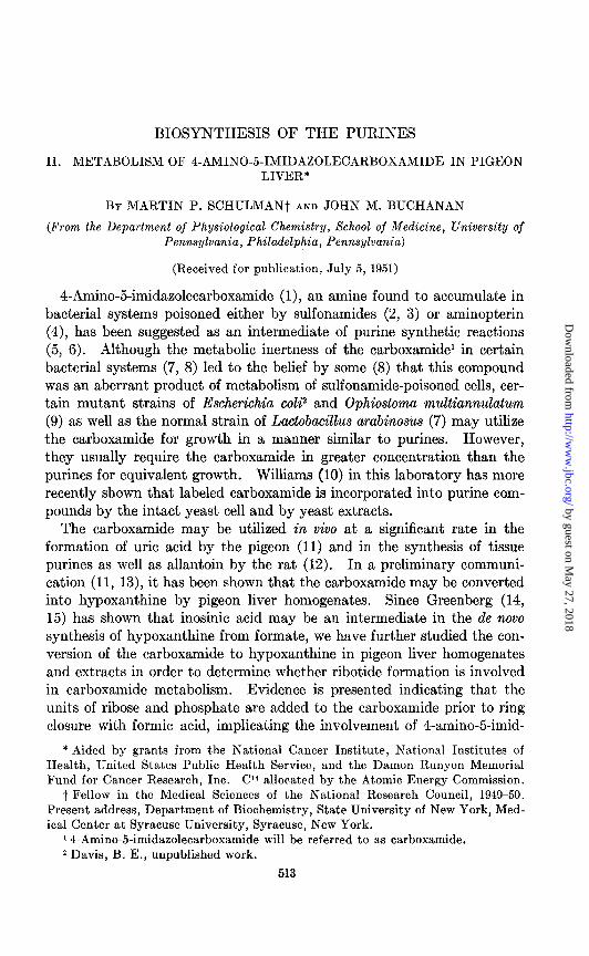
Isolation Procedures;3 Inosinic Acid-After adjustment of the pH to 9.0 with NaOH, the supernatant was placed on a column of Dowex 1 (chloride) of 300 to 500 mesh, 11 ml. in volume and 12 cm. in length. The resin was prepared by preliminary washings with N HCI, water, 5 per cent sodium carbonate, water, and N HCl in the order named. The resin was then exhaustively washed with water until the washings were chloride-free and neutral to litmus. After passage of the supernatant through the column at a rate of 0.5 to 1.0 ml. per minute, the column was washed with water until the optical density of the effluent at 250 rnp decreased to 1 to 2 units per ml. when measured in a cell with a 1 cm. light path. HCl(O.005 N) was then passed through the column to elute the purine compounds. A typical elution curve in which optical density is plotted against effluent volume is shown in Fig. 1, where it may be seen that inosinic acid was sharply separated from hypoxanthine. In other experiments involving both hy- poxanthine and inosine, these two compounds were eluted at the same time from the column, and were therefore not separable on the Dowex 1 resin. The identity of materials eluted from the column may be ascertained by determining the ratio of light absorption at 250 to 265 m/l. These ratios for inosinic acid, inosine, and hypoxanthine are respectively 2.22, 2.31,
3 We wish to thank Dr. Waldo E. Cohn for providing us with unpublished informa- tion on the separation of inosinic acid from inosine and hypoxanthine on the Dowex 1 resin column.
by guest on May 27, 2018
http://ww
w.jbc.org/
Dow
nloaded from

M. P. SCHULMAN AND J. M. BUCHANAN 515
and 1.81. There is a component eluted from the column between inosinic acid and hypoxanthine which is a natural constituent of tissues and has a ratio of absorption at wave-lengths 250 to 265 rnp of approximately 1.
The inosinic acid fraction eluted from the resin column was neutralized with an excess of solid barium carbonate, heated to boiling, filtered while hot, and the filtrate concentrated to dryness in vacua at 40”. The barium inosinate was taken up in a small volume of water and allowed to crystallize overnight in the cold room. These crystals were washed with small por- tions of cold water and then recrystallized from water and again washed.
HYPOXANTHINE HYPOXANTHINE FRACTION FRACTION
l5- 15-
z z z z INOSINIC ACID INOSINIC ACID
% % FRACTION FRACTION
IO - IO -
I I I I I I 400 500 600 400 500 600
, , 700 700 800 800 900 900 1000 1000 1100 1100 1200 1200 1300 1300 1400 1400
VOLUME OF EFFLUENT IN ML. VOLUME OF EFFLUENT IN ML.
FIG. 1. Separation of inosinic acid from hypoxanthine on the Dowex 1 resin col- FIG. 1. Separation of inosinic acid from hypoxanthine on the Dowex 1 resin col- umn. umn. 0, E at 250 rnp; X, E at 265 rnp. 0, E at 250 rnp; X, E at 265 rnp. Volume of resin column, 11 ml.; 12 cm. in Volume of resin column, 11 ml.; 12 cm. in length; length; 300 to 500 mesh. 300 to 500 mesh.
After drying they were then ready for radioactivity and spectrophotometric analysis.
Separation of Hypoxanthine and Inosine-In experiments in which non- radioactive inosinic acid and inosine were incubated together, appreciable amounts of this latter compound had been converted into hypoxanthine. The “hypoxanthine fraction” from the resin (Fig. l), therefore, contained both inosine and hypoxanthine. This fraction was neutralized with NaOH, evaporated to a small volume under reduced pressure, added to sufficient n-butanol so that a saturated butanol solution was obtained, and then placed on a starch column (17). 400 gm. of purified potato starch (Amend Drug and Chemical Company, New York) were employed in columns 5.2 cm. in diameter. The hypoxanthine and inosine were eluted from the
by guest on May 27, 2018
http://ww
w.jbc.org/
Dow
nloaded from
516 BIOSYNTHESIS OF PURINES. II
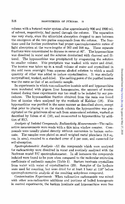
column with a butanol-water system after approximately 800 and 1800 ml. of solvent, respectively, had passed through the column. The separation was very sharp, since the ultraviolet absorption dropped to zero between the emergence of the two purine compounds from the column. The frac- tions used for further purification had proper spectrophotometric ratios of light absorption at the wave-lengths of 262 and 248 mp. These separate fractions were concentrated to dryness in vacua at 40”. The hypoxanthine was dissolved in water and the solution decolorized with charcoal and fil- tered. The hypoxanthine was precipitated by evaporating the solution to smaller volume. This precipitate was washed with water and dried. The inosine was taken up in a small volume of water and crystallized in the cold room overnight from aqueous alcohol solution to which a small quantity of ether was added to induce crystallization. It was similarly recrystallized, washed, and dried. The melting point of the purified inosine was the same as that of an authentic sample.
In experiments in which non-radioactive inosinic acid and hypoxanthine were incubated with pigeon liver homogenates, the amount of inosine formed during these experiments was too small to be isolated by our pro- cedures. The hypoxanthine fraction obtained from the resin was found free of inosine when analyzed by the methods of Kalckar (18). This hypoxanthine was purified in the same manner as described above, except that prior to placing it on the starch column the hypoxanthine was pre- cipitated as the gelatinous silver salt from ammoniacal solution, washed as described by Edson et al. (19), and reconverted to hypoxanthine by addi- tion of HCI.
Analysis of Isolated Compounds; Radioactivity Measurements-The radio- active measurements were made with a thin mica window counter. Com- pounds were usually plated directly without conversion to barium carbo- nate. The samples were plated on small weighed metal planchets (4.9 sq. cm. in area), counted to a standard error of 5 per cent, and corrected for self-absorption.
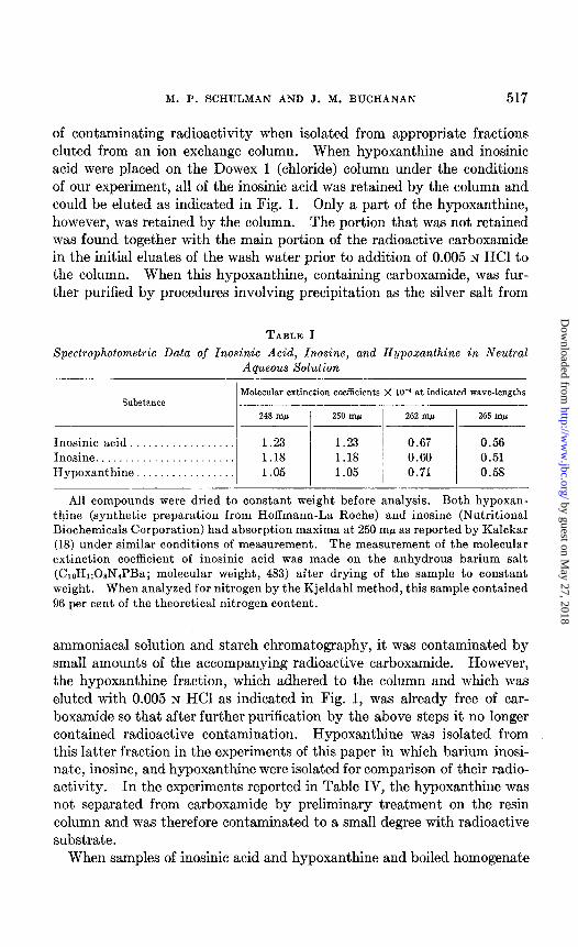
Spectrophotometric Analysis-All the compounds which were analyzed for radioactivity were dissolved in water and routinely analyzed with the Beckman model DU spectrophotometer. In all instances the compounds isolated were found to be pure when compared to the molecular extinction coefficients of authentic samples (Table I). Barium inosinate crystallizes from water with water of crystallization (20). Samples of the hydrate were used for counting, but were dried for at least 1 hour at 110” before spectrophotometric analysis of the resulting anhydrous compound.
Contamination Experiments-When radioactive carboxamide was mixed with other non-radioactive additions and portions of boiled homogenate in control experiments, the barium inosinate and hypoxanthine were free
by guest on May 27, 2018
http://ww
w.jbc.org/
Dow
nloaded from
M. P. SCHULMAN AND J. M. BUCHANAN 517
of contaminating radioactivity when isolated from appropriate fractions eluted from an ion exchange column. When hypoxanthine and inosinic acid were placed on the Dowex 1 (chloride) column under the conditions of our experiment, all of the inosinic acid was retained by the column and could be eluted as indicated in Fig. 1. Only a part of the hypoxanthine, however, was retained by the column. The portion that was not retained was found together with the main portion of the radioactive carboxamide in the initial eluates of the wash water prior to addition of 0.005 N HCl to the column. When this hypoxanthine, containing carboxamide, was fur- ther purified by procedures involving precipitation as the silver salt from
TABLE I
Spectrophotometric Data of Inosinic Acid, Inosine, and Hypoxanthine in Neutral
Substance
Inosinic acid. ................. Inosine ........................ Hypoxanthine .................
Aqueous Solution
Molecular extinction coefficients X lo-’ at indicated wave-lengths
248 m/s 250 mp 262 mp 265 m/.l
1.23 1.23 0.67 0.56 1.18 1.18 0.60 0.51 1.05 1.05 0.71 0.58
All compounds were dried to constant weight before analysis. Both hypoxan- thine (synthetic preparation from Hoffmann-La Roche) and inosine (Nutritional Biochemicals Corporation) had absorption maxima at 250 rnp as reported by Kalckar (18) under similar conditions of measurement. The measurement of the molecular extinction coefficient of ihosinic acid was made on the anhydrous barium salt (C&Hn08NIPBa; molecular weight, 483) after drying of the sample to constant weight. When analyzed for nitrogen by the Kjeldahl method, this sample contained 96 per cent of the theoretical nitrogen content.
ammoniacal solution and starch chromatography, it was contaminated by small amounts of the accompanying radioactive carboxamide. However, the hypoxanthine fraction, which adhered to the column and which was eluted with 0.005 N HCl as indicated in Fig. 1, was already free of car- boxamide so that after further purification by the above steps it no longer contained radioactive contamination. Hypoxanthine was isolated from this latter fraction in the experiments of this paper in which barium inosi- nate, inosine, and hypoxanthine were isolated for comparison of their radio- activity. In the experiments reported in Table IV, the hypoxanthine was not separated from carboxamide by preliminary treatment on the resin column and was therefore contaminated to a small degree with radioactive substrate.
When samples of inosinic acid and hypoxanthine and boiled homogenate
by guest on May 27, 2018
http://ww
w.jbc.org/
Dow
nloaded from
518 BIOSYNTHESIS OF PURINES. II
were mixed with either radioactive formate or glycine and isolated by the above procedures, they were free of contaminating radioactivity.
Preparationof Materials Used-4-Amino-5-imidazolecarboxamide4 labeled in the 4 position with Cl4 was synthesized by the method of Miller, Gurin, and Wilson (12). C14-Hypoxanthine was prepared biologically by incu- bating glycine-2-C14 with pigeon liver homogenates. Hypoxanthine carrier was added to the homogenates at the conclusion of the experiment and the hypoxanthine precipitated with ammoniacal silver nitrate. Free hypoxan- thine was obtained from the silver ammoniacal salt and purified by passage through a starch column as described above. Inosinic acid was prepared as the barium salt by reacting adenosine-5-phosphate (Schwarz Labora- tories) with adenylic acid deaminase (21) in a succinate buffer at pH 5. The solution was made acid with HCl to pH 2, neutralized with an excess of solid BaC03, heated to boiling, and filtered. Upon evaporation of the solution and chilling overnight, barium inosinate crystallized out. The product was recrystallized before use, washed with alcohol and ether, and dried at 110”. Inosine was obtained from the Nutritional Biochemicals Corporation; hypoxanthine, prepared synthetically, from Hoffmann-La Roche; and glycine-l-C’4 and glycine-2-C4 from Tracerlab, Inc.
RESULTS AND DISCUSSION
Preliminary X&dies with 4-Amino-5-imidaxolecarboxamide with Homog- enates and Extracts-Incubation of 4-amino-5-imidazolecarboxamide with pigeon liver homogenates results in a disappearance of the compound dur- ing the course of the incubation. Usually the carboxamide was not me- tabolized appreciably by the tissue without the addition of cY-ketoglutarate. This may be seen in the experiments of Table II in which the disappearance of the carboxamide, as measured by the Bratton-Marshall method for diazotizable amine (22), was stimulated several fold by the addition of or-ketoglutarate. Occasionally, inactive tissue preparations were encoun- tered and more infrequently the carboxamide was metabolized without cr-ketoglutarate. In these cases, the oc-ketoglutarate exerted no additional effect on the metabolism of the carboxamide, a fact which might be ex- plained by the presence of cr-ketoglutarate or its metabolic equivalents in sufficient concentration in the tissue preparation to cause optimal me- tabolism of the carboxamide without added substrate. It may also be seen in Table II that the disappearance proceeds well in the cell-free super- natant of the homogenate and is not dependent on the presence of the
4 We wish to thank Dr. S. Gurin and Dr. C. S. Miller of this department for samples of radioactive and non-radioactive 4-amino-5-imidaxolecarboxamide, and Dr. E. M. Schultz of Sharp and Dohme Company for samples of the non-radioactive com- pound.
by guest on May 27, 2018
http://ww
w.jbc.org/
Dow
nloaded from
M. P. SCHULMAN AND J. M. BUCHANAN 519
mitochondria or microsomes or nuclear material. The preparation re- mained active upon standing for short periods of time at 0”.
Conversion of P4-Carboxamide to C14-Hypoxanthine in Homogenates and Extracts--When labeled carboxamide was incubated with pigeon liver ho- mogenates for a period of 60 to 90 minutes, the hypoxanthine isolated after the addition of carrier was found to be appreciably radioactive. In Experi- ment 1, Table III, sufficient data were collected to estimate the percentage of the metabolized carboxamide which was converted into hypoxanthine.
In Experiment 1, Table III, approximately 77 per cent of the carbox- amide metabolized was converted into hypoxanthine. In Experiments 2
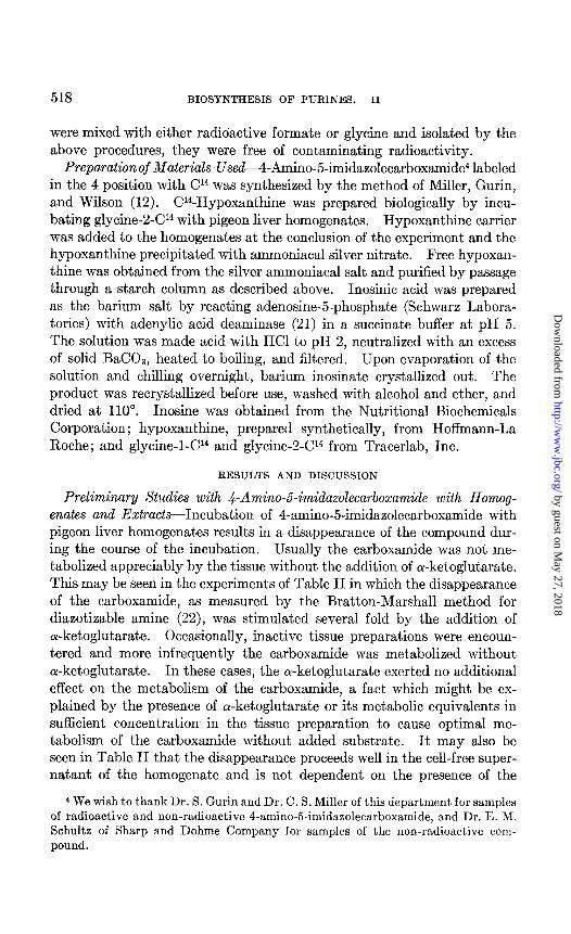
TABLE II
Effect of or-Ketoglutarate on Carboxamide Metabolism and Comparison of Disappearance of Carboxamide in Homogenates and Clari$ed Extracts of
Pigeon Liver
The values are given in micromoles.
system
Homogenate
Extract Recombined homogenate
Carboxamide disappearing a-K&o- glutarak. Experi- Experi- Experi- Experi- Experi- Experi-
ment 1 merit 2 merit 3 merit 4 merit 5 merit 6 _____ -__
- 0.10 0.12 0.00 + 0.52 0.49 0.39 0.39 0.54 + 0.58 0.72 0.29 + 0.57 0.45 0.58
All the vessels contained initially 3 to 4 jbM of carboxamide, 2 ml. of 1:2 homoge- nate, and 30 PM of or-ketoglutarate when indicated. Vessels of Experiments 4, 5, and 6 contained in addition 15 PM of sodium formate and 5 PM of glycine; final volume of all the vessels, 2.4 ml.; time of incubation, 1 hour; 38’.
and 3 reported in Table III, the amount of hypoxanthine produced by the tissue during incubation was not measured, so that the values for the amount of hypoxanthine formed from the labeled carboxamide are calcu- lated on the basis of the amount of added carrier only and are therefore minimal values.
It is of importance to note that in Experiment 3, Table III, the homoge- nate was centrifuged at 100,000 X g for 20 minutes and the reaction was carried out by the enzymes of the clear supernatant. This tracer experi- ment as well as the non-radioactive experiments recorded in Table II demonstrate that the disappearance of the carboxamide and its conversion to hypoxanthine are catalyzed by enzymes of the soluble protein fraction of the pigeon liver and are independent of the insoluble protein of the homogenate.
by guest on May 27, 2018
http://ww
w.jbc.org/
Dow
nloaded from
520 BIOSYNTHESIS OF PURINES. II
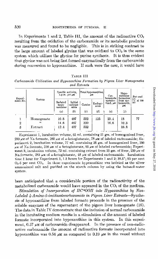
In Experiments 1 and 2, Table III, the amount of the radioactive CO2 resulting from the oxidation of the carboxamide or its metabolic products was measured and found to be negligible. This is in striking contrast to the large amount of labeled glycine that was oxidized to COZ in the same system which utilizes the glycine for purine synthesis. It is thus evident that glycine was not being first formed enzymatically from the carboxamide during conversion to hypoxanthine. If such were the case, it would have
TABLE III
Carboxamide Utilization and Hypoxanthine Formation by Pigeon Liver Homogenates and Extracts
system Experi- ment No
Specific activity, c.p.m. per pY
Isolated ho-
xanthine
Initial :arboxa-
mide
(a) @I
1 Ilomogenate 10.8 407 2 “ 14.6 467 3 Extract 12.4 407
-
‘1 rota1 hypoxanthine I*p
Carrier Endoge- now
(6) (a
552 125 330 552
Hy o- xan&e Per cent formation cf) from car- - x 100 boxamide, cc)
IrM.
18 77 10.3 16.8
Experiment 1, incubation volume, 65 ml. containing 21 gm. of homogenized liver, 210 PM of Na formate, 260 PM of a-ketoglutarate, 78 &M of labeled carboxamide; Ex- periment 2, incubation volume, 77 ml. containing 25 gm. of homogenized liver, 250 PM of Na formate, 310 FM of cu-ketoglutarate, 93 PM of labeled carboxamide; Experi- ment 3, incubation volume, 72 ml. containing extract from 23 gm. of liver, 230 PM of Na formate, 285 /IM of a-ketoglutarate, 48 pM of labeled carboxamide. Incubation time 1 hour for Experiment 3, 1.5 hours for Experiments 1 and 2; 38.5”; 95 per cent OS-5 per cent COZ. In these experiments hypoxanthine was isolated as the silver ammoniacal salt and purified on the starch column by using the butanol-water system.
been anticipated that a considerable portion of the radioactivity of the metabolized carboxamide would have appeared in the CO, of the medium.
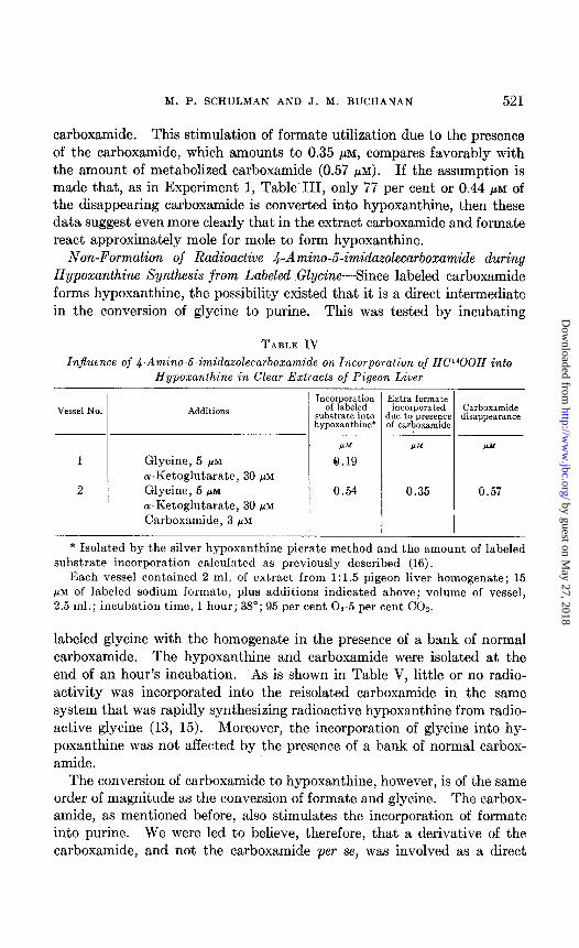
Stimulation of Incorporation of HC1400H into Hypoxanthine by Non- Labeled .&Amino-54midazolecarboxamide in Pigeon Liver Extracts-Synthe- sis of hypoxanthine from labeled formate proceeds in the presence of the soluble enzymes of the supernatant of the pigeon liver homogenate (16). The data in Table IV demonstrate that the inclusion of normal carboxamide in the incubating medium results in a stimulation of the amount of labeled formate incorporated into hypoxanthine in this system. In this experi- ment, 0.57 pM of carboxamide disappeared. In the presence of non-radio- active carboxamide the amount of radioactive formate incorporated into hypoxanthine was 0.54 pM as compared to 0.19 pM in the vessel without
by guest on May 27, 2018
http://ww
w.jbc.org/
Dow
nloaded from
M. P. SCHULMAN AND J. M. BUCHANAN 521
carboxamide. This stimulation of formate utilization due to the presence of the carboxamide, which amounts to 0.35 ,UM, compares favorably with the amount of metabolized carboxamide (0.57 PM). If the assumption is made that, as in Experiment 1, Table III, only 77 per cent or 0.44 PM of the disappearing carboxamide is converted into hypoxanthine, then these data suggest even more clearly that in the extract carboxamide and formate react approximately mole for mole to form hypoxanthine.
Non-Formation of Radioactive 4-Amino-5-imidazolecarboxamide during Hypoxanthine Synthesis from Labeled GEycine-Since labeled carboxamide forms hypoxanthine, the possibility existed that it is a direct intermediate in the conversion of glycine to purine. This was tested by incubating
TABLE IV
Injluence of a-Amino-&imidazolecarboxamide on Incorporation of HF400H into Hypoxanthine in Clear Extracts of Pigeon Liver
Vessel No.
I
2
Additions
Glycine, 5 PM
ol-Ketoglutarate, 39 PM
Glycine, 5 PM
a-Ketoglutarate, 39 PM
Carboxamide, 3 PM
Incorporation Extra formate of labeled
substrate into incorporated
hypoxanthine’ due to presence of carboxamide
w
8.19 PM
0.35 0.54
Carboxamide disappearance
I.r‘+J
0.57
* Isolated by the silver hypoxanthine picrate method and the amount of labeled substrate incorporation calculated as previously described (16).
Each vessel contained 2 ml. of extract from 1:1.5 pigeon liver homogenate; 15 PM of labeled sodium formate, plus additions indicated above; volume of vessel, 2.5 ml.; incubation time, 1 hour; 38”; 95 per cent 02-5 per cent CO,.
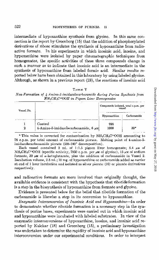
labeled glycine with the homogenate in the presence of a bank of normal carboxamide. The hypoxanthine and carboxamide were isolated at the end of an hour’s incubation. As is shown in Table V, little or no radio- activity was incorporated into the reisolated carboxamide in the same system that was rapidly synthesizing radioactive hypoxanthine from radio- active glycine (13, 15). Moreover, the incorporation of glycine into hy- poxanthine was not affected by the presence of a bank of normal carbox- amide.
The conversion of carboxamide to hypoxanthine, however, is of the same order of magnitude as the conversion of formate and glycine. The carbox- amide, as mentioned before, also stimulates the incorporation of formate into purine. We were led to believe, therefore, that a derivative of the carboxamide, and not the carboxamide per se, was involved as a direct
by guest on May 27, 2018
http://ww
w.jbc.org/
Dow
nloaded from
522 BIOSYNTHESIS OF PURINES. II
intermediate of hypoxanthine synthesis from glycine. In this same con- nection is the report by Greenberg (15) that the addition of phosphorylated derivatives of ribose stimulates the synthesis of hypoxanthine from radio- active formate. In his experiments in which inosinic acid, inosine, and hypoxanthine were isolated by paper chromatographic techniques from homogenates, the specific activities of these three compounds change in such a manner as to indicate that inosinic acid is an intermediate in the synthesis of hypoxanthine from labeled formic acid. Similar results re- ported below have been obtained in this laboratory by using labeled glycine. Although, as shown in a previous report (23), the reactions of inosinic acid
TABLE V
Non-Formation of a-Amino-J-imidazolecarboxamide during Purine Synthesis from NHd2HdY”OOH in Pigeon Liver Homogenates
Compounds isol~~s~~total c.p.m. per
Vessel No.
Hypoxanthine Carboxamide
1 Control 998 2 4-Amino-5-imidazolecarboxamide, 6 PM 1008 30*
* This value is corrected for contamination by NHzCHzC400H amounting to 78 o.p.m. per total amount of carboxamide picrate. Melting point of 4-amino-5- imidazolecarboxamide picrate (238-240” decomposition).
Each vessel contained 2 ml. of 1:1.5 pigeon liver homogenate; 5.4 PM of NHzCHLWOOH (specific activity, 7850 c.p.m. per PM of glycine), 15 PM of sodium formate, 30 PM of or-ketoglutarate, plus the addition of carboxamide in Vessel 2. Incubation volume, 2.5 ml.; 10 mg. of hypoxanthine or carboxamide added as carrier at end of 1 hour incubation and isolated as silver picrate (16) or picrate derivatives respectively.
and radioactive formate are more involved than originally thought, the available evidence is consistent with the hypothesis that ribotide formation is a step in the biosynthesis of hypoxanthine from formate and glycine.
Evidence is presented below for the belief that ribotide formation of the carboxamide is likewise a step in its conversion to hypoxanthine.
Enzymatic Interconversion of Inosinic Acid and Hypoxanthine-In order to demonstrate whether ribotide formation is a necessary step in the syn- thesis of purine bases, experiments were carried out in which inosinic acid and hypoxanthine were incubated with labeled substrates. In view of the enzymatic interconversions of hypoxanthine, inosine, and inosinic acid re- ported by Kalckar (18) and Greenberg (15), a preliminary investigation was undertaken to determine the rapidity of inosinic acid and hypoxanthine interconversion under our experimental conditions. In order to interpret
by guest on May 27, 2018
http://ww
w.jbc.org/
Dow
nloaded from
M. P. SCHULMAN AND J. M. BUCHANAN 523
our data from experiments with labeled carboxamide and glycine, it would be necessary that a complete equilibration of inosinic acid and hypoxan- thine did not occur in the course of the incubation. Thus, if hypoxanthine had been formed from labeled glycine and carboxamide directly without ribotide involvement, the isolated hypoxanthine should have had a greater specific activity than the isolated inosinate, provided that equilibrium be- tween inosinate and hypoxanthine had not occurred. On the other hand, if ribotide formation had been involved in the formation of the completed purine skeleton, the inosinic acid should have had a greater specific activity than the hypoxanthine.
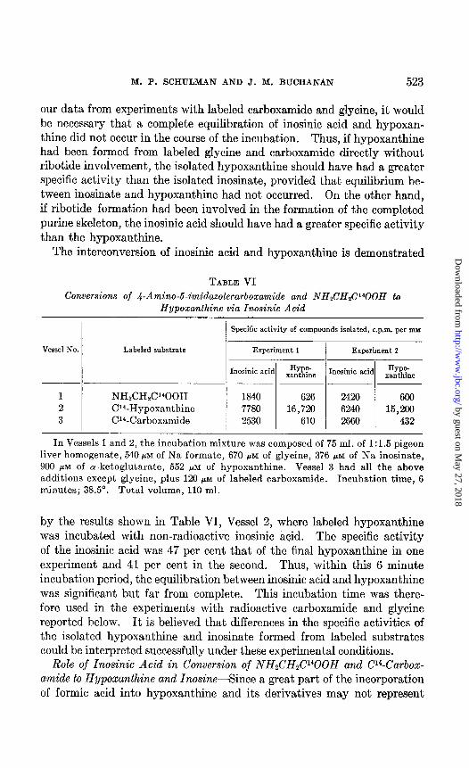
The interconversion of inosinic acid and hypoxanthine is demonstrated
TABLE VI
Conversions of a-Amino-6-imidazolecarboxamide and NHzCHzCIQOH to Hypoxanthine via Inosinic Acid
Specific activity of compounds isolated, c.p.m. per IDY
Vessel No.
1 2 3
Labeled substrate
NHzCHzCPOOH Cr4-Hypoxanthine Cl4-Carboxamide
Experiment 1 Experiment 2
Inosinic acid HY o- Tl. Inosinic acid HYPE-
xant me xanthine
1840 626 2420 600 7780 16,720 6240 15,200
2530 610 2660 432
In Vessels 1 and 2, the incubation mixture was composed of 75 ml. of 1:1.5 pigeon liver homogenate, 540 FM of Na formate, 670 PM of glycine, 376 PM of Na inosinate, 900 pM of cu-ketoglutarate, 552 PM of hypoxanthine. Vessel 3 had all the above additions except glycine, plus 129 pM of labeled carboxamide. Incubation time, 6 minutes; 38.5”. Total volume, 110 ml.
by the results shown in Table VI, Vessel 2, where labeled hypoxanthine was incubated with non-radioactive inosinic acid. The specific activity of the inosinic acid was 47 per cent that of the final hypoxanthine in one experiment and 41 per cent in the second. Thus, within this 6 minute incubation period, the equilibration between inosinic acid and hypoxanthine was significant but far from complete. This incubation time was there- fore used in the experiments with radioactive carboxamide and glycine reported below. It is believed that differences in the specific activities of the isolated hypoxanthine and inosinate formed from labeled substrates could be interpreted successfully under these experimental conditions.
Role of Inosinic Acid in Conversion of NHL’H&‘1400H and C14-Carbox- amide to Hypoxanthine and Inosine-Since a great part of the incorporation of formic acid into hypoxanthine and its derivatives may not represent
by guest on May 27, 2018
http://ww
w.jbc.org/
Dow
nloaded from
524 BIOSYNTHESIS OF PURINES. II
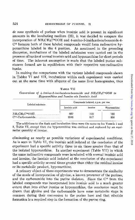
de novo synthesis of purines when inosinic acid is present in significant amounts in the incubating medium (23), it was decided to compare the incorporation of NH&H&‘400H and 4-amino-5-imidaaolecarboxamide-4- Cl4 because both of these labeled compounds would form radioactive hy- poxanthine labeled in the 4 position. As mentioned in the preceding discussion, incubations of the labeled substrates were carried out in the presence of banks of normal inosinic acid and hypoxanthine for short periods of time. The inherent assumption is made that the labeled purine sub- stances formed are in equilibrium with their respective non-radioactive banks.
In making the comparisons with the various labeled compounds shown in Tables VI and VII, incubations within each experiment were carried out at the same time with aliquots of the same liver homogenates, thus
TABLE VII
Conversions of Q-Amino-6-imidazolecarboxamide and NHzCHzCl@OH to Hypoxanthine and Inosine via Inosinic Acid
Compounds isolated, c.p.m. per my Labeled substrate
Inosinic acid Inosine Hypoxanthine
NH&H&POOH 667 123 135 CW-Carboxamide. 2040 817 555
The additions to the flask and incubation time were the same as for Vessels 1 and 3, Table VI, except that the hypoxanthine was omitted and replaced by an equi- molar quantity of inosine.
eliminating as nearly as possible variations of experimental conditions. As is seen in Table VI, the inosinic acid isolated at the conclusion of the experiment had a specific activity three to six times greater than that of the isolated hypoxanthine. In another experiment (Table VII) in which the same radioactive compounds were incubated with normal inosinic acid and inosine, the inosinic acid isolated at the conclusion of the experiment had a specific activity several times greater than either the residual inosine or its metabolic product, hypoxanthine.
A primary object of these experiments was to demonstrate the similarity of the mode of incorporation of glycine, a known precursor of the purines, and the carboxamide into the purine skeleton. Since the Cl4 from the labeled compounds was incorporated into inosinic acid to a much greater extent than into either inosine or hypoxanthine, the conclusion must be drawn that glycine and the carboxamide have some metabolic steps in common during their conversion to the purine base and that ribotide formation is a required step in the formation of the purine ring.
by guest on May 27, 2018
http://ww
w.jbc.org/
Dow
nloaded from
M. P. SCHULMAN AND J. M. BUCHANAN 525
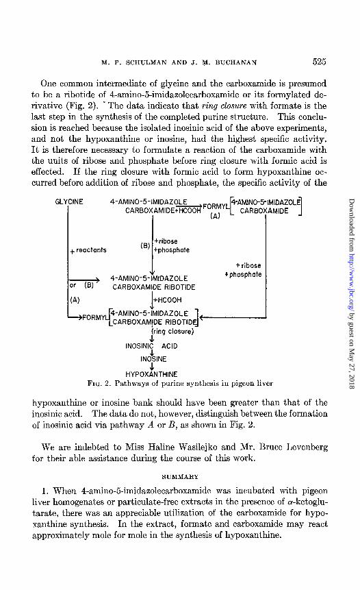
One common intermediate of glycine and the carboxamide is presumed to be a ribotide of 4-amino-5-imidazolecarboxamide or its formylated de- rivative (Fig. 2). . The data indicate that ring closure with formate is the last step in the synthesis of the completed purine structure. This conclu- sion is reached because the isolated inosinic acid of the above experiments, and not the hypoxanthine or inosine, had the highest specific activity. It is therefore necessary to formulate a reaction of the carboxamide with the units of ribose and phosphate before ring closure with formic acid is effected. If the ring closure with formic acid to form hypoxanthine oc- curred before addition of ribose and phosphate, the specific activity of the
4-AMINO-5-IMIDAZOLE CARBOXAMIDE RIBOTIDE
f ribose +phosphate
RIBOTIDE - 1 INOSINIC ACID
INO&INE J
HYPOXANTHINE
AMIDE 1 GLYCINE 4-AMINO-5-IMIDAZOLE
CARBOXAMIDE+HCOOH.Fo~~
I
FIG. 2. Pathways of purine synthesis in pigeon liver
hypoxanthine or inosine bank shauld have been greater than that of the inosinic acid. The data do not, however, distinguish between the formation of inosinic acid via pathway A or B, as shown in Fig. 2.
We are indebted to Miss Haline Wasilejko and Mr. Bruce Levenberg for their able assistance during the course of this work.
SUMMARY
1. When 4-amino-5-imidazolecarboxamide was incubated with pigeon liver homogenates or particulate-free extracts in the presence of cu-ketoglu- tarate, there was an appreciable utilization of the carboxamide for hypo- xanthine synthesis. In the extract, formate and carboxamide may react approximately mole for mole in the synthesis of hypoxanthine.
by guest on May 27, 2018
http://ww
w.jbc.org/
Dow
nloaded from
526 BIOSYNTHESIS OF PURINES. II
2. Radioactive 4-amino-5-imidazolecarboxamide is not formed from NH&HLY400H in pigeon liver homogenates.
3. When either radioactive glycine or carboxamide was incubated with pigeon liver homogenates in the presence of normal inosinic acid and inosine (or hypoxanthine), the specific activity of the inosinic acid after short incubation was considerably greater than that of the residual inosine or hypoxanthine.
4. These results indicate that (a) inosinic acid is an intermediate in the synthesis of hypoxanthine from radioactive glycine and from 4-amino-5- imidazolecarboxamide and that the units of ribose and phosphate are added to the compounds prior to ring closure in the 2 position of the purine base with formic acid. (b) Although the carboxamide is probably not an inter- mediate of purine synthesis from glycine, it is converted to a non-purine intermediate which may be 4-amino-5-imidazolecarboxamide ribotide or its formylated derivative.
BIBLIOGRAPHY
1. Shive, W., Ackermann, W. W., Gordon, M., Getzendaner, M. E., and Eakin, R. E., J. Am. Chem. Sot., 69,725 (1947).
2. Fox, C. L., Jr., Proc. Sac. Exp. Biol. and Med., 61, 102 (1942). 3. Stetten, M. R., and Fox, C. L., Jr., J. Biol. Chem., 161, 333 (1945). 4. Woolley, D. W., and Pringle, R. B., J. Am. Chem. Sac., 72, 634 (1950). 5. Shive, W., and Roberts, E. C., J. Biol. Chem., 162, 463 (1946). 6. Ravel, J. M., Eakin, R. E., and Shive, W., J. BioZ. Chem., 172, 67 (1948). 7. Shive, W., Ann. New York Acad. SC., 62, 1212 (1950). 8. Gots, J. S., Federation Proc., 9, 178 (1950). 9. Fries, N., Bergstrom, S., and Rottenberg, M., Physiol. Planfarum, 2, 210 (1949).
10. Williams, W. J., Federation Proc., 10, 270 (1951). 11. Schulman, M. P., Buchanan, J. M., and Miller, C. S., Federation Proc., 9, 225
(1950). 12. Miller, C. S., Gurin, S., and Wilson, D. W., Science, 112, 654 (1950). 13. Buchanan, J. M., J. Cell. and Camp. Physiol., 38, suppl. 1, 143 (1951). 14. Greenberg, G. R., Federation Proc., 9, 179 (1950). 15. Greenberg, G. R., J. BioZ. Chem., 190, 611 (1951). 16. Schulman, M. P., Sonne, J. C., and Buchanan, J. M., J. BioZ. Chem., 196, 499
(1952). 17. Edman, P., Acta them. Stand., 2, 592 (1948). 18. Kalckar, H. M., J. BioZ. Chem., 167,429 (1947). 19. Edson, N. L., Krebs, H. A., and Model, A., Biochem. J., 30, 1380 (1936). 20. Levene, P. A., and Tipson, R. S., J. BioZ. Chem., 111, 313 (1935). 21. Kalckar, H. M., J. BioZ. Chem., 167, 461 (1947). 22. Bratton, A. C., and Marshall, E. K., Jr., J. BioZ. Chem., 128, 537 (1939). 23. Schulman, M. P., and Buchanan, J. M., Federation Proc., 10, 244 (1951).
by guest on May 27, 2018
http://ww
w.jbc.org/
Dow
nloaded from
Martin P. Schulman and John M. BuchananIN PIGEON LIVER
4-AMINO-5-IMIDAZOLECARBOXAMIDEMETABOLISM OF
BIOSYNTHESIS OF THE PURINES: II.
1952, 196:513-526.J. Biol. Chem.
http://www.jbc.org/content/196/2/513.citation
Access the most updated version of this article at
Alerts:
When a correction for this article is posted•
When this article is cited•
to choose from all of JBC's e-mail alertsClick here
ml#ref-list-1
http://www.jbc.org/content/196/2/513.citation.full.htaccessed free atThis article cites 0 references, 0 of which can be
by guest on May 27, 2018
http://ww
w.jbc.org/
Dow
nloaded from


![University of Groningen Antimalarial Drug Discovery ......lack the de novo purine synthesis pathway and take up host cell purines for growth [2, 8]. Inhibition of this pathway was](https://img.pdfslide.us/doc/110x75/5e30b84fcfea694521705426/university-of-groningen-antimalarial-drug-discovery-lack-the-de-novo-purine.jpg)