Embed Size (px)
Citation preview
Insciences Journal Volume 1, Issue 1, Pages 65-79, 2011
DOI:10.5640/insc.010165
ISSN: 1664-171X
Biosynthesis of Silver Nanoparticles by Fungus Trichoderma Reesei (A Route for Large Scale Production of AgNPs)
Khabat Vahabi (1), G.Ali Mansoori
(2) and Sedighe Karimi (3)
Department of BioEngineering, University of Illinois at Chicago, (M/C 063) Chicago, IL 60607-7052 USA
Emails: (1) [email protected]; (2) [email protected]; (3) [email protected]
Abstract: One of the requirements for advancement of nanotechnology is the development
of reliable experimental protocols for the synthesis of nanomaterials over a range of
biological compositions, sizes and high monodispersity. An attractive possibility of green
nanotechnology is to use micro organisms in the synthesis of nanoparticles. Recently, the
utilization of biological systems, especially fungi, has emerged as a novel method for the
synthesis of nanoparticles. Nanoparticles are considered as fundamental molecular building
blocks for nanotechnology. They are the starting points for preparing many nanostructured
materials and devices. In this paper we report the extracellular biosynthesis of silver
nanoparticles (AgNPs) by using a fungus named Trichoderma Reesei (also known as
Hypocrea jecorina). In the biosynthesis of AgNPs by this fungus, the fungus mycelium is
exposed to the silver nitrate solution. That prompts the fungus to produce enzymes and
metabolites for its own survival. In this process the toxic Ag+ ions are reduced to the none
toxic metallic AgNPs through the catalytic effect of the extracellular enzyme and
metabolites of the fungus. Absorption UV Visible light spectroscopy is used to follow up
with the reaction process. Fluorescence emission spectroscopy is used to produce detailed
information on the progress of reduction of silver nitrate (formation of silver nanoparticles)
on the nanosecond timescale. Fourier transform infrared spectroscopy is used for
quantitative analyses of the reaction products. Our measurements indicate that extracellular
biosynthesis of AgNPs by Trichoderma reesei produces AgNPs with the diameters in the
range of 5 50 nm. Trichoderma Reesei is an environmentally friendly fungus, and it is well
known for its formation of extracellular enzyme and metabolites in very large amounts,
much higher than other fungi. The present process is an excellent candidate for industrial
scale production of silver nanoparticles.
65
Keywords: Extracellular biosynthesis; Extracellular enzyme; Extracellular metabolitem; Fungus;
Green nanotechnology; Large scale production; Silver nanoparticle; Trichoderma reesei.
1. Introduction:
Nanotechnology is the application of science and technology to control matter at the molecular
level. At the nanoscale level, the properties of matter are significantly different from their macroscopic
bulk properties. Nanotechnology is also referred to the ability for designing, characterization,
production and application of structures, devices and systems by controlling shape and size at the
nanometer scale [1].
Nanoparticles are viewed as the fundamental building blocks of nanotechnology [1, 2]. They are
the starting points for preparing many nanostructured materials and devices. Their synthesis is an
important component of the rapidly growing research efforts in nanoscience and nanoengineering [1].
The nanoparticles of a wide range of materials can be prepared by a number of methods. In synthesis
and assembly strategies of nanoparticles or nanomaterials, precursors from liquids, solid or gas phase
are used [1].
Currently, there is a growing need to using environmentally friendly nanoparticles that do not
produce toxic wastes in their process synthesis protocol. To achieve this, we are inclined to shift to
benign synthesis processes, which happen to be mostly of biological nature [1]. This is the theme of
nanobiotechnology, which has many advantages such as ease with which the process can be scaled up,
economic viability, possibility of easily covering large surface areas by suitable growth of the mycelia,
etc. Another advantage of nanobiotechnology is the development of reliable processes for the
synthesis of nanomaterials over a range of sizes (with good monodispersity) and chemical
composition.
It is well known that biological entities like microorganisms and living cells are the best examples
of machines that possess operating parts at the nanoscale level and perform a number of jobs ranging
from generation of energy to extraction of targeted materials at a very high efficiency [3]. The
utilization of such microorganisms like bacteria, fungi, herbal extracts and yeasts in the synthesis of
nanoparticles is a relatively recent activity. It is known that certain bacteria, yeasts and now fungi play
an important role in remediation of toxic metals through reduction of the metal ions so long as they are
not toxic in other ways. For example, environmentally friendly microorganisms could minimize the
toxicity in the process of metallic nanoparticle production by reduction of the metal ions or by
formation of insoluble complexes with metal ions (e.g. metal sulfides) in the form of colloidal particles
[4]. Accordingly, these environmentally friendly biological systems may be considered as benign
nanofactories. It must be pointed out that many such microorganisms are biologically poisonous to
humans, animals and plants, and care must be taken in their choice for production of nanoparticles.
Nanoparticle synthesis is an important component of rapidly growing research efforts in nanoscale
science and engineering. Biotechnology approach towards the synthesis of nanoparticles has many
advantages, such as ease with which the process can be scaled up, economic viability, possibility of
easily covering large surface areas by suitable growth of the mycelia, and its green chemistry nature
provided the microorganism medium is safe.
Some of the examples of the use of biological entities in the synthesis of nanoparticles of different
chemical compositions include the following:
66
K. Vahabi, G.A. Mansoori, S. KarimiBiosynthesis of Silver Nanoparticles by Fungus Trichoderma Reesei (A Route for Large Scale Production of AgNPs)
Insciences J. 1(1): 65-79, 2011
i. Ribosomes for biosynthesis of gold nanoparticles [5];
ii. Bacteria for production of cadmium sulfide [6, 7], zinc sulfide [8], magnetite [9], iron sulfide
[10] and silver [11-13] nanoparticles;
iii. Yeasts for production of lead sulfide and cadmium sulfide [7] nanoparticles;
iv. Production of silver nanoparticles using Emblica Officinalis herbal fruit extract [14], production
of gold nanoparticles using lemongrass extract [15] and synthesis of nanoparticles of variable
morphology using leaves of different plants, sprouts, roots [16] and stems of live alfalfa plants [17].
v. Application of fungi for production of silver nanoparticles, which is the emphasis of the present
paper. Production of silver nanoparticles through fungi has several advantages over the above-
mentioned approaches. They include tolerance towards high metal nanoparticle concentration in the
medium, easy management in large-scale production of nanoparticles, good dispersion of nanoparticle
and much higher amounts of protein expressions. Compared to bacterial broth, fungal broth can be
easily filtered by filter press or similar commonly used equipment, thus saving considerable
investment costs for specialized equipment which may be needed for other methods. As a result, for
large-scale production of nanoparticles fungi is preferred over other methods.
1.1. Mechanism of nanoparticle production through fungi
It is demonstrated that using the dissimilatory properties of an eukaryotic organism such as fungi
may be used to biosynthesize and grow nanoparticles. It is shown that certain fungi have the ability of
producing extracellular metabolites that serve as agent for their own survival when exposed to such
environmental stresses like toxic materials (such as metallic ions), predators and temperature variations
[4].
In the biosynthesis of metal nanoparticles by a fungus, the fungus mycelium is exposed to the metal
salt solution. That prompts the fungus to produce enzymes and metabolites for its own survival. In this
process the toxic metal ions are reduced to the none-toxic metallic solid nanoparticles through the
catalytic effect of the extracellular enzyme and metabolites of the fungus.
The presence of hydrogenase in fungi, such as Fusarium oxysporum [18], Trichoderma reesei [19]
and Trichoderma viride, was demonstrated with washed cell suspensions that had been grown
aerobically or anaerobically in a medium with glucose and salts amended with nitrate [20]. The nitrate
reductase was apparently essential for ferric iron reduction [21]. Many fungi that exhibit these
characteristic properties, in general, are capable of reducing Au (III) or Ag (I) [22]. Besides these
extracellular enzymes, several naphthoquinones [23-25] and anthraquinones [26] with excellent redox
properties, were reported in Fusarium oxysporum that could act as electron shuttle in metal reductions
[28-30]. Specifically the following results towards production of nanoparticles have been achieved
using fungi:
i. Biosynthesis of magnetite using the fungus fusarium oxysporum and Verticillium species [31].
ii. Production of gold nanotriangles by actinomycete, which is a bacteria resembling fungi [32];
iii. Intracellular synthesis of gold and silver nanoparticles in Verticillium fungal cells [11, 12, 33].
iv. Extracellular production of gold, silver and bimetallic Au-Ag alloy nanoparticles by the fungus
Fusarium oxysporum [34 38]. It has been observed that the exposure of aqueous solutions of metal
salts or a mixture of metal salts to Fusarium oxysporum resulted in extracellular formation of
nanoparticles of dimensions 5–50 nm and alloy nanoparticles of dimensions 8–14 nm [37 40].
67
K. Vahabi, G.A. Mansoori, S. KarimiBiosynthesis of Silver Nanoparticles by Fungus Trichoderma Reesei (A Route for Large Scale Production of AgNPs)
Insciences J. 1(1): 65-79, 2011
v. Extracellular production of silver nanoparticles using the fungus aspergillus fumigatus [41].
vi. Production of silver nanoparticle as a result of the reduced state of pretreated fungus Phoma
Species [42].
vii. Extracellular enzymatic reduction of MnO2, nitrate, selenite and ferric ions using fungus
Trichoderma reesei [29].
In the present paper we report extracellular production of silver nanoparticles using Trichoderma
reesei (also known as Hypocrea jecorina). In what follows, the main advantages of Trichoderma
reesei over other fungi are reported.
1.2. Advantages of Trichoderma reesei over other fungi
For industrial applications, fungi should have certain properties which include high production of
specific enzymes or metabolite, high growth rate, easy handling in large-scale production and low-cost
requirement for production procedures [43]. Trichoderma reesei is well known for its formation of
extracellular enzyme in very large amounts, up to 100 g/L [44], which is much higher compared with
other fungi. It is also shown that Trichoderma reesei produces a wide variety of extracellular enzymes
and metabolites such as industrial production of glucosidase, paracelsin, protein, acetyl xylem asterase,
cellobiohydrolase D, cellulose, hemicellulase, cell wall lytic enzyme, β-glucosidase, β-1, 3-glucanase,
and glucose at industrial scale [43]. It should also be mentioned that Trichoderma reesei is the best
studied cellulolytic fungus. It is widely used for the large-scale gene transformation and other
biotechnology industries dealing with overexpression of extracellular enzymes [45]. Here we report
our experimental study of the extracellular synthesis of silver nanoparticles using Trichoderma reesei.
2. Experimental Procedure and Materials
In this study, six different strains of Trichoderma reesei were used for experimentation. The fungal
inoculates were prepared in potato dextrose agar (PDA) media (a common microbiological media for
culturing fungus) at 28°C in Petri plates.
For the synthesis of nanoparticles, the fungus was grown in 200 mL bottles each containing 100 mL
of GC medium (composed of 0.5 % glucose and 0.4 % casein hydrolysate) and at 25–28°C under
continuous mixing condition by a magnetic stirrer (rotary shaker IKA KS 260 basic) at 150 rpm for 72
hours. The reason to use the GC medium is because the growth yield of Trichoderma reesei is greater
in glucose-casein hydrolysate broth than in other media. Casein hydrolysate is a mixture of amino
acids and peptides produced by enzymatic or acid hydrolysis of casein.
The mycelial (vegetative part of the fungus) mass was then separated from the culture broth by
sterile paper filter, and the settled mycelia were washed thrice with sterile distilled water. The
harvested mycelial mass was then used for the synthesis of silver nanoparticles.
2.1. Biosynthesis of silver nanoparticles
In a typical biosynthesis production scheme of silver nanoparticles, 10g of Trichoderma reesei
fungus wet biomass was mixed with a 100 ml aqueous solution of 1 mM silver nitrate (AgNO3). Then
the mixture was placed in a 100 rpm rotating shaker at 28 °C for 120 hours duration. In this process
silver nanoparticles were produced through reduction of the silver ions to metallic silver.
68
K. Vahabi, G.A. Mansoori, S. KarimiBiosynthesis of Silver Nanoparticles by Fungus Trichoderma Reesei (A Route for Large Scale Production of AgNPs)
Insciences J. 1(1): 65-79, 2011
2.2. Characterization of nanoparticles
The reduction of silver ions was routinely monitored by visual inspection of the solution as well as
by measuring the UV-Visible spectra of the solution by periodic sampling of aliquots (2 mL) of the
aqueous component. The UV-Vis spectroscopy measurements were recorded on a Shimadzu dual-
beam spectrophotometer (model UV-1601 PC) operated at a resolution of 1 nm. The fluorescence
measurements were carried out on a Perkin-Elmer LS 50B luminescence spectrophotometer.
In order to perform Fourier transform infrared spectroscopic (FTIR) studies of the results the films
of nanoparticles were produced on Si(111) substrates by drop-coating the metal nanoparticle solution.
The FTIR system used in this study was a Shimadzu FTIR-8201 PC instrument. It was run in the
diffuse reflectance mode at a resolution of 4 cm–1
.
The nanoparticle films were also formed on carbon coated copper grids (40 µm × 40 µm mesh size)
and transmission electron microscopy (TEM) images of the films were scanned on a JEOL 1200 EX
instrument operated at an accelerated voltage of 120 kV.
3. Results and Discussion
3.1. Extracellular Synthesis of Silver Nanoparticles
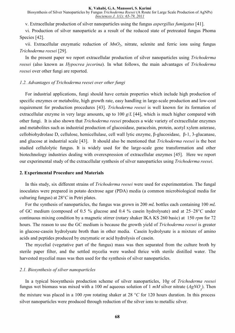
Figure 1A shows a bottle of the fungal cells after removal from the culture medium and before
immersion in 1 mM AgNO3 solution. The pale yellow color of the fungal cells can clearly be observed
in Figure 1A. A picture of the bottle containing the fungal cells after immersion in 1 mM AgNO3
solution for 72 hours is shown in Figure 1B. It can be observed that the previous pale yellow color of
the reaction mixture is changed to the brownish color after 72 hours of reaction. The appearance of a
yellowish-brown color in solution containing the biomass is a clear indication of the formation of
silver nanoparticles in the reaction mixture. The color of the solution is due to the excitation of surface
plasmon vibrations (essentially the vibration of the group conduction electrons) in the silver
nanoparticles.
Figure 1: Picture of bottles containing Trichoderma reesei biomass before (A) and after (B) exposure
to Ag+ ions for 72 h.
69
K. Vahabi, G.A. Mansoori, S. KarimiBiosynthesis of Silver Nanoparticles by Fungus Trichoderma Reesei (A Route for Large Scale Production of AgNPs)
Insciences J. 1(1): 65-79, 2011
3.2. Optical Spectroscopy Measurements
Optical spectroscopy is widely used for the characterization of nanomaterials. In the present study
we use three different spectroscopy techniques to fully characterize the silver nanoparticles we have
produced. They include absorption UV-Visible light spectroscopy, fluorescence emission spectroscopy
and Fourier transform infrared spectroscopy.
3.2.1. Ultraviolet-Visible (UV-Vis) Spectroscopy
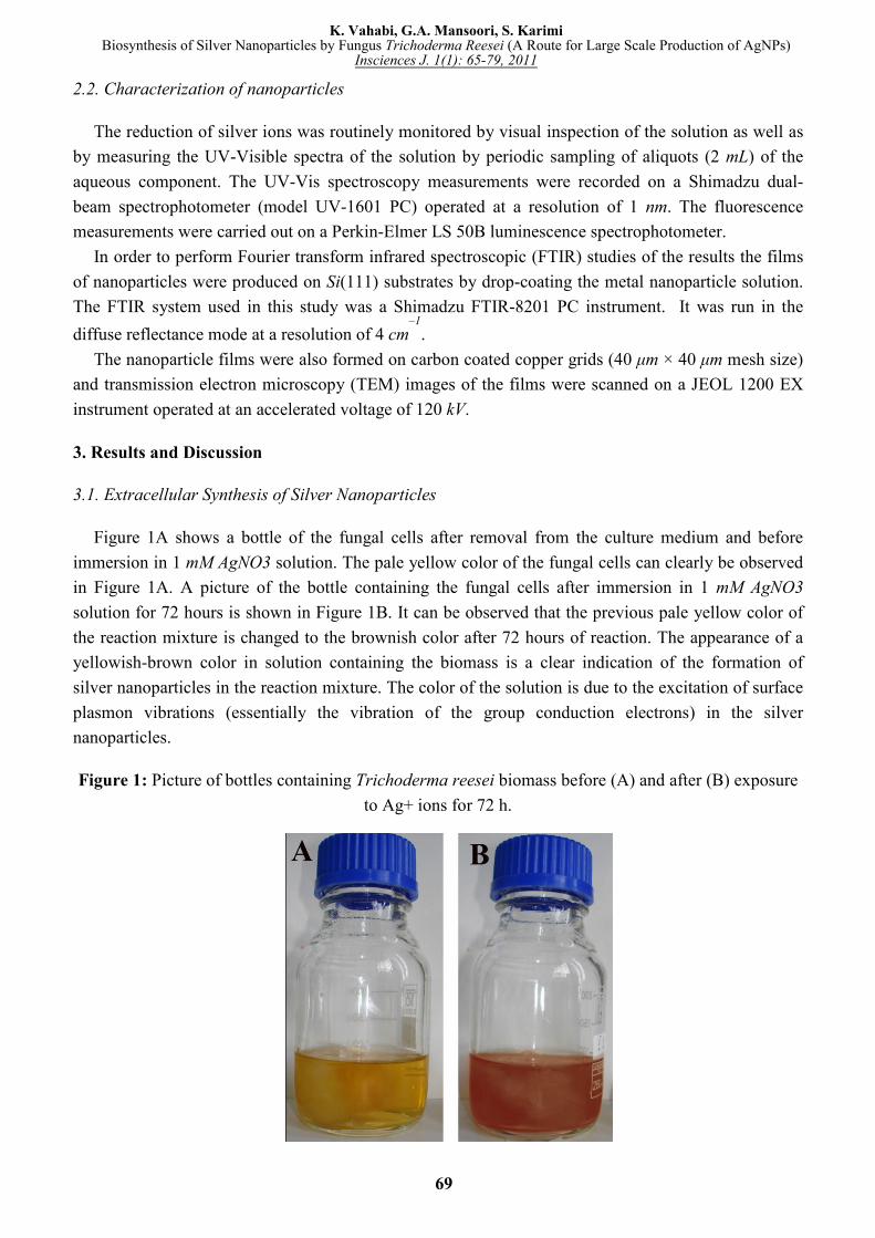
We use UV-Vis spectroscopy to follow up with the reaction process. The spectra recorded from the
Trichoderma reesei reaction vessel at different reaction times are reported in Figure 2. The time at
which the aliquots were removed for measurement is indicated next to the respective curves. The
strong surface plasmon resonance centered at ca. 414-420 nm, is characteristic of colloidal silver. This
peak increased from 414 to 420 nm as the reaction proceeded. The spectra clearly show the increase in
intensity of silver solution with time, indicating the formation of increased number of silver
nanoparticles in the solution. According to this figure, there is no appreciable change in the UV-Vis
spectra of the reaction product after 72 hours indicative of the fact that reaction came to equilibrium at
about 72 hours. It should be pointed out that the reaction was allowed to proceed for about one month.
Interestingly, the solution was extremely stable even after a month of reaction, with no evidence of
aggregation of particles.
Figure 2: UV-Vis spectra recorded at various times (12, 24,…120 hours) after the start of the reaction
of 100 ml of 1 mM AgNO3 solution with 10 g of Trichoderma reesei wet biomass.
70
K. Vahabi, G.A. Mansoori, S. KarimiBiosynthesis of Silver Nanoparticles by Fungus Trichoderma Reesei (A Route for Large Scale Production of AgNPs)
Insciences J. 1(1): 65-79, 2011

y
3.2.2. Fluorescence Emission Spectroscopy
Fluorescence spectroscopy is one of the widely used spectroscopic techniques in the fields of
nanobiotechnology [34, 38], biochemistry and molecular biophysics today [46]. Fluorescence
spectroscopy can provide detailed information on the behavior of macrom
timescale [46].
In this technique, light of some wavelength is directed onto a specimen, prompting the transition of
the electron from the ground to excited state, which then undergoes a non
and the excited electron moves to a more stable excited level. After a characteristic time in the excited
state, the electron returns to the ground state by emitting the characteristic wavelength in the form of
light. This emitted energy can be used to provide qu
about chemical composition, structure, impurities, kinetic process and energy transfer [47].
In the process of dissociation of silver nitrate, it appears that a reductase enzyme (nitrate reductase)
is responsible for the reduction of Ag+ ions and the subsequent formation of metallic silver
nanoparticles. The same observation was reported with another fungus, Fusarium oxysporum, and it
was pointed out that nitrate reductase was responsible for the reduction o
formation of silver nanoparticles [29]. In Figure 3 we report the nitrate reductase through the reaction
of nitrite with 2,3-diaminophthalene (DAN
of fluorescence intensity at 405 nm and 490 nm
diaminonapthotriazole and excess DAN
increased with the addition of a 0.1% KNO
Figure 3: Fluorescence emission spectra for the reaction of nitrite with 2,3
emission spectra the curves A and B were, respectively: fungal filtrate and fungal filtrate and 0.1%
KNO3 solution. The maximum excitation wave
Nanotechnol
3.2.2. Fluorescence Emission Spectroscopy
Fluorescence spectroscopy is one of the widely used spectroscopic techniques in the fields of
nanobiotechnology [34, 38], biochemistry and molecular biophysics today [46]. Fluorescence
spectroscopy can provide detailed information on the behavior of macromolecules on the nanosecond
In this technique, light of some wavelength is directed onto a specimen, prompting the transition of
the electron from the ground to excited state, which then undergoes a non-radiative internal relaxation,
excited electron moves to a more stable excited level. After a characteristic time in the excited
state, the electron returns to the ground state by emitting the characteristic wavelength in the form of
light. This emitted energy can be used to provide qualitative and sometimes quantitative information
about chemical composition, structure, impurities, kinetic process and energy transfer [47].
In the process of dissociation of silver nitrate, it appears that a reductase enzyme (nitrate reductase)
nsible for the reduction of Ag+ ions and the subsequent formation of metallic silver
nanoparticles. The same observation was reported with another fungus, Fusarium oxysporum, and it
was pointed out that nitrate reductase was responsible for the reduction of Ag+ ions and the subsequent
formation of silver nanoparticles [29]. In Figure 3 we report the nitrate reductase through the reaction
diaminophthalene (DAN-reagent). The emission spectrum exhibits two major peaks
sity at 405 nm and 490 nm, corresponding to the emission maximum of 2,3
diaminonapthotriazole and excess DAN-reagent, respectively. The intensity of these two bands
increased with the addition of a 0.1% KNO3 solution, confirming the presence of nitrate red
Fluorescence emission spectra for the reaction of nitrite with 2,3-diaminophthalene. In the
emission spectra the curves A and B were, respectively: fungal filtrate and fungal filtrate and 0.1%
solution. The maximum excitation wavelength was at 375 nm.
Fluorescence spectroscopy is one of the widely used spectroscopic techniques in the fields of
nanobiotechnology [34, 38], biochemistry and molecular biophysics today [46]. Fluorescence
olecules on the nanosecond
In this technique, light of some wavelength is directed onto a specimen, prompting the transition of
radiative internal relaxation,
excited electron moves to a more stable excited level. After a characteristic time in the excited
state, the electron returns to the ground state by emitting the characteristic wavelength in the form of
alitative and sometimes quantitative information
about chemical composition, structure, impurities, kinetic process and energy transfer [47].
In the process of dissociation of silver nitrate, it appears that a reductase enzyme (nitrate reductase)
nsible for the reduction of Ag+ ions and the subsequent formation of metallic silver
nanoparticles. The same observation was reported with another fungus, Fusarium oxysporum, and it
f Ag+ ions and the subsequent
formation of silver nanoparticles [29]. In Figure 3 we report the nitrate reductase through the reaction
reagent). The emission spectrum exhibits two major peaks
corresponding to the emission maximum of 2,3-
reagent, respectively. The intensity of these two bands
solution, confirming the presence of nitrate reductase.
diaminophthalene. In the
emission spectra the curves A and B were, respectively: fungal filtrate and fungal filtrate and 0.1%
length was at 375 nm.
71
K. Vahabi, G.A. Mansoori, S. KarimiBiosynthesis of Silver Nanoparticles by Fungus Trichoderma Reesei (A Route for Large Scale Production of AgNPs)
Insciences J. 1(1): 65-79, 2011
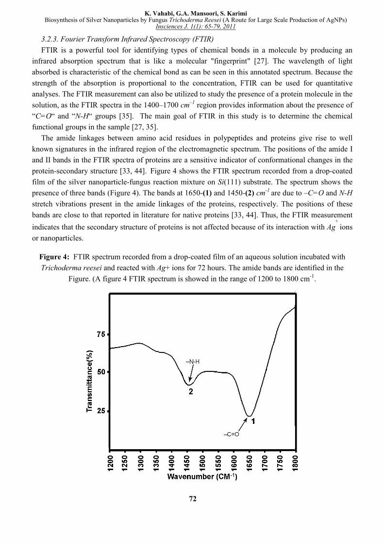
3.2.3. Fourier Transform Infrared Spectroscopy (FTIR)
FTIR is a powerful tool for identifying types of chemical bonds in a molecule by producing an
infrared absorption spectrum that is like a molecular "fingerprint" [27]. The wavelength of light
absorbed is characteristic of the chemical bond as can be seen in this annotated spectrum. Because the
strength of the absorption is proportional to the concentration, FTIR can be used for quantitative
analyses. The FTIR measurement can also be utilized to study the presence of a protein molecule in the
solution, as the FTIR spectra in the 1400–1700 cm–1
region provides information about the presence of
“C=O“ and “N-H“ groups [35]. The main goal of FTIR in this study is to determine the chemical
functional groups in the sample [27, 35].
The amide linkages between amino acid residues in polypeptides and proteins give rise to well
known signatures in the infrared region of the electromagnetic spectrum. The positions of the amide I
and II bands in the FTIR spectra of proteins are a sensitive indicator of conformational changes in the
protein-secondary structure [33, 44]. Figure 4 shows the FTIR spectrum recorded from a drop-coated
film of the silver nanoparticle-fungus reaction mixture on Si(111) substrate. The spectrum shows the
presence of three bands (Figure 4). The bands at 1650-(1) and 1450-(2) cm-1
are due to –C=O and N-H
stretch vibrations present in the amide linkages of the proteins, respectively. The positions of these
bands are close to that reported in literature for native proteins [33, 44]. Thus, the FTIR measurement
indicates that the secondary structure of proteins is not affected because of its interaction with Ag+
ions
or nanoparticles.
Figure 4: FTIR spectrum recorded from a drop-coated film of an aqueous solution incubated with
Trichoderma reesei and reacted with Ag+ ions for 72 hours. The amide bands are identified in the
Figure. (A figure 4 FTIR spectrum is showed in the range of 1200 to 1800 cm-1
.
72
K. Vahabi, G.A. Mansoori, S. KarimiBiosynthesis of Silver Nanoparticles by Fungus Trichoderma Reesei (A Route for Large Scale Production of AgNPs)
Insciences J. 1(1): 65-79, 2011
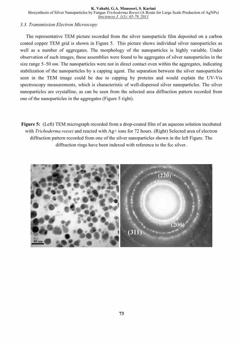
3.3. Transmission Electron Microscopy
The representative TEM picture recorded from the silver nanoparticle film deposited on a carbon
coated copper TEM grid is shown in Figure 5. This picture shows individual silver nanoparticles as
well as a number of aggregates. The morphology of the nanoparticles is highly variable. Under
observation of such images, these assemblies were found to be aggregates of silver nanoparticles in the
size range 5–50 nm. The nanoparticles were not in direct contact even within the aggregates, indicating
stabilization of the nanoparticles by a capping agent. The separation between the silver nanoparticles
seen in the TEM image could be due to capping by proteins and would explain the UV-Vis
spectroscopy measurements, which is characteristic of well-dispersed silver nanoparticles. The silver
nanoparticles are crystalline, as can be seen from the selected area diffraction pattern recorded from
one of the nanoparticles in the aggregates (Figure 5 right).
Figure 5: (Left) TEM micrograph recorded from a drop-coated film of an aqueous solution incubated
with Trichoderma reesei and reacted with Ag+ ions for 72 hours. (Right) Selected area of electron
diffraction pattern recorded from one of the silver nanoparticles shown in the left Figure. The
diffraction rings have been indexed with reference to the fcc silver.
73
K. Vahabi, G.A. Mansoori, S. KarimiBiosynthesis of Silver Nanoparticles by Fungus Trichoderma Reesei (A Route for Large Scale Production of AgNPs)
Insciences J. 1(1): 65-79, 2011
4. Conclusions
In this research, we have shown for the first time the use of Trichoderma reesei in the extracellular
synthesis of silver nanoparticles. In the biosynthesis of metal nanoparticle by a fungus, enzymes are
produced which reduce a salt to its metallic solid nanoparticles through the catalytic effect. Compared
to other filamentous fungus, the Trichoderma reesei is considered to be the most efficient extracellular
enzyme producer, and has a long history in the production of industrial enzymes [44].
Extracellular secretion of enzymes offers the advantage of obtaining large quantities in a relatively
pure state, free from other cellular proteins associated with the organism, and can be easily processed
by filtering of the cells and isolating the enzyme for nanoparticles synthesis from cell free filtrate. Our
measurements indicate that extracellular biosynthesis of silver nanoparticle by Trichoderma reesei
produces AgNPs with the diameters in the range of 5 50 nm. In Table 1 we compare the size ranges,
methods of AgNP produced through various fungi, together with the environmental, biological and
economical implications of the use of each fungus. According to this table biosynthesis of silver
nanoparticles by fungus Trichoderma Reesei is preferred from the points of view of safety, economy
and the large scale production potential.
As discussed above, we can biosynthesize silver nanoparticles on a large scale through Trichoderma
reesei, which is a major advantage over other fungus methods. It should be mentioned that
Trichoderma reesei is not known to be harmful to humans. According to previous studies on
Trichoderma reesei, the production of extracellular enzyme and nanoparticles in this fungus is more
efficient than other fungi. It is also shown that Trichoderma reesei has easier and cheaper cultivation
requirements and higher growth rates on both industrial and laboratory scales, thereby having a lower
cost in large scale production. It should be pointed out that large scale production of silver
nanoparticles by other techniques, such as chemical vapor deposition, irradiation, and liquid solution
reduction, usually produces particles larger than a few micrometers in size. These other techniques
also involve lower production yields and higher expenses [14, 27, 33] compared to large scale
biosynthesis through Trichoderma reesei. Because of the significant commercial value of the findings
reported in this paper a patent application [54] is submitted on this subject.
Acknowledgements
The authors appreciate the very helpful review of this paper and comments by Prof. Thomas W.
George.
74
K. Vahabi, G.A. Mansoori, S. KarimiBiosynthesis of Silver Nanoparticles by Fungus Trichoderma Reesei (A Route for Large Scale Production of AgNPs)
Insciences J. 1(1): 65-79, 2011
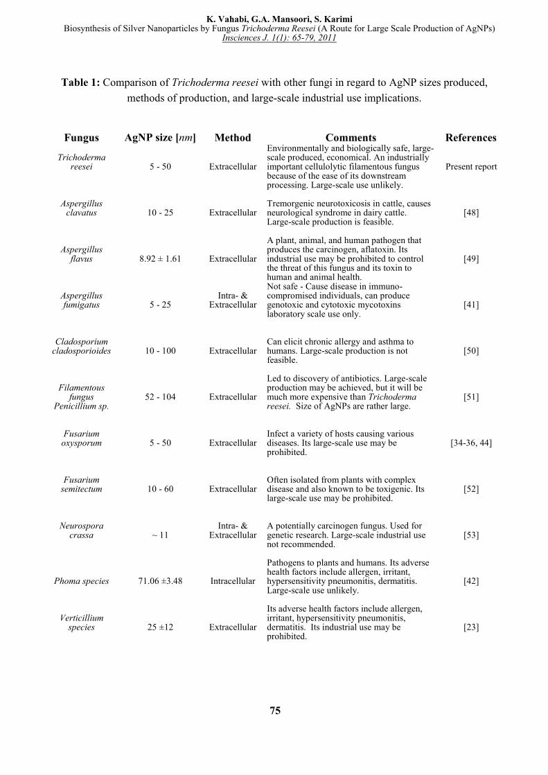
Table 1: Comparison of Trichoderma reesei with other fungi in regard to AgNP sizes produced,
methods of production, and large-scale industrial use implications.
Fungus AgNP size [nm] Method Comments References
Trichoderma reesei 5 - 50 Extracellular
Environmentally and biologically safe, large-scale produced, economical. An industrially important cellulolytic filamentous fungus because of the ease of its downstream processing. Large-scale use unlikely.
Present report
Aspergillus clavatus 10 - 25 Extracellular
Tremorgenic neurotoxicosis in cattle, causes neurological syndrome in dairy cattle. Large-scale production is feasible.
[48]
Aspergillus flavus 8.92 ± 1.61 Extracellular
A plant, animal, and human pathogen that produces the carcinogen, aflatoxin. Its industrial use may be prohibited to control the threat of this fungus and its toxin to human and animal health.
[49]
Aspergillus fumigatus 5 - 25
Intra- & Extracellular
Not safe - Cause disease in immuno-compromised individuals, can produce genotoxic and cytotoxic mycotoxins laboratory scale use only.
[41]
Cladosporium cladosporioides 10 - 100 Extracellular
Can elicit chronic allergy and asthma to humans. Large-scale production is not feasible.
[50]
Filamentous fungus
Penicillium sp. 52 - 104 Extracellular
Led to discovery of antibiotics. Large-scale production may be achieved, but it will be much more expensive than Trichoderma reesei. Size of AgNPs are rather large.
[51]
Fusarium oxysporum 5 - 50 Extracellular
Infect a variety of hosts causing various diseases. Its large-scale use may be prohibited.
[34-36, 44]
Fusarium semitectum 10 - 60 Extracellular
Often isolated from plants with complex disease and also known to be toxigenic. Its large-scale use may be prohibited.
[52]
Neurospora crassa ~ 11
Intra- & Extracellular
A potentially carcinogen fungus. Used for genetic research. Large-scale industrial use not recommended.
[53]
Phoma species 71.06 ±3.48 Intracellular
Pathogens to plants and humans. Its adverse health factors include allergen, irritant, hypersensitivity pneumonitis, dermatitis. Large-scale use unlikely.
[42]
Verticillium species 25 ±12 Extracellular
Its adverse health factors include allergen, irritant, hypersensitivity pneumonitis, dermatitis. Its industrial use may be prohibited.
[23]
75
K. Vahabi, G.A. Mansoori, S. KarimiBiosynthesis of Silver Nanoparticles by Fungus Trichoderma Reesei (A Route for Large Scale Production of AgNPs)
Insciences J. 1(1): 65-79, 2011
References
[1] Mansoori, GA , 2005 Principles of Nanotechnology – Molecular-Based Study of Condensed Matter
in Small Systems, World Scientific Pub. Co., Hackensack, NJ.
[2] Mansoori GA, George TF, Zhang G and Assoufid L, 2007 Molecular Building Blocks for
Nanotechnology, Springer, New York.
[3] Goodsell DS 2004 Bionanotechnology: Lessons from Nature, Wiley-Liss, Hoboken, New York.
[4] Mehra RK, Winge DR 1991 Metal Ion Resistance in Fungi: Molecular Mechanisms and their
Regulated Expression. J. Cell. Biochem, 45 30-40.
[5] Pavel IS 2005 Assembly of Gold Nanoparticles by Ribosomal Molecular Machines. PHD thesis,
The University of Texas at Austin.
[6] Smith PR, Holmes JD, Richardson DJ, Russell DA, Sodeau JR 1998 Photophysical and
Photochemical Characterization of Bacterial Semiconductor Cadmium Sulfide Particles. J. Chem.
Soc., Faraday Trans, 94 1235-1241.
[7] Kowshik M, Deshmukh N, Vogel W, Urban J, Kulkarni SK, Paknikar KM 2002 Microbial
Synthesis of Semiconductor CdS Nanoparticles, their Characterization, and their Use in the
Fabrication of an Ideal Diode. Biotechnol. Bioengineer, 78 583-587.
[8] Labrenz M, Druschel GK, Thomsen-Ebert T, Gilbert B, Welch SA, Kemner KM, Logan GA,
Summons RE, De Stasio G, Bond PL, Lai B, Kelly SD, Banfield JF 2000 Formation of Sphalerite
(ZnS) Deposits in Natural Biofilms of Sulfate-Reducing Bacteria. Science, 290 1744-1747.
[9] Lovley DR, Stolz JF, Nord GL, Phillips EJP 1987 Anaerobic Production of Magnetite by a
Dissimilatory Iron-Reducing Microorganism. Nature, 330 252-254.
[10] Watson JHP, Cressey BA, Roberts AP, Ellwood DC, Charnock JM 2000 Structural and Magnetic
Studies on Heavy-Metal-Adsorbing Iron Sulphide Nanoparticles Produced by Sulphate-Reducing
Bacteria J. Magn. Mater, 214 13-30.
[11] Kowshik M, Vogel W, Urban J, Kulkarni SK, Paknikar KM 2002 Extracellular Synthesis of
Silver Nanoparticles by a Silver-Tolerant Yeast Strain MKY3. Adv. Mater., 14 812-815.
[12] Naik RR, Stringer SJ, Agarwal G, Jones SE, Stone MO 2002 Biomimetic Synthesis and
Patterning of Silver Nanoparticles. Nat Mater, 1 169-172.
[13] Klaus T, Joerger R, Olsson E, Claes G, Granqvist R 1999 Silver-Based Crystalline Nanoparticles,
Microbially Fabricated. Microbiology Appl Phys Sci, 96(24) 13611–13614.
[14] Balaprasad A, Chinmay D, Ahmad A, Sastry M 2005 Biosynthesis of Gold and Silver
Nanoparticles Using Emblica Officinalis Fruit Extract, Their Phase Transfer and Transmetallation
in an Organic Solution. J Nanosci Nanotechnol, 5(7) 1665-1671.
[15] Shankar S, Rai A, Ankamwar B, Singh A, Ahmad A, Sastry M 2004 Biological Synthesis of
Triangular Gold Nanoprisms. Nature Materials, 3 482-488.
[16] Shankar SS, Ahmad A, Sastry M 2003 Geranium Leaf Assisted Biosynthesis of Silver
Nanoparticles Biotechnol. Prog. 19 1627-1631.
[17] Gardea JL, Torresdey E, Gomez JR, Peralta-Videa JG, Parsons H, Troiani M, Yacaman J: 2003
Alfalfa Sprouts A Natural Source for the Synthesis of Silver Nanoparticles. Langmuir, 19 1357-
1361.
76
K. Vahabi, G.A. Mansoori, S. KarimiBiosynthesis of Silver Nanoparticles by Fungus Trichoderma Reesei (A Route for Large Scale Production of AgNPs)
Insciences J. 1(1): 65-79, 2011
[18] Gilbert B, Zhang H, Huang F, Finnegan MP, Waychunas GA, Banfield JF 2003Special Phase
Transformation and Crystal Growth Pathways Observed in Nanoparticles. Geochem. Trans, 4 20-
25.
[19] Rautio J, Smit BA, Wiebe M, Penttilä M, Saloheimo M 2006 Transcriptional Monitoring of
Steady State and Effects of Anaerobic Phases in Chemostat Cultures of the Filamentous Fungus
Trichoderma Reesei. BMC Genomics, 7 247-249.
[20] Chovanec P, Kalinak M, Liptaj T, Pronayova N, Jakubik T, Hudecova D, Varecka L 2005 Study
of Trichoderma Viride Metabolism under Conditions of the Restriction of Oxidative Processes.
Can. J. Microbiol, 51(10) 853-862.
[21] Ottow JCG, Von Klopotek A 1969 Enzymatic Reduction of Iron Oxide by Fungi. Appl.
Microbiol, 18 41-43.
[22] Lloyd JR 2003 Microbial Reduction of Metals and Radionuclides. FEMS Microbial. Rev, 27 411-
425.
[23] Medentsev AG, Alimenko VK 1998 Naphthoquinone Metabolites of the Fungi. Photochemistry,
47 935-959.
[24] Duran N, Teixeira MFS, De Conti R, Esposito E 2002 Ecological-Friendly Pigments from Fungi.
Crit Rev Food Sci Nutr, 42 53-66.
[25] Bell AA, Wheeler MH, Liu J, Stipanovic RD, Puckhaber LS, Orta H 2003 United States
Department of Agriculture-Agricultural Research Service Studies on Polyketide Toxins of
Fusarium Oxysporum f sp Vasinfectum: Potential Targets for Disease Control. Pest Manag Sci, 59
736-747.
[26] Baker RA, Tatum JH 1998 Novel Anthraquinones from Stationary Cultures of Fusarium
Oxysporum. J Ferment Bioeng, 85 359-361.
[27] Senapati S 2005 Biosynthesis and Immobilization of Nanoparticles and their Applications. Ph.D.
thesis, University of Pune.
[28] Misko TP, Schilling RJ, Salvemini D, Moore WM, Currie MG 1993 A Fluorometric Assay for the
Measurement of Nitrite in Biological Samples. Anal Biochem, 214 11-16.
[29] Klittich CJR, Leslie JF 1988 Nitrate Reduction Mutants of Fusarium Moniliforme (gibberella-
fujikuroi). Genetics, 118 417-423.
[30] Kumar CV, McLendon GL 1997 Nanoencapsulation of Cytochrome c and Horseradish Peroxidase
at the Galleries of Alpha-Zirconium Phosphate. Chem Mater, 9 863-870.
[31] Bharde A, Rautaray D, Bansal
V, Ahmad A, Sarkar
I, Mohammad Yusuf S, Sanyal M, Sastry M
2006 Extracellula Biosynthesis of Magnetite using Fungi. Small, 2(1) 135-41.
[32] Ahmad A, Senapati S, Khan MI, Kumar R, Sastry M 2003 Extracellular Biosynthesis of
Monodisperse Gold Nanoparticles by a Novel Extremophilic Actinomycete, Thermomonospora
sp. Langmuir, 19(8) 3550.
[33] Mukherjee P, Ahmad A, Mandal D, Senapati S, Sainkar SR, Khan MI, Ramani R, Parischa R,
Ajaykumar PV, Alam M, Sastry M, Kumar R 2001 Bioreduction of AuCl4-ions by the Fungus,
Verticillium sp. And Surface Trapping of the Gold Nanoparticles formed. Angew Chem Int Edu,
40 3585-3588.
[34] Durán N, Marcato, PD, Alves OL, Souzaand G, Esposito E 2005 Mechanistic Aspects of
Biosynthesis of Silver Nanoparticles by Several Fusarium Oxysporum Strains. Journal of
Nanobiotechnology, 3:8 doi:10.1186/1477-3155-3-8.
77
K. Vahabi, G.A. Mansoori, S. KarimiBiosynthesis of Silver Nanoparticles by Fungus Trichoderma Reesei (A Route for Large Scale Production of AgNPs)
Insciences J. 1(1): 65-79, 2011
[35] Senapati S, Ahmad A, Khan MI, Sastry M, Kumar R 2005 Extracellular Biosynthesis of Bimetallic
Au-Ag Alloy Nanoparticles. Small, 1(5) 517-20.
[36] Souza GIH, Marcato PD, Duran N, Esposito E 2004 Utilization of Fusarium Oxysporum in the
Biosynthesis of Silver Nanoparticles and its Antibacterial Activities. In IX National Meeting of
Environmental Microbiology Curtiba, PR (Brazil) Abstract pag. 25.
[37] Mukherjee P, Senapati S, Mandal D, Ahmad A, Khan MI, Kumar R, Sastry M 2002 Extracellular
Synthesis of Gold Nanoparticles by the Fungus Fusarium Oxysporum. Chem Biochem, 3 461-463.
[38] Ahmad A, Mukherjee P, Senapati S, Mandal D, Khan MI, Kumar R, Sastry M 2003 Extracellular
Biosynthesis of Silver Nanoparticles using the Fungus Fusarium Oxysporum. Colloid Sorf B, 28
313-318.
[39] Mukherjee P, Ahmad A, Mandal D, Senapati S, Sainkar SR, Khan MI, Parischa R, Ajayakumar
PV, Alam M, Kumar R, Sastry M 2001 Fungusmediated Synthesis of Silver Nanoparticles and
their Immobilization in the Mycelial matrix: A Novel Biological approach to Nanoparticle
Synthesis. Nano Let, 1 515-519.
[40] Sastry M, Ahmad A, Islam NI, Kumar R 2003 Biosynthesis of Metal Nanoparticles using Fungi
and Actinomycete. Urent Sci, 85 162-170.
[41] Bhainsa KC, D'Souza SF 2006 Extracellular Biosynthesis of Silver Nanoparticles using the
Fungus Aspergillus Fumigatus. Colloids Surf B Biointerfaces. 47(2) 160-164.
[42] Chen JC, Lin ZH and Ma XX 2003 Evidence of the Production of Silver Nanoparticles via
Pretreatment of Phoma sp.3.2883 with silver nitrate. Lett Appl Microbiol, 37 105–108
[43] Durand H, Clanet M, Tiraby G 1988 Genetic Improvement of Trichoderma Reesei for Large Scale
Cellulase Production. Enzyme Microb Technol, 10 341–346.
[44] Oksanen T, Pere J, Paavilainen L, Buchert J, Viikari L 2000 Treatment of Recycled Kraft Pulps
with Trichoderma Reesei Hemicellulases and Cellulases. J Biotechnol, 78(1) 39–44.
[45] Archer DB, Jeenes DJ, Mackenzie DA 1994 Strategies for Improving Heterologous Protein
Production from Filamentous Fungi. Antonie Leeuwenhoek, 65 245–250.
[46] Joseph R, Lakowicz Sf, Grattonnll E, Cherek H, Badri P, Maliwal S, Gabor Laczko SS 1984
Determination of Time-resolved Fluorescence Emission Spectra and Anisotropies of a
Fluorophore-Protein Complex Using Frequency-Domain Phase-modulation Fluorometry. The
Journal of Biological Chemistry, 259 (17) 10967-10972.
[47] Lakowicz, JR 1983 Principles of Fluorescence Spectroscopy, Plenum Press, New York.
[48] Verma VC, Kharwar RN and Gange AC 2010 Biosynthesis of Antimicrobial Silver Nanoparticles
by the Endophytic Fungus Aspergillus Clavatus. Nanomedicine, 5(1) 33-40.
[49] Vigneshwaran, N, Ashtaputrea, N.M, Varadarajana, P.V., Nachanea, R.P., Paralikara, K.M. and
Balasubramanyaa, R.H. 2007 Biological Synthesis of Silver Nanoparticles using the Fungus
Aspergillus Flavus, Materials Letters, 61(6), 1413-1418.
[50] Balaji DS, Basavaraja S, Deshpande R, Bedre Mahesh D, Prabhakar BK and Venkataraman A
2009 Extracellular Biosynthesis of Functionalized Silver Nanoparticles by Strains of
Cladosporium Cladosporioides Fungus. Colloids and Surfaces B: Biointerfaces, 68(1), 88-92.
[51] Hemath Naveen KS, Kumar G, Karthik L, Bhaskara Rao KV 2010 Extracellular Biosynthesis of
Silver Nanoparticles using the Filamentous Fungus Penicillium sp. Archives of Applied Science
Research, 2(6) 161-167.
78
K. Vahabi, G.A. Mansoori, S. KarimiBiosynthesis of Silver Nanoparticles by Fungus Trichoderma Reesei (A Route for Large Scale Production of AgNPs)
Insciences J. 1(1): 65-79, 2011
[52] Basavaraja S, Balaji SD, Lagashetty A, Rajasab AH and Venkataraman A 2008 Extracellular
Biosynthesis of Silver Nanoparticles using the Fungus Fusarium Semitectum. Materials Research
Bulletin, 43(5) 1164-1170.
[53] Castro-Longoria E, Vilchis-Nestor AR and Avalos-Borja M 2011Biosynthesis of Silver, Gold and
Bimetallic Nanoparticles using the Filamentous Fungus Neurospora Crassa. Colloids and Surfaces
B: Biointerfaces, 83(1) 42-48.
[54] Mansoori GA, SYNTHESIS OF NANOPARTICLES BY FUNGI, 2010, Patent application
number: 20100055199, IPC8 Class: AA01N5916FI, USPC Class: 424618,
www.faqs.org/patents/app/20100055199.
79
K. Vahabi, G.A. Mansoori, S. KarimiBiosynthesis of Silver Nanoparticles by Fungus Trichoderma Reesei (A Route for Large Scale Production of AgNPs)
Insciences J. 1(1): 65-79, 2011