Embed Size (px)
Citation preview
J Clin Pathol 1993;46:437-440
Use of Probemix and OmniProbe biotinylatedcDNA probes for detecting HPV infection inbiopsy specimens from the genital tract
I Zehbe, E Rylander, A Strand, E Wilander
AbstractAims-To compare two commerciallyavailable pan probes for the identifica-tion of human papillomavirus (HPV)DNA expression in histological sectionsand to type the HPV positive cases.Methods-97 formalin fixed, paraffin waxembedded biopsy specimens from thegenital tract were tested for HPV positiv-ity with in situ hybridisation usingbiotinylated cDNA pan probes-Probe-mix (Enzo) and OmniProbe (Digene).The HPV positive cases were furthertested with HPV types 6/11, 16/18, and31I33I35I51, and the HPV type was relatedto the histological diagnosis. Formalinfixed, HeLa cells (10-50 HPV 18 copiesper cell) and SiHa cells (1-2 HPV 16copies per cell) were used as referencecell lines.Results-32% of the specimens gave pos-itive nucleic signals with both Probemixand OmniProbe. Of these, 84% could befurther characterised with regard toHPV types 6I11, 16/18, and 31I33I35I51; 4%of all cases were positive with eitherProbemix or OmniProbe. The concor-dance of these probes was high, 96% alto-gether. HeLa cells stained positive butSiHa cells did not.Conclusion-There is no difference be-tween Probemix and OmniProbe for thegeneral detection of HPV. The meandetection limit of these probes is about 20copies a cell.
(7 Clin Pathol 1993;46:437-440)
Department ofPathology andCytology, UniversityHospital, S-751 85Uppsala, SwedenI ZehbeE WilanderDepartment ofObstetrics andGynaecologyE RylanderDepartment ofDermatology andVenereologyA StrandCorrespondence to:Dr Ingeborg Zehbe
Accepted for publication4 November 1992
Human papillomavirus (HPV) is associatedwith anogenital tract lesions. Although certainHPV types (such as 6/11) occur mainly inbenign lesions such as condyloma acumina-tum, others (16/18) increase the risk of devel-oping anogenital malignancies."
In situ hybridisation with biotinylatedcDNA probes is a frequently used molecularhybridisation method that permits the deter-mination of HPV type in relation to the mor-phological appearance of the lesion.57 AcDNA pan probe for HPV detection is an
adjunct to the histopathological diagnosis andcan be used for screening. Tissues positivewith this probe may be assayed subsequentlyusing type specific probes for HPV.
MethodsEighty patients, aged between 15 and 51
years, with HPV suspect or manifest lesionswere studied. Ninety seven formalin fixed,paraffin wax embedded tissue samples fromthe genital tract were processed for histo-pathological diagnosis and in an additionalsequence tested for HPV positivity with insitu hybridisation using biotinylated cDNApan probes (Enzo Diagnostics Inc, New York,Beltsville, USA (Probemix) and Digene,Maryland, USA (OmniProbe)). Both probesuse a mixture of HPV specific probes formu-lated to detect most HPV types associatedwith anogenital lesions. With Probemix nofurther details are known as to what typesexactly are covered, but OmniProbe detectsHPV types 6, 11, 16, 18, 31, 33, 35, 42, 43,44, 45, 51, 52 and 56. Enzo Diagnosticsoffers Probemix as a complete kit includingreagents for alkaline phosphatase detectionand HPV positive biopsy controls.OmniProbe, however, may be purchased withpositive DNA controls and negative plasmidcontrols only. Both control systems wereapplied according to the manufacturers' rec-ommendations and found to work satisfacto-rily. To assess the sensitivity of the variousprobes applied, we used formalin fixed HeLacells (10-50 HPV 18 copies a cell), a giftfrom Dr Eva Rupp, The Ludwig CancerInstitute, Uppsala, Sweden and SiHa cells(1-2 HPV 16 copies a cell), HTB 35, ATCC.Biopsy specimens that stained positive witheither of these two general HPV probes werealso typed for HPV 6/11 (Enzo and Digene),16/18 (Enzo and Digene), 18 (Enzo),31/33/51 (Enzo) and 31/33/35 (Digene).HPV types 42, 43, 44, 45, 52 and 56 are notyet commercially available.
Briefly, 4-6,um sections on organosilanecoated slides were treated with proteinase K(P-0390; Sigma) at a concentration of0-l mg/ml SSPE buffer for 15 minutes at37°C and the endogenous peroxidase activityblocked in aqueous 3% H202. Slides weredehydrated in 99% ethanol, air dried, anddenaturated for 5 minutes at 94°C. Hybrid-isation was carried out in a humid chamberovernight (pan probes) and for two hours(type specific probes to prevent cross-hybridi-sation) at 37°C. Posthybridisation wasperformed in 50% formamid/2 x SSPE(final concentration) for 10 minutes at 37GC,followed by enzymatic detection with bio-tinylated horseradish peroxidase bound tostreptavidin (K 377 A-B; Dakopatts) 1 in 100for 30 minutes at 37°C and visualisation withdiaminobenzidine (DAB)-H202. The DAB-
437
on 25 Decem
ber 2018 by guest. Protected by copyright.
http://jcp.bmj.com
/J C
lin Pathol: first published as 10.1136/jcp.46.5.437 on 1 M
ay 1993. Dow
nloaded from
Zehbe, Rylander, Strand, Wilander
Table 1 HPV negative and HPVpositive cases stainedwith both pan probes in relation to histological diagnosis
Histological HPVnegative HPVpositivediagnosis biopsies biopsies Total
Negative findings 41 0 41Hyperplasia 6 1 7Condyloma
acuminatum 0 9 9Flat condyloma 10 6 16SILI 0 2 2SIL II 2 9 12SIL III 7 6 12Total 66 33 99
H202 detection system was better than thatusing alkaline phosphatase. Finally, the slideswere counterstained in Mayer's haema-toxylin, rinsed in graded ethanols and xylene,and placed on to coverslips.5 HeLa and SiHacells were treated in exactly the same manner.
The following classification was used forthe morphological diagnosis: 1 normal squa-mous epithelium and inflammatory or reac-
tive changes; 2 hyperplasia, referring to a
thickening of the squamous mucosa, but lack-ing koilocytosis and nuclear atypia; threecondyloma acuminatum and flat condyloma.Four SIL (squamous intraepithelial lesion)I-III with or without koilocytosis correspond-ing to mild, moderate, and severe dysplasia or
carcinoma in situ (CIS).
ResultsPAN-PROBESOf the 97 biopsy specimens tested, 27showed a positive reaction for HPV with bothProbemix and OmniProbe. Two sectionswere also positive only with Probemix and
two only with OmpniProbe. The total numberof positive specimens with both pan probeswas 31 (32%). The concordance of theseprobes was high, 96% altogether. The sensi-tivity of both probes was considered to be10-50 copies a cell because formalin fixedHeLa cells stained positive with either panprobe whereas SiHa cells stained negative. Ofthe 31 positive biopsy specimens 26 (84%)could also be identified for HPV types 6/11,16/18, 18 and 31/33/35/51. Of 66 HPV nega-
tive biopsy specimens 41 were categorisedaccording to group 1. The remaining HPVnegative biopsy specimens were hyperplasias(n = 6), flat condylomas (n = 10), SIL II withkoilocytosis (n = 1), SIL II without koilocyto-sis (n = 1) and SIL III without koilocytosis (n= 7). Results are summarised in tables 1 and2.
TYPE SPECIFIC HPV PROBES AND MORPHOLOGYThe type specific probes differed only inbiopsy specimens with HPV positivity for31/33/35/51 (cases 11, 15, 26 and 28). Thereason for this is most probably the fact thatEnzo combines HPV types 31/33/51 andDigene HPV types 31/33/35. HeLa cellsshowed an equal staining intensity with thetype specific probes for 16/18 (Enzo andDigene) and 18 (Enzo) compared with thepan probes. SiHa cells stained negative withthe type specific probes 16/18 (Enzo andDigene) as was seen with both pan probes.HPV types 6/11 were found in lesions
morphologically categorised as condylomaacuminatum (n = 9), hyperplasia (n = 1), andflat condyloma (n = 1). HPV 16 (and nega-tive for HPV 18) was seen in four cases: flat
Table 2 HPVpositive cases and interrelation between morphology and HPV types in individual biopsy specimens
Pan probes HPV typingAge Biopsy Histological
Case No (yr) site diagnosis ENZO Digene ENZO Digene
1 51 vagina SIL II wo k + + - 31/33/35vulva SIL II w k + + - 31/33/35
2 15 portio SIL II w k + + 31/33/51 31/33/353 20 portio* Flat condyloma + + 18 16/18
Condyloma acuminatum + + 6/11 6/114 21 vulva Flat condyloma + + 16 16/185 42 portio Condyloma acuminatum + + 6/11 6/116 28 portio SIL II w k + + 16 16/187 22 vagina Hyperplasia + + 6/11 6/118 20 portio Flat condyloma + + 6/11 6/119 20 portio Condyloma acuminatum + + 6/11 6/1110 22 portio Condyloma acuminatum + - 6/11 6/1111 32 portio SIL III wo k - + 31/33/51 -
12 44 cervix SILIIIwo k + + - -
13 29 portio* Flat condyloma/ + + 31/33/51 31/33/35Condyloma acuminatum + + 6/11 6/11
14 23 portio SIL II w k + + 31/33/51 31/33/3515 21 portio SIL II w k + + 31/33/51 -
16 31 vagina Condyloma acuminatum + + 6/11 6/1117 29 portio SIL II w k + + 16 16/1818 21 portio Condylomaacuminatum + + 6/11 6/1119 20 portio SIL II wo k + + 18 16/18
portio SILIIw k - + - -
20 26 portio Condyloma acuminatum + + 6/11 6/1121 45 portio SILIwk + - -
22 26 penis SIL I wo k + + 31/33/51 31/33/3523 34 portio SIL II w k + + 31/33/51 31/33/3524 19 portio Condyloma acuminatum + + 6/11 6/1125 34 portio SIL III wo k + + 16 16/1826 32 portio SIL II wo k + + 31/33/51 -
27 23 penis Flat condyloma + + - -
28 46 portio Flat condyloma + + - 31/33/3529 40 portio SIL III wo k + + 18 16/18
*Two HPV types were identified in these patients in different areas of the epithelium. SIL, squamous intraepithelial lesion;SIL w k, squamous intraepithelial lesion with koilocytosis; SIL wo k, squamous lesion without koilocytosis. The biopsyspecimens of tests 4, 7, 21, and 27 were also negative for HPV DNA.
438
on 25 Decem
ber 2018 by guest. Protected by copyright.
http://jcp.bmj.com
/J C
lin Pathol: first published as 10.1136/jcp.46.5.437 on 1 M
ay 1993. Dow
nloaded from
Use ofProbemix and OmniProbe biotinylated cDNA probes for detection ofHPV infection
,W.
0
Ki &P
I$-*U
I ., % .4 *
I.
4.t-1 -
*.
I C
t
fA
j .f. 1A9
.r
I
Figure 1 Portio biopsy specimen with condylo?(Digene); detected with DAB-H ,.
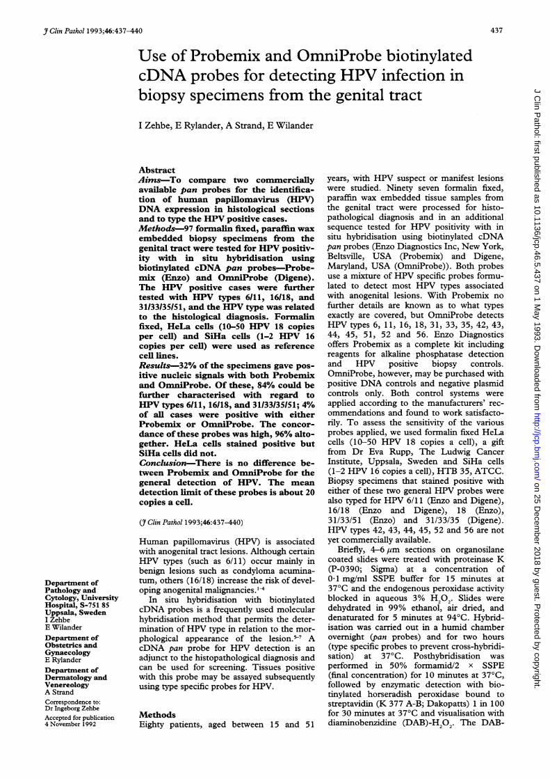
natum and HPV 18 in an area with flatcondyloma (case 3) (figs 1 and 2). The twoother positive HPV 18 biopsy specimens wereseen in an SIL II and SIL III lesion without
* _ ; JF .*. |>*f koilocytosis. Most of the biopsy specimens,v v saltogether 11, stained positive with
31/33/35/51. One of those turned out to bedouble positive with HPV types 6/11 inepithelium showing condyloma acuminatumand with HPV types 31/33/35/51 in epitheli-
.-o4.ts um with flat condyloma (case 13). The otherswere diagnosed as follows: flat condyloma (n
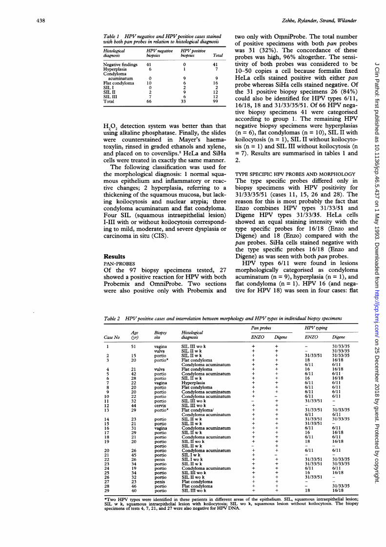
0W* * c-t = 1), SIL I without koilocytosis (n = 1) (fig
3), SIL II with koilocytosis (n = 4), SIL IIwithout koilocytosis (n = 1) and SIL III with-out koilocytosis (n = 3). Biopsy specimenswith negative findings such as normal mucosaand inflammatory changes were all HPV neg-
tna acuminatum positive for HPV 6/11 ative.All controls used were satisfactory. Low
risk HPV types 6/11 were more often found inepithelium with condyloma acuminatum thanin squamous intraepithelial lesions of varying
4,<-̂L fs severity. Flat condylomas showed mainly pos-itive signals of either high risk HPV types16/18 or medium risk HPV types 31/33/35/51. The fact that some specimens werereactive with the pan probes but negative withthe type specific probes, may indicate either
5- - the presence of yet uncharacterised HPVtypes or HPV types 42-45, 52, and 56 includ-ed in Digene's OmniProbe.
.0c .6t ~
I lw
.0.,
-# >..0
to*a~ -W
* ,, i-s i;2 e, . e;. ^ e.e s
. _ . f;v r
.v 4 v
, _ -^ 9
Figure 2 A different part of the epithelium representingflat condyloma positive fo18 (ENZO); detected with DAB-H20,.
9,I.
.~
atf §
, a,
Figure 3 A penile SIL I lesion without koilocytosis positive with HPV31133/35(Digene); detected with DAB-H 2*.
condyloma (n = 1), SIL II with koilc(n = 2), and SIL III without koilc(n = 1). HPV 18 was found in threeOne of these was also positive for HP)6/11 in a different area of the epitiHPV 6/11 was present in a condyloma
7-l.ss DiscussionEvaluation of methods to detect possibleHPV in biopsy specimens is frequentlyrequested as a complement to ordinary histo-pathological diagnosis. The purpose is not
)r HPV only to demonstrate the presence of HPVHPV DNA in general, but rather to obtain infor-
mation of the specific HPV types expressed.This is of variable prospective clinical impor-tance. Because a considerable number of theknown HPV types may invade the genitalmucosa, several sequential sections from eachindividual biopsy specimen are needed toanalyse all the different HPV types. For thisreason a pan probe represents a technicalimprovement. Pan probes can be applied forthe screening of biopsy specimens with regardto HPV DNA expression. Sections with apositive HPV DNA reaction are tested withdifferent type specific cDNA probes in a sec-ond sequence, while in negative specimens nofurther hybridisation tests are necessary, thusreducing workload and saving costs. Thisapplied to 68% of the biopsy specimens inour study.
Probemix and OmniProbe are of the samediagnostic value for the general detection ofHPV. The sensitivity of the pan probes isabout 10-50 copies a cell as formalin fixedHeLa cells showed positive signals in the
lcytosis present study. Furthermore, HeLa cells had)cytosis the same staining intensity with the pan ascases. well as the HPV type specific 18 probe. This
V types indicates that specimens that are negativeielium. with the pan probes are also unreactive withacumi- the type specific probes.
439
0
4
4k
,t
I
"
Z"
I 4
on 25 Decem
ber 2018 by guest. Protected by copyright.
http://jcp.bmj.com
/J C
lin Pathol: first published as 10.1136/jcp.46.5.437 on 1 M
ay 1993. Dow
nloaded from
Zehbe, Rylander, Strand, Wilander
A relatively high proportion of the squa-mous intraepithelial lesions expressed HPVtypes 31/33/35/51 in the present study. Thisdiscrepancy with respect to previous pub-lished findings may be explained by the factthat these HPV types have not been studiedas systematically as HPV types 16/18.A considerable number of our specimens
were HPV negative, despite the fact that theclinician suspected HPV positive lesionsfrom the presence of distinctly outlinedacetowhite areas of the mucosa. It was recent-ly suggested that acetowhite patches maysometimes be associated with other viral,bacterial, or fungal infections rather thanHPV alone.9
In situ hybridisation does not seem toindicate low intensity HPV infection (lessthan 20 copies a cell) and for that reason,tissues harbouring only a few HPV DNAcopies will not be within the detection limit.Among these may be latent infections as wellas advanced squamous intraepithelial lesionsand invasive cancer.410 More sensitivemethods for HPV identification have beenpresented, however, which, like in situhybridisation, allow tissue morphology to bepresent for histopathological diagnosis.Among these new methods, the in situ poly-merase chain reaction (PCR) appearspromising. 11-2 Using this method, 1-2 copiesof HPV DNA a cell can be recognised andthe genital changes and the role of HPV intheir pathogenesis examined further.
This study was supported by grants from the Swedish CancerFoundation (CaF), the Lion's Cancer Foundation, andSelander's Foundation.
1 Broker TR, Botchan MT. Papillomaviruses-retrospec-tives and prospectives. In: Botchan MT, Grodzicker T,Sharp P, eds. Cancer cells New York: Cold SpringHarbor Laboratory, 1986:17-36.
2 zur Hausen H, Schneider A. The role of papillomavirusesin human anogenital cancer. The Papovaviridae. In:Salzman NP, Howley P, eds. The papillomaviruses. NewYork: Plenum Press, 1987:245-63.
3 Reid R. The biology and significance of human papillo-mavirus infections in the genital tract. Yale J Biol Med1988;61:307.
4 Schneider A, Meinhardt G, Kirchmayr R, Schneider V.Prevalence of human papillomavirus genomes in tissuesfrom the lower genital tract as detected by molecular insitu hybridization. Int J' Gynecol Pathol 1991; lO: 1.
5 Warford A, In situ hybridisation: a new tool in pathology.Med Lab Sci 1988;45:381.
6 Nuovo GJ, Friedman D, Richart RM. In situ hybridiza-tion analysis of human papillomavirus DNA segregationpatterns in lesions of the female genital tract. GynecolOncol 1990;36:256.
7 Unger ER, Hammer ML, Chenggis ML. Comparison of 35Sand biotin as labels for in situ hybridization: Use of anHPV model system. J Histochem Cytochem 1991;39:145.
8 Zehbe I, Rylander E, Strand A, Wilander E. In situhybridization for the detection of human papillomavirus(HPV) in gynaecological biopsies. A study of two com-mercial kits. Anticancer Res 1992;12:1383.
9 Wikstom A, Hedblad M-A, Johansson B, et al. The aceticacid test in evaluation of subclinical genital papilloma-virus infection: A comparative study on penoscopy,histopathology, virology and scanning electron micro-scopy findings. Genitouin Med 1992;68:90.
10 Nuovo GJ. A comparison of slot blot, Southern blot andin situ hybridisation analysis for human papillomavirusDNA in genital tract lesions. Obstet Gynecol 1989;74:74.
11 Nuovo GJ, Gallery F, MacConnell P, Becker J, Bloch W.An improved technique for the in situ detection ofDNAafter polymerase chain reaction amplification. Am JPathol 1991;139:1239.
12 Nuovo GJ, Margiotta M, MacConnell P, Becker J. Rapidin situ detection of PCR-amplified HIV-1 DNA. DiagnMol Pathost (in press).
440
on 25 Decem
ber 2018 by guest. Protected by copyright.
http://jcp.bmj.com
/J C
lin Pathol: first published as 10.1136/jcp.46.5.437 on 1 M
ay 1993. Dow
nloaded from