Embed Size (px)
Citation preview
Research ArticleBiopsy of Different Oral Soft Tissues Lesions by KTP andDiode Laser: Histological Evaluation
Umberto Romeo,1 Claudia Russo,1 Gaspare Palaia,1 Rossella Lo Giudice,1
Alessandro Del Vecchio,1 Paolo Visca,2 Guido Migliau,1 and Alberto De Biase1
1 Department of Oral and Maxillofacial Sciences, “Sapienza” University of Rome, Via Caserta 6, 00161 Rome, Italy2 Department of Cytology and Cellular Diagnostics, Regina Elena Institute, Via Elio Chianesi 53, 00144 Rome, Italy
Correspondence should be addressed to Gaspare Palaia; [email protected]
Received 29 July 2014; Accepted 9 September 2014; Published 27 October 2014
Academic Editor: Samir Nammour
Copyright © 2014 Umberto Romeo et al. This is an open access article distributed under the Creative Commons AttributionLicense, which permits unrestricted use, distribution, and reproduction in any medium, provided the original work is properlycited.
Introduction. Oral biopsy aims to obtain clear and safe diagnosis; it can be performed by scalpel or laser.The controversy in this latterapplication is the thermal alteration due to tissue heating.The aim of this study is the histological evaluation of margins of “in vivo”biopsies collected by diode and KTP lasers.Material andMethods. 17 oral benign lesions biopsies weremade by diode 808 nm (SOL,DenMatItalia, Italy) and KTP 532 nm (SmartLite, DEKA, Italy). Samples were observed at OMLEICADM2000; margin alterationswere evaluated through Leica Application Suite 3.4. Results. Epithelial and connective damages were assessed for each pathologywith an average of 0.245mm and a standard deviation of ±0.162mm in mucoceles, 0.382mm ± 0.149mm in fibromas, 0.336mm ±0.106mm in hyperkeratosis, 0.473mm ± 0.105mm in squamous hyperplasia, 0.182mm in giant cell granuloma, and 0.149mm inmelanotic macula. Discussion. The histologic aspect of lesions influenced the response to laser, whereas the greater inflammationand cellularity were linked with the higher thermal signs. Many artifacts were also associated to histologic procedures. Conclusion.Both tested lasers permitted sure histologic diagnosis. However, it is suggested to enlarge biopsies of about 0.5mm, to avoid thermalalterations, especially in inflammatory lesions like oral lichen planus.
1. Introduction
A biopsy is a diagnostic procedure which consists in takinga tissue fragment to subject it to a histological examinationand, therefore, to obtain a diagnosis of certainty that can orcannot confirm the suspicion clinical diagnostic [1].
Biopsies can be classified according to the used material,the clinician timing, the lesion site, and the used techniquethat can be distinguished in incisional and excisional biopsies.The incisional biopsy involves the removal of a representativeportion of the lesion and a portion of healthy tissue adjacentto it [2–4]; while the excisional biopsy consists in the removalof the whole lesion allowing, at the same time, carrying outboth a diagnostic and therapeutic procedure [4, 5].
The biopsy is generally indicated for the following:
(i) recognizing neoplastic, preneoplastic, and other softtissue diseases;
(ii) identifying the origin of ulcers that do not heal withintwo weeks;
(iii) defining the nature of lesions that do not regress aftertherapy;
(iv) removing lesions of the right dimensions and verify-ing their nature.
Nowadays it is possible to performoral biopsies using twodifferent tools, the scalpel and the laser.
The scalpel allows obtaining a tissue fragment character-ized by the presence of well-defined peri-incisional marginswith no structural alterations. However, this surgery alwaysrequires anesthesia and sutures, and the operative field is notbloodless.
The laser devices most commonly used in oral softtissues surgery are the diode (600–980 nm), the potassiumtitanyl phosphate (KTP, 532 nm), the carbon dioxide laser(CO2, 10600 nm), the neodymium-doped yttrium aluminum
Hindawi Publishing Corporatione Scientific World JournalVolume 2014, Article ID 761704, 6 pageshttp://dx.doi.org/10.1155/2014/761704
2 The Scientific World Journal
garnet (Nd:YAG, 1064 nm), and the erbium-doped yttriumaluminum garnet (Er:YAG 2940 nm).
Lasers, used for biopsies execution, have several advan-tages than the scalpel. In fact, they consent to obtain a goodhemostasis, bloodless field and a faster healing, above allduring the initial phases [6].
However, due to the thermal effects of the laser, incisionalmargins of tissue samples can be altered, creating doubtsabout the effectiveness of this method in the diagnosisof systemic disease [7]. If the use of this tool has manyadvantages over the cold blade, the risk of jeopardizing theoutcome of histological analysis, due to laser thermal effectson peri-incisional area, still raises doubts. Actual scientificliterature does not reveal in vivo studies concerning theevaluation of peri-incisional biopsy taken with the laser.Pathological tissues “in vivo,” compared to those “ex vivo,”are characterized by a higher concentration of liquid, lowercell cohesion, and normal or pathological amounts of blood(e.g., in inflammatory or autoimmune diseases).
This consideration could lead to an improvement incutting ability of the laser that could permit the parametersapplied to be reduced with less damage to cut margins buton the other hand to higher local heat buildup with largerthermal artefacts.
The aim of this “in vivo” study is to analyze the tissuefragments removed by laser surgery, to assess the epithelialand connective tissue damage caused by its thermal effects.
2. Materials and MethodsSeventeen patients (8F/9M), affected by oral benign patholo-gies, have been subjected to oral excisional biopsy. In somecases, lesions have been treated using an incisional biopsybecause of their site or their size. All tissue samples have beenremoved by the same operator in order to execute a properbiopsy thanks to his experience and knowledge in laser toolsand biological tissue characteristics.
Biopsies have been performed using two different wave-lengths with the following parameters: diode laser 808 nm(SOL, DenMatItalia, Italy), power: 2W in CW, fluence:2400 J/cm2, fiber spot: 320 𝜇m; KTP laser 532 nm (SmartLite,DEKA, Italy), power: 1.5W in PW, fluence: 212 J/cm2, fiberspot: 300 𝜇m. Parameters have been selected considering theright execution of the surgical intervention and the patientcompliance never exceeding 5 minutes.
Local anesthesia with 1.8mL of mepivacaine solution(Mepivacaina Pierrel, 30mg/mL, injection solution 1.8mL,Pierrel Spa, Milan, Italy) without vasoconstrictor was per-formed around the area of the lesion before the beginning ofeach surgical intervention, injecting the solution at a distanceof 0,5 cm from lesions margins. The excised lesions size wasbetween 0,5 and 1 cm of diameter. Twomucoceles were takenout by diode laser and one by KTP laser; 5 fibromas wereexcised by diode; 3 hyperkeratosis lesions were removed bydiode and 1 by KTP; the 3 oral lichen planus, the melanoticmacula, and the oral giant cell granuloma were removedby diode laser. After surgery, the samples were sent to thepathologist, for the histological evaluation and diagnosis, ina single-blind mode. No suture or medication was applied
and the wound was left to heal by secondary intention. Allbiopsy samples were fixed in a 10% neutral-buffered formalinsolution, embedded in paraffin, sectioned, and stained withhematoxylin-eosin for conventional histopathological evalu-ation.
Tissue fragments were again observed through the useof an optical microscope LEICA DM 2000, 5x and 10xmagnification, and thanks to an appropriate software (LeicaApplication Suite version 3.4) quantitatively and qualitativelymarginal alterations, due to the thermal action of lasers,have been evaluated. Quantitative evaluation carried outa measurement in millimeters and statistical analysis wascarried out by calculating the arithmetic mean and standarddeviation (a measure of the dispersion of data around theexpected value) of the different values, while the involvementof epithelial and connective tissue in thermal alterations hasbeen evaluated in the qualitative aspect. In every oral pathol-ogy, connective and epithelial damage have been evaluated interms of charring and coarctation, since in many cases it wasimpossible to evaluate them separately.
3. ResultsFollow-up at 7 and 21 days showed a complete recovery of thewound, without any complications or pain.
The presence of peripheral alterations has not influencedhistological analysis: for all samples, it was possible to obtaina certainty diagnosis. Histological examination showed threemucoceles, five fibromas, four hyperkeratotic lesions, threeoral lichen planus, one giant cells granuloma, and one mela-notic macula.
Peripheral damage has been individually evaluated foreach disease, considering that the morphological and struc-tural characteristics of the various lesions could stronglyinfluence the tissues response to the laser action.
Graphs have been realized to show the trend of the mea-sures, and statistical analysis has been carried out calculatingthe mean and standard deviation of the different values.Moreover, in each histological group the same parametershave been evaluated.
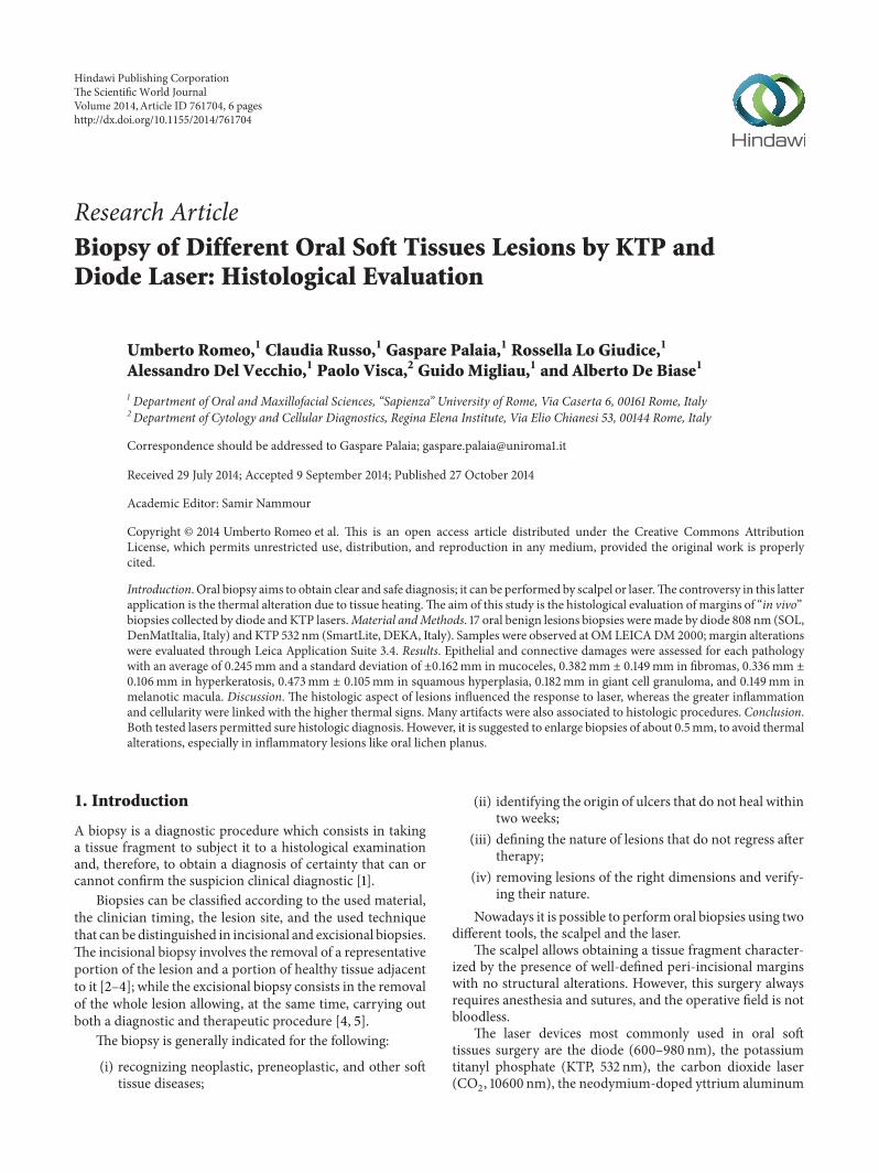
The histological evaluation of peri-incisional margins inwhich themicroscopic analysis was compatible with the diag-nosis ofmucocele showed a damage average of 0.245mmwitha standard deviation of ±0.162mm (Figures 1 and 2; Table 1).Only in one case the epithelium was not visible because thedamage was exclusively assessed to the connective tissue.
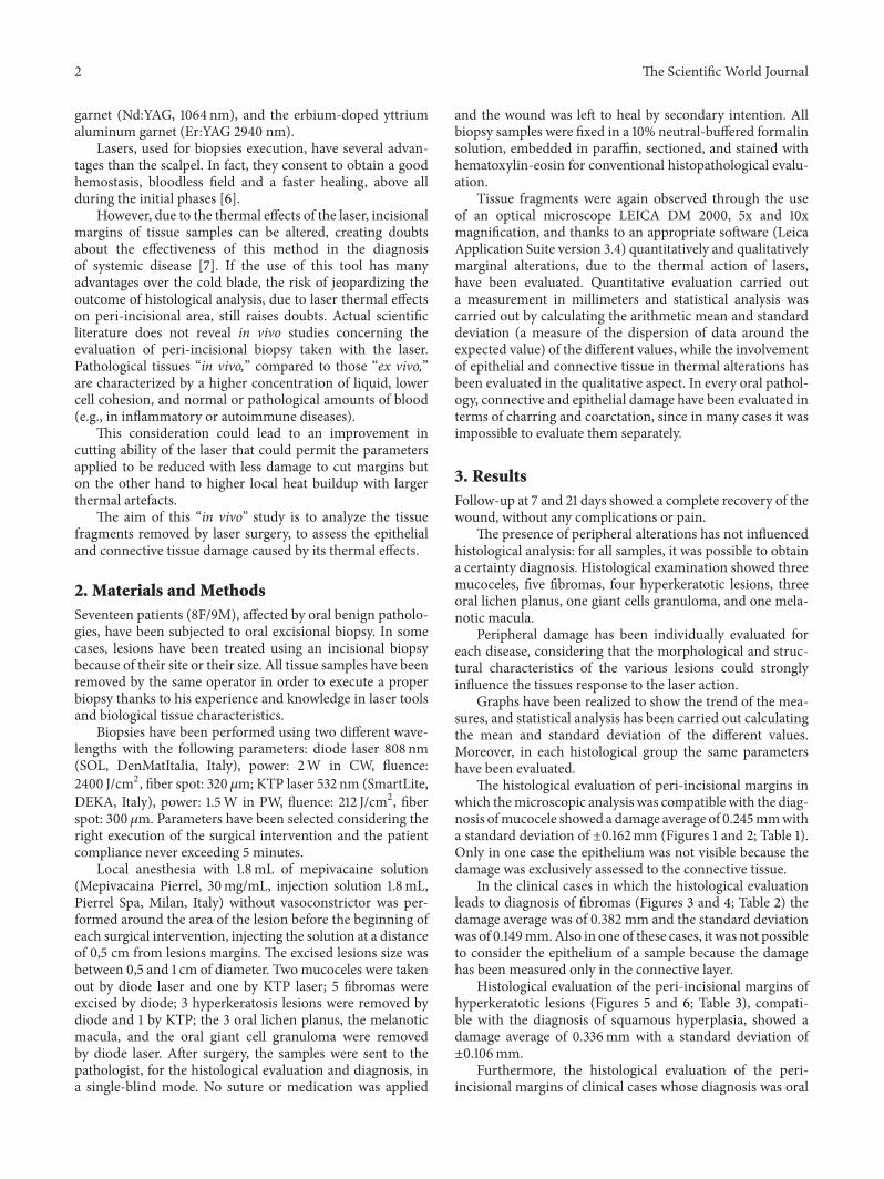
In the clinical cases in which the histological evaluationleads to diagnosis of fibromas (Figures 3 and 4; Table 2) thedamage average was of 0.382 mm and the standard deviationwas of 0.149mm.Also in one of these cases, it was not possibleto consider the epithelium of a sample because the damagehas been measured only in the connective layer.
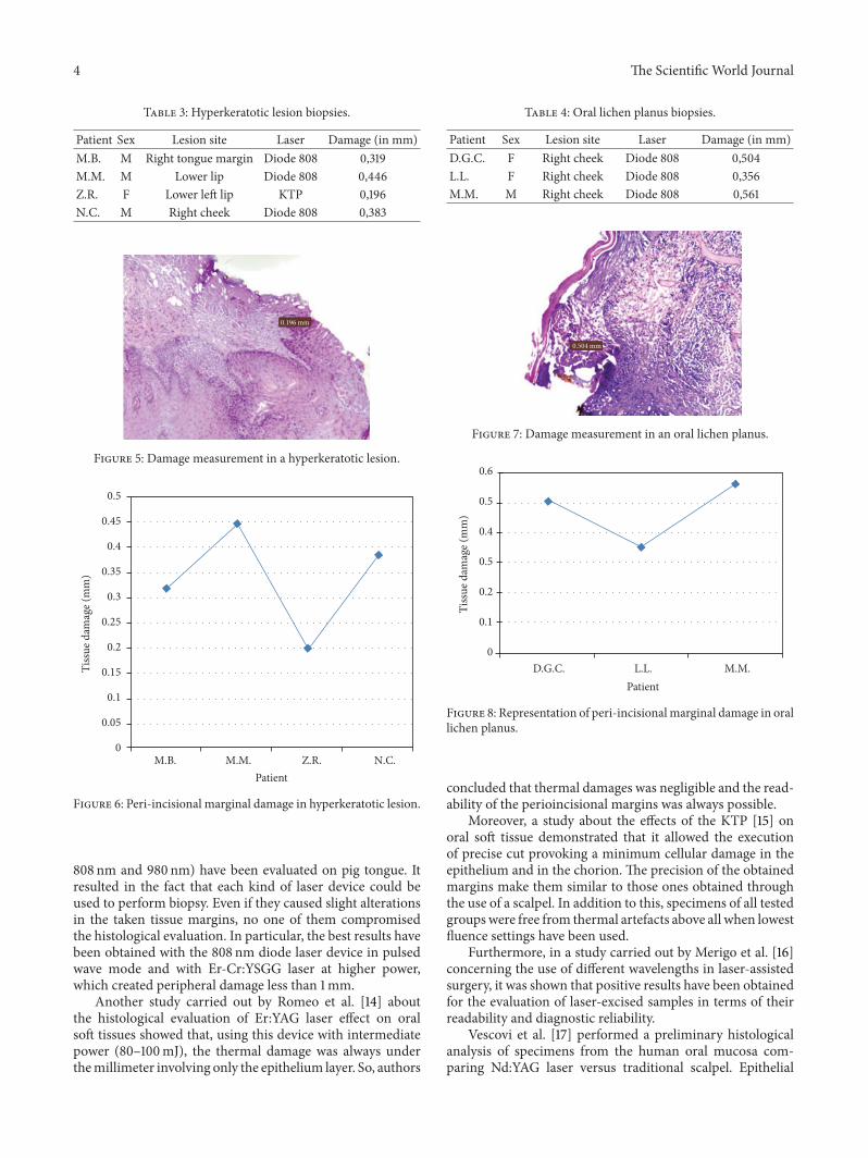
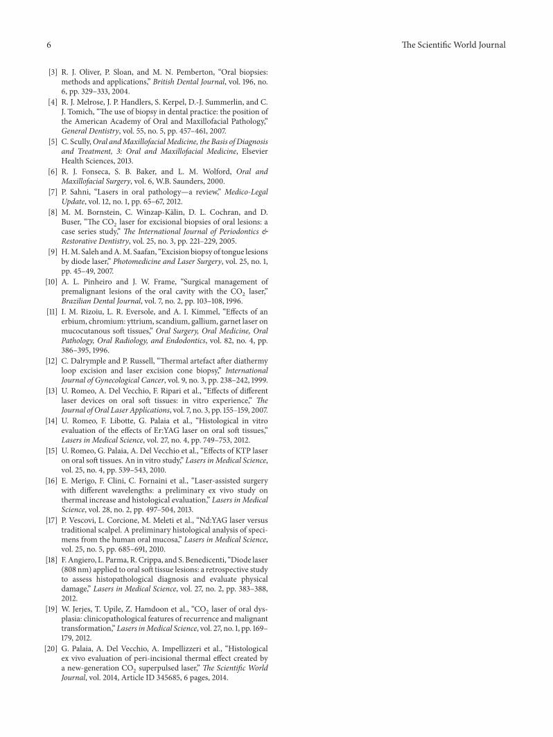
Histological evaluation of the peri-incisional margins ofhyperkeratotic lesions (Figures 5 and 6; Table 3), compati-ble with the diagnosis of squamous hyperplasia, showed adamage average of 0.336mm with a standard deviation of±0.106mm.
Furthermore, the histological evaluation of the peri-incisional margins of clinical cases whose diagnosis was oral
The Scientific World Journal 3
Table 1: Mucocele biopsies.
Patient Sex Lesion site Laser Damage (in mm)D.C. F Inferior lip Diode 808 0,442N.F. F Upper left lip KTP 0,213S.S. M Inferior lip Diode 808 0,102
0.422 mm
Figure 1: Damage measurement in a mucocele.
0.450.4
0.350.3
0.250.2
0.150.1
0.05
D.C. N.F. S.S.0
Patient
Tiss
ue d
amag
e (m
m)
Figure 2: Peri-incisional marginal damage in mucocele.
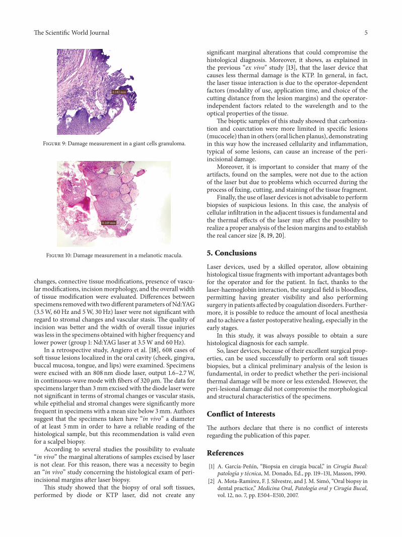
lichen planus demonstrated a damage average of 0.473mmwith a standard deviation of ±0.105mm (Figures 7 and 8;Table 4).
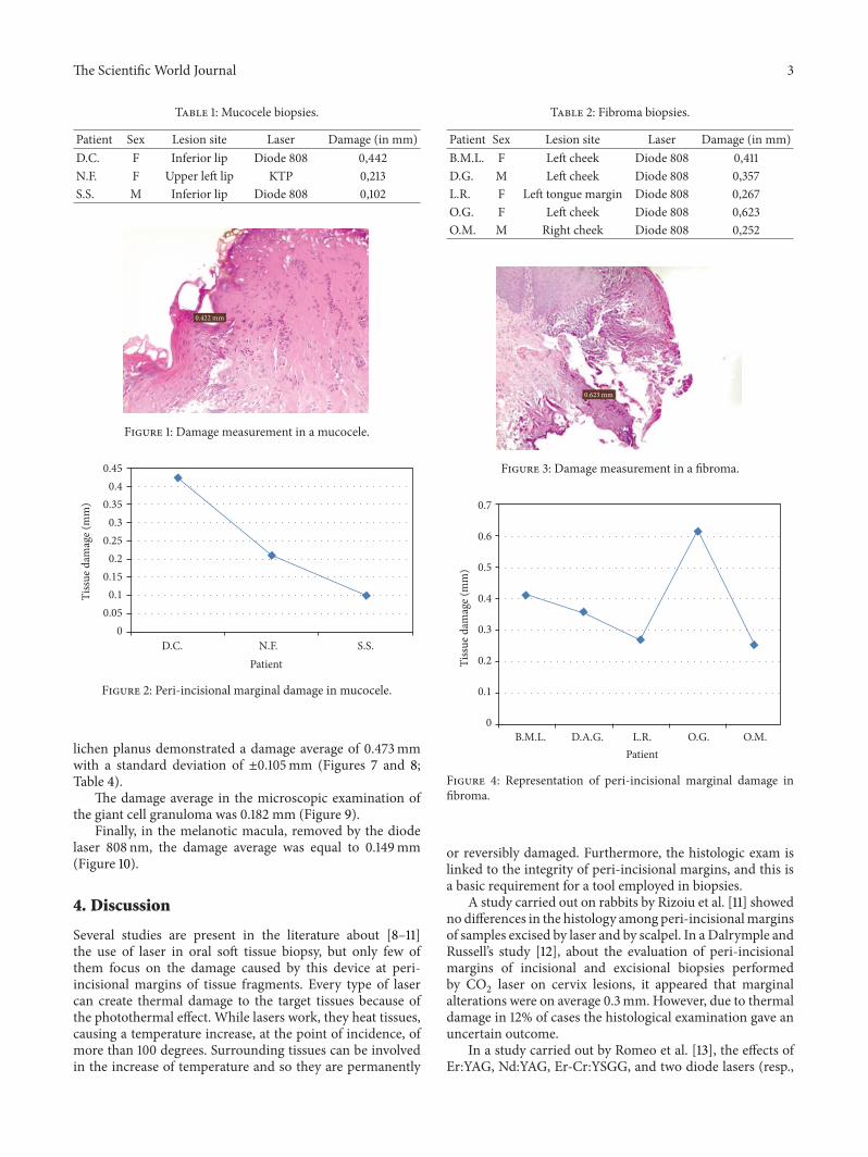
The damage average in the microscopic examination ofthe giant cell granuloma was 0.182 mm (Figure 9).
Finally, in the melanotic macula, removed by the diodelaser 808 nm, the damage average was equal to 0.149mm(Figure 10).
4. Discussion
Several studies are present in the literature about [8–11]the use of laser in oral soft tissue biopsy, but only few ofthem focus on the damage caused by this device at peri-incisional margins of tissue fragments. Every type of lasercan create thermal damage to the target tissues because ofthe photothermal effect. While lasers work, they heat tissues,causing a temperature increase, at the point of incidence, ofmore than 100 degrees. Surrounding tissues can be involvedin the increase of temperature and so they are permanently
Table 2: Fibroma biopsies.
Patient Sex Lesion site Laser Damage (in mm)B.M.L. F Left cheek Diode 808 0,411D.G. M Left cheek Diode 808 0,357L.R. F Left tongue margin Diode 808 0,267O.G. F Left cheek Diode 808 0,623O.M. M Right cheek Diode 808 0,252
0.623 mm
Figure 3: Damage measurement in a fibroma.
0.5
0.4
0.3
0.2
0.1
0
0.6
0.7
O.M.O.G.L.R.D.A.G.B.M.L.
Tiss
ue d
amag
e (m
m)
Patient
Figure 4: Representation of peri-incisional marginal damage infibroma.
or reversibly damaged. Furthermore, the histologic exam islinked to the integrity of peri-incisional margins, and this isa basic requirement for a tool employed in biopsies.
A study carried out on rabbits by Rizoiu et al. [11] showedno differences in the histology among peri-incisionalmarginsof samples excised by laser and by scalpel. In a Dalrymple andRussell’s study [12], about the evaluation of peri-incisionalmargins of incisional and excisional biopsies performedby CO
2laser on cervix lesions, it appeared that marginal
alterations were on average 0.3mm. However, due to thermaldamage in 12% of cases the histological examination gave anuncertain outcome.
In a study carried out by Romeo et al. [13], the effects ofEr:YAG, Nd:YAG, Er-Cr:YSGG, and two diode lasers (resp.,
4 The Scientific World Journal
Table 3: Hyperkeratotic lesion biopsies.
Patient Sex Lesion site Laser Damage (in mm)M.B. M Right tongue margin Diode 808 0,319M.M. M Lower lip Diode 808 0,446Z.R. F Lower left lip KTP 0,196N.C. M Right cheek Diode 808 0,383
0.196 mm
Figure 5: Damage measurement in a hyperkeratotic lesion.
0.45
0.5
0.4
0.35
0.3
0.25
0.2
0.15
0.1
0.05
M.B. M.M. Z.R. N.C.0
Tiss
ue d
amag
e (m
m)
Patient
Figure 6: Peri-incisional marginal damage in hyperkeratotic lesion.
808 nm and 980 nm) have been evaluated on pig tongue. Itresulted in the fact that each kind of laser device could beused to perform biopsy. Even if they caused slight alterationsin the taken tissue margins, no one of them compromisedthe histological evaluation. In particular, the best results havebeen obtained with the 808 nm diode laser device in pulsedwave mode and with Er-Cr:YSGG laser at higher power,which created peripheral damage less than 1mm.
Another study carried out by Romeo et al. [14] aboutthe histological evaluation of Er:YAG laser effect on oralsoft tissues showed that, using this device with intermediatepower (80–100mJ), the thermal damage was always underthemillimeter involving only the epithelium layer. So, authors
Table 4: Oral lichen planus biopsies.
Patient Sex Lesion site Laser Damage (in mm)D.G.C. F Right cheek Diode 808 0,504L.L. F Right cheek Diode 808 0,356M.M. M Right cheek Diode 808 0,561
0.504 mm
Figure 7: Damage measurement in an oral lichen planus.
0.6
0.5
0.4
0.5
0.2
0.1
0D.G.C. L.L. M.M.
Tiss
ue d
amag
e (m
m)
Patient
Figure 8: Representation of peri-incisionalmarginal damage in orallichen planus.
concluded that thermal damages was negligible and the read-ability of the perioincisional margins was always possible.
Moreover, a study about the effects of the KTP [15] onoral soft tissue demonstrated that it allowed the executionof precise cut provoking a minimum cellular damage in theepithelium and in the chorion. The precision of the obtainedmargins make them similar to those ones obtained throughthe use of a scalpel. In addition to this, specimens of all testedgroupswere free from thermal artefacts above all when lowestfluence settings have been used.
Furthermore, in a study carried out by Merigo et al. [16]concerning the use of different wavelengths in laser-assistedsurgery, it was shown that positive results have been obtainedfor the evaluation of laser-excised samples in terms of theirreadability and diagnostic reliability.
Vescovi et al. [17] performed a preliminary histologicalanalysis of specimens from the human oral mucosa com-paring Nd:YAG laser versus traditional scalpel. Epithelial
The Scientific World Journal 5
0.182 mm
Figure 9: Damage measurement in a giant cells granuloma.
0.149 mm
Figure 10: Damage measurement in a melanotic macula.
changes, connective tissue modifications, presence of vascu-larmodifications, incisionmorphology, and the overall widthof tissue modification were evaluated. Differences betweenspecimens removedwith two different parameters ofNd:YAG(3.5W, 60Hz and 5W, 30Hz) laser were not significant withregard to stromal changes and vascular stasis. The quality ofincision was better and the width of overall tissue injurieswas less in the specimens obtainedwith higher frequency andlower power (group 1: Nd:YAG laser at 3.5W and 60Hz).
In a retrospective study, Angiero et al. [18], 608 cases ofsoft tissue lesions localized in the oral cavity (cheek, gingiva,buccal mucosa, tongue, and lips) were examined. Specimenswere excised with an 808 nm diode laser, output 1.6–2.7W,in continuous-wave mode with fibers of 320𝜇m.The data forspecimens larger than 3mm excised with the diode laser werenot significant in terms of stromal changes or vascular stasis,while epithelial and stromal changes were significantly morefrequent in specimens with a mean size below 3mm. Authorssuggest that the specimens taken have “in vivo” a diameterof at least 5mm in order to have a reliable reading of thehistological sample, but this recommendation is valid evenfor a scalpel biopsy.
According to several studies the possibility to evaluate“in vivo” the marginal alterations of samples excised by laseris not clear. For this reason, there was a necessity to beginan “in vivo” study concerning the histological exam of peri-incisional margins after laser biopsy.
This study showed that the biopsy of oral soft tissues,performed by diode or KTP laser, did not create any
significant marginal alterations that could compromise thehistological diagnosis. Moreover, it shows, as explained inthe previous “ex vivo” study [13], that the laser device thatcauses less thermal damage is the KTP. In general, in fact,the laser tissue interaction is due to the operator-dependentfactors (modality of use, application time, and choice of thecutting distance from the lesion margins) and the operator-independent factors related to the wavelength and to theoptical properties of the tissue.
The bioptic samples of this study showed that carboniza-tion and coarctation were more limited in specific lesions(mucocele) than in others (oral lichen planus), demonstratingin this way how the increased cellularity and inflammation,typical of some lesions, can cause an increase of the peri-incisional damage.
Moreover, it is important to consider that many of theartifacts, found on the samples, were not due to the actionof the laser but due to problems which occurred during theprocess of fixing, cutting, and staining of the tissue fragment.
Finally, the use of laser devices is not advisable to performbiopsies of suspicious lesions. In this case, the analysis ofcellular infiltration in the adjacent tissues is fundamental andthe thermal effects of the laser may affect the possibility torealize a proper analysis of the lesionmargins and to establishthe real cancer size [8, 19, 20].
5. Conclusions
Laser devices, used by a skilled operator, allow obtaininghistological tissue fragments with important advantages bothfor the operator and for the patient. In fact, thanks to thelaser-haemoglobin interaction, the surgical field is bloodless,permitting having greater visibility and also performingsurgery in patients affected by coagulation disorders. Further-more, it is possible to reduce the amount of local anesthesiaand to achieve a faster postoperative healing, especially in theearly stages.
In this study, it was always possible to obtain a surehistological diagnosis for each sample.
So, laser devices, because of their excellent surgical prop-erties, can be used successfully to perform oral soft tissuesbiopsies, but a clinical preliminary analysis of the lesion isfundamental, in order to predict whether the peri-incisionalthermal damage will be more or less extended. However, theperi-lesional damage did not compromise the morphologicaland structural characteristics of the specimens.
Conflict of Interests
The authors declare that there is no conflict of interestsregarding the publication of this paper.
References
[1] A. Garcıa-Penın, “Biopsia en cirugıa bucal,” in Cirugıa Bucal:patologıa y tecnica, M. Donado, Ed., pp. 119–131, Masson, 1990.
[2] A. Mota-Ramırez, F. J. Silvestre, and J. M. Simo, “Oral biopsy indental practice,” Medicina Oral, Patologıa oral y Cirugıa Bucal,vol. 12, no. 7, pp. E504–E510, 2007.
6 The Scientific World Journal
[3] R. J. Oliver, P. Sloan, and M. N. Pemberton, “Oral biopsies:methods and applications,” British Dental Journal, vol. 196, no.6, pp. 329–333, 2004.
[4] R. J. Melrose, J. P. Handlers, S. Kerpel, D.-J. Summerlin, and C.J. Tomich, “The use of biopsy in dental practice: the position ofthe American Academy of Oral and Maxillofacial Pathology,”General Dentistry, vol. 55, no. 5, pp. 457–461, 2007.
[5] C. Scully,Oral andMaxillofacialMedicine, the Basis of Diagnosisand Treatment, 3: Oral and Maxillofacial Medicine, ElsevierHealth Sciences, 2013.
[6] R. J. Fonseca, S. B. Baker, and L. M. Wolford, Oral andMaxillofacial Surgery, vol. 6, W.B. Saunders, 2000.
[7] P. Sahni, “Lasers in oral pathology—a review,” Medico-LegalUpdate, vol. 12, no. 1, pp. 65–67, 2012.
[8] M. M. Bornstein, C. Winzap-Kalin, D. L. Cochran, and D.Buser, “The CO
2laser for excisional biopsies of oral lesions: a
case series study,” The International Journal of Periodontics &Restorative Dentistry, vol. 25, no. 3, pp. 221–229, 2005.
[9] H.M. Saleh andA.M. Saafan, “Excision biopsy of tongue lesionsby diode laser,” Photomedicine and Laser Surgery, vol. 25, no. 1,pp. 45–49, 2007.
[10] A. L. Pinheiro and J. W. Frame, “Surgical management ofpremalignant lesions of the oral cavity with the CO
2laser,”
Brazilian Dental Journal, vol. 7, no. 2, pp. 103–108, 1996.[11] I. M. Rizoiu, L. R. Eversole, and A. I. Kimmel, “Effects of an
erbium, chromium: yttrium, scandium, gallium, garnet laser onmucocutanous soft tissues,” Oral Surgery, Oral Medicine, OralPathology, Oral Radiology, and Endodontics, vol. 82, no. 4, pp.386–395, 1996.
[12] C. Dalrymple and P. Russell, “Thermal artefact after diathermyloop excision and laser excision cone biopsy,” InternationalJournal of Gynecological Cancer, vol. 9, no. 3, pp. 238–242, 1999.
[13] U. Romeo, A. Del Vecchio, F. Ripari et al., “Effects of differentlaser devices on oral soft tissues: in vitro experience,” TheJournal of Oral Laser Applications, vol. 7, no. 3, pp. 155–159, 2007.
[14] U. Romeo, F. Libotte, G. Palaia et al., “Histological in vitroevaluation of the effects of Er:YAG laser on oral soft tissues,”Lasers in Medical Science, vol. 27, no. 4, pp. 749–753, 2012.
[15] U. Romeo, G. Palaia, A. Del Vecchio et al., “Effects of KTP laseron oral soft tissues. An in vitro study,” Lasers inMedical Science,vol. 25, no. 4, pp. 539–543, 2010.
[16] E. Merigo, F. Clini, C. Fornaini et al., “Laser-assisted surgerywith different wavelengths: a preliminary ex vivo study onthermal increase and histological evaluation,” Lasers in MedicalScience, vol. 28, no. 2, pp. 497–504, 2013.
[17] P. Vescovi, L. Corcione, M. Meleti et al., “Nd:YAG laser versustraditional scalpel. A preliminary histological analysis of speci-mens from the human oral mucosa,” Lasers in Medical Science,vol. 25, no. 5, pp. 685–691, 2010.
[18] F.Angiero, L. Parma, R.Crippa, and S. Benedicenti, “Diode laser(808 nm) applied to oral soft tissue lesions: a retrospective studyto assess histopathological diagnosis and evaluate physicaldamage,” Lasers in Medical Science, vol. 27, no. 2, pp. 383–388,2012.
[19] W. Jerjes, T. Upile, Z. Hamdoon et al., “CO2laser of oral dys-
plasia: clinicopathological features of recurrence andmalignanttransformation,” Lasers inMedical Science, vol. 27, no. 1, pp. 169–179, 2012.
[20] G. Palaia, A. Del Vecchio, A. Impellizzeri et al., “Histologicalex vivo evaluation of peri-incisional thermal effect created bya new-generation CO
2superpulsed laser,” The Scientific World
Journal, vol. 2014, Article ID 345685, 6 pages, 2014.

Submit your manuscripts athttp://www.hindawi.com
Hindawi Publishing Corporationhttp://www.hindawi.com Volume 2014
Oral OncologyJournal of
DentistryInternational Journal of
Hindawi Publishing Corporationhttp://www.hindawi.com Volume 2014
Hindawi Publishing Corporationhttp://www.hindawi.com Volume 2014
International Journal of
Biomaterials
Hindawi Publishing Corporationhttp://www.hindawi.com Volume 2014
BioMed Research International
Hindawi Publishing Corporationhttp://www.hindawi.com Volume 2014
Case Reports in Dentistry
Hindawi Publishing Corporationhttp://www.hindawi.com Volume 2014
Oral ImplantsJournal of
Hindawi Publishing Corporationhttp://www.hindawi.com Volume 2014
Anesthesiology Research and Practice
Hindawi Publishing Corporationhttp://www.hindawi.com Volume 2014
Radiology Research and Practice
Environmental and Public Health
Journal of
Hindawi Publishing Corporationhttp://www.hindawi.com Volume 2014
The Scientific World JournalHindawi Publishing Corporation http://www.hindawi.com Volume 2014
Hindawi Publishing Corporationhttp://www.hindawi.com Volume 2014
Dental SurgeryJournal of
Drug DeliveryJournal of
Hindawi Publishing Corporationhttp://www.hindawi.com Volume 2014
Hindawi Publishing Corporationhttp://www.hindawi.com Volume 2014
Oral DiseasesJournal of
Hindawi Publishing Corporationhttp://www.hindawi.com Volume 2014
Computational and Mathematical Methods in Medicine
ScientificaHindawi Publishing Corporationhttp://www.hindawi.com Volume 2014
PainResearch and TreatmentHindawi Publishing Corporationhttp://www.hindawi.com Volume 2014
Preventive MedicineAdvances in
Hindawi Publishing Corporationhttp://www.hindawi.com Volume 2014
EndocrinologyInternational Journal of
Hindawi Publishing Corporationhttp://www.hindawi.com Volume 2014
Hindawi Publishing Corporationhttp://www.hindawi.com Volume 2014
OrthopedicsAdvances in