Embed Size (px)
Citation preview
Bioscience Reports, Vol. 22, No. 2, April 2002 ( 2002)
OVERVIEW
Biophysical Aspects of Using Liposomes as Delivery Vehicles
Anne S. Ulrich1
Liposomes are used as biocompatible carriers of drugs, peptides, proteins, plasmic DNA,antisense oligonucleotides or ribozymes, for pharmaceutical, cosmetic, and biochemicalpurposes. The enormous versatility in particle size and in the physical parameters of thelipids affords an attractive potential for constructing tailor-made vehicles for a wide rangeof applications. Some of the recent literature will be reviewed here and presented from abiophysical point of view, thus providing a background for the more specialized articles inthis special issue on liposome technology. Different properties (size, colloidal behavior,phase transitions, and polymorphism) of diverse lipid formulations (liposomes, lipoplexes,cubic phases, emulsions, and solid lipid nanoparticles) for distinct applications (parenteral,transdermal, pulmonary, and oral administration) will be rationalized in terms of commonstructural, thermodynamic and kinetic parameters of the lipids. This general biophysicalbasis helps to understand pharmaceutically relevant aspects such as liposome stability dur-ing storage and towards serum, the biodistribution and specific targeting of cargo, and howto trigger drug release and membrane fusion. Methods for the preparation and characteriz-ation of liposomal formulations in ûitro will be outlined, too.
KEY WORDS: Biophysics; drug delivery; lipid; liposome.
ABBREVIATIONS: CHEMS, cholesterylhemisuccinate; CHOL, cholesterol; DC Choles-terol, 3β-(N-(N ′,N ′,-dimethylaminoethane)-carbimol) cholesterol; DPMC, 1,2-dimyristoyl-sn-glycero-3-phosphatidylcholine; DMRIE, 1,2-dimyristoyloxypropyl-3-dimethyl-hydroxy-ethyl ammonium; DODAc, dioctadecyldimethylammonium chloride; DOGS, diocta-decylamidoglycyl spermine; DOPE, 1,2-dioleoyl-sn-glycero-3-phosphatidylethanolamine;DOSPA, 2,3-dioleoyloxy-N-(2(sperminecarboxamide)ethyl)-N,N-dimethyl-1-propanannin-ium; DOTAP, 1,2-dioleoyloxy-3-(trimethylammonio) propane; DOTMA, N-(2,3-(dioleoyloxy)propyl-N,N,N-trimethyl ammonium; DPPS, 1,2-dipalmitoyl-sn-glycero-3-phosphatidylserine; DPSG, N-(succinyl)dipalmitoyl phosphatidylethanolamine; DSPG, 1,2-distearoyl-sn-glycero-3-phosphatidylglycerol; GMO, glycerol monoleate; N-maleyl-DOPE,N-(maleyl)dioleoyl phosphatidylethanolamine; OA, oleic acid.
LIPOSOME COMPOSITION AND SIZE
Many reviews and book chapters have focused on the application of liposomesfor drug delivery, gene therapy, and immunization. Only a few recent papers shallbe pointed out here [1–18], even if this may not pay justice to the pioneers in someareas. When using liposomes as drug carriers the main aim is to reduce toxic sideeffects in sensitive organs such as heart and kidneys and to target specific tissuessuch as tumors. By optimizing the lipid composition, liposomal size, membrane
1Friedrich-Schiller-University of Jena, Institute of Molecular Biology, Winzerlaer Str. 10, 07745 Jena,Germany. E-mail: [email protected]
129
0144-8463�02�0400-0129�0 2002 Plenum Publishing Corporation
130 Ulrich
fluidity, surface charge, and steric stabilization, it is possible to extend the thera-peutic index of liposomal carriers over that of the corresponding conventional for-mulations. Likewise, when designing novel cationic lipids for gene therapy, theirability to mediate transfection can be attributed to several physical factors, includingthe spontaneous condensation of the DNA due to electrostatic interactions, the netpositive charge of the system that may promote association with negatively chargedcell surfaces, as well as the fusogenic properties of the lipids which can destabilizethe plasma membrane or endosomal compartment. Finally, the rationale behind theuse of liposomes for immunization purposes relies on their ability to deliver an anti-gen into selected immune cells and to stimulate an immune response. Before examin-ing the pharmaceutical behavior of these different types of liposomal carriers interms of their physicochemical parameters, some general information and basicdefinitions will be summarized in the following paragraphs.
Lipid Structure and Assembly
The amphilic molecules used for the preparation of liposomes are derived fromor based on the structure of biological membranes lipids, as summarized in Fig. 1[1, 3, 10–12, 19–21]. Two hydrocarbon chains are usually esterified to a glycerolbackbone (‘‘glycerolipids’’, or ‘‘plasmalogens’’ in the case of an α-β unsaturatedether), or they constitute the hydrophobic ceramide moiety (‘‘sphingolipids’’). Thishydrophobic part is linked to a hydrophilic headgroup containing either a phosphate(‘‘phospholipids’’) or some carbohydrate units (‘‘glycolipids’’). Biologically relevantlipid headgroups are either zwitterionic [phosphatidylcholine (PC), phosphatidyle-thanolamine (PE), sphingomyelin (SM)], negatively charged [phosphatidic acid (PA),phosphatidylglycerol (PG), phosphatidylserine (PS), phosphatidylinositol (PI), car-diolipin (CL), substituted glycolipids such as monosiagloganglioside (GM1)], orentirely uncharged [unsubstituted glycolipids]. Saturated acyl chains typically varyin length from 10 carbons (lauryl), 12 (myristoyl), 14 (palmitoyl) to 16 (stearoyl),and the longer 18-carbon chains are usually unsaturated with one (oleoyl), two (lino-leyl) or three (linolenyl) cis-double bonds. Positively charged lipids are custom-mademolecules, based on the same structural principles, which are designed to condensethe DNA and to interact with oppositely charged biological membranes. Examplesof cationic amphiphiles include DOTAP, DOTMA, DODAC, DC-Chol, DMRIE,DOSPA, DOGS, amongst many others, seen in Fig. 1. Cholesterol is readily incor-porated into lipid assemblies up to 50%, whereas purely hydrophobic lipids such astriacylglycerides (TAG) do not disperse in water, unless they are surrounded by asurfactant monolayer (e.g. phospholipid, detergent) to form an emulsion or a solidlipid nanoparticle (SLN).
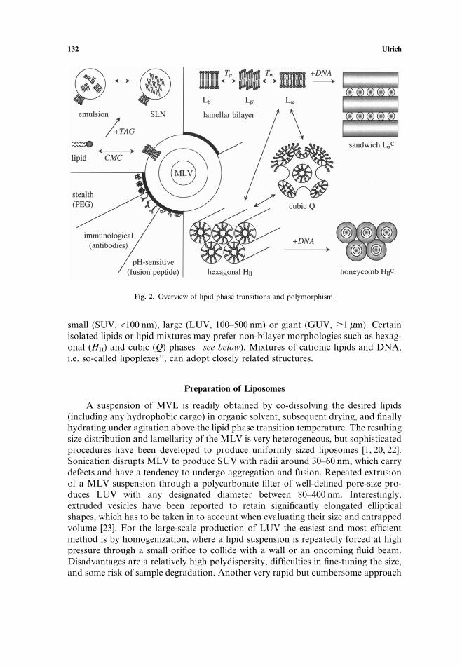
Amphiphilic lipids are poorly soluble in water as monomers, with a low criticalmicelle concentration (CMC) typically between 10−8 and 10−12 M, depending on thehydrocarbon chain length. While single-chain lipids (lysolipids, free unsaturated acylchains, detergents, etc.) spontaneously assemble into micelles, most membrane-derived lipids tend to be driven into bilayers. The resulting lamellar structures formclosed vesicles, i.e. liposomes, as illustrated in Fig. 2. One commonly distinguishesbetween multilamellar vesicles (MLV, 0.1–10 µm), and unilamellar ones that can be
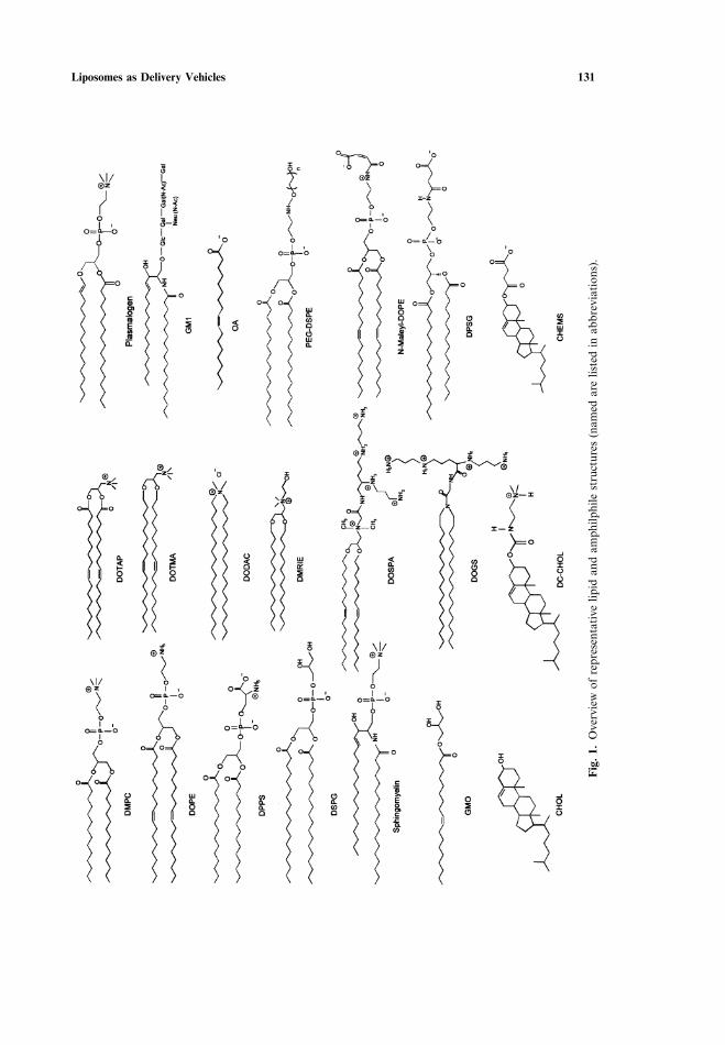
Liposomes as Delivery Vehicles 131
Fig
.1.
Ove
rvie
wof
repr
esen
tative
lipid
and
amph
ilphi
lest
ruct
ures
(nam
edar
elis
ted
inab
brev
iation
s).
132 Ulrich
Fig. 2. Overview of lipid phase transitions and polymorphism.
small (SUV, <100 nm), large (LUV, 100–500 nm) or giant (GUV, ¤1 µm). Certainisolated lipids or lipid mixtures may prefer non-bilayer morphologies such as hexag-onal (HII) and cubic (Q) phases –see below). Mixtures of cationic lipids and DNA,i.e. so-called lipoplexes’’, can adopt closely related structures.
Preparation of Liposomes
A suspension of MVL is readily obtained by co-dissolving the desired lipids(including any hydrophobic cargo) in organic solvent, subsequent drying, and finallyhydrating under agitation above the lipid phase transition temperature. The resultingsize distribution and lamellarity of the MLV is very heterogeneous, but sophisticatedprocedures have been developed to produce uniformly sized liposomes [1, 20, 22].Sonication disrupts MLV to produce SUV with radii around 30–60 nm, which carrydefects and have a tendency to undergo aggregation and fusion. Repeated extrusionof a MLV suspension through a polycarbonate filter of well-defined pore-size pro-duces LUV with any designated diameter between 80–400 nm. Interestingly,extruded vesicles have been reported to retain significantly elongated ellipticalshapes, which has to be taken in to account when evaluating their size and entrappedvolume [23]. For the large-scale production of LUV the easiest and most efficientmethod is by homogenization, where a lipid suspension is repeatedly forced at highpressure through a small orifice to collide with a wall or an oncoming fluid beam.Disadvantages are a relatively high polydispersity, difficulties in fine-tuning the size,and some risk of sample degradation. Another very rapid but cumbersome approach
Liposomes as Delivery Vehicles 133
relies on dissolving a mixture of lipid and hydrophobic carbon in ethanol or ether,which is then injected into an appropriate buffer to form a heterogeneous mixtureof SUV, LUV or MLV, depending on concentration.
Hydrophobic and amphiphilic drugs can be readily incorporated into liposomesusing the methods above [24, 25], but for water-soluble compounds there are moreefficient approaches to achieve a high yield of encapsulation [22]. For example, ves-icles can be passively charged using reverse phase evaporation (REV), by injectingan aqueous solution of the drug rapidly into an organic phase containing the lipid.The resulting ‘‘water-in-oil’’ emulsion is sonicated and partially dried down to asemi-solid gel, which is then converted by vigorous shaking into a concentratedsuspension of vesicles in water (typically 0.1 to 1.0 µm diameter, with up to 50%entrapment). Alternatively, dehydration–rehydration vesicles (DRV) can be pro-duced at large-scale by mixing an aqueous solution of the drug with a suspensionof ‘‘empty’’ LUV. Freeze-drying and rehydration induces fusion between adjacentmembranes and the solute is taken up by the resulting MLV (0.1 to 2.0 µm diameter,with up to 80% entrapment). Another, more gentle method for the passive entrap-ment or reconstitution of sensitive proteins and membrane-anchored ligands (e.g.for targeting purposes or antigen presentation) relies on dissolving the lipids andcargo have in a detergent. Subsequent dilution or detergent removal by dialysis,biobeads, or gel filtration leads to the formation of liposomes, although it is stilldifficult to control the reconstitution mechanism rationally [26]. As opposed to pass-ive loading, active loading procedures exploit differences in the partition coefficientof a drug on pH and ionic strength [27]. For example, basic compounds carryingamino groups are relatively lipophilic at high pH by hydrophilic at low pH. bysetting up a pH gradient across the liposomal membrane (either by adding base tovesicles prepared at low pH, or by loading the vesicles with (NH4)2SO4 which willrelease NH3 and thus turn acidic inside), the drug will actively diffuse into the lipo-some and be trapped.
Preparation of Cationic Lipid-DNA Complexes
Early attempts to incorporate large DNA fragments or plasmids into conven-tional zwitterionic or anionic liposomes were rather inefficient. Only in the presenceof Ca2+ does negatively charged DOPS accommodate DNA in stable particles ofseveral µm diameter, exhibiting a cochleate structure (a continuous multilayeredsheet rolled up in a cylinder) [28]. The use of cationic amphiphiles to condense anddeliver DNA, on the other hand, is one of the most popular non-viral approachesfor gene therapy [2, 5, 6, 8, 9, 13, 15, 16, 18, 29]. It is well known that the concen-trations of lipid and DNA, the ionic strength and temperature of the suspendingmedium, the order of addition, and the rate of mixing affect the resulting lipoplexsize and homogeneity. One of the most critical parameters for the colloidal stabilityand transfection activity of lipoplexes is the initial charge ratio of cationiclipid:DNA. Highly positively charged complexes, where the DNA is completelysequestered, exhibit a relatively uniform diameter of about 100–450 nm, and a simi-lar sized distribution is observed when complexes are prepared with an excess ofDNA. Empirically, the most uniform results are obtained when rapidly adding the
134 Ulrich
limited component to the one in excess, whereas very slow mixing or the reverseorder frequently leads to precipitation [16]. At either charge ratio far from 1:1, theresulting high colloidal stability is attributed to the net surface charge. On the otherhand, when neutral complexes are prepared, very heterogeneous particles areobtained (350–1200 nm diameter). Since the lipid-DNA assembly is governed bymultivalent electrostatic interactions, macroscopic aggregation tends to bekinetically controlled and irreversible, while thermodynamic equilibrium can onlybe locally established at the microscopic level [30]. It is not surprising that differenttransfection activities have been reported for any given liposome composition,depending on the use of MLV, LUV, or SUV. With a carefully controlled mixingprocedure, however, uniform complexes with a narrow size distribution could bereproducibly obtained [5], irrespective of the type of liposomes used [31]. The trans-fection activity of these particles was dependent only on the final size (300–2000 nmdiameter) which determines the corresponding mode of cellular uptake (see below).Remarkably, their biological activity was not affected by altering the overall lipid�DNA charge ratio following the initial condensation [30], suggesting that the initialsteps of mixing are most critical.
Particle Size Characterization
Spectroscopic methods for describing particle size and lipid assembly have beenrecently reviewed [14]. Both turbidimetry and light scattering are based on the samephysical phenomenon, despite the different instrumentation required. Turbidity isreadily measured in conventional spectrophotometers by determining the opticaldensity (OD) typically at 400 nm [2]. Even though it is not possible to estimate theparticle size, this method is very useful for quickly checking the reproducibility ofpreparations or for monitoring the solubilization and reconstitution of vesicles.Light scattering methods, on the other hand, are more sensitive as they detect the90°-scattering, which can be measured either under steady-state conditions in a fluo-rimeter, or dynamically in a designated instrument equipped with a laser. The lattermethod, also called quasielastic light scattering or photon correlation spectroscopy(PCS), analyzes the intensity of the scattered light in the millisecond time regimethrough autocorrelation analysis. The Brownian motion of the particles induces abroadening of the spectrum that is related to their size and shape [23]. Dynamiclight scattering is thus the most popular method to calculate the mean hydrodynamicradius of suspended particles and their polydispersity index, and it coves a size rangefrom a few nanometers to several µm [13, 14]. An alternative approach to determinethe distribution profile is afforded by field-flow-fractionation (FFF). The separationprinciple relies on the differential behavior of particles under laminar flow whenthey are exposed to a perpendicular field, which may affect their mass (sedimentationFFF), size (cross-flow FFF), or charge (electric-field FFF) [32].
The homogeneity and morphology of liposomes and lipoplexes can also bevisualized by electron microscopy (EM) [33–37]. Negative-stain EM gives a straight-forward impression of the particle size distribution (provided there are no stainingartefacts due to pH, ions, osmolarity), although the lamellarity and morphology ofthe lipid are difficult to assess. In freeze-fracture EM, the hydrophobic monolayer
Liposomes as Delivery Vehicles 135
faces are exposed and depicted in detail by the shadowed replicas. These imagesreadily reveal the packing geometries of lamellar and hexagonal phases as well asrippled morphologies (see below). Cryo-EM, finally, is a powerful approach to vis-ualize the three-dimensional geometry and the DNA-load of vesicular structurestrapped within a thin layer of ice, even though the contrast is comparatively low.
Colloidal Stability and ζ-potential
Following the entrapment of a drug, antigen, or DNA, the physical stability ofa liposome formulation is determined by its colloidal behavior and its ability toretain the cargo for long periods during storage. The liposomes should ideallyremain intact upon dilution or changes in the ionic strength, as typically encounteredduring administration. From a thermodynamical point of view it is notable thatplain liposomes, and lipoplexes in particular, are not at equilibrium but representkinetically trapped systems. Hence, their structures are relatively stable upondilution, whereas thermodynamically reversible systems such as micelles and micro-emulsions would immediately aggregate or disintegrate [38]. According to the basicDLVO (Derjaguin–Landau–Verwey–Overbeek) theory, a system will be stable insimple electrolyte solutions if the electrostatic repulsion between two particles islarger than their van der Waals attraction. Charged liposomes are thus most suitablestored at low ionic strength, but aggregation may occur at high lipid concentrationsor in the presence of multivalent ions with high affinities (e.g. PS with Ca2+) [1, 39].Coating uncharged colloidal particles with non-ionic hydrophilic polymers, such asPEG (see below), renders them stable against nonspecific interactions and self-aggre-gation, because the hydration repulsion and the steric barrier prevent closeapproach.
The electrical properties of liposomal surfaces are conveniently examined bymicroelectrophoresis, which yields the ζ-potential and the surface charge density ascharacteristic parameters [39, 40]. Even uncharged’’ liposomes prepared from purePC possess non-zero ζ-potentials over a wide range of ionic strengths. In most elec-trolyte solutions the ζ-potential of PC tends to be negative due to an anion layeradsorbed to the raised zwitterionic headgroup dipoles. The ζ-potential is sensitiveto lipid phase transitions, the adsorption of amphiphiles and proteins, steric stabiliz-ation with uncharged PEG, and the presence of surface modifications—hence it is auseful parameter for monitoring liposome stability and for checking the reproduc-ibility of batches.
Chemical stability of the lipids during storage is another point of concern,especially against hydrolysis, and in the case of unsaturated lipid chains also againstoxidation. Liposomes can be stored frozen or as lyophilized powders, but it is essen-tial to re-check their size distribution, morphology, and entrapped cargo before use.A cryoprotectant, such as trehalose, is suitably added to avoid phase transitions andmembrane fusion [27].
To characterize lipid identity, purity, and quantity, MALDI-TOF offer severaladvantages over conventional chromatographic methods such as TLC, GC, or LC[13]. Normal-phase HPLC is also suitable for analyzing lipid mixtures, which can
136 Ulrich
be detected by evaporative light scattering rather than UV absorption. Similar ana-lytical methods apply to the characterization of lipid-protein and lipid-DNA com-plexes [6].
LIPID PHASE TRANSITIONS AND POLYMORPHISM
Lipid Chain-melting Transition
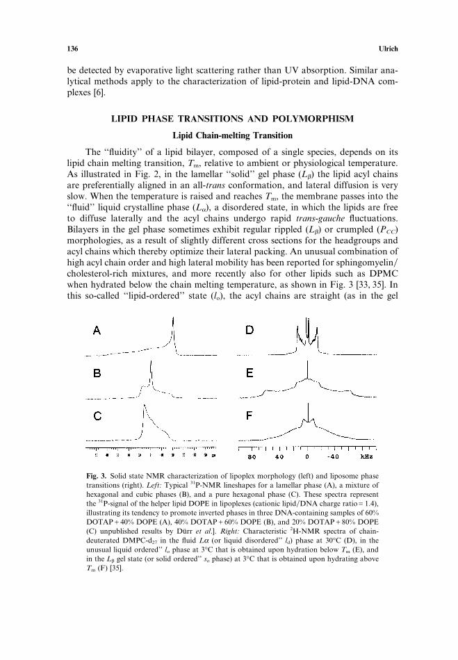
The ‘‘fluidity’’ of a lipid bilayer, composed of a single species, depends on itslipid chain melting transition, Tm, relative to ambient or physiological temperature.As illustrated in Fig. 2, in the lamellar ‘‘solid’’ gel phase (Lβ) the lipid acyl chainsare preferentially aligned in an all-trans conformation, and lateral diffusion is veryslow. When the temperature is raised and reaches Tm, the membrane passes into the‘‘fluid’’ liquid crystalline phase (Lα), a disordered state, in which the lipids are freeto diffuse laterally and the acyl chains undergo rapid trans-gauche fluctuations.Bilayers in the gel phase sometimes exhibit regular rippled (Lβ) or crumpled (PCC)morphologies, as a result of slightly different cross sections for the headgroups andacyl chains which thereby optimize their lateral packing. An unusual combination ofhigh acyl chain order and high lateral mobility has been reported for sphingomyelin�cholesterol-rich mixtures, and more recently also for other lipids such as DPMCwhen hydrated below the chain melting temperature, as shown in Fig. 3 [33, 35]. Inthis so-called ‘‘lipid-ordered’’ state (lo), the acyl chains are straight (as in the gel
Fig. 3. Solid state NMR characterization of lipoplex morphology (left) and liposome phasetransitions (right). Left: Typical 31P-NMR lineshapes for a lamellar phase (A), a mixture ofhexagonal and cubic phases (B), and a pure hexagonal phase (C). These spectra representthe 31P-signal of the helper lipid DOPE in lipoplexes (cationic lipid�DNA charge ratioG1.4),illustrating its tendency to promote inverted phases in three DNA-containing samples of 60%DOTAPC40% DOPE (A), 40% DOTAPC60% DOPE (B), and 20% DOTAPC80% DOPE(C) unpublished results by Durr et al.]. Right: Characteristic 2H-NMR spectra of chain-deuterated DMPC-d27 in the fluid Lα (or liquid disordered’’ ld) phase at 30°C (D), in theunusual liquid ordered’’ lo phase at 3°C that is obtained upon hydration below Tm (E), andin the Lβ gel state (or solid ordered’’ so phase) at 3°C that is obtained upon hydrating aboveTm (F) [35].
Liposomes as Delivery Vehicles 137
phase) but the lipid molecules as a whole undergo rapid diffusion in the plane ofthe membrane (as in the fluid phase). It remains to be investigated whether liposomesin the lo state may exhibit an enhanced toughness and elasticity.
The lipid chain melting transition temperature of membranes increases withaclyl chain length (DMPC: 24°C, DPPC: 42°C, and DPSC: 55°C), with the degree ofchain saturation (DOPC: −15°C), and with loss of headgroup hydration (dehydratedDPMC: ∼40°C). Many natural membrane lipids (e.g. egg PC) typically possess anunsaturated chain in the sn-1 position and a saturated one in sn-2, such that theyare fluid at physiological temperature. The addition of cholesterol increases disorderamongst the acyl chains in the gel phase, whereas the fluid phase becomes moreordered. At 50% cholesterol the membrane is saturated and the lipid phase transitioncompletely abolished. Cholesterol is thus used as a universal membrane sealer, mini-mizing the bilayer permeability and providing it with improved mechanical andcohesive strength, but exceptions to this general rule have been reported [3, 11].
Depth Profile of the Bilayer
A lipid membrane may be regarded as a composit of layers with very distinctcharacteristics. Much information has been derived from computer simulations[3, 11, 41], as discussed by Saiz et al. in this issue [42]. Basically, the outermost layerconsists of perturbed water, which has a considerably lower dielectric constant thanthe bulk and is less capable of forming hydrogen bonds with approaching com-pounds. The second layer contains water, lipid headgroups, and the upper paths ofsome acyl chains, as the hydrophilic�hydrophobic interface is smeared-out over asignificant depth. This layer is available for non-covalent interactions with drugs,and is likely to play a role in the folding of peptides and proteins. The third layerconsists of conformationally ordered acyl-chain segment, which impose an aniso-tropic potential on hydrophobic molecules penetrating the membrane. Finally, theinnermost layer consists of the acyl chain termini and is as conformationally dis-ordered as liquid decane.
Lipid bilayers in the fluid state readily accommodate hydrophobic drugs andanesthetics, whose solubility correlates with their octanol-water partition coef-ficients. In the gel state, on the other hand, hydrophobic compounds are less solublein membranes and tend to be expelled, especially in the case of saturated acyl chainsthat will minimize their packing defects. The temperature-dependent solubility ofdrugs plays an important role in the production of solid lipid nanoparticles, whichconsist of a solid lipid core (usually triacylglyceride) that is emulsified by a phosphol-ipid monolayer or amphilphilic polymer, as illustrated in Fig. 2. Loading is achievedby melting the neutral matrix lipid and dissolving the drug in the emulsion. Uponcooling, the differential solubility of the drug in the two lipid phases (fluid and gel)and in the aqueous buffer can lead to a relative enrichment in either the core of theSLN (useful for sustained release) or in its periphery (useful for a burst phase) [43].To enhance the loading capacity of a SLN, more complex lipid mixtures need to beemployed, containing mono-, di- and tricylglycerides with fatty acids of differentchain lengths, which form less perfect crystals with many imperfections to accommo-date the drug.
138 Ulrich
Lateral Phase Separation
The lateral organization of a lipid bilayer can be rather inhomogenous andplays a major role in drug penetration and membrane permeability [3]. Static lateralphase separation is frequently encountered in lipid mixtures with very different acylchain lengths, or when certain headgroups couple to extramembraneous surfaces.Dynamic structural heterogeneity is much harder to characterize, but is manifest asdensity fluctuations close to the chain-melting phase transition, or as compositionalfluctuations in moderately-matched lipid mixtures. Since the molecular packing islaterally disturbed at the interfaces between the domains, these boundaries willenhance the penetration of drugs and peptides into the membrane and greatlyincrease bilayer permeability. A dynamic heterogeneity also implies a softening ofthe membrane and a decrease in its bending rigidity, which in turn affects its capacityto undergo fusion, vesiculation, or interaction with other surfaces. these effects havebeen exploited to design permeability enhances and fusogenic systems that can beactively triggered by a change in temperature (see below).
A related phase separation phenomenon can be triggered by the strong interac-tion between Ca2+ ions and negatively charged PS headgroups. A fluid-fluid phasecoexistence has been reported after addition of Ca2+ to a mixture of PCCPS due tothe formation of cis-complexes in the bilayer phase. Vesicles consisting of pure PSare driven to fuse by the addition of Ca2+, which is attributed to the formation oftrans-complexes in apposing bilayers, being accompanied by the formation of localgel-phase domains. Likewise, the interaction of cationic amphiphiles with DNA mayaffect their mixing behavior with uncharged helper lipids and possibly promote lat-eral phase-separation [3]. A dehydration-induced lipid phase-transition upon lipo-plex assembly has nevertheless been ruled out, even though a significant release ofwater and counterions occurs upon intimate DNA-lipid contact [44].
Lipid Polymorphism
Lipid polymorphism is an essential aspect to consider in the rational design ofliposomes for drug delivery and gene therapy, and has been well reviewed [7, 9, 19].Besides the lamellar phase many lipids and surfactants can also adopt other morhpo-logies, such as the inverted hexagonal phase (HII) or various cubic phases (Q), sum-marized in Fig. 2. The hexagonal phase consists of an array of cyclindrical rods,wherein the lipids are oriented with their headgroups towards the aqueous core.Cubic phases are made up of bicontinuous surfaces, such that every point of theinternal surface of this macroscopic assembly has access to the external aqueousspace via aqueous channels (about 5 nm diameter), and any lipid molecule mayexchange places with another in the same monolayer simply by diffusion [45].
The preference of lipids for certain geometries is conveniently rationalized by‘‘molecular shape’’ arguments. Lipids with a large headgroup and a small hydro-carbon cross-section have a cone-like geometry, self-assemble into micelles, and aresaid to exhibit positive membrane curvature. Lipids that are cylindrical in shape,having nearly equal headgroup to hydrocarbon area, self-assemble into bilayers.Finally, lipids with small headgroups adopt ‘‘inverted’’ phases such as HII and Q,
Liposomes as Delivery Vehicles 139
are said to exhibit negative membrane curvature. Examples of cone-shape lipids aredetergents, lysolipids, and ganglioside GM1, whereas most of the natural membranelipids are cylindrical. The tendency to form inverted phases is pronounced for purePE, diaclyglycerol (DAG), PA (with Ca2+, or at low pH), PS (with Ca2+), and CL(with Ca2+). Long and unsaturated acyl chains have particularly large cross-sections,especially at high temperature, whereas the removal of water will reduce the effectiveheadgroup volume.
Unsaturated PE lipids in isolation prefer the inverted HII phase, e.g. DOPEundergoes an Lα→HII transition at 10°C. This tendency is supposed to play a keyrole in the delivery of drugs and DNA across cellular membranes, as DOPE facili-tates the formation of highly curved intermediates that are necessary for membranefusion [12, 16, 19]. Unsaturated PEs will adopt a lamellar bilayer structure in thepresence of stabilizing lipids such as PCs and PEG-lipid constructs. However, oncethe stabilizing function is removed, for example by pH-triggering (see below), alamellar-to-hexagonal phase transition can be triggered that is accompanied byrelease of contents and lipid mixing, thus being reminiscent of membrane fusionevents. On the other hand, by inducing a positive curvature in the outer monolayerof vesicles, for example by the addition of lysolipids or detergents, it is possible tosuppress their ability to participate in fusion. Cationic amphiphiles, in particular,display a very interesting phase behavior with regard to the delivery of DNA, asthey are usually stable as bilayers in isolation or when supplemented with neutralhelper lipids. However, when they are combined with negatively charged lamellarlipids (e.g. DODAC plus CHEMS), the mixture will adopt an inverted HII phase.This transition is attributed to the formation of ion pairs and an effective reductionin headgroup volumes.
The formation of cubic phases, finally, is attributed to a moderately ‘‘inverted’’molecular shape, as exemplified by glycerol monooleate (GMO) [45]. At a reducedwater content of 20% (w�w), on raising the temperature this lipid goes through thesequence Lα→Q→HII→ inverted micelles (see Fig. 2). Likewise, the addition ofwater to its Lα phase at room temperature triggers conversion into the very viscouscubic phase. This transition is suitably exploited in using GMO as a matrix for drugdelivery, since the fluid lamellar phase has a sufficiently low viscosity to be readilyinjected into a cavity, where it will take up water and transform into a stiff gel.
It must be generally noted that any hydrophobic drug, amphiphilic compoundor proteinaceous cargo is likely to affect the phase diagram of its liposomal carrier,hence great care has to be taken to characterize the corresponding behavior of theloaded system [24, 25, 46–48].
DNA-lipid Structures
The structures of cationic liposome-DNA complexes have been compre-hensively characterized in homogeneous bulk samples by X-ray diffraction and bytheoretical means [49–51]. There exist two basic architectures, LC
α and HCII, which
resemble the pure lipid phases as shown in Fig. 2. The lamellar architecture com-prises of parallel strands of DNA sandwiched between fluid lipid bilayers. Replace-ment of DOPC by DOPE in DOTAP-containing liposomes was shown to lead to a
140 Ulrich
transition from the multilamellar ‘‘sandwich’’ structure to an inverted ‘‘honeycomb’’phase, where single DNA strands are coated by a lipid monolayers and arranged ina hexagonal array [9, 16, 18]. Another, so-called ‘‘spaghetti and meatball’’morphology has been described by EM in terms of a single DNA strand that iscovered by a lipid bilayer and connects larger lipid�DNA aggregates.
The small suspended lipoplex particles used in transfection are less likely topossess a homogeneous structure. Invaginated bilamellar liposomes, as describedby Smyth Templeton in this issue [52], represent one of the few well-characterizedmorphologies that have been observed by cryo-EM for several cationic lipids afterextrusion [1, 2]. It has been suggested that the initial interaction of DNA with theouter surface of a unilamellar vesicle induces a contraction of its outer lipid layerdue to charge neutralization. This in turn leads to an ivnersion of the vesicle aroundthe DNA, such that a bilamellar vesicular structure is formed. The DNA is engulfedin the innermost compartment, where it appears to be well protected and highlyeffective in transfection.
Methods for Characterizing Lipid Phases and Transitions
Differential scanning calorimetry (DSC) is used to determine the onset tempera-ture of the lipid chain-melting and other phase transitions, and the area under thecurve (i.e. the enthalpy) is representative of the cooperativity [44]. Since the tran-sition temperature is sensitive to additives in the bilayer, it is a suitable parameterto monitor drug-lipid interactions and to check for break-down products or impurit-ies. To determine the total amount of water in concentrated lipid dispersions, it ispossible to analyze the area under the ice-melting peak [53]. The related techniqueof high sensitivity titration calorimetry finally can provide accurate thermodynamicparameters of a drug partitioning into a membrane [47].
Both nuclear magnetic resonance (NMR) and electron spin resonance (ESR)spectroscopy are powerful tools to examine the local structural and motionalfeatures of designated lipid segments, thus providing an overall picture of lipid mor-phology as well as detailed insights into the local molecular architecture. Technicaldetails and applications have been extensively reviewed [14, 20, 21, 24, 47, 54–56].For example, the solid state 31P-NMR signal of phospholipids is a straightforwardindicator of lamellar, hexagonal and micellar�cubic phases. Not only pure liposomesbut also lipoplexes have been characterized that way, as illustrated in Fig. 3. [Durret al., in preparation]. Additionally, the local order and mobility of individualmolecular segments can be studied either by selectively deuterating them for 2H-NMR or by labelling them with a nitroxide radical for ESR. Characteristic 2H-NMR spectra of the lipid acyl chains are useful for distinguishing the gel phase, thefluid phase, and the unusual liquid ordered’’ state, as depicted in Fig. 3. [35]. Analy-sis of deuterated lipid headgroups has shown that the alignment of their dipolemoments sensitively reflects the association of charged molecules as well as waterwith the membrane surface [57, 58]. Likewise, the penetration and localization ofhydrophobic drugs and anesthetics (see Xu et al., in this issue [59]), as well as thelocal conformation, orientation and lipid-perturbation of an amphiphilic peptide ortransmembrane protein can be described in detail by solid state NMR (55, 60–62].
Liposomes as Delivery Vehicles 141
Fluorescence spectroscopy provides many complementary approaches forstudying bilayer re-arrangements such as vesicle leakage, membrane fusion, andhydrophobic exposure [12, 15, 18, 63]. An extensive arsenal of highly sensitive water-soluble or lipid-anchored dyes is available for monitoring the escape of aqueouscontents by de-quenching of ANTS and DPX, the mixing of lipids by resonance-energy transfer between rhodamine-PE and NBD-PE, and the dehydration-inducedspectral shifts of ANS or laurdan [44].
SURFACE MODIFICATIONS OF LIPOSOMES
Steric Stabilization
One of the most critical aspects in drug delivery and gene therapy is to improveliposome stability and enhance their circulation times in the blood. It has beendemonstrated that coating the lipoplexes with neutral or negatively charged poly-mers can enhance their stability. For example, including ganglioside GM1 into lipo-somes leads to significantly enhanced stability in serum. A major improvement wasachieved by coating with polyethyleneglycol (PEG), as illustrated in Fig. 2. Thishydrophilic polymer prevents liposomal aggregation during storage and particularlyin serum after administration, as discussed in more detail by Allen et al. in thisissue [64] (see also [7, 8, 12]). The incorporation of PEG-PE into so-called ‘‘stealth’’liposomes establishes a steric barrier that delays their recognition and clearance fromthe blood stream by the mononuclear phagocyte system. Circulation times of con-ventional liposomes (typically several minutes) have been increased this way up tomany hours. It has even been possible to insert PEG-coupled lipids into the outerleaflet of preformed liposomes and lipoplexes, causing little loss of the entrappedcontents.
Optimum stabilization is typically achieved with 5–10% PEG-PE with a molec-ular mass in the range 1000–2000 Da. At lower concentrations the polymer chainconfiguration changes from a so-called brush-structure to a mushroom-structure,hence the surface tends to remain fully covered. A general drawback of PEGylatedliposomes is their reduced ability to approach the target membrane and undergofusion. To circumvent this limitation, various liposome formulations have beendesigned to shed their PEG coat during circulation. For example, PEG can beattached to short acyl chains which readily escape from the vesicle, or an acceptorpopulation of uncoated vesicles can be co-administered as a sink’’. alternatively,PEG can be actively unmasked upon acidification in the endosome, using acid-labilechemical linkages (see below).
Use of Fusogenic Peptides and pH-titrable Polymers
Enveloped viruses, such as influenza or HIV, carry fusogenic proteins on theirsurface by which they enter the host cell [12]. Membrane destabilization and fusionis accomplished by a short amphiphilic sequence, the fusion peptide, located eitherat the tip or in an internal region of the protein, which gets exposed upon receptorrecognition or at acid pH upon endocytosis. Such synthetic peptides can trigger
142 Ulrich
fusion of liposomes in ûitro, in many cases in a pH-dependent manner [63]. Theiractivity is attributed to a conformational change and possibly oligomerization of thepeptide [37]. In an attempt to mimick nature, liposomes have been equipped withfusion peptides, as for example discussed by Bungener et al. in this issue [65] andothers [7, 12, 15]. Anchoring the peptide to the lipid bilayer via a myristic chain wasshown to greatly enhance the fusogenic action and may provide a convenient wayof incorporating the peptides into vesicular carriers, as illustrated in Fig. 3. Lipo-plexes, too, have exhibited improved transfection activities when mixed with chargedfusion peptides. A drawback may concern the risk of an immunogenic response inûiûo, besides the relatively high cost of synthetic peptides.
An alternative, more cost-effective and less immunogenic approach relies on theadsorption of pH-titrable polymers to the liposomal surface [12]. Poly-histidine andpoly-lysine, for example, become positively charged upon lowering the pH and havebeen shown to destabilize and fuse negatively charged membranes in ûitro. Poly-(amidoamine)s undergo a marked conformational change from a relatively coiledhydrophobic structure at neutral pH to a relaxed hydrophilic structure at acidic pH,and may be thus exploited as endosomolytic agents. Catinoic poly)ethyleneimine)can induce non-leaky fusion at pH <7, and is even being investigated as a genedelivery vesicle on its own. Interestingly, its activity as a transfection enhancer hasbeen attributed to its high buffering capacity, which may raise the endlysosomal pHand inactivate the lytic enzymes, before ultimately the osmolarity increases and thelysosome bursts. Polyanions, bearing carboxyl groups, are useful for destabilizingand fusing both negatively charged as well as uncharged membranes, due to theirhydrophobic character at low pH. Promising pH-sensitive polyanions are, forexample, poly(acrylic acid) derivatives and succinylated poly(glycidol)s. Further trig-gering approaches using synthetic polymers are based on the temperature-dependentlipid affinity of N-isoproplylacrylamide, which undergoes a solubility change atambient temperature. Altogether, there remains much potential to be explored withsynthetic polymers and many open questions concerning their behavior in combi-nation with liposomal carriers in ûiûo.
Ligand-targeted Liposomes
Ligand-targeted liposomes and lipoplexes offer a vast potential for directing thecargo to designated cell types in ûiûo, using surface-bound site-specific ligands[4, 8, 66]. Many different kinds of ligands are available, such as antibodies (see Fig.2), receptors, peptides, vitamins, oligonucleotides (aptamers), or carbohydrates asdescribed by Park in this issue [67]. Large numbers of ligands can be positionedonto the liposome surface, and the resulting multivalency strongly enhances thebinding affinity. Ligands of different types can be combined onto the same carrier,providing a more precise target selection. Since lipid assemblies are usually dynamicstructures, surface-coupled ligands have a high motional freedom to position them-selves for optimal substrate-interactions. In stealth liposomes, however, the extendedPEG molecules (∼50–75 A for PEG2000) would prevent the recognition of cell surfacereceptors, but ligands have been successfully attached to the distal ends of the PEGchains to space them far from the vesicle surface, as described by Maruyama in this
Liposomes as Delivery Vehicles 143
issue [68]. Besides reaching selected cellular targets, surface displayed proteins arealso particularly useful for antigen presentation and immunization purposes.
LIPOSOMAL DELIVERY, DRUG RELEASE AND TRANSFECTION
Liposome Stability
The capacity of liposomes to retain their cargo in ûitro depends mainly onthe lipid composition and temperature [10]. A general sequence of hydrophilicsolute permeability is: waterHsmall non-electrolytesHanionsHcations≡large non-electrolytesHlarge poly-electrolytes. Membrane permeability is highest at the lipidphase transition, and is generally lower in the gel phase than in the fluid phase [3].Stability against leakage has thus been promoted using phospholipids that remainin the solid phase at physiological temperatures, such as TPPC and DSPC. Incorpor-ation of cholesterol generally enhances bilayer stability against leakage, whereas lipidimpurities (e.g. lysolipids, unsaturated fatty acids, uneven chain lengths, etc.) canweaken the bilayers and cause loss of contents. The use of multilamellar carriersretards leakage to some extent, but polymerized liposomes (e.g. made of 2,4-octa-decadienoyl chains) are ultimately the most resistant [69].
Under physiological conditions, solute leakage depends not only on the intrinsicmembrane permeability but also in its interactions with components of the biologicalfluids, which strongly affect liposome clearance as discussed by Ishida et al. in thisissue [70]. For example, in blood the lipid molecules can be transferred from theliposomal membrane to plasma lipoproteins (HDL, LDL, etc.). This escape is par-ticularly pronounced for fluid liposomes (e.g. DOPC) or those containing single- orshort-chain lipids, which distintegrate rapidly and release their contents within a fewminutes after intravenous injection. The longest lifetimes have been generallyobserved for gel-phase vesicles that are small (∼50 nm) and carry no net charge. Theincorporation of cholesterol minimizes lipid exchange, and circulation periods aresignificantly enhanced by incorporating PEG-DSPE in liposomes and lipoplexes.
Since hydrophobic compounds are readily accommodated in the fluid hydro-carbon region of a bilayer, liposomes might be intuitively considered to be excellentcarriers for lipophilic cargo. However, it has been realized that hydrophobic solutesare often rapidly (within minutes) depleted from their carriers by exchange mechan-isms, leading to their equilibration amongst all other lipidic structures in the circu-lation (lipoproteins, erythrocyte membranes, etc.) [25, 47, 48]. If solubility permits,hydrophobic drugs may be preferably accommodated in gel state MLV or in multi-layered solid cochlates [28]. Alternatively, solid lipid nanoparticles offer a stablehydrophobic environment with a high-loading capacity [32, 43].
Delivery of Drugs and Antigens from Liposomes
The exact mechanism of liposome breakdown remains speculative, once it hasbeen trapped on the cell surface. A drug may simply leak out and traverse the plasmamembrane by diffusion or pore formation. Alternatively, the liposome may eitherfuse directly with the plasma membrane, or it may be taken up by endocytosis or
144 Ulrich
phagocytosis. In receptor-mediated endocytosis, small particles (<150 nm diameter)bind to cell surface receptors and are taken up by clathrin-coated pits to form coatedvesicles. After internalization the clathrin coat is removed and the vesicle fuses withlysosomes, which induces the breakdown of the lipids and release of their contents.Large particles (>150 nm), on the other hand, are taken up principally by phago-cytosis, which is usually limited to specific cells such as macrophages but can beinduced in many other cell types with appropriate ligands. In both cases, the lipo-some could either be degraded in the low pH environment, or it could fuse directlywith the endosomal or lysosomal membrane. The actual route of intracellular pro-cessing is highly relevant for antigen presentation purposes. In ûitro, conventionalliposomes have been found to deliver their proteinaceous contents to the lysosome,such that antigens are processed for presentation via the major histocompatibilitycomplex (MHC) II pathway [71]. On the other hand, pH-sensitive liposomes havebeen observed to deliver antigens to antigen-presenting cells via the cytosolic MHCI pathway [72]. In ûitro, however, the situation is significantly more complex, andfurther details about using liposomes for immunization are covered in this issue byBungener et al. [65], by Chikh et al. [73], by Zhou and Neutra [74], and by Wilsonet al. [75].
One of the most important tools for triggering liposomal delivery is to exploitthe pH-sensitivity of the endocytotic pathway, and excellent reviews have been pre-sented [7, 12, 19]. Endosomes and lysosomes can reach pH values below 5.0, andliposomes can be in principle by internalized by any kind of cell when suitablytargeted. There are four different classes of pH-sensitive liposomes, two of whichhave already been discussed above, namely those containing fusogenic peptides andthose covered with acid-titratable synthetic polymers. An alternative, lipid-basedapproach relies on the combination of polymorphic lipids such as DOPE with mildlyacidic amphiphiles that act as stabilizers at neutral pH [12]. For example, oleic acid(OA) or CHEMS have a cylindrical shape in their charged state, which keeps DOPEin a lamellar phase and prevents liposome aggregation due to electrostatic repulsion.Protonation of the weakly acidic lipid, however, renders its headgroup less hydro-philic. As a result, its shape becomes conical and reinforces the HII preference ofDOPE, and moreover the colloidal suspension will aggregate due to the resultantloss of electrostatic and hydration repulsion. Yet another kind of pH-triggeringmechanism involves ‘‘caged’’ lipid-derivatives with specifically engineered chemicalbonds that are hydrolyzed by acid-catalysis (see Fig. 1). For example, the lipidheadgroup of N-maleyl-DOPE has been covalently modified such that cleavageleaves behind plain DOPE which favors membrane fusion. Likewise, the lipid chainsof acid-sensitive plasmalogens are hydrolyzable to produce membrane destabilizingfatty aldehydes and lysolipids [7].
Light-activation, too, can be used to trigger a chemical reaction between plas-mologens and free radical species that are generated from a burst of singlet oxygen,following illumination in the presence of a suitable sensitizer. Other, enzymatic trig-gering mechanisms have been based on the simultaneous administration of two kindsof liposomes. For example, Ca2+ has been encapsulated in a photo-oxidatively trig-gered subpopulation of vesicles, which upon release stimulates the hydrolysis of thesecond liposome population by an exogenous Ca2+-dependent phospholipase A2 [12].
Liposomes as Delivery Vehicles 145
Thermosensitive liposomes, finally, rely on a relatively sharp phase transition in thelipid phase at temperatures only a few degrees above 37°C. Packing defects observedat the gel-to-liquid crystalline phase transition result in increased permeability toentrapped solutes or drugs [3]. Such ‘‘thermosomes’’ require an external stimulationby heat in a spatially well-defined region, e.g. around a tumor, but whole-bodyhypothermia is also being pursued in order to trap distant metastases.
Transfection Mechanisms of Lipoplexes
The mechanisms of DNA transfer from the lipoplex to the nucleus remainrather elusive and are difficult to control. The initial release of DNA into the cyto-plasm is generally attributed to an intrinsic ability of the cationic lipids to destabalizethe plasma membrane or the endosomal compartment (depending on the mode ofuptake). Specifically, a flip-flop of anionic lipids from the cytoplasmic leaflet to thelumenal leaflet of the endosome appears to result in the formation of charge-neutralion pairs, thereby releasing the DNA from the complex [9, 16]. An additional mem-brane-destabilizing role has been attributed to the resulting mixture of cationic withanionic lipids, which exhibits a high tendency to undergo fusion due to the effectivedecrease in headgroup size and hydration as an ion-pair [8, 19].
When DOPE is used as a helper lipid, cationic liposomes generally mediatehigher levels of transfection than those containing DOPC. This observation is attri-buted to the tendency of DOPE to undergo a lamellar-to-hexagonal phase transitionin a pH-, composition- or temperature-dependent manner, which is likely to facilitatefusion with or destabilization of the target membrane. Additionally, it has beenproposed that the amine group of PE may compete with the cationic lipids for theDNA phosphate backbone, thus assisting disassembly of the complex and allowingthe DNA to escape from the endosome [15, 16, 76]. Cholesterol has been used as analternative helper lipid, resulting in the formation of more stable complexes in ûitrothan those containing DOPE. Since the beneficial effects of different helper lipidsvary with the cell lines and depend on the exact conditions in ûiûo, it is not straight-forward to draw general conclusions from biophysical parameters in ûitro. As illus-trated by Hirko et al. in this issue [77], there are many further biological barriers tobe overcome and different environments to be encountered in ûiûo [66]. For example,it has been shown that DNA which is coated by liposomes remains protected againstnucleuses while traversing the cytosol, whereas the concomitant condensation�com-paction of the DNA appears to be unfavorable for gene expression once it reachesthe nucleus.
Administration of Liposome Formulations
Different applications may require distinct release kinetics and different par-ticles sizes [4, 22]. To deliver tomographic imaging agents, for example, a rapid burstwill achieve maximum contrast between the circulation system and the target tissues.For the sustained delivery of therapeutic agents, on the other hand, liposomes shouldprovide a continuous source of material to be taken up by the target site overextended periods of time. Following an intravenous injection, the first capillaries to
146 Ulrich
be encountered by a delivery vehicle are in the lung tissue, which tends to mechan-ically entrap large particles such as MLV or serum-induced aggregates. The lungthus represents a unique site for drug delivery if large liposomes are employed, butcare has to be taken to avoid pulmonary emboli. Administration of liposomal for-mulations to the epithelial cells in the lower airways of the lung by nebulization, onthe other hand, requires smaller particles (∼300 nm) that are in the respirable range[78].
The other ‘‘first pass’’ organ to be encountered after systemic administration isthe liver, which can thus be targeted with fast-release liposomes. Large vesicles aregenerally more rapidly eliminated from the bloodstream, whereas small ones circu-late for longer, especially those with neutral or positively charged, rigid bilayers.Liver, lung and spleen may be circumvented using stealth liposomes. Although lipo-somes do not usually pass through the endothelial barrier of the vascular system,under pathological conditions small particles (<100 nm) are able to extravasate atsites of inflammation and through the poorly formed neovasculatore of some solidtumors [8]. Regardless of the circulation time of an intravenously injected liposomaldose, however, the majority will eventually be taken up by the mononuclear phago-cyte system. Using alternate parenteral routes, such as intraperitoneal, subcutaneousor intramuscular, large liposomes generally tend to be retained longer at the site ofinjection, while small ones are mainly taken up by the draining lymph system.
Transdermal drug delivery requires small and highly fluid liposomes (100–200 nm) that are able to squeeze through the intercellular regions of the stratumcorneum. Surfactants can be included to increase the elasticity of the liposomes,together with PEGylated lipids or charged amphiphiles that improve colloidal stab-ility [79]. Rapid disintegration of the liposomes into mixed micelles may furtherenhance the skin permeation [46]. The epidermal lipid barrier itself can be softenedby adding permeability enhances such as oleic acid or azone, which decrease thelipid acyl chain order presumably by increasing the lateral phase inhomogeneity [3].
Oral administration of liposomes is limited to very stable formulations thatsurvive the low pH of the stomach and the bile salts and hydrolytic enzymes of theintestine, as discussed by Zhou and Neutra in this issue [74]. In the gut, compara-tively large particles of several µm size can be taken up by Peyer’s patches, whichare part of the lymphoid tissue. Polymerized MVL have been sown to retain theirhydrophilic cargo for extended periods over 24 hr [69], and solid cochleates are alsosufficiently stable to deliver oligonucleotides and proteins orally [28]. Solid lipidnanoparticles, too, are promising vesicles for hydrophobic compounds, offering ahigh loading capacity and slow release kinetics of up to a week [32, 43].
A novel lipid-based material for intramuscular, subcutaneous, periodontal,vaginal, and post-surgical implants is provided by cubic phases, which serve as amacroscopic matrix for the local delivery of hydrophobic as well as hydrophilicdrugs and enzymes [45]. The release is typically controlled by diffusion within thegel, but is limited by biodegradation and by an initial burst phase during injectionof the lipid in the lamellar state.
Besides liposomes, emulsions, and solid lipid particles, other carrier systems arebeing developed, based on polymeric or ceramic particles, offering complementarymodes of delivery according to their respective physical properties.
Liposomes as Delivery Vehicles 147
ACKNOWLEDGMENTS
The author acknowledges financial support within SFB 19 from the DeutscheForschungsgemeinshaft, is grateful to share results with Ulrich Durr, Leticia Magda-leno and Stephen Grage, and thanks Parvesh Wadhwani for help with the figures.
REFERENCES
1. Lasic, D. D. (1997) Liposomes in Gene Deliûery. CRC Press, Boca Raton.2. Smyth Templeton, N. and Lasic, D. D. (1999) New directions in liposome gene delivery. Mol. Biotech-
nol. 11:175–80.3. Mouritsen, O. G. and Jorgensen, K. (1998) A new look at lipid-membrane structure in relation to
drug research. Pharm. Res. 15:1507–1519.4. Forssen, E. and Willis, M. (1998) Ligand-targeted liposomes. Adûanced Drug Deliûery 29:249–271.5. Zelphati, O. et al. (1998) Stable and monodisperse lipoplex formulations for gene delivery. Gene Ther.
5:1272–1282.6. Ferrari, M. E. et al. (1998) Analytical methods for the characterization of cationic lipid-nucleic acid
complexes. Hum. Gene Ther. 9:341–351.7. Gerasimov, O. V. et al. (1999) Cytosolic drug delivery using pH- and light-sensitive liposomes. Adû.
Drug. Deliû. Reû. 38:317–338.8. Chonn, A. and Cullis, P. R. (1998) Recent advances in liposome technologies and their applications
for systemic gene delivery. Adû. Drug. Deliû. Reû. 30:73–83.9. Maurer, N. et al. (1999) Lipid-based systems for the intracellular delivery of genetic drugs. Mol.
Membr. Biol. 16:129–140.10. Langner, M. and Kra, T. E. (1995) Liposome-based drug delivery systems. Pol. J. Pharmacol. 51:211–
222.11. Langner, M. (2000) Effect of liposome molecular composition on its ability to carry drugs. Pol. J.
Pharmacol. 52:3–14.12. Drummond, D. C., Zignani, M., and Leroux, J. (2000) Current status of pH-sensitive liposomes in
drug delivery. Prog. Lipid Res. 39:409–460.13. Hutchins, B. (2000) Characterization of plasmids and formulations for non-viral gene therapy. Curr.
Opin. Mol. Ther. 2:131–135.14. Goni, F M. and Alonso, A. (2000) Spectroscopic techniques in the study of membrane solubilization,
reconstitution, and permeabilization by detergents. Biochim. Biophys. Acta. 1508:51–68.15. Pedroso de Lima, M. C. et al. (1999) Gene delivery mediated by cationic liposomes: from biophysical
aspects to enhancement of transfection. Mol. Membr. Biol. 16:103–109.16. Pedroso de Lima, M. C. et al. (2001) Cationic lipid-DNA complexes in gene delivery: from biophysics
to biological applications. Adû. Drug. Deliû. Reû. 47:277–294.17. Lasic, D. D. and Papahadjopoulos, D. (1998) Medical Applications of Liposomes. Elsevier,
Amsterdam.18. Regelin, A. E. et al. (2000) Biophysical and lipofection studies of DOTAP analogs. Biochim. Biophys.
Acta. 1464:151–164.19. Hafez, I. M. and Cullis, P. R. (2001) Roles of lipid polymorphism in intracellular delivery. Adû. Drug
Deliû. Reû. 47:139–148.20. New, R. (1994) Liposomes—A Practical Approach (B. D. H. D. Rickwood, ed.) Vol. 58. IRL Press
at Oxford University Press.21. Gennis, R. B. (1989) Biomembranes: Molecular Structure and Function (C. Cantor, ed.) Springer
Verlag.22. Frezard, F. (1999) Liposomes: from biophysics to the design of peptide vaccines. Braz. J. Med. Biol.
Res. 32:181–189.23. Jin, A. J. et al. (1999) Light scattering characterization of extruded lipid vesicles. Eur. Biophys. J.
28:187–199.
148 Ulrich
24. Peleg-Shulman, T. et al. (2001) Characterization of steric alloys stabilized cisplatin liposomes bynuclear magnetic resonance. Biochim. Biophys. Acta. 1510:278–291.
25. Fatouros, D. G. and Antimisiaris, S. G. (2001) Physicochemical properties of liposomes incorporatinghydrachlorothiazide and chlorothiazide. J. Drug Target 9:61–74.
26. Ollivon, M. et al. (2000) Vesicle reconstitution from lipid-detergent mixed micelles. Biochim. Biophys.Acta. 1508:34–50.
27. Winden, E. C. A. v., Zuidam, N. J., and Crommelin, D. J. A. (1998) Strategies for large scale pro-duction and optimized stability of pharmaceutical liposomes developed for parenteral use. In: MedicalApplications of Liposomes (D. D. Lasic and D. Papahadjopoulos, eds.) Elsevier Science.
28. Zarif, L. and Mannino, R. J. (2000) Cochleates. Lipid-based vesicles for gene delivery-concept,achievements and future development. Adû. Exp. Med. Biol. 465:83–93.
29. Murphy, E. A. et al. (2000) Compaction of DNA in an anionic micelle environment followed byassembly into phosphatidylcholine liposomes. Nucleic Acids Res. 28:2986–2992.
30. Zuidam, N. J. et al. (1999) Lamellarity of cationic liposomes and mode of preparation of lipoplexesaffect transfection efficiency. Biochim. Biophys. Acta. 1419:207–20.
31. Ross, P. C. and Hui, S. W. (1996) Lipoplex size is the major determinant of in ûitro lipofectionefficiency. Gene. Ther. 6:651–659.
32. Mehnert, W. and Mader, K. (2001) Solid lipid nanoparticles: production, characterization and appli-cations. Adû. Drug Deliû. Reû. 47:165–196.
33. Meyer, H. W., Bunjes, H., and Ulrich, A. S. (1999) Morphological transitions of brain sphingomyelinare determined by the hydration protocol: ripples re-arrange in plane, and sponge-like networksdistintegrate into small vesicles. Chem. Phys. Lipids 99:111–123.
34. Meyer, H. W. and Richter, W. (2001) Freeze-fracture studies on lipids and membranes. Micron32:615–44.
35. Meyer, H. W. et al. (2000) Hydration of DMPC and DPPC at 4 degrees C produces a novel subgelphase with convex-concave bilayer curvatures. Chem. Phys. Lipids 105:149–166.
36. Meyer, H. W. et al. (1998) Minimal radius of curvature of lipid bilayers in the gel phase state corre-spond to the dimension of biomembrane structures ‘‘caveolae’’. J. Struct. Biol. 124:77–87.
37. Ulrich, A. S. et al. (1999) Ultrastructural characterization of peptide-induce membrane fusion andpeptide self-assembly in the lipid bilayer. Biophys. J. 77:829–841.
38. Chung, H. et al. (2001) Oil components modulate physical characteristics and function of the naturaloil emulsions as drug or gene delivery system. J. Control Release 71:339–350.
39. Jones, M. N. (1995) The surface properties of phospholipid liposome systems and their characteris-ation. Adû. Colloid Interface Sci. 54:93–128.
40. Radko, S. P. and Chrambach, A. (1999) Capillary electrophoresis of subcellular-sized particles. J.Chromatogr. B. Biomed. Sci. Appl. 722:1–10.
41. Tielman, D. P., Marrink, S. J., and Berendsen, H. J. C. (1997) A computer’s perspective of mem-branes: molecular dynamics studies of lipid bilayer systems. Biochim. Biophys. Acta. 1331:235–270.
42. Saiz, L., Bandyopadhyay, S., and Klein, M. L. (2001) Towards an Understanding of complex Bio-logical Membranes from Atomistic Molecular Dynamics Simulations. Biosci. Reps. 22:151–173.
43. Muller, R. H., Mader, K., and Gohla, S. (2000) Solid lipid nanoparticles (SLN) for controlled drugdelivery—a review of the state of the art. Eur. J. Pharm. Biopharm. 50:161–177.
44. Hirsch-Lerner, D. and Barenholz, Y. (1999) Hydration of lipoplexes commonly used in gene delivery:follow-up by laurdan fluorescence changes and quantification by differential scanning calorimetry.Biochim. Biophys. Acta. 1461:47–57.
45. Shah, J. C., Sadhale, Y., and Chilukuri, D. M. (2001) Cubic phase gels as drug delivery systems.Adû. Drug. Deliû. Reû. 47:229–250.
46. Stoye, I., Schroder, K., and Muller-Goymann, C. C. (1998) Transformation of a liposomal dispersioncontaining ibuprofen lysinate and phospholipids into mixed micelles—physico-chemical characteriz-ation and influence on drug permeation through excized human stratum corneum. Eur. J. Pharm.Biopharm. 46:191–200.
47. Fahr, A. and Seelig, J. (2001) Liposomal formulations of Cyclosporin A: a biophysical approach topharmacokinetics and pharmadocynamics. Crit. Reû. Ther. Drug. Carrier Syst. 18:141–172.
48. Ardhammar, M. et al. (1999) In ûitro membrane penetration of modified peptide nucleic acid (PNA).J. Biomol. Struct. Dyn. 17:33–40.
Liposomes as Delivery Vehicles 149
49. Radler, J. O. et al. (1997) Structure of DNA-cationic liposome complexes: DNA intercalation inmultilamellar membranes in distinct interhelical packing regimes. 275:810–814.
50. Koltover, I. et al. (1998) An inverted hexagonal phase of cationic liposome-DNA complexes relatedto DNA release and delivery. Science 281:78–81.
51. May, S., Harries, D., and Beh-Shaul, A. (2000) The phase behavior of cationic lipid-DNA complexes.Biophys. J. 78:1681–1697.
52. Templeton, N. S. (2002) Cationic Liposome-Mediated Gene Delivery in ûiûo. Biosci. Reps. 22:283–295.
53. Ulrich, A. S., Sami, M., and Watts, A. (1994) Hydration of DOPC bilayers by differential scanningcalorimetry. Biochim. Biophys. Acta. 1191:225–230.
54. Ulrich, A. S., and G. S. L. (1998) Solid state 2H-NMR. In: Solid State: NMR of polymers (I. Andoand T. Asakura, eds.) Elsevier, Amsterdam, 190–211.
55. Grage, S. L. et al. (2001) Solid state 19F-NMR of biomembranes. In: Perspectiûes on Solid StateNMR in Biology (S. Kiihne and H. J. M. d. Groot, eds.) Kluwer Academic Press, p. 83–91.
56. Ulrich, A. S. (2000) High resolution 1H and 19F solid state NMR. In: Encyclopedia of Spectroscopyand Spectrometry (J. Linden, G. Tranter, and G. Holmes, eds.). Academic Press, p. 813–825.
57. Ulrich, A. S. and Watts, A. (1994) Molecular response of the lipid headgroup to bilayer hydrationmonitored by 2H-NMR. Biophys. J. 66:1441–1449.
58. Ulrich, A. S. and Watts, A. (1994) Lipid headgroup hydration studied by 2H-NMR: a link betweenspectroscopy and thermodynamics. Biophys. Chem. 49:39–50.
59. Xu. Y., Yushmanov, V. E., and Tang, P. (2002) NMR Studies of Drug Interaction with Membranesand Membrane-Associated Proteins.
60. Salgado, J. et al. (2001) Membrane-bound structure and alignment of the antimicrobial beta-sheetpeptide gramicidin S derived from angular and distance constraints by solid state 19F-NMR. J.Biomol. 21:191–208.
61. Watts, A., Ulrich, A. S., and Middleton, D. A. (1995) Membrane protein structure: the contributionand potential of novel solid state NMR approaches. Mol. Membr. Biol. 12:233–246.
62. Ulrich, A. S. et al. (1995) Re-orientation of retinal in the M-photointermediate of bacteriorhodopsin.Nat. Struct. Biol. 2:190–192.
63. Ulrich, A. S. et al. (1998) Membrane fusion is induced by a distinct peptide sequence of the seaurchin fertilization protein bindin. J. Biol. Chem. 273:16748–16755.
64. Allen, C. et al. (2002) Controlling the Physical Behavior and Biological Performance of LiposomesFormulation through Use of Surface Grafted Poly(ethylene glycol). Biosci. Reps. 22:225–250.
65. Bungener, L. et al. (2002) Delivery of Protein Antigens to the Immune System by Fusion-ActiveVirosomes: A Comparison with Liposomes and ISCOMS.
66. Bally, M. B. et al. (1999) Biological barriers to cellular delivery of lipid-based DNA carriers. Adû.Drug Deliû. Reû. 38:291–315.
67. Park, Y. S. (2002) Tumor-Directed Targeting of Liposomes. Biosci. Reps. 22:267–281.68. Maruyama, K. (2002). PEG-Immunoliposome.69. Okada, J., Cohen, S., and Langer, R. (1995) In ûitro evaluation of polymerized liposomes as an oral
drug delivery system. Pharm. Res. 12:576–582.70. Ishida, T., Harashima, H., and Kiwada, H. (2002) Liposome Clearance.71. Leserman, L. and Barois, N. (1998) Major histocompatibility complex class II molecules, liposomes
and antigen presentation. In: Medical Applications of Liposomes (D. D. Lasic and D. Papahad-jopoulos, eds.). Elsevier, p. 25–45.
72. Rao, M. and Alving, c. R. (1998) Class I presentation of liposomal antigens. In: Medical Applicationsof Liposomes (D. D. Lasic and D. Papahadjopoulos, eds.). Elsevier, p. 15–23.
73. Chikh, G. and Schutze-Redelmeir, M.-P. (2002) Liposomal Delivery of CTL Epitopes to DendriticCells. Biosci. Reps. 22:339–353.
74. Zhou, F. and Neutra, M. R. (2002) Antigen Delivery to the Mucosa-Associated Lymphoid TissueUsing Liposomes as a Carrier. Biosci. Reps. 22:355–369.
75. Wilson, A., Pitt, B., and Li, S. (2002) Complex Role of CpG in Liposomal Delivery of DNA Oligonu-cleotides. Biosci. Reps. 22:309–322.
76. Harvie, P., Wong, F. M. P., and Bally, M. B. (1998) Characterization of lipid DNA interactions.Biophys. J. 75:1040–1051.
150 Ulrich
77. Hirko, A. C. et al. (2002) Plasmid Based Delivery Systems in Rat Brain. Biosci. Reps. 22:297–308.78. Abu-Dahab, R., Schafer, U. F., and Lehr, C. (2001) Lectin-functionalized liposomes for pulmonary
drug delivery: effect of nebulization on stability and bioadhesion. Eur. J. Pharm. Sci. 14:37–46.79. van der Bergh, B. A. et al. (2001) Elasticity of vesicles assessed by electron spin resonance, electron
microscopy and extrusion measurements. Int. J. Pharm. 217:13–24.