Embed Size (px)
Citation preview


BIOMIMETIC, BIORESPONSIVE, AND BIOACTIVE
MATERIALS


BIOMIMETIC, BIORESPONSIVE, AND BIOACTIVE
MATERIALS
An Introduction to Integrating Materials
with Tissues
Edited by
Matteo SantinGary Phillips
Brighton Studies in Tissue-Mimicry and Aided Regeneration (BrightSTAR)School of Pharmacy and Biomolecular Sciences
University of BrightonBrighton, UK
A JOHN WILEY & SONS, INC., PUBLICATION
Copyright © 2012 by John Wiley & Sons, Inc. All rights reserved.
Published by John Wiley & Sons, Inc., Hoboken, New Jersey.Published simultaneously in Canada.
No part of this publication may be reproduced, stored in a retrieval system, or transmitted in any form or by any means, electronic, mechanical, photocopying, recording, scanning, or otherwise, except as permitted under Section 107 or 108 of the 1976 United States Copyright Act, without either the prior written permission of the Publisher, or authorization through payment of the appropriate per-copy fee to the Copyright Clearance Center, Inc., 222 Rosewood Drive, Danvers, MA 01923, (978) 750-8400, fax (978) 750-4470, or on the web at www.copyright.com. Requests to the Publisher for permission should be addressed to the Permissions Department, John Wiley & Sons, Inc., 111 River Street, Hoboken, NJ 07030, (201) 748-6011, fax (201) 748-6008, or online at http://www.wiley.com/go/permissions.
Limit of Liability/Disclaimer of Warranty: While the publisher and author have used their best efforts in preparing this book, they make no representations or warranties with respect to the accuracy or completeness of the contents of this book and specifically disclaim any implied warranties of merchantability or fitness for a particular purpose. No warranty may be created or extended by sales representatives or written sales materials. The advice and strategies contained herein may not be suitable for your situation. You should consult with a professional where appropriate. Neither the publisher nor author shall be liable for any loss of profit or any other commercial damages, including but not limited to special, incidental, consequential, or other damages.
For general information on our other products and services or for technical support, please contact our Customer Care Department within the United States at (800) 762-2974, outside the United States at (317) 572-3993 or fax (317) 572-4002.
Wiley also publishes its books in a variety of electronic formats. Some content that appears in print may not be available in electronic formats. For more information about Wiley products, visit our web site at www.wiley.com.
Library of Congress Cataloging-in-Publication Data:
Biomimetic, bioresponsive, and bioactive materials: an introduction to integrating materials with tissues/edited by Matteo Santin, Gary Phillips. p. cm. Includes index. ISBN 978-0-470-05671-4 (hardback) 1. Biomimetic polymers. 2. Biomimetics. 3. Tissues–Mechanical properties. I. Santin, Matteo. II. Phillips, Gary. QD382.B47B56 2012 660.6–dc23 2011021420
Printed in the United States of America.
10 9 8 7 6 5 4 3 2 1

CONTENTS
Preface xi
Contributors xiii
1 HISTORYOFBIOMIMETIC,BIOACTIVE,ANDBIORESPONSIVEBIOMATERIALS 1Matteo Santin and Gary Phillips
1.1 The First Generation of Biomaterials: The Search for “The Bioinert” 1
1.1.1 Bioinert: Myth, Reality, or Utopia? 4
1.2 The Second Generation of Biomaterials: Biomimetic, Bioresponsive, Bioactive 5
1.2.1 Hydroxyapatite (HA) and Bioglass®: Cell Adhesion and Stimulation 6
1.2.2 Collagen, Fibrin Glue, and Hyaluronic Acid Hydrogels: Presenting the ECM 6
1.2.3 Chitosan and Alginate: Replacing the ECM 9
1.2.4 Poly(Lactic/Glycolic) Acid Copolymers: Encouraging Tissue Remodeling by Safe Biodegradation 10
1.2.5 Porous Metals: Favoring Mechanical Integration 11
1.3 The Third-Generation Biomaterials: Biomimicking Natural Bioactive and Bioresponsive Processes 13
1.3.1 Principal Phases of Tissue Regeneration 14
1.3.1.1 Cell Adhesion: The Cornerstone of Tissue Regeneration 16
1.3.1.2 Mechanisms of Tissue Mineralization 19
1.4 Principles of Biomimesis and Bioactivity 21
1.4.1 Biomimicking of the ECM 22
1.4.2 Biomimicking of Cell Membrane Components 24
1.4.3 Biomimicking Cell Signaling Pathways 24
1.4.3.1 Modulation of the Growth Factor Signaling by Gene Expression: Bioactive Gene Delivery Systems 25
v

vi� CONTENTS
1.5 Bioactive Biomaterials from Different Natural Sources 26
1.5.1 Silk Fibroin 26
1.5.2 Soybean-Based Biomaterials 27
1.6 Scope of This Book 29
References 30
2 SOFTTISSUESTRUCTUREANDFUNCTIONALITY 35Gabriela Voskerician
2.1 Overview 35
2.2 Epithelial Tissue 36
2.2.1 Background 36
2.3 The Skin 37
2.3.1 Structure and Functionality 37
2.3.2 Repair, Healing, and Renewal 42
2.4 Muscle Tissue 46
2.4.1 Background 46
2.4.2 Skeletal Muscle 48
2.4.2.1 Structure and Functionality 48
2.4.2.2 Repair, Healing, and Renewal 50
2.4.3 Smooth Muscle 51
2.4.3.1 Structure and Functionality 51
2.4.3.2 Repair, Healing, and Renewal 52
2.4.4 Cardiac Muscle 54
2.4.4.1 Structure and Functionality 54
2.4.4.2 Repair, Healing, and Renewal 55
2.5 Connective Tissue 56
2.5.1 Background 56
2.5.2 Embryonic Connective Tissue 57
2.5.3 Connective Tissue Proper 58
2.5.3.1 Cells of the Connective Tissue Proper 59
2.5.3.2 Connective Tissue Proper Fibers 60
2.5.3.3 Ground Substance 63
2.5.4 Specialized Connective Tissues 64
2.5.4.1 Structure and Function 64
2.5.4.2 Repair, Healing, and Renewal of Hyaline Cartilage 66
2.6 The Foreign Body Response 68
Exercises/Questions for Chapter 2 76
References 76

CONTENTS� vii
3 HARDTISSUESTRUCTUREANDFUNCTIONALITY 81Antonio Merolli and Paolo Tranquilli Leali
3.1 Definition of Hard Tissues 81
3.2 Articular Cartilage 81
3.2.1 Structure of the Articular Cartilage 82
3.2.2 Specific Mechanism Repair of the Articular Cartilage 83
3.3 Bone Tissue 84
3.3.1 The Structure of the Bony Tissues 85
3.3.2 The Functions of Bone Tissue 86
3.3.3 Cell Types Involved in Bone Homeostasis: The Osteoblasts and the Osteoclasts 88
3.3.4 Ossification, Turnover, and Remodeling 89
3.3.5 Bone Composite Structure and Its Effect on Mechanical Performance 91
3.4 Concluding Remarks 92
Exercises/Questions for Chapter 3 92
References 93
4 BIOMEDICALAPPLICATIONSOFBIOMIMETICPOLYMERS:THEPHOSPHORYLCHOLINE-CONTAININGPOLYMERS 95Andrew L. Lewis and Andrew W. Lloyd
4.1 Historical Perspective 95
4.2 Synthesis of PC-Containing Polymers 97
4.3 Physicochemical Properties of PC-Containing Polymers 98
4.3.1 Antifouling Mechanisms of Action 98
4.3.2 Swelling Phenomena and Structural Aspects of PC Coatings 100
4.4 Stability and Mechanical Property Considerations 102
4.4.1 PC Coatings and Surface Treatments 102
4.4.2 Bulk Hydrogels and Blends 104
4.5 Biological Compatibility 105
4.5.1 Interactions with Proteins, Eukaryotic Cells, and Bacteria 105
4.5.2 Interaction with Other Tissues 107
4.6. Applications of PC Polymers 107
4.6.1 Cardiovascular Applications 107
4.6.1.1 PC-Coated Coronary Stents 108
4.6.1.2 Vascular Grafts 108
4.6.1.3 Extracorporeal Circuits 109

viii� CONTENTS
4.6.2 Ophthalmic Applications 110
4.6.2.1 Intraocular Lenses 110
4.6.2.2 Contact Lenses 111
4.6.2.3 Other Ocular Devices 112
4.6.3 Anti-Infective Applications 112
4.6.3.1 Urological Devices 112
4.6.3.2 Tympanostomy Tubes 112
4.6.4 Orthopedic Applications 113
4.6.5 Biosensors and Diagnostics 113
4.6.6 Separation Systems 115
4.6.7 PC Polymers for Drug Delivery 116
4.6.7.1 Drug Delivery Coatings 116
4.6.7.2 Gel-Based Drug Delivery Systems 119
4.6.7.3 Nano/Micro Particulate Drug and Gene Delivery 119
4.6.7.4 Drug Conjugates 122
4.6.8 Emerging Applications 122
4.7 Summary 123
Exercises/Questions for Chapter 4 124
References 125
5 BIOMIMETIC,BIORESPONSIVE,ANDBIOACTIVEMATERIALS:INTEGRATINGMATERIALSWITHTISSUE 141Roberto Chiesa and Alberto Cigada
5.1 Introduction 141
5.2 Mandatory Requirements for Metals as Implantable Materials 142
5.2.1 Stiffness 142
5.2.2 Strength 143
5.2.3 Corrosion Resistance 144
5.2.3.1 General Corrosion 144
5.2.3.2 Crevice Corrosion 145
5.2.3.3 Fretting Corrosion 145
5.2.3.4 Galvanic Corrosion 145
5.3 Biocompatibility of Metals 145
5.3.1 ISO Standardized Metal Family 146
5.3.1.1 Stainless Steels 146
5.3.1.2 Cobalt Alloys 148
5.3.1.3 Titanium and Titanium Alloys 149

CONTENTS� ix
5.4 Surface Treatments of Metals for Biomedical Applications 150
5.4.1 Cathodic Deposition Treatments 152
5.4.2 Anodic Oxidation 152
Exercises/Questions for Chapter 5 157
References 157
6 CERAMICS 161Montserrat Espanol, Román A. Pérez, Edgar B. Montufar, and Maria-Pau Ginebra
6.1 Historical Perspective 161
6.2 Biostable Ceramics 162
6.2.1 Alumina 163
6.2.2 Zirconia 164
6.3 Bioactive and Resorbable Ceramics 165
6.3.1 Basic Concepts 165
6.3.2 Glasses and Glass–Ceramics 166
6.3.2.1 Physicochemical Properties of Bioactive Glasses 167
6.3.2.2 Silicate-Based Glasses 168
6.3.2.3 Phosphate-Based Glasses 170
6.3.2.4 Processing of Glass and Glass–Ceramics 170
6.3.3 Calcium Phosphates 172
6.3.3.1 Physicochemistry of Calcium Phosphates 172
6.3.3.2 Processing of Calcium Orthophosphates 175
6.3.4 New Trends in Bioactive and Resorbable Materials Integration 178
Exercises/Questions for Chapter 6 183
References 184
7 BIOFUNCTIONALBIOMATERIALSOFTHEFUTURE 191Mário Barbosa, Gary Phillips, and Matteo Santin
7.1 Clinically Led Next Generation Biomaterials 191
7.1.1 Wound Dressings and Dermal Substitutes 192
7.1.2 Vascular Grafts and Cardiovascular Stents 193
7.1.3 Joint Implants and Cartilage Tissue Engineering 194
7.1.4 Bone Fillers 195
7.1.5 Nerve Guides 195
7.1.6 Ophthalmologic Devices 195
7.2 Biomacromolecule-Inspired Biomaterials 196
7.2.1 Artificial Laminin 196
7.2.2 Artificial Elastin 197

x� CONTENTS
7.2.3 Artificial Collagen 197
7.2.4 GAG- and PGN-Mimicking Biomaterials 197
7.3 Nanostructured Biomimetic, Bioresponsive, and Bioactive Biomaterials 198
7.3.1 Nanofabrication of Biomaterials 198
7.3.1.1 2D Techniques 199
7.3.1.2 3D Techniques 199
7.3.1.3 Polymeric Dendrimers 200
7.3.1.4 Self-Assembling Peptides 201
7.4 Conclusions 202
Exercises/Questions for Chapter 7 203
References 203
Index 207

xi
In the last 40 years, clinicians, industrialists, and patients have witnessed and experi-enced one of the most exciting advances achieved by modern science and technology; the research and development of medical devices. The coming together of bioengineer-ing and materials science has played a fundamental role in the production of devices that are able to save the lives of many patients worldwide or significantly improve the quality of life for patients in those countries where the incidence of aging and lifestyle-related diseases have become a paramount social issue. Learning lessons from other fields of materials science, biomedical devices have been designed which are able to restore functionality in limbs and in the cardiovascular system as well as to replace the functions of compromised organs.
The relatively recent integration of molecular and cell biologists in the search for high-performance biomaterials and biomedical devices has led to the transformation of this research field from an engineering and materials science-based discipline into a truly interdisciplinary area of investigation. In the past few decades, the need to address the host response toward the implanted materials and to encourage tissue repair at their surfaces has sparked a new research approach in which biological processes have been studied in the presence of the challenge of an artificial surface. As a result, research worldwide has been driven by the search for biomaterials and devices specifically designed for targeted clinical applications, and new biochemical and cellular pathways have been identified.
An understanding of the finely tuned dependence of the activity of immunocom-petent and tissue cells on the surrounding environment has led to a paradigm shift in the biomaterials field where the goal of tuning tissue response toward biomimetic, bioresponsive, and bioactive biomaterials has widely been accepted by the scientific community.
As a truly interdisciplinary community, we are now witnessing and experiencing a new era for our discipline where the concepts of biomimicry, bioresponsiveness, and bioactivity are associated not only to the production of new biomedical devices, but also to biomaterials able to drive the complete regeneration of tissues and organs, the integrity of which has been compromised by trauma, disease, or aging.
The present book aims to mark this era by illustrating the advances made thus far and critically discussing the challenges that still need to be faced. The book aims not only to provoke the thoughts of the experts, but also to stimulate a new generation of young students and scientists who will certainly be the protagonists of the future prog-ress in this field. By presenting lessons from successful and unsuccessful stories and
PREFACE

xii� Preface
by critically assessing the state-of-the-art at research and clinical level, the editors of this book have aimed to provide the community with their contribution and to stimulate new research questions that will be able to open new routes of exploration.
The editors would like to express their gratitude to John Wiley & Sons for believ-ing in this initiative and for supporting them throughout their editorial journey. The most profound gratitude goes to all those valuable colleagues who have given their expertise and availability to this project, thus making it possible; it is a further testimony to the value of many years of collaborations spent together in exciting research projects.
It is hoped that the reader of this book will find its reading a rewarding experience and appreciate its various sections as well as the colored illustration of the figures that can be accessed through ftp://ftp.wiley.com/public/sci_tech_med/biomimetic_ bioresponsive.
Matteo SantinGary Phillips

xiii
CONTRIBUTORS
Mário A. Barbosa, Biomaterials Division, INEB-Instituto de Engenharia Biomédica, Universidade do Porto, Porto, Portugal
Roberto Chiesa, Dipartimento di Chimica, Materiali e Ingegneria Chimica, Politec-nico di Milano, Milano, Italy
Alberto Cigada, Dipartimento di Chimica, Materiali e Ingegneria Chimica, Politec-nico di Milano, Milano, Italy
Montserrat Espanol, Universitat Politècnica de Catalunya, Department of Materials Science and Metallurgical Engineering, Barcelona, Spain
Maria-Pau Ginebra, Universitat Politècnica de Catalunya, Department of Materials Science and Metallurgical Engineering, Barcelona, Spain
Paolo Tranquilli Leali, Department of Orthopaedic Surgery, University of Sassari, Sassari, Italy
Andrew L. Lewis, Biocompatibles UK Ltd, Farnham, Surrey, UK
Andrew W. Lloyd, Dean Faculty of Science and Engineering, University of Brighton, Moulsecooomb, Brighton, UK
Antonio Merolli, Orthopaedics and Hand Surgery, The Catholic University of Rome, Rome, Italy
Edgar B. Montufar, Universitat Politècnica de Catalunya, Department of Materials Science and Metallurgical Engineering, Barcelona, Spain
Román A. Pérez, Universitat Politècnica de Catalunya, Department of Materials Science and Metallurgical Engineering, Barcelona, Spain
Gary Phillips, Brighton Studies in Tissue-mimicry and Aided Regeneration (Bright-STAR), School of Pharmacy and Biomolecular Sciences, University of Brighton, Brighton, UK
Matteo Santin, Brighton Studies in Tissue-mimicry and Aided Regeneration (Bright-STAR), School of Pharmacy and Biomolecular Sciences, University of Brighton, Brighton, UK
Gabriela Voskerician, Department of Biomedical Engineering, Case Western Reserve University, Cleveland, OH, and Krikorjan, Inc., Menlo Park, CA, and Biodesign Innovation Group, Stanford University, Stanford, CA


1
1.1 THE FIRST GENERATION OF BIOMATERIALS: THE SEARCH FOR “THE BIOINERT”
Since it was first perceived that artificial and natural materials could be used to replace damaged parts of the human body, an “off-the-shelf” materials selection approach has been followed. These materials, now referred to as “first-generation” biomaterials, tended to be “borrowed” from other disciplines rather than being specifically designed for biomedical applications, and were selected on the basis of three main criteria: (1) their ability to mimic the mechanical performances of the tissue to be replaced, (2) their lack of toxicity, and (3) their inertness toward the body’s host response [Hench & Polack 2002].
Following this approach, pioneers developed a relatively large range of implants and devices, using a number of synthetic and natural materials including polymers, metals, and ceramics, based on occasional earlier observations and innovative approaches by clinicians. Indeed, many of these devices are still in use today (Figure 1.1A–J). A typical example of this often serendipitous development process was the use of poly(methyl methacrylate) (PMMA) to manufacture intraocular and contact lenses. This material (Table 1.1) was selected following observations made by the clini-cian Sir Harold Ridley that fragments of the PMMA cockpit that had penetrated into
1HISTORY OF BIOMIMETIC,
BIOACTIVE, AND BIORESPONSIVE BIOMATERIALS
Matteo Santin and Gary Phillips
Biomimetic, Bioresponsive, and Bioactive Materials: An Introduction to Integrating Materials with Tissues, First Edition. Edited by Matteo Santin and Gary Phillips.© 2012 John Wiley & Sons, Inc. Published 2012 by John Wiley & Sons, Inc.

Figure 1.1. Examples of medical implants and their components. (A–F) Orthopedic implant
components: (A) femor head, (B) hip socket, (C) titanium stent coated with porous titanium
foam, (D) titanium stent coated with hydroxapatite coating, (E) knee implant components.
(F–J) Other types of biomedical implants: (F) vascular graft, (G) coronary stent, (H) ureteral
stent (insert shows detail of the device pig-tail end), (I) intrauterine device, (J) wound
dressing.
A F
B G
C H
D I
E J

THE FIrsT GEnErATIOn OF BIOmATErIAls: THE sEArCH FOr “THE BIOInErT” 3
TABLE 1.1. Chemical Structure of Typical Polymeric Biomaterials
Polymer Structure
Polystyrenen
Poly(vinyl chloride)n
Cl
Poly(2-hydroxyethyl methacrylate)
O
O
OH
n
Poly(methyl methacrylate)n
O
O
Chronoflex 80A poly(urethane) -(-PHECD-DesmW-BD-)-n
Hydrothane poly(urethane) Polyethylene glycol polyurethane
PFPM/PEHA75/25
CH2(CF2)3CF2H
nn
OO
OO
Poly(octofluoropentyl methacrylate/ethylhexyl methacrylate)
the eyes of World War II pilots induced a very low immune response (see Section 1.3.1) [Williams 2001].
Also against the backdrop of the Second World War, a young Dutch physician named Willem Kolff pioneered the development of renal replacement therapies by taking advantage of a cellophane membrane used as sausage skin to allow the dialysis of blood from his uremic patients against a bath of cleansing fluid [Kolff 1993]. Later, in the early 1960s, John Charnley, learning about the progress materials science had made in obtaining mechanically resistant metals and plastics, designed the first hip joint prosthesis able to perform satisfactorily in the human body [Charnley 1961]. These are typical examples of how early implant materials were selected; however, it was soon
4 HIsTOry OF BIOmImETIC, BIOACTIvE, AnD BIOrEspOnsIvE BIOmATErIAls
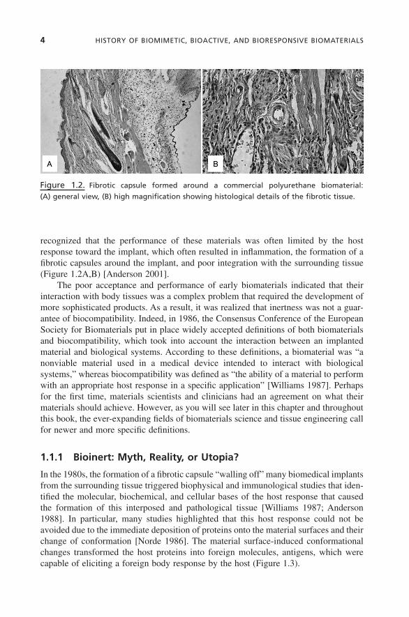
recognized that the performance of these materials was often limited by the host response toward the implant, which often resulted in inflammation, the formation of a fibrotic capsules around the implant, and poor integration with the surrounding tissue (Figure 1.2A,B) [Anderson 2001].
The poor acceptance and performance of early biomaterials indicated that their interaction with body tissues was a complex problem that required the development of more sophisticated products. As a result, it was realized that inertness was not a guar-antee of biocompatibility. Indeed, in 1986, the Consensus Conference of the European Society for Biomaterials put in place widely accepted definitions of both biomaterials and biocompatibility, which took into account the interaction between an implanted material and biological systems. According to these definitions, a biomaterial was “a nonviable material used in a medical device intended to interact with biological systems,” whereas biocompatibility was defined as “the ability of a material to perform with an appropriate host response in a specific application” [Williams 1987]. Perhaps for the first time, materials scientists and clinicians had an agreement on what their materials should achieve. However, as you will see later in this chapter and throughout this book, the ever-expanding fields of biomaterials science and tissue engineering call for newer and more specific definitions.
1.1.1 Bioinert: Myth, Reality, or Utopia?
In the 1980s, the formation of a fibrotic capsule “walling off” many biomedical implants from the surrounding tissue triggered biophysical and immunological studies that iden-tified the molecular, biochemical, and cellular bases of the host response that caused the formation of this interposed and pathological tissue [Williams 1987; Anderson 1988]. In particular, many studies highlighted that this host response could not be avoided due to the immediate deposition of proteins onto the material surfaces and their change of conformation [Norde 1986]. The material surface-induced conformational changes transformed the host proteins into foreign molecules, antigens, which were capable of eliciting a foreign body response by the host (Figure 1.3).
Figure 1.2. Fibrotic capsule formed around a commercial polyurethane biomaterial:
(A) general view, (B) high magnification showing histological details of the fibrotic tissue.

BIOmImETIC, BIOrEspOnsIvE, BIOACTIvE 5
Biomaterial surfaces contacted by blood, saliva, urine, cerebrospinal and peritoneal fluids, or tears cannot avoid interactions with proteins and other molecules that are naturally contained in the overlying body fluid [Santin et al. 1997]. As a consequence, the implant surface is rapidly covered by a biofilm that masks the material surface and dictates the host response. It is clear, therefore, that as a result of these processes, no biomaterial can be considered to be totally inert. However, although they are subjected to a continuous turnover, it is a fact that proteins (and more broadly, all tissue macro-molecules) retain their native conformation during the different phases of tissue forma-tion and remodeling. Hence, for the past two decades the scientific community has striven for the development and synthesis of a new generation of biomaterials that are able to control protein adsorption processes and/or tissue regeneration around the implant.
1.2 THE SECOND GENERATION OF BIOMATERIALS: BIOMIMETIC, BIORESPONSIVE, BIOACTIVE
In conjunction with the findings regarding the biochemical and cellular bases of host response toward implants, material scientists began their search for biomimetic, bio-responsive, and bioactive materials capable of controlling interactions with the sur-rounding biological environment and that could participate in tissue regeneration processes.
Figure 1.3. schematic representation of protein denaturation upon adsorption on biomate-
rial surface: (a) soluble protein approaches biomaterial surface, (b) protein adsorbs to material
surface, (c) protein starts to unfold through interactions with material surface, (d) protein
acquires an antigenic conformation.

6 HIsTOry OF BIOmImETIC, BIOACTIvE, AnD BIOrEspOnsIvE BIOmATErIAls
The move toward the synthesis and engineering of this type of biomaterial was initiated by the discovery of ceramic biomaterials that were proven to favor the integra-tion of bony tissue in dental and orthopedic applications [Clarke et al. 1990], as well as by the use of synthetic or natural polymers [Raghunath et al. 1980; Giusti et al. 1995]. Second-generation metals also emerged that were able to improve the integration with the surrounding tissue.
1.2.1 Hydroxyapatite (HA) and Bioglass®: Cell Adhesion and Stimulation
The ability of HA, and Bioglass to integrate with the surrounding bone in orthopedic and dental applications strictly depends on the physicochemical properties of these two types of ceramics, which will be described in Chapter 7. Here, it has to be mentioned that since their early discovery and use in surgery, the good integration of these bio-materials with bone, the osteointegration, depends on mechanisms of different nature that have triggered new concepts/definitions and new technological targets among scientists.
Although HA osteointegration can intuitively be attributed to their ability to mimic the bone mineral phase (see Chapter 3), the mechanisms underlying Bioglass-induced bone formation are not as clearly identifiable. It is known that HA favors the deposition of new bone on its surface by supporting osteoblast adhesion and by promoting the chemical bonding with the bone mineral phase [Takeshita et al. 1997]. Furthermore, the ability of HA to induce bone formation when implanted intramuscularly in animals, allegedly via the differentiation of progenitor cells, clearly shows their osteoinductive potential; indeed, osteoinductivity is defined as the ability of a biomaterial to form ectopic bone. Conversely, Bioglass osteoinductivity seems to be intrinsic to its degrada-tion process whereby (1) growth factors remain trapped within the gel phase formed during the degradation of the material and, consequently, released to the cells upon complete material dissolution; (2) structural proteins of the extracellular matrix (ECM) such as fibronectin form strong bonds with particles of the degrading material; and (3) silicon ions stimulate osteoblast (and allegedly progenitor cell) differentiation and, subsequently, the production of new bone [Xynos et al. 2000]. Regardless of the type of ceramic used, it is now widely recognized that the topographical features of these types of biomaterials are also fundamental to their bioactivity. For example, the absence of porosity or porosity of different sizes may lead to no osteointegration or to only poor bone formation [Hing et al. 2004].
1.2.2 Collagen, Fibrin Glue, and Hyaluronic Acid Hydrogels: Presenting the ECM
The use of collagen, fibrin, and hyaluronan, which are all natural components of the ECM, was born from scientists’ intuition that tissue cells recognize these biopolymers as natural substrates to form new tissue.
Fundamental to the application of these biological materials was an appreciation of their physicochemical and biological properties. Collagen is the most ubiquitous

BIOmImETIC, BIOrEspOnsIvE, BIOACTIvE 7
structural protein in the human body and the principal constituent of ECM in connective tissues [Rivier & Sadoc 2006]. It consists of a tightly packed structure composed of three polypeptide chains that wind together to form a triple helix [Rivier & Sadoc 2006]. These collagen molecules then associate to form collagen fibrils. A number of reviews are available on the structure of the different types of collagen found throughout the body [Engel & Bachinger 2005; Rivier & Sadoc 2006]. Collagen plays a key role in the wound healing process and the development of cartilage and tendons, and it is known that collagen can favor the formation of HA on its structure, thus inducing bone mineralization [Zhai & Cui 2006]. As part of the ECM, collagen provides a suitable milieu for cell proliferation, migration, and differentiation during the production of new tissue via its biodegradation and tissue remodeling. Collagen is, therefore, a natural biomaterial whose inherent potential has been exploited by biomaterials scientists in ligament replacement and other tissue engineering applications [Rothenburger 2001; Gentleman et al. 2003, 2006; Boccafoschi 2005; Kutschka et al. 2006], and collagen types I and IV have been commercialized as dermal substitutes [Jones et al. 2006].
The use of fibrin as a biomaterial was founded on the fact that fibrin clots are self-assembling networks with biological and physicochemical attributes that have the potential to be used in a number of biomedical applications. Three-dimensional (3D) porous fibrin networks are formed through a series of events during the blood coagula-tion cascade, resulting in the formation of a biopolymer gel material. The structure of the gel is determined by the thrombin-mediated conversion of fibrinogen to fibrin and the subsequent self-assembly of the fiber network [Helgerson et al. 2004]. Fibrin glue saw its first application as a surgical adhesive, but in the emerging era of tissue engi-neering, it has been suggested by many scientists as a suitable gel for cell encapsulation (Figure 1.4a–c) [Bach et al. 2001]. This is due to the fact that fibrin clots provide a structural scaffold that allows the adhesion, proliferation, and migration of cells impor-tant in the wound healing process and, when associated with proteins as a clot, has intrinsic biological properties that support and control, to some extent, cell differentia-tion. Fibrin-based biomaterials also benefit from the fact that they are naturally remod-eled and resorbed as part of the fibrinolytic processes associated with the cellar deposition of a new ECM as part of the normal wound healing processes [Helgerson et al. 2004] (see Section 1.3.1).
Hyaluronan, one of the main components of cartilage (see Chapter 3), has been chemically modified and commercialized to favor cartilage and skin regeneration (see Chapter 6) [Barbucci et al. 1993]. Hyaluronan consists of a single polysaccharide chain with no peptide in its primary structure, and it has a molecular weight that reaches millions of Daltons [Fraser et al. 1997]. The biological properties of this molecule are imparted by specific hyaluronan binding sites present in other ECM molecules and on the surface of cells [Fraser et al. 1997]. A number of proteins exist—the hyaladherins—that have the ability to recognize hyaluronan and result in the binding of hyaluronan molecules with proteoglycans to reinforce the structure of the ECM [Fraser et al. 1997; Day & Prestwich 2002]. At the molecular and cellular levels, it is now known that these biomolecules are able to support tissue regeneration because of the presence of specific bioligands that are able to recognize receptors on the cell membrane which, in turn, stimulates cell functions [Turley et al. 2002] (see Section 1.3.1.1).

8 HIsTOry OF BIOmImETIC, BIOACTIvE, AnD BIOrEspOnsIvE BIOmATErIAls
Furthermore, the physicochemical and biochemical properties of the three mole-cules discussed here can favor the interaction with other tissue components, forming organized macromolecular structures capable of conferring on tissues their specific mechanical properties (see Chapter 2). As previously mentioned, it is known that col-lagen can favor the formation of HA on its structure, thus inducing bone mineralization [Zhai & Cui 2006], and that hyaluronan is capable of interacting with other proteins to form macromolecular structures that are able to retain a relatively high water content. This high water content thus acts as an effective shock absorber in cartilage and ocular tissues [Fraser et al. 1997]. However, these substrates have shown some drawbacks and limitations. Although they provided the regenerating tissue with some important proper-ties, some others were missing. As mentioned above, one of the main benefits of using these biopolymers in clinical applications is that they promote biorecognition. However, in most cases, this biorecognition is not specific for the type of cells that need to be targeted to induce tissue regeneration. For example, collagen and fibrin, as well as other important ECM proteins (e.g., fibronectin), present in their structure the arginine–glycine–aspartic acid (RGD) sequence that is recognized by most tissue cells as well as by inflammatory cells such as monocytes or macrophages [Phillips & Kao 2005]. As a result of this relatively broad spectrum of cell recognition, collagen-based bioma-
Figure 1.4. Collagen deposition by osteoblasts encapsulated in a fibrin hydrogel. Incubation
times: (a) 24 hours, (b) 48 hours, and (c) 72 hours.
24 hours 48 hours
72 hours
a
c
b
BIOmImETIC, BIOrEspOnsIvE, BIOACTIvE 9
terials have been shown to induce an immune response in patients, which often leads to the formation of fibrotic tissue (see Section 1.1). In addition, collagen-based implants, either extracted from mammalian sources or from recombinant bacteria, may not rep-resent the composition of the real ECM and miss some components required to regulate the process of tissue regeneration. For example, it has been proven that physiological skin ECM collagen presents, on its surface, proteins such as α1-microglobulin, which is capable of modulating the activity of resident macrophages [Santin & Cannas 1999]. The absence of this protein in pathological tissues (e.g., scar tissue) and collagen implants seems to lead to a collagen-induced activation of immunocompetent cells. The modulating action of the α1-microglobulin is likely to be only one aspect of a multi-faceted process leading to the regulation of the immunocompetent cell activity in con-nective tissues. Therefore, collagen-based implants, although representing a step forward in developing biomaterials for tissue regeneration, address the problem in a relatively simplistic manner. A plethora of immunomodulators are present in physio-logical tissues, which may need to be taken into account to improve the performance of the collagen-based biomaterials.
Similarly, hyaluronan is recognized by cell receptors such as CD44, which are present on the membrane of both tissue and inflammatory cells. The role of this poly-saccharide in nature is tuned by its molecular weight [Mytar et al. 2001; Teder et al. 2002]. It has been proven that low molecular weight hyaluronan is fundamentally proinflammatory and angiogenic, thus promoting the formation of granular tissue. Conversely, relatively high molecular weight hyaluronan seems to prevent angiogenesis and inflammation. Thus far, at the clinical level, relatively high molecular weight hyal-uronan and its ester derivatives have been used, but not enough information has been collected to optimize the molecular weight of this polysaccharide. More accurate studies may be able to define the appropriate physicochemical characteristics of hyaluronan-based biomaterials to encourage some degree of vascularization and inflam-mation, which are required for a physiological regeneration.
Finally, although the use of fibrin glue as an adhesive material in surgery is wide-spread and successful, the tissue regeneration potential of this natural hydrogel has been proven to be limited unless key growth factors are loaded in its mesh. As for collagen and hyaluronan, this is not surprising considering that the main function of fibrin is to stop the bleeding and provide the damaged tissue with a temporary scaffold for its repair.
Each of the biopolymers mentioned in this section have reached the market and provided good, although not always satisfactory, clinical performances. Nevertheless, the use of these materials in clinics has opened the door to the development of biomi-metic biomaterials able to mimic the structure, biochemistry, and biofunctionality of tissue components.
1.2.3 Chitosan and Alginate: Replacing the ECM
As previously mentioned, the ECM is a structural, 3D network consisting of a number of macromolecules and polyelectrolytes including fibronectin, proteoglycan, collagen, laminin, and glycosaminoglycans. This macromolecular architecture mediates the