Embed Size (px)
Citation preview
Biomedical and Psychosocial Factors Associated with Pain
and Disability after Peripheral Nerve Injury
by
Christine B. Novak
A thesis submitted in conformity with the requirements for the degree of Doctorate of Philosophy
Institute of Medical Science University of Toronto
© Copyright by Christine B. Novak, 2010
ii
Biomedical and Psychosocial Factors Associated with Pain and
Disability after Peripheral Nerve Injury
Christine B. Novak
Doctorate of Philosophy
Institute of Medical Science University of Toronto
2010
Abstract
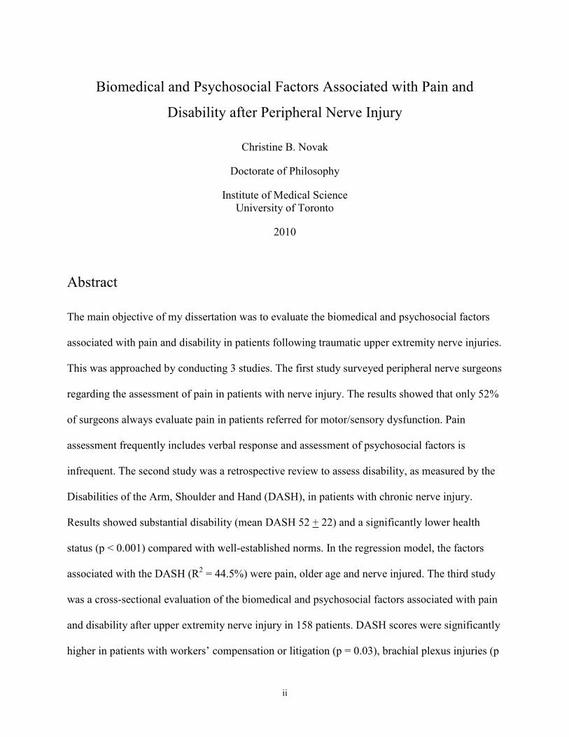
The main objective of my dissertation was to evaluate the biomedical and psychosocial factors
associated with pain and disability in patients following traumatic upper extremity nerve injuries.
This was approached by conducting 3 studies. The first study surveyed peripheral nerve surgeons
regarding the assessment of pain in patients with nerve injury. The results showed that only 52%
of surgeons always evaluate pain in patients referred for motor/sensory dysfunction. Pain
assessment frequently includes verbal response and assessment of psychosocial factors is
infrequent. The second study was a retrospective review to assess disability, as measured by the
Disabilities of the Arm, Shoulder and Hand (DASH), in patients with chronic nerve injury.
Results showed substantial disability (mean DASH 52 + 22) and a significantly lower health
status (p < 0.001) compared with well-established norms. In the regression model, the factors
associated with the DASH (R2 = 44.5%) were pain, older age and nerve injured. The third study
was a cross-sectional evaluation of the biomedical and psychosocial factors associated with pain
and disability after upper extremity nerve injury in 158 patients. DASH scores were significantly
higher in patients with workers’ compensation or litigation (p = 0.03), brachial plexus injuries (p
iii
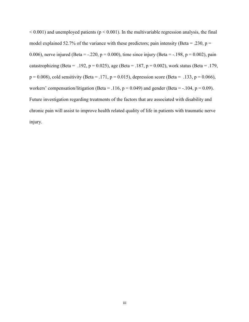
< 0.001) and unemployed patients (p < 0.001). In the multivariable regression analysis, the final
model explained 52.7% of the variance with these predictors; pain intensity (Beta = .230, p =
0.006), nerve injured (Beta = -.220, p = 0.000), time since injury (Beta = -.198, p = 0.002), pain
catastrophizing (Beta = .192, p = 0.025), age (Beta = .187, p = 0.002), work status (Beta = .179,
p = 0.008), cold sensitivity (Beta = .171, p = 0.015), depression score (Beta = .133, p = 0.066),
workers’ compensation/litigation (Beta = .116, p = 0.049) and gender (Beta = -.104, p = 0.09).
Future investigation regarding treatments of the factors that are associated with disability and
chronic pain will assist to improve health related quality of life in patients with traumatic nerve
injury.
iv
Acknowledgements
A PhD is a journey of hard work, perseverance, persistence, determination and good luck. I am
extremely grateful to everyone who walked with me and helped me on this long, educational,
sometimes tortuous and definitely rewarding journey.
Thank you to Joel Katz who was a wonderful supervisor and mentor throughout my doctorate
journey. Thank you to the other members of my Program Advisory Committee, Dimitri
Anastakis and Dorcas Beaton for their assistance and unwavering support. Thank you to Susan
Mackinnon who has been a wonderful mentor and friend for many years. Each generously
provided me with the benefits of their expertise and resources. Their knowledge, insight and
support were invaluable and I could not have successfully completed this dissertation without
their assistance. This has been a long, educational journey at a very difficult time in my life and I
thank each of them for their support and encouragement. These individuals each in their unique
way generously provided me with the necessary assistance, resources and expertise to complete
my dissertation and successfully achieve my doctorate degree. I shall be forever grateful.
I am very thankful for the financial support that I received for this dissertation. I would like to
acknowledge the support of the Canadian Institutes of Health Research through a Doctoral
Fellowship Award, and the American Association for Hand Surgery for the Research Grant
Award.
I am extremely grateful to everyone who assisted in the successful completion of my dissertation
studies. Thank you to all of those who completed the questionnaires and gave of their time to

v
participate in each study. Thank you to all of the individuals who assisted with these studies from
the University of Toronto Hand Program and the Division of Plastic & Reconstructive Surgery,
Washington University School of Medicine in St. Louis, particularly Linda Dvali, Susan
Kaminski, Lynn Nowen, Tina Orth, Kathy Pavlovic and all of the hand therapists who helped me
in so many ways. Thank you to Patrick Gullane, Kathleen Nicholson and the Wharton Head &
Neck Foundation for their support and assistance with this journey from the beginning. It is
through the generosity of all of these individuals that I was able to successfully complete this
project.
Most importantly, thank you to all of my family, Jim, Brenda, James, Alison, Laura, Lindsay and
my parents, John and Mary, who have provided my strongest support. They have always
supported my decisions, assisted me in every phase of my life and shared laughter and tears with
me along the way.
This dissertation is dedicated to my brother, who taught me about the virtues of perseverance,
determination and tenacity, how to continue with honour in the face of adversity and most
importantly to never give up.
vi
Table of Contents
Title Page…………………………………………………………………………………..………i
Abstract……………………………………………………………………………………………ii
Acknowledgements ........................................................................................................................ iv
Table of Contents ........................................................................................................................... vi
List of Tables................................................................................................................................... x
List of Figures ................................................................................................................................ xi
List of Abbreviations.....................................................................................................................xii
Chapter 1 Introduction .................................................................................................................... 1
1 Introduction ................................................................................................................................ 1
Chapter 2 Review of Literature....................................................................................................... 4
2 Background ................................................................................................................................ 4
2.1 Occurrence of Upper Extremity Peripheral Nerve Injury ................................................... 4
2.2 Neuropathic Pain in Patients with Upper Extremity Nerve Injury...................................... 6
2.2.1 Abstract ................................................................................................................... 8
2.2.2 Introduction ............................................................................................................. 9
2.2.3 Methods................................................................................................................. 10
2.2.4 Results ................................................................................................................... 11
2.2.5 Discussion ............................................................................................................. 16
2.2.6 Limitations ............................................................................................................ 31
2.2.7 Future Directions................................................................................................... 32
2.3 Classification of Peripheral Nerve Injury.......................................................................... 33
2.4 Recovery Following Peripheral Nerve Injury ................................................................... 35
vii
2.4.1 Traumatic Injury to the Brachial Plexus ............................................................... 35
2.4.2 Traumatic Injury to the Median, Ulnar, Radial, and/or Digital Nerves ................ 37
2.5 Measurement of Outcome ................................................................................................. 39
2.5.1 Disabilities of the Arm, Shoulder and Hand (DASH)........................................... 42
2.5.2 Neuropathic Pain ................................................................................................... 44
2.5.3 Measures of Pain ................................................................................................... 46
2.5.4 Cold Sensitivity..................................................................................................... 50
2.5.5 Measures of Cold Sensitivity ................................................................................ 52
2.6 Disablement Frameworks.................................................................................................. 55
2.7 Psychosocial Factors ......................................................................................................... 60
2.7.1 Pain Catastrophizing ............................................................................................. 61
2.7.2 Depression Symptoms........................................................................................... 66
2.7.3 Post-traumatic Stress Symptoms........................................................................... 67
2.8 Summary ........................................................................................................................... 70
Chapter 3 Objectives & Hypotheses ............................................................................................. 72
3 Main Objective ......................................................................................................................... 72
3.1 Specific Questions............................................................................................................. 72
3.2 Hypotheses ........................................................................................................................ 73
Chapter 4 Materials and Methods ................................................................................................. 75
4 Materials and Methods ............................................................................................................. 75
4.1 Research Ethics Board Approval ...................................................................................... 75
Chapter 5 Study 1.......................................................................................................................... 76
5 Evaluation of Pain Measurement Practices and Opinions of Peripheral Nerve Surgeons ....... 77
5.1 Abstract ............................................................................................................................. 77
5.2 Introduction ....................................................................................................................... 78
viii
5.3 Methods............................................................................................................................. 79
5.3.1 Data Analysis ........................................................................................................ 80
5.4 Results ............................................................................................................................... 81
5.4.1 Questionnaire Responses to Pain Assessment and Treatment .............................. 81
5.5 Discussion ......................................................................................................................... 85
Chapter 6 Study 2.......................................................................................................................... 90
6 Patient Reported Outcome Following Peripheral Nerve Injury ............................................... 91
6.1 Abstract ............................................................................................................................. 91
6.2 Introduction ....................................................................................................................... 92
6.3 Material & Methods .......................................................................................................... 93
6.3.1 Measures................................................................................................................ 94
6.3.2 Data Analysis ........................................................................................................ 95
6.4 Results ............................................................................................................................... 97
6.4.1 Univariate Variables Associated with DASH ....................................................... 99
6.4.2 Multivariable Regression .................................................................................... 101
6.5 Discussion ....................................................................................................................... 103
Chapter 7 Study 3........................................................................................................................ 106
7 Biomedical and Psychosocial Factors Associated with Pain and Disability after Peripheral Nerve Injury ........................................................................................................................... 107
7.1 Abstract ........................................................................................................................... 107
7.2 Introduction ..................................................................................................................... 108
7.3 Methods........................................................................................................................... 110
7.3.1 Subjects ............................................................................................................... 110
7.3.2 Testing Protocol .................................................................................................. 111
7.3.3 Statistical Methods .............................................................................................. 114
7.4 Results ............................................................................................................................. 116
ix
7.4.1 Univariate Statistical Analyses............................................................................ 119
7.4.2 Multivariable Regression Analysis ..................................................................... 124
7.5 Discussion ....................................................................................................................... 129
Chapter 8 General Discussion ..................................................................................................... 136
8 General Discussion................................................................................................................. 136
8.1 Disability ......................................................................................................................... 138
8.2 Neuropathic Pain ............................................................................................................. 141
8.3 Cold Sensitivity............................................................................................................... 144
8.4 Psychosocial Factors ....................................................................................................... 146
8.4.1 Pain Catastrophizing ........................................................................................... 147
8.4.2 Symptoms of Depression .................................................................................... 149
8.4.3 Post-traumatic Stress Symptoms......................................................................... 151
8.5 Strengths & Limitations .................................................................................................. 152
8.6 Future Directions............................................................................................................. 155
References ................................................................................................................................... 158
Appendix A ................................................................................................................................. 199
Appendix B ................................................................................................................................. 207
Appendix C ................................................................................................................................. 212
Copyright Acknowledgements.................................................................................................... 214
x
List of Tables
Table 2.1: Characteristics of Included Studies
Table 2.2: Measures to Assess Neuropathic Pain
Table 5.1: Factors Important in the Development of Chronic Neuropathic Pain
Table 5.2: First Non-operative Interventions Used For Patients with Chronic Neuropathic Pain
Table 5.3: Patient Characteristics Frequently Seen More Than Six Months Following Nerve
Injury
Table 6.1: Mean Values for the DASH Scores and SF-36 Domains and Composite Scores
Table 6.2: Evaluation of Correlational Relationships Between the Predictor Factors and the
DASH
Table 6.3: Preliminary Multivariable Regression Model With Seven Predictor Variables
Table 6.4: Final Multivariable Regression Model Using Manual Backward Elimination
Table 7.1 Patient Demographics
Table 7.2 Summary of Questionnaire Scores
Table 7.3 Univariate Analyses Between DASH, Pain Intensity, Cold Sensitivity and Independent
Variables
Table 7.4 Correlational Relationship Between the DASH Scores and Other Questionnaires
Table 7.5 Correlation Coefficients Between Variables in the Regression Analysis
Table 7.6 Summary of Regression Models
Table 7.7 Final Multivariable Regression Model Using Manual Backward Elimination
xi
List of Figures
Figure 2.1: The International Classification of Functioning, Disability and Health (ICF) Model
Figure 6.1: Comparison of SF-36 domains and the physical and mental component scores
between nerve injured patients and Canadian normative data
Figure 6.2: Comparison of level of nerve injury and the outcome of the DASH
xii
List of Abbreviations
analysis of variance ANOVA
Cold Intolerance Severity Scale CISS
Cold Sensitivity Scale CSS
Diagnostic and Statistical Manual of Mental Disorders DMS
Disabilities of the Arm, Shoulder and Hand DASH
dorsal root entry zone DREZ
electromyography EMG
Hospital Anxiety and Depression Scale HADS
International Association for the Study of Pain IASP
International Classification of Functioning, Disability and Health ICF
magnetic resonance imaging MRI
McGill Pain Questionnaire MPQ
Medical Research Council MRC
millimetre mm
numeric rating scales NRS
odds ratio OR
Pain Catastrophizing Scale PCS
Pain Rating Index PRI
xiii
Post-traumatic Stress Disorder PTSD
Post-traumatic Stress Disorder Checklist - Civilian version PCL-C
Present Pain Intensity PPI
standard deviation sd
Short Form McGill Pain Questionnaire SF-MPQ
Short Form Medical Outcomes 36 SF-36
United States US
verbal rating scale VRS
visual analog scale VAS
World Health Organization WHO
1
Chapter 1 Introduction
1 Introduction
Traumatic peripheral nerve injury may result in substantial morbidity and the cost of
traumatic hand and nerve injuries may be substantial to both the individual and society.
Recovery following nerve injury is variable and is thought to be primarily dependent upon
the severity of the nerve injury, nerve regeneration and capacity of the motor endplates or
sensory end organs to reinnervate. However, many injuries appear to be of similar severity
and yet recovery and patient functional outcome is variable. Outcome studies following
nerve injury typically have reported physical impairment as related to the recovery of motor
fibres and sensory end organs in terms of muscle strength, touch sensation, two-point
discrimination. In contrast, symptoms of cold sensitivity and pain intensity are reported less
frequently. The influence of these biomedical factors related to physical impairment may
account for only a portion of the functional recovery following nerve injury. Studies that
have evaluated other musculoskeletal disorders have identified the importance and influences
of psychosocial factors on outcome. These factors have been mostly disregarded in the
assessment of patients following nerve injury.
The World Health Organization (WHO) has developed the International Classification of
Functioning, Disability and Health (ICF) to provide a conceptual framework for defining
health and disability. This model incorporates body structures and functions (physiological
function), activity and participation in the context of life domains with consideration of the
2
impact of contextual (environmental and personal) factors (World Health Organization,
2001; Jette, 2009). In the context of the patient with a peripheral nerve injury, this model
takes into consideration the interaction between the condition after injury (physical
impairment and activity performance) and the personal and environmental factors which may
affect outcome.
Multiple biomedical factors, including motor or sensory dysfunction, pain, and cold
sensitivity, may be present after nerve injury. These limitations in body function or structure
may be defined as physical impairments and may result in disability as defined by an activity
or participation limitation. However, psychosocial factors may also be associated with the
resultant disability. Previous studies of patients with distal radius fractures, osteoarthritis and
nerve pathologies have shown evidence of an association between psychosocial factors (such
as pain catastrophizing, anxiety and depression), pain intensity and patient satisfaction
(Bailey, Vaskutas, Fox, Baum, & Mackinnon, 2009; Lozano Calderon, Paiva, & Ring, 2008a;
Lozano Calderon, Souer, Jupiter, & Ring, 2008b; Souer, Lozano Calderon, & Ring, 2008).
The association of the biomedical and psychosocial factors in patients with peripheral nerve
injury and the relative contributions of these factors to patient disability have yet to be
determined. Identification of the factors that are associated with disability and functional
outcome will assist in the development of more comprehensive and efficacious treatment
strategies for patients with incomplete recovery and morbidity associated with traumatic
upper extremity nerve injury.
In my dissertation, I will evaluate the biomedical and psychosocial factors associated with
pain and disability following traumatic upper extremity nerve injury. In Chapter 2, the review
of the relevant literature and background will be presented. This chapter includes a published
3
review and critical analysis of the literature related to the assessment of neuropathic pain and
reported outcome following nerve injury (Novak, C. B. & Katz, J. (2010). Neuropathic Pain
in Patients with Upper Extremity Nerve Injury. Physiotherapy Canada, 62, 190-201). In
Chapter 3, the specific objectives and hypotheses of the my dissertation are presented. The
three studies which evaluated these hypotheses and which comprise the empirical portion of
my dissertation are presented in Chapter 5 (Novak, Anastakis, Beaton & Katz (2009).
Evaluation of Pain Measurement Practices and Opinions of Peripheral Nerve Surgeons.
Hand, 4, 344-349.), Chapter 6 (Novak, Anastakis, Beaton & Katz (2009). Patient Reported
Outcome Following Peripheral Nerve Injury. Journal of Hand Surgery, 34A, 281-287.), and
Chapter 7 (Novak, C. B., Beaton, D. E., Anastakis, D. J., Mackinnon, S. E. & Katz, J.,
(2010). Biomedical and Psychosocial Factors Associated with Pain and Disability After
Peripheral Nerve Injury). The dissertation concludes with a general discussion in Chapter 8
relating the findings of these studies to the literature, of the strengths and limitations of the
three studies presented and of future directions for clinical practice and research with patients
after peripheral nerve injury.
4
Chapter 2 Review of Literature
2 Background
Traumatic peripheral nerve injury may result in morbidity associated with motor or sensory
impairment and/or pain as a direct result of trauma to the nerve. The cost of traumatic hand
and nerve injuries may be substantial to both the individual and society (Dias & Garcia-Elias,
2006; Rosberg, Carlsson, & Dahlin, 2005a; Rosberg et al., 2005b).
2.1 Occurrence of Upper Extremity Peripheral Nerve Injury
Peripheral nerve dysfunction may occur from various etiologies related to traumatic and non-
traumatic causes. Nerve injuries resulting from trauma vary in severity and as a consequence,
the requisite treatment and ultimate recovery will also vary. The prevalence of traumatic
upper extremity peripheral nerve injuries appears to be relatively small compared to other
trauma injuries, although the exact number of nerve injuries is difficult to determine. Midha
(1997) reported 60 patients with brachial plexus injuries from a total of 4,538 trauma patients
that were seen at a level 1 trauma center in Ontario, Canada over a nine year period. Noble,
Munro, Prasad & Midha (1998) reported 5,777 trauma patients seen between January 1986 to
November 1996 and there were 200 upper and lower extremity nerve injuries in 162 patients
(2.8 %). Beghi, Kurland, Mulder & Nicolosi (1985) reviewed the available records from
1970 to 1981 at the Mayo Clinic. The authors reported 579 clinic records related to the
brachial plexus and in 44 cases, brachial plexus nerve injury was the result of trauma or
5
compression (the total records available at the Mayo Clinic were not stated). McAllister,
Gilbert, Calder & Smith (1996) evaluated the etiology and pattern of nerve injury in 813
patients with upper extremity trauma. Injuries were more prevalent in males (74.2%), in
younger patients and were associated with sharp lacerations. In a study from Sweden,
Ekholm et al. (2006) reported 8.5% of injuries to the humerus resulted in a radial nerve palsy.
In a systematic review of the management of radial nerve palsy following humeral fracture
by Shao, Harwood, Grotz, Limb & Giannoudis (2005), the prevalence of radial nerve injury
was reported to be 11.8%.
Recovery following peripheral nerve injury may vary from complete restoration of motor
and/or sensory function to complete residual paresis and numbness. The costs associated with
peripheral nerve injuries may vary relative to the severity of nerve injury, recovery and
treatment. In patients with traumatic forearm and hand injuries, Rosberg et al (2005a)
reported higher disability and costs (medical and personal) in patients with more severe
injuries. In another study by Rosberg et al. (2005b), substantial cost was reported for
treatment of patients with median and/or ulnar nerve injuries and the cost increased with
concomitant tendon injuries and in patients who changed jobs. Peripheral nerve injuries
result in a substantial cost to the individual and society, although the exact cost is difficult to
determine and may be influenced by a number of factors related to the injury, individual and
society.
Neuropathic pain following traumatic peripheral nerve injury is variable and when present,
may be severe in intensity. The International Association for the Study of Pain (IASP)
defines “neuropathic pain” as pain resulting from a lesion or disease affecting the
somatosensory system (Loeser & Treede, 2008). It is difficult to determine the prevalence of
6
neuropathic pain caused by traumatic nerve injury. In the surgical literature, those studies
which have reported the prevalence of traumatic peripheral nerve injuries do not include data
regarding neuropathic pain. In the pain literature, neuropathic pain associated with peripheral
sensory neuropathies are more frequently reported compared to neuropathic pain as a result
of traumatic peripheral nerve injuries. In a population survey from the United Kingdom, a
45% prevalence of chronic pain was reported in the general population and 8% of these
individuals had neuropathic pain, although the exact origin of the pain was not stated
(Torrance, Smith, Bennett, & Lee, 2006). A community based study from Austria reported a
3.3% prevalence of neuropathic pain and in the majority of cases, non-traumatic etiologies
were identified as the cause of the pain (Gustorff et al., 2008). Many of these individuals
from Austria reported pain related activity restriction, sleep disturbance, depression and
anxiety. Hayes, Browne, Lantry & Burstal (2002) identified 51 patients with acute
neuropathic pain out of a total 4,888 new patients who were seen at a 500-bed Australian
hospital over a two and a half year period. Of the 51 patients with neuropathic pain, 43%
involved a traumatic injury. The prevalence of peripheral nerve injuries including the
brachial plexus may be small compared to the overall prevalence of traumatic injuries but the
morbidity associated with these injuries may be substantial with significant cost to the
individual and society at large.
2.2 Neuropathic Pain in Patients with Upper Extremity Nerve Injury
This section presents a review and critical analysis of the published literature dealing with
outcomes of patients following traumatic upper extremity nerve injuries. This review of the
literature has been accepted for publication in Physiotherapy Canada, (Novak, C. B. & Katz,
7
J. (2010). Neuropathic Pain in Patients with Upper Extremity Nerve Injury. Physiotherapy
Canada, 62:109-201). Permission to reproduce this manuscript in my dissertation was
granted by the publisher. In this literature review, I was specifically interested in the
following questions: In outcome studies of patients with traumatic nerve injury, is
neuropathic pain reported when present and if reported, what type of assessments, measures
or questionnaires are utilized? Are valid measures of upper extremity disability, such as the
Disabilities of the Arm, Shoulder and Hand (DASH), used in outcome studies of patients
with upper extremity nerve injury?
8
Reprinted with permission from Physiotherapy Canada (University of Toronto Press).
Novak, C. B. & Katz, J. (2010). Neuropathic Pain in Patients with Upper Extremity Nerve
Injury. Physiotherapy Canada, 62:109-201.
2.2.1 Abstract
Purpose: The purpose of this review was to present an analysis of the literature of the
outcome studies reported in patients following traumatic upper extremity nerve injuries
(excluding amputation), to assess the presence of an association between neuropathic pain
and outcome in patients following traumatic upper extremity nerve injuries, and to provide
recommendations for inclusion of more comprehensive outcome measures by clinicians who
treat these patients.
Summary of Key Points: A Medline and CINAHL literature search retrieved 48 articles. This
review identified very few studies of patients with peripheral nerve injury that reported
neuropathic pain. When pain was reported, visual analog or numeric rating scales were most
frequently used, and standardized questionnaires measuring pain or psychosocial function
were rarely administered. Recent evidence shows substantial long term disability and pain in
patients following peripheral nerve injury.
Recommendation: To better understand neuropathic pain in patients following peripheral
nerve injury, future outcome studies should include valid, reliable measures of physical
impairment, pain, disability, health related quality of life, and psychosocial functioning.
9
2.2.2 Introduction
The International Association for the Study of Pain (IASP) defines neuropathic pain as pain
resulting from a lesion or disease in the peripheral or central nervous system (Cruccu et al.,
2004; Loeser et al., 2008; Merskey & Bogduk, 1994). Within this broad categorization,
various etiologies may cause “neuropathic pain”. Neuropathic pain may occur as a result of
trauma, central nervous lesions, or diseases such as diabetic peripheral neuropathy, herpetic
nerve lesions, or multiple sclerosis, and each etiology has different implications with regard
to assessment and treatment. Many of the studies and reviews in the literature have evaluated
neuropathic pain as it relates to disease states or limb amputation, but few have included
other traumatic peripheral nerve injuries. Although it is commonly believed that traumatic
upper extremity nerve injury may be associated with poor outcome that is often related to
pain, most studies report only physical impairment related to motor and/or sensory recovery.
Few studies report neuropathic pain following nerve injury or the impact of the resultant
physical impairments on the patient (Ahmed-Labib, Golan, & Jacques, 2007; Choi, Novak,
Mackinnon, & Kline, 1997; Novak, Anastakis, Beaton, & Katz, 2009b).
The purpose of this review was to present an overview of the literature of the outcome
studies reported in patients following traumatic upper extremity nerve injuries (excluding
amputation). We were specifically interested in assessing the presence of an association
between neuropathic pain and outcome in patients following traumatic upper extremity nerve
injuries and in providing recommendations for inclusion of more comprehensive outcome
measures by clinicians.
10
2.2.3 Methods
The following specific questions were investigated: In outcome studies of patients with
traumatic nerve injury, is persistent pain systematically recorded? Is neuropathic pain
reported when present? If it is reported, what types of assessments are used? Are valid
measures of upper extremity disability, such as the Disabilities of the Arm, Shoulder and
Hand (DASH), used in outcome studies of patients with upper extremity nerve injury?
2.2.3.1 Search Strategy
The literature for the present review was obtained by an electronic search of Ovid Medline
databases (1950–2009), CINAHL (1979–2009), the Cochrane Database of Systematic
Reviews (2005-2009), and subsequent review of the reference lists of retrieved articles. The
search included both subject headings and keywords and included articles up to November
2009. It was limited to the English language and adults, and it excluded amputation injuries,
case reports and abstracts.
To assess the presence of the terms “neuropathic pain” and “health related quality of life
outcome” in studies of patients with traumatic nerve injury, the initial Medline search
included “neuropathic pain” AND “quality of life”. The term “quality of life” was used
because it is a Medline subject heading and because, particularly in the surgical literature,
this is the term commonly used to refer to disability and health related quality of life. We
then performed a more extensive Medline search to include broader terms. The search
strategy used the following terms: “brachial plexus OR radial nerve OR median nerve OR
ulnar nerve” AND “recovery of function OR treatment outcome” AND “pain”. To include all
11
peripheral nerves, we used the following terms: “peripheral nerve” AND “pain OR pain
measurement” AND “disability OR disability evaluation” AND “arm OR arm injuries OR
hand OR hand injuries OR upper extremity”. The search in the Cochrane Database of
Systematic Reviews used the following terms: “nerve injury” OR “brachial plexus” OR
“median nerve” OR “ulnar nerve” OR “radial nerve”.
2.2.4 Results
The initial Medline search included “neuropathic pain” AND “quality of life” and this search
found 79 citations. Based on the titles and abstracts, 59 articles described neuropathic pain
resulting from spinal cord injuries, amputations, low back, lower extremity, or non-injury
etiologies (e.g., cancer, diabetic neuropathy, herpes, multiple sclerosis); these articles were
excluded. The other 20 articles were retrieved for closer examination. None of the 20 articles
reported outcomes related only to patients with traumatic upper extremity nerve injury. Eight
of the 20 reported outcomes in patients with neuropathic pain resulting from various
etiologies, including small samples of patients with nerve injury. The search was also
performed in CINAHL, but no additional articles were retrieved.
A more extensive Medline search included broader terms: “brachial plexus OR radial nerve
OR median nerve OR ulnar nerve” AND “recovery of function OR treatment outcome” AND
“pain”. This search retrieved 330 citations. Review of the titles and abstracts found 31
citations that appeared to be related to traumatic upper extremity nerve injuries, and these
were retrieved for closer examination. The search was also performed in CINAHL; no
additional articles were retrieved.
12
To include all peripheral nerves, the terms were: “peripheral nerve” AND “pain OR pain
measurement” AND “disability OR disability evaluation” AND “arm OR arm injuries OR
hand OR hand injuries OR upper extremity”. This search retrieved 387 citations, including
24 articles related to traumatic upper extremity nerve injuries. Because the DASH (Beaton et
al., 2001; Beaton, Wright, Katz, & Upper Extremity Collaborative Group, 2005; Davidson,
2004; Gummesson, Atroshi, & Ekdahl, 2003; Dias, Rajan, & Thompson, 2008; Gummesson,
Ward, & Atroshi, 2006; Hudak, Amadio, & Bombardier, 1996; SooHoo, McDonald, Seiler,
& McGillivary, 2008) questionnaire is commonly used to measure upper extremity outcome,
a separate search was conducted using “disability” or “DASH” by using the following search
terms: “arm injuries OR brachial plexus OR radial nerve OR median nerve OR ulnar nerve”
AND “recovery of function OR treatment outcome” AND “disability OR DASH”. This
search retrieved 108 citations. A review of the titles and abstracts yielded 16 articles that
evaluated outcome following nerve injury with consideration of disability and five articles
that included evaluation with the DASH. The remaining five articles were not primarily
reports of nerve injury outcome but reports of various orthopaedic surgical procedures where
nerve related complications were reported. The search was also performed in CINAHL; no
additional articles were retrieved.
The search in the Cochrane Database of Systematic Reviews retrieved 102 citations. There
was one citation relevant to the treatment of radial nerve injuries, but only the protocol was
published.
13
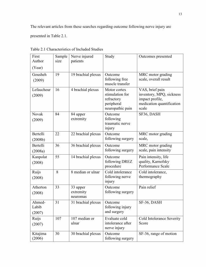
The relevant articles from these searches regarding outcome following nerve injury are
presented in Table 2.1.
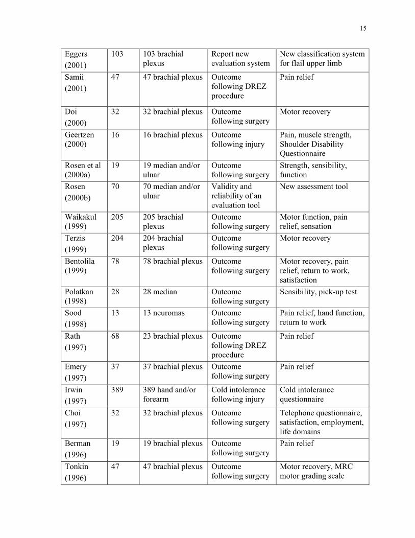
Table 2.1 Characteristics of Included Studies
First Author
(Year)
Sample size
Nerve injured patients
Study Outcomes presented
Gousheh
(2009)
19 19 brachial plexus Outcome following free muscle transfer
MRC motor grading scale, overall result
Lefaucheur
(2009)
16
4 brachial plexus Motor cortex stimulation for refractory peripheral neuropathic pain
VAS, brief pain inventory, MPQ, sickness impact profile, medication quantification scale
Novak
(2009)
84 84 upper extremity
Outcome following traumatic nerve injury
SF36, DASH
Bertelli
(2008b)
22 22 brachial plexus Outcome following surgery
MRC motor grading scale,
Bertelli
(2008a)
36 36 brachial plexus Outcome following surgery
MRC motor grading scale, pain intensity
Kanpolat
(2008)
55 14 brachial plexus Outcome following DREZ procedure
Pain intensity, life quality, Karnofsky Performance Scale
Ruijs
(2008)
8 8 median or ulnar Cold intolerance following nerve injury
Cold intolerance, thermography
Atherton
(2008)
33 33 upper extremity neuromas
Outcome following surgery
Pain relief
Ahmed-Labib
(2007)
31 31 brachial plexus Outcome following injury and surgery
SF-36, DASH
Ruijs
(2007)
107 107 median or ulnar
Evaluate cold intolerance after nerve injury
Cold Intolerance Severity Score
Kitajima (2006)
30 30 brachial plexus Outcome following surgery
SF-36, range of motion
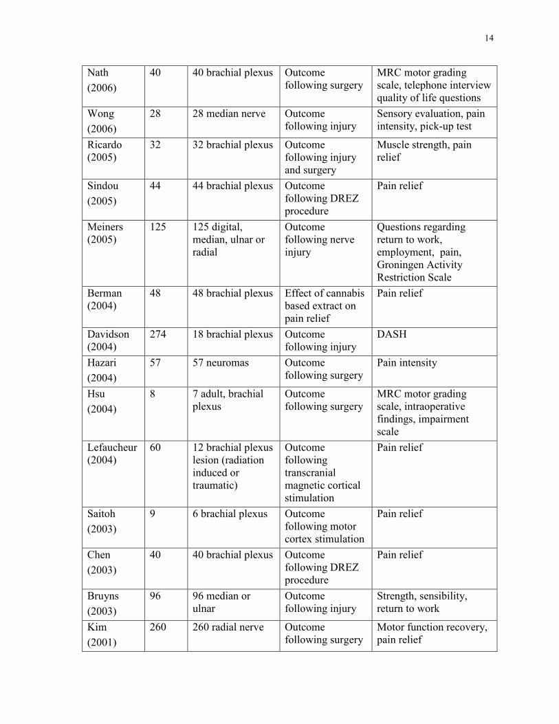
14
Nath
(2006)
40 40 brachial plexus Outcome following surgery
MRC motor grading scale, telephone interview quality of life questions
Wong
(2006)
28 28 median nerve Outcome following injury
Sensory evaluation, pain intensity, pick-up test
Ricardo (2005)
32 32 brachial plexus Outcome following injury and surgery
Muscle strength, pain relief
Sindou
(2005)
44 44 brachial plexus Outcome following DREZ procedure
Pain relief
Meiners (2005)
125 125 digital, median, ulnar or radial
Outcome following nerve injury
Questions regarding return to work, employment, pain, Groningen Activity Restriction Scale
Berman (2004)
48 48 brachial plexus Effect of cannabis based extract on pain relief
Pain relief
Davidson (2004)
274 18 brachial plexus Outcome following injury
DASH
Hazari
(2004)
57 57 neuromas Outcome following surgery
Pain intensity
Hsu
(2004)
8 7 adult, brachial plexus
Outcome following surgery
MRC motor grading scale, intraoperative findings, impairment scale
Lefaucheur (2004)
60 12 brachial plexus lesion (radiation induced or traumatic)
Outcome following transcranial magnetic cortical stimulation
Pain relief
Saitoh
(2003)
9 6 brachial plexus Outcome following motor cortex stimulation
Pain relief
Chen
(2003)
40 40 brachial plexus Outcome following DREZ procedure
Pain relief
Bruyns
(2003)
96 96 median or ulnar
Outcome following injury
Strength, sensibility, return to work
Kim
(2001)
260 260 radial nerve Outcome following surgery
Motor function recovery, pain relief
15
Eggers
(2001)
103 103 brachial plexus
Report new evaluation system
New classification system for flail upper limb
Samii
(2001)
47 47 brachial plexus Outcome following DREZ procedure
Pain relief
Doi
(2000)
32 32 brachial plexus Outcome following surgery
Motor recovery
Geertzen (2000)
16 16 brachial plexus Outcome following injury
Pain, muscle strength, Shoulder Disability Questionnaire
Rosen et al (2000a)
19 19 median and/or ulnar
Outcome following surgery
Strength, sensibility, function
Rosen
(2000b)
70 70 median and/or ulnar
Validity and reliability of an evaluation tool
New assessment tool
Waikakul (1999)
205 205 brachial plexus
Outcome following surgery
Motor function, pain relief, sensation
Terzis
(1999)
204 204 brachial plexus
Outcome following surgery
Motor recovery
Bentolila (1999)
78 78 brachial plexus Outcome following surgery
Motor recovery, pain relief, return to work, satisfaction
Polatkan (1998)
28 28 median Outcome following surgery
Sensibility, pick-up test
Sood
(1998)
13 13 neuromas Outcome following surgery
Pain relief, hand function, return to work
Rath
(1997)
68 23 brachial plexus Outcome following DREZ procedure
Pain relief
Emery
(1997)
37 37 brachial plexus Outcome following surgery
Pain relief
Irwin
(1997)
389 389 hand and/or forearm
Cold intolerance following injury
Cold intolerance questionnaire
Choi
(1997)
32 32 brachial plexus Outcome following surgery
Telephone questionnaire, satisfaction, employment, life domains
Berman
(1996)
19 19 brachial plexus Outcome following surgery
Pain relief
Tonkin
(1996)
47 47 brachial plexus Outcome following surgery
Motor recovery, MRC motor grading scale
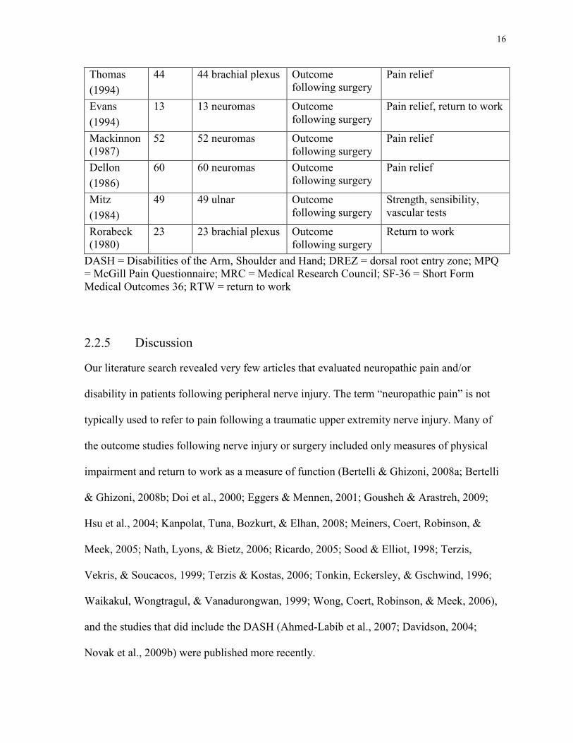
16
Thomas
(1994)
44 44 brachial plexus Outcome following surgery
Pain relief
Evans
(1994)
13 13 neuromas Outcome following surgery
Pain relief, return to work
Mackinnon (1987)
52 52 neuromas Outcome following surgery
Pain relief
Dellon
(1986)
60 60 neuromas Outcome following surgery
Pain relief
Mitz
(1984)
49 49 ulnar Outcome following surgery
Strength, sensibility, vascular tests
Rorabeck (1980)
23 23 brachial plexus Outcome following surgery
Return to work
DASH = Disabilities of the Arm, Shoulder and Hand; DREZ = dorsal root entry zone; MPQ = McGill Pain Questionnaire; MRC = Medical Research Council; SF-36 = Short Form Medical Outcomes 36; RTW = return to work
2.2.5 Discussion
Our literature search revealed very few articles that evaluated neuropathic pain and/or
disability in patients following peripheral nerve injury. The term “neuropathic pain” is not
typically used to refer to pain following a traumatic upper extremity nerve injury. Many of
the outcome studies following nerve injury or surgery included only measures of physical
impairment and return to work as a measure of function (Bertelli & Ghizoni, 2008a; Bertelli
& Ghizoni, 2008b; Doi et al., 2000; Eggers & Mennen, 2001; Gousheh & Arastreh, 2009;
Hsu et al., 2004; Kanpolat, Tuna, Bozkurt, & Elhan, 2008; Meiners, Coert, Robinson, &
Meek, 2005; Nath, Lyons, & Bietz, 2006; Ricardo, 2005; Sood & Elliot, 1998; Terzis,
Vekris, & Soucacos, 1999; Terzis & Kostas, 2006; Tonkin, Eckersley, & Gschwind, 1996;
Waikakul, Wongtragul, & Vanadurongwan, 1999; Wong, Coert, Robinson, & Meek, 2006),
and the studies that did include the DASH (Ahmed-Labib et al., 2007; Davidson, 2004;
Novak et al., 2009b) were published more recently.
17
The IASP defines “neuropathic pain” as resulting from a lesion or disease in the peripheral or
central nervous system, and pain following a traumatic peripheral nerve injury would
therefore be classified as neuropathic pain. However, traumatic peripheral nerve injuries
excluding amputation injuries are not frequently included in the neuropathic pain literature,
and there have been few reports of patients with peripheral nerve injuries included among
other more common etiologies (Dworkin, Jensen, Gammaitoni, Olaleye, & Galer, 2007;
Jensen, Chodroff, & Dworkin, 2007; Meyer-Rosberg et al., 2001b; Meyer-Rosberg et al.,
2001a; Toth, Lander, & Wiebe, 2009). In a literature review by Jensen et al. (2007),
neuropathic pain was negatively associated with health related quality of life and stronger
associations were demonstrated when pain specific measures were compared to more generic
measures. Numerous etiologies were included in the review by Jensen et al. and there was no
differentiation between types of lesions. An overview of neuropathic pain by Dworkin et al.
(2007) identified nine common peripheral neuropathic pain syndromes. Traumatic injury was
not listed among these neuropathic pain syndromes. Meyer-Rosberg et al. (2001b) evaluated
the burden of illness in patients with neuropathic pain and among their sample of patients
was a small number with traumatic peripheral nerve injury. The authors reported a high level
of pain with significantly lower SF-36 scores in all domains compared to normative data, but
information was not presented specifically on the patients with traumatic peripheral nerve
injury.
In the surgical literature, studies that report outcomes following peripheral nerve injuries
rarely report information about pain (e.g., pain quality, intensity, frequency of episodes,
duration). In our survey of peripheral nerve surgeons, only 52% reported formally assessing
pain in patients referred primarily for motor or sensory dysfunction following nerve injury,
18
and in patients referred for pain, the most frequent method of assessing pain was a verbal
patient response (Novak, Anastakis, Beaton, & Katz, 2009a). This lack of detailed
assessment of neuropathic pain in patients with nerve injury parallels the underrepresentation
of pain assessment in the surgical literature following traumatic peripheral nerve injury.
The paucity of citations in the literature regarding neuropathic pain resulting from traumatic
peripheral nerve injury may be related to the relatively small number of cases of nerve injury
compared to other causes of neuropathic pain and other types of trauma. An urban population
survey from the United Kingdom reported a 45% prevalence of chronic pain, with 8% of
neuropathic origin (Torrance et al., 2006). The causes of neuropathic pain were not reported
in this study. A 3.3% prevalence of neuropathic pain was reported in a study from Austria, in
which the majority of subjects identified non-traumatic etiologies as the cause of the pain
(Gustorff et al., 2008). The exact prevalence of traumatic nerve injuries is difficult to
determine (Ekholm, Ponzer, Tornkvist, Adami, & Tidermark, 2008; Midha, 1997; Noble,
Munro, Prasad, & Midha, 1998). Midha (1997) reported that 4,538 trauma patients were seen
from January 1986 to December 1994 at a level 1 trauma center in Ontario, Canada,
including 60 patients with brachial plexus injuries. Noble et al. (1998) reported the
prevalence of upper and lower extremity peripheral nerve injuries from the same institution.
From January 1986 to November 1996, 5,777 trauma patients were seen; 200 nerve injuries
were identified in 162 patients (2.8%). An epidemiological study of humeral fractures from
Sweden reported that only 8.5% of injuries involved a radial nerve palsy (Ekholm et al.,
2006). These studies reveal a low prevalence of peripheral nerve injuries compared to the
overall prevalence of traumatic injuries; however, the morbidity associated with these
19
injuries may be severe, and early comprehensive assessment and intervention is essential for
optimal outcomes.
2.2.5.1 Outcome Measures
Studies that have evaluated outcome following peripheral nerve injury have routinely
focused on physical impairment, including sensory and motor dysfunction (Bertelli et al.,
2008a; Bertelli et al., 2008b; Bruyns et al., 2003; Doi et al., 2000; Gousheh et al., 2009; Hsu
et al., 2004; Mitz, Meriaux, & Vilain, 1984; Nath et al., 2006; Polatkan, Orhun, Polatkan,
Nuzumlali, & Bayri, 1998; Ricardo, 2005; Rosen, Dahlin, & Lundborg, 2000a; Terzis et al.,
1999; Tonkin et al., 1996; Wong et al., 2006). Outcome measures following motor nerve
injury usually include manual muscle testing with the Medical Research Council (MRC)
grading system, amount of weight that can be lifted or subjective grading by the researchers
on scales ranging from “excellent” to “poor” (Bengtson et al., 2008). Patient functional
assessment and/or pain evaluation are rarely included in outcome studies. The few studies
that have reported pain predominantly included patients following brachial plexus nerve
injuries (Bentolila, Nizard, Bizot, & Sedel, 1999; Berman, Birch, & Anand, 1998; Bertelli et
al., 2008a; Bruxelle, Travers, & Thiebaut, 1988; Geertzen, Groothoff, Nicolai, & Rietman,
2000; Htut, Misra, Anand, Birch, & Carlstedt, 2006; Kato, Htut, Taggart, Carlstedt, & Birch,
2006; Kanpolat et al., 2008; Samii, Bear-Henney, Ludeman, Tatagiba, & Blomer, 2001;
Waikakul et al., 1999). In these studies, traumatic injuries, root avulsions, and injuries
proximal to the dorsal root ganglion were associated with more pain; surgical intervention
and the timing of surgery relative to injury were identified as important factors in alleviating
pain. These outcome studies reported pain intensity and frequency; however, validated
20
patient report questionnaires to assess the impact of the pain or impairment on the patient
were not included.
Riess, Cogbill, Patel, Lambert & Mathiason (2007) reported significant short and long term
disability following scapulothoracic dissociation compared to patients with brachial plexus
nerve injuries. Outcome was evaluated by telephone interview; interviews included basic
questions about upper extremity strength and work. No validated outcome measures were
used in this study, and participants were not asked about pain. In another telephone survey,
Choi et al. (1997) contacted 32 patients with brachial plexus injury and administered quality
of life questions from the US General Social Survey. Moderately high general life
satisfaction and quality of life were reported; 75% of patients reported “significant pain” and
38% were using pain medications.
Rating scales and composite scores have been introduced for the assessment of sensibility,
motor function and impairment following nerve injury. In general, these rating scales and
composite scores have placed very little emphasis on pain, including pain associated with
cold sensitivity. The MRC scale (Medical Research Council of the U.K., 1976) is a six-point
scale ranging from 0 to 5 based on the function of the muscle against gravity or with manual
resistance; modifications of this scale have been described. Highet and Zachary introduced a
scale to categorize recovery of sensibility that was later modified by Mackinnon and Dellon
(1988). This scale includes a range from “no sensibility” to “complete recovery” and
considers touch, two-point discrimination, and pain response. The composite score
introduced by Rosen and Lundborg (2000b) includes three domains (sensory, motor,
pain/discomfort); cold intolerance and hyperaesthesia are ranked on a numeric scale, and
these two parameters make up the pain/discomfort domain. Aberg et al. (2007) presented a
21
method for clinical evaluation following peripheral nerve injury. They investigated the
applicability of a battery of clinical tests in a small sample of 15 patients with median nerve
injuries and 15 control subjects. The tests in this clinical assessment were sensory recovery
(two-point discrimination, cutaneous pressure thresholds, pin prick, thermal thresholds,
sensory nerve conduction velocity and amplitude), motor recovery (manual muscle testing,
grip and pinch strength, motor nerve conduction velocity and amplitude, needle
electromyography), and functional recovery (four questions about function, pain, cold
intolerance and dysaesthesia; DASH; motor performance test; Sollerman hand function test;
sensorimotor test). Only one question addressed pain (present or absent) and one question
addressed cold intolerance (present or absent), and the study included no quantification of
their intensity, frequency, or impact on functional outcome.
2.2.5.2 Measurement of Neuropathic Pain
Pain is a subjective experience that is best evaluated by subjective patient report. Various
approaches have been described for assessing neuropathic pain, ranging from simple verbal
rating scales (VRS), numeric rating scales (NRS), and visual analogue scales (VAS) to multi-
item, multidimensional questionnaires that measure the quality and intensity of pain. The
VAS, NRS, and VRS, which usually provide a unidimensional measure of pain intensity (or
pain affect, depending on the scale anchors), are commonly used to measure pain in the
clinical setting. Introduced by Melzack (1975), the McGill Pain Questionnaire (MPQ) is the
most frequently used and cited pain questionnaire. However, only one outcome study used
the MPQ to assess pain following treatment for neuropathic pain (Lefaucheur et al., 2009).
22
The MPQ, developed by Melzack (1975) to obtain quantitative and qualitative measures of
the experience of pain, yields two global scores: the pain rating index (PRI) and the present
pain intensity (PPI). The PRI is the sum of the rank values of the 75 words chosen from 20
sets of qualitative words, each containing two to six adjectives that describe the sensory,
affective, and evaluative properties of pain. The lists of pain descriptors are read to patients,
who are asked to choose the word in each category that best describes their pain at the
moment. The PPI is rated on a scale of 0 (none) to 5 (excruciating). The short form MPQ
(SF-MPQ) was developed by Melzack (1987) for use when time is limited and when more
information is required than is provided by unidimensional measures such as the VAS. The
SF-MPQ consists of 15 adjectives from the sensory (n = 11) and affective (n = 4) categories
of the original MPQ. Each adjective is rated on a four-point scale.
The selection of the optimal treatment approach and/or medication may be optimized by
differentiating between nociceptive and neuropathic pain (Bennett et al., 2007). A
modification of the SF-MPQ has been recently published that is reliable and valid for patients
with neuropathic and non-neuropathic pain (Dworkin et al., 2009). The two main differences
between the SF-MPQ and the modified SF-MPQ are the addition of seven adjectives relevant
to neuropathic pain and the inclusion of a 10-point numeric rating scale to rate the intensity
of each descriptor. Each version of the MPQ has been shown to have at least adequate
psychometric properties, and each is a reliable and valid measure of acute and chronic pain
(Melzack & Katz, 2006). Other questionnaires that have been described for assessment of
neuropathic pain include the Neuropathic Pain Scale (Galer & Jensen, 1997), the Pain
Quality Assessment Scale (Jensen et al., 2006), and the PainDetect (Freynhagen, Baron,
Gockel, & Tolle, 2006). The Neuropathic Pain Scale is a 10-item scale in which patients are
23
asked to rank various dimensions (intensity, quality, allodynia) of pain on an 11-point NRS.
This scale was validated with a diverse group of patients that included those with peripheral
nerve injury, and it has been shown to be sensitive to alterations in the quality and intensity
of neuropathic pain (Galer et al., 1997; Jensen, 2004; Jensen et al., 2005). However, it was
limited to those patients who attended a chronic pain clinic, and, as outlined by the authors, it
may not represent all of the pain qualities that patients with neuropathic pain experience. The
Pain Quality Assessment Scale is a 20-item questionnaire modified from the Neuropathic
Pain Scale to include more descriptors to differentiate neuropathic and non-neuropathic pain.
This scale was validated in 40 patients with carpal tunnel syndrome; this group did not
include other etiologies of neuropathic pain (Jensen et al., 2006). The PainDetect is 20-item
questionnaire developed to evaluate the qualities associated with neuropathic pain
(Freynhagen et al., 2006). Patients are asked to rank the degree of their symptoms on a scale
of 0 to 10 for different qualities of pain. A higher score indicates more pain. This
questionnaire was validated in a sample of patients with chronic low back pain; and the
group did not include patients with a traumatic nerve injury. The authors reported a
sensitivity of 85% and specificity of 80% in classifying patients with neuropathic pain
(Freynhagen et al., 2006). Although these findings indicate moderate sensitivity and
specificity, this measure has not been validated for use in patients with neuropathic pain
following traumatic nerve injury. None of these neuropathic pain questionnaires has been
universally accepted, and none has been used exclusively in patients with traumatic
peripheral nerve injury.
24
2.2.5.3 Assessment of Disability and Health Status
Biopsychosocial models of disablement and health linking the biomedical, social and
personal perspectives have been developed by the World Health Organization (WHO) and by
others including Nagi, Verbrugge and Jette (Jette, 2006; Verbrugge & Jette, 1994; World
Health Organization, 2001). Based on Nagi’s model, Verbrugge and Jette (1994) described
the disablement process as a pathway between active pathology, impairment, functional
limitations, and disability, with consideration of other individual and risk factors (Jette &
Haley, 2005). Within the framework of the International Classification of Function,
Disability and Health (ICF) model developed by the WHO, body structures and functions
(physiological function), activity, and participation are considered in the context of life
domains with interaction between the contextual environmental and personal factors (World
Health Organization, 2001; Jette, 2009). In terms of nerve injury, this model takes into
consideration the interaction between the condition after injury (physical impairment and
activity performance, including participation) and contextual (personal and environmental)
factors.
Generic health measures such as the Short Form Medical Outcomes 36 (SF-36) were
designed to assess health status. Responses to the SF-36 may be calculated in eight domains
and/or summarized in a physical and a mental component score (Brazier et al., 1992; Garratt,
Ruta, Abdalla, Buckingham, & Russell, 1993; Hopman et al., 2000; McHorney & Raczek,
1993; Ware & Sherbourne, 1992). Meyer-Rosberg et al. (2001a) compared scores on the SF-
36 and the Nottingham Health Profile for a diverse group of patients with neuropathic pain.
They found that these patients had poorer scores compared to normative values and patients
with high levels of pain scored worse on both measures. In a retrospective chart review,
25
patients with traumatic upper extremity nerve injuries had a significantly lower health status
in all SF-36 domains and component scores (Hopman et al., 2000; Novak et al., 2009b).
Ahmed-Labib et al. (2007) reported significantly worse health status in all SF-36 scores
except general health, vitality, and the mental component score, as well as a higher level of
disability, in patients following brachial plexus injury and reconstructive surgery. Based on
correlational analysis, the authors concluded that root avulsion injuries and delayed surgical
repair were associated with poorer functional outcome. Kitajima, Doi, Hattori, Takka &
Estrella (2006) evaluated 30 patients with brachial plexus nerve injuries with a minimum
follow-up of 12 months. Compared to the Japanese normative data, the nerve injured patients
had a significantly lower health status (physical function, bodily pain, role physical and
physical composite score). Generic questionnaires are useful for assessing general health
status, but they may be limited in the assessment of upper extremity outcome. Disease-
specific questionnaires such as the DASH may be more sensitive to diagnoses and
pathologies affecting the upper extremity.
The DASH is a 30-item patient report measure to assess upper extremity disability that has
established psychometric properties (Beaton et al., 2001; Beaton et al., 2005; Dias, Bhowal,
Wildin, & Thompson, 2001; Gummesson et al., 2003; Gummesson et al., 2006; Hudak et al.,
1996; SooHoo et al., 2008). Although the DASH is the most validated measure of upper
extremity disability, it was not commonly used in the outcome studies of patients with nerve
injury found in our literature search. Studies that evaluated disability with nerve injury
reported high DASH scores (Ahmed-Labib et al., 2007; Bushnell, McWilliams, Whitener, &
Messer, 2008; Davidson, 2004; Ekholm et al., 2008; Novak et al., 2009b; Topel et al., 2009).
In the evaluation of patients following an upper extremity nerve injury, substantial disability
26
was found, and this was predicted by pain, older age, and brachial plexus injury (Novak et
al., 2009b). Davidson (2004) used the DASH to evaluate 274 patients following upper
extremity traumatic injuries, including amputations and brachial plexus injuries. High levels
of disability were reported, and these were significantly higher in patients with brachial
plexus injuries (Davidson, 2004). Ahmed-Labib et al. (2007) evaluated 31 patients following
surgery for a brachial plexus injury; assessment included the DASH and SF-36. These
patients reported high levels of disability, and their scores for six of the eight SF-36 domains
were significantly worse compared to normative data. Topel et al. (2009) evaluated 33
patients following upper extremity arterial trauma. Patients with concomitant nerve injury
(81%) had more functional deficits, with a significantly higher DASH score and lower SF-36
physical composite score. Patients with a radial nerve palsy following humeral fractures were
evaluated by Ekholm et al. (2008) using patient reported outcome, including the DASH and
the SF-36; most patients reported low disability levels and good health status. Following
digital nerve repair, Bushnell et al. (2008) reported low levels of disability as assessed by the
QuickDASH. Wong, Fung, Cuh & Chan (2007) evaluated 146 patients following traumatic
hand injuries, both before participation in a rehabilitation program and at discharge. Both
DASH scores and QuickDASH scores were included in the data analysis, which showed a
high correlation (r = 0.96) between these scores at admission and discharge. There was a
significant improvement in DASH scores at discharge; patients who did not return to work
reported significantly more disability at both admission and discharge (p < 0.05).
27
2.2.5.4 Considerations of Cold Sensitivity and Contextual (Psychosocial) Factors
2.2.5.4.1 Cold Sensitivity
Cold sensitivity, described as pain or discomfort, stiffness, sensory disturbance, and colour
changes with exposure to cold, is frequently reported following traumatic upper extremity
injuries (Graham & Schofield, 2008; Irwin, Gilbert, Terenghi, Smith, & Green, 1997; Novak
et al., 2009a; Ruijs, Jaquet, Brandsma, Daanen, & Hovius, 2008; Vaksvik, Hetland, Rokkum,
& Holm, 2009). The terms “cold sensitivity” and “cold intolerance” have been used
interchangeably in the literature; in this review, the term “cold sensitivity” is used, except in
cases where reference is made to published studies whose authors have used the term “cold
intolerance”. The symptoms of cold sensitivity are often attributed to poor outcome
following traumatic peripheral nerve injuries and have been reported more frequently in
patients with digital amputations and replantations (Backman, Nystrom, Backman, & Bjerle,
1993; Campbell & Kay, 1998; Collins, Novak, Mackinnon, & Weisenborn, 1996; Craigen,
Kleinert, Crain, & McCabe, 1999; Dabernig, Hart, Schwabegger, Dabernig, & Harpf, 2006;
Graham et al., 2008; Irwin et al., 1997; Lithell, Backman, & Nystrom, 1998; McCabe,
Mizgala, & Glickman, 1991; Nancarrow, Rai, Sterne, & Thomas, 1996; Nylander, Nylander,
& Lassvik, 1987; Ruijs, Jaquet, Daanen, & Hovius, 2006; Ruijs, Jaquet, Van Riel, Daanen, &
Hovius, 2007; Vaksvik et al., 2009). Several studies have supported the continuation of cold
sensitivity symptoms in patients with hand injuries and nerve injuries and following
replantation (Campbell et al., 1998; Collins et al., 1996; Nancarrow et al., 1996; Povlsen,
Nylander, & Nylander, 1995a; Povlsen, Nylander, & Nylander, 1995b; Vaksvik et al., 2009).
Campbell and Kay (1998) evaluated 176 patients following hand injuries, 73% of whom
reported cold related symptoms; most of these were related to pain. Graham and Schofield
28
(2008) evaluated patients more than two years after hand injury; most of these patients (90%
of trauma cases) reported cold intolerance, and only 9% reported an improvement over time.
Long term cold intolerance in patients with hand injuries was evaluated by Nancarrow et al.
(1996); 69% of patients reported cold intolerance, and 97% of these patients continued to
have symptoms five years after injury. Collins et al. (1996) reported long term follow-up of
patients after upper extremity nerve injury. Among patients who were at least five years post
injury, 76% reported cold intolerance, and 87% of these patients reported moderate or severe
symptoms. Dabernig et al. (2006) evaluated patients after digital replantation, with a mean
follow-up time of five years. The mean DASH score was 11 (out of a total possible score of
100), and cold intolerance was reported by 87% of patients. In a diverse group of patients
with neuropathic pain that included traumatic nerve injuries, cold-evoked pain was rated as
the most intense pain (Meyer-Rosberg et al., 2001b).
Evaluation of cold sensitivity in the literature is variable and includes verbal scales, patient
report questionnaires, and physical assessment (Carlsson, Cederlund, Hoglund, Lundborg, &
Rosen, 2008; Collins et al., 1996; Craigen et al., 1999; Graham et al., 2008; Irwin et al.,
1997; McCabe et al., 1991; Ruijs et al., 2006; Traynor & MacDermid, 2008). Traynor and
MacDermid (2008) compared cold immersion and a patient report questionnaire in healthy
control subjects; while both physical and subjective assessments were reliable, they were not
significantly correlated with each other. Objective tests such as cold immersion,
measurement of rewarming, or arterial pressures may adequately assess vascular status, but
these types of assessments provide no indication of the pain and cold symptoms perceived by
the patient (Backman et al., 1993; Nylander et al., 1987; Povlsen et al., 1995a; Povlsen et al.,
1995b; Traynor et al., 2008). Patient report questionnaires such as the Cold Sensitivity
29
Severity Scale, introduced by McCabe et al. (1991) and the Cold Intolerance Symptom Scale
(Irwin et al., 1997; Ruijs et al., 2006; Ruijs et al., 2007) introduced by Irwin et al. (1997)
provide an opportunity for patients to rank their symptoms on a numeric scale. In both scales,
a higher score indicates a greater degree of cold sensitivity.
2.2.5.4.2 Contextual (Psychosocial) Factors
While pain questionnaires and rating scales (verbal and numerical) can assess pain intensity,
quality, and frequency and are often used in the surgical literature (Atherton, Fabre, Anand,
& Elliot, 2008; Berman, Anand, Chen, Taggart, & Birch, 1996; Berman, Symonds, & Birch,
2004; Chen, Lu, & Yeh, 2003; Dellon & Mackinnon, 1986; Emery, Blondet, Mertens, &
Sindou, 1997; Evans & Dellon, 1994; Hazari & Elliot, 2004; Kim, Kam, Chandika, Tiel, &
Kline, 2001a; Lefaucheur et al., 2004; Mackinnon & Dellon, 1987; Rath et al., 1997;
Rorabeck, 1980; Saitoh et al., 2003; Sindou, Blondet, Emery, & Mertens, 2005; Thomas &
Kitchen, 1994), these types of measures do not evaluate the psychosocial factors that are
often associated with neuropathic pain. The European Federation of Neurological Societies
presented guidelines for the assessment of neuropathic pain (Cruccu et al., 2004). A baseline
assessment can be achieved with NRS, VRS, or VAS, and more in-depth assessment can
include pain descriptors, temporal factors, and functional impact (Cruccu et al., 2004; Gilron,
Watson, Cahill, & Moulin, 2006). In our recent survey of peripheral nerve surgeons, 75% of
surgeons reported that they quantitatively assess pain in patients referred for pain following
nerve injury, but very few surgeons used a validated questionnaire to assess pain in these
patients (Novak et al., 2009a). Although it is recognized that psychosocial factors may
contribute to poor outcome and ongoing neuropathic pain, these factors are rarely reported in
30
the surgical literature following traumatic peripheral nerve injuries (Doornberg et al., 2005;
Lindenhovius, Buijze, Kloen, & Ring, 2008; Lozano Calderon et al., 2008a; Novak et al.,
2009b; Souer et al., 2008).
Associated contextual (psychosocial) factors have been shown to play an important role in
the experience of chronic pain in other populations. In particular, the role of depression
(Nicholson & Verma, 2004), fear-avoidance (Leeuw et al., 2007), pain catastrophizing
(Edwards, Bingham, Bathon, & Haythornthwaite, 2006), and post-traumatic stress disorder
(PTSD) symptoms (Asmundson, Coons, Taylor, & Katz, 2002), in other chronic pain
populations warrants serious consideration and study in patients with traumatic peripheral
nerve injuries. Given the traumatic nature of the injuries these patients have sustained, we
believe that it is essential to thoroughly evaluate not only pain but also PTSD symptoms.
PTSD typically develops after exposure to an event or situation that is perceived to be
threatening to the physical or emotional integrity of an individual. DSM-IV-TR diagnostic
criteria (American Psychiatric Association, 2000) for PTSD cover three symptom clusters:
(1) re-experiencing the traumatic event (e.g., nightmares and “flashbacks”); (2) emotional
numbing (e.g., feeling detached from others) and avoidance of thoughts, feelings and
activities associated with the trauma; and (3) increased arousal (e.g., insomnia, exaggerated
startle reflex, hypervigilance). Recent data show that chronic pain and PTSD are strongly
associated (Asmundson et al., 2002; Asmundson & Katz, 2008). One possible reason for the
high co-morbidity of PTSD and chronic pain may be the substantial symptom overlap
common to both disorders, including anxiety and hyperarousal, attentional biases, avoidant
behaviours, emotional lability, and elevated somatic focus. The overlap in symptoms
suggests that the two disorders may be mutually maintaining or may share an underlying
31
psychological vulnerability that places some individuals with a greater likelihood of
developing one or both disorders. Anxiety sensitivity has been identified as one of the trait
vulnerability factors that predispose individuals to develop chronic pain, PTSD or both
(Asmundson et al., 2002). The intractability of the two disorders is not surprising when
viewed in the context of mutual maintenance and shared vulnerability models. This
underscores the importance of screening for both disorders when either one is present,
especially in patients who have sustained a traumatic nerve injury, given the painful and
traumatic nature of the precipitating event.
2.2.6 Limitations
The major limitation of this review is the paucity of literature reporting outcomes following
nerve injury beyond physical impairment. Assessment following nerve injury should include
measures of physical impairment such as range of motion, strength and sensibility. Pain
assessment with questionnaires such as the modified MPQ to assess both neuropathic and
nociceptive pain will provide valuable information beyond pain intensity. Additional patient
report questionnaires such as the DASH will provide information about upper extremity
disability, and questionnaires to evaluate for symptoms of depression, fear-avoidance, pain
catastrophizing, and PTSD symptoms will be useful in identifying concomitant psychosocial
factors that may affect outcome.
32
2.2.7 Future Directions
The assessment of pain in patients with neuropathic pain secondary to traumatic peripheral
nerve injuries has lagged behind that of patients with neuropathic pain of other etiologies.
The relative lack of information extends beyond measures of pain per se; as described in this
review, there is very little information on the associated upper extremity disability, pain
disability or health status in patients with traumatic peripheral nerve injuries. Assessment in
future studies should include measures of physical impairment, pain, associated contextual
(personal and environmental) factors, and functional outcomes, such as disability, to provide
a more comprehensive patient evaluation and the opportunity to maximize patient outcome
and minimize morbidity following nerve injury.
33
2.3 Classification of Peripheral Nerve Injury
This section reviews the commonly used systems for classifying nerve injury. Traumatic
nerve injuries vary in severity of nerve injury relative to the severity of the trauma. Nerve
injury will result in a number of changes at the level of the injury, the distal and proximal
nerve segment, the distal sensory/motor end organ, the cell body and the central cortex. Two
classification systems for nerve injury are commonly cited (Seddon, 1943; Sunderland,
1978). Seddon (1943) introduced a classification that consisted of 3 levels; neurapraxia,
axonotmesis and neurotmesis. Sir Sydney Sunderland (1978) expanded the classification to
include five degrees of nerve injury (I to V) and each degree reflects an increase in severity
of the nerve injury. A first degree injury which is similar to Seddon’s neurapraxia is a
temporary conduction block as a result of demyelination of the nerve. Once the nerve has
remyelinated and the conduction block is resolved, motor and/or sensory function will return.
With a first degree injury, complete recovery is expected within 12 weeks following injury.
A second degree injury involves more severe trauma and injury to the nerve. It is similar to
the type of injury described by Seddon as an axonotmesis. Distal and proximal axonal
degeneration occur and changes are evident in the electrodiagnostic studies with indications
of muscle fibre denervation. Nerve regeneration will occur at the rate of approximately one
millimetre (mm) per day. With a second degree injury, complete recovery is expected since
the endoneurial sheath is intact, unless the rate of muscle degeneration exceeds the rate of
nerve regeneration; that is if the injury is too far proximal for muscle reinnervation to occur
before complete muscle degeneration has occurred and thus reinnervation is not possible. A
third degree injury involves more severe trauma and similar to a second degree injury,
proximal and distal axonal degeneration will occur. However, in contrast to a second degree
34
injury, the endoneurial tubes are not intact with a third degree injury. Therefore the
regenerating axons may not reach their original end organs and incomplete recovery is
anticipated. Patients with these types of injuries will benefit from motor and sensory
reeducation to maximize functional outcome. A fourth degree injury is a neuroma-in-
continuity and this neuroma will preclude the axons from advancing distally beyond the level
of injury. A fifth degree injury is complete nerve transection. Fourth and fifth degree injuries
require surgical intervention to restore neural continuity. Nerve regeneration will then
proceed similar to the recovery seen with a third degree injury at the rate of approximately
one mm per day with an advancing Tinel’s sign. A sixth degree nerve injury is the term used
to describe a mixed nerve injury which involves varying degrees of nerve injury (I to V)
within the same nerve (Mackinnon & Dellon, 1988).
Sunderland II, III, IV and V degree injuries involve axonal degeneration proximal and distal
to the site of nerve injury and denervation of the sensory end organs and muscle fibres
(Mackinnon et al., 1988). Recovery of motor and sensory function is dependent upon
reinnervation of the motor end plates and sensory receptors. Sensory recovery is possible for
many years following nerve injury, however the quality of sensibility may be compromised
with long delays prior to reinnervation of the sensory end organs (Mackinnon et al., 1988).
Reinnervation of the muscle fibres requires that the motor axons reach the target muscle
within a critical period of time (Fu & Gordon, 1995; Lien, Cederna, & Kuzon, 2008). The
exact duration of time in which motor fibre reinnervation is possible is unknown. However in
an animal model, motor recovery substantially declined when the surgical repair occurred
greater than one month following injury (Kobayashi et al., 1997). However the critical time
period for reinnervation is related to the type of nerve injury; shorter for transection nerve
35
injuries (Sunderland V degree injury) compared to in-continuity nerve lesions such as a crush
injury which may result in a Sunderland II and III degree injury (Mackinnon et al., 1988).
Motor recovery depends upon a viable source of regenerating motor axons and better
recovery occurs with shorter durations of muscle denervation.
Reinnervation of the motor fibres and sensory end organs following nerve injury are
dependent upon the degree of nerve injury, patient and injury factors. However for some
patients, the recovery that occurs is less than optimal and remains a challenging problem.
2.4 Recovery Following Peripheral Nerve Injury
Traumatic peripheral nerve injury results in a number of peripheral and central nervous
system responses. Recovery following nerve injury depends upon timely nerve regeneration
and reinnervation of the distal sensory and motor targets and correct remapping of the cortex.
Management of patients following nerve injury includes both non-operative and operative
treatments and there are many reports of outcome following these treatments. Assessment
following nerve injury includes patient or surgeon subjective assessment, patient reported
questionnaires and/or objective measures such as brain imaging, electromyography or nerve
conduction studies and there are no widely accepted standardized methods to report outcome.
2.4.1 Traumatic Injury to the Brachial Plexus
Traumatic injury to the brachial plexus may cause avulsion of multiple roots or injury to a
single trunk, division, or nerve, and may result in substantial physical impairment due to loss
36
of motor and/or sensory function or pain. There is no standard evaluation to assess patients
following brachial plexus nerve injury and there is wide variation in the methods used to
report outcome and recovery. Recovery of motor function following brachial plexus nerve
injury or surgery is most often reported using the MRC scale, a modification of the MRC
scale or a surgeon graded outcome. In patients with no recovery following injury, surgical
intervention using nerve grafts and/or nerve transfer to restore distal shoulder, elbow and in
some cases hand function have been used. Reported outcomes have ranged from incomplete
to complete recovery of distal shoulder, elbow and hand function. In most studies, individual
surgeon assessment or a muscle grading system was used, although these muscle grading
systems have not been uniformly utilized (Bentolila et al., 1999; Bertelli & Ghizoni, 2007;
Bertelli et al., 2008b; Cardenas-Mejia, O'Boyle, Chen, & Chuang, 2007; Chuang, Yeh, &
Wei, 1992; Chuang, Lee, Hashem, & Wei, 1995; Chuang, Epstein, Yeh, & Wei, 1993;
Ferraresi, Garozzo, & Buffatti, 2004; Gousheh et al., 2009; Gu, 2007; Haninec, Samal,
Tomas, Houstava, & Dubovy, 2007; Hsu et al., 2004; Htut, Misra, Anand, Birch, & Carlstedt,
2007; Jivan, Kumar, Wiberg, & Kay, 2009; Kim, Cho, Tiel, & Kline, 2003;
Leechavengvongs, Witoonchart, Uerpairojkit, Thuvasethakul, & Ketmalasiri, 1998;
Mackinnon, Novak, Myckatyn, & Tung, 2005; Merrell, Barrie, Katz, & Wolfe, 2001;
Moiyadi, Devi, & Nair, 2007; Novak, Mackinnon, & Tung, 2003; Oberlin et al., 1994;
Richardson, 1997; Riess, Cogbill, Patel, Lambert, & Mathiason, 2007; Suzuki, Doi, Hattori,
& Pagsaligan, 2007; Terzis et al., 1999; Terzis et al., 2006; Tonkin et al., 1996; Tung, Novak,
& Mackinnon, 2003; Waikakul et al., 1999; Zheng, Hou, Gu, Shi, & Guan, 2008). Hand
sensation was reported in patients with brachial plexus avulsion injuries by Hattori et al.
(2009) and the evaluation was performed using vibration and pressure thresholds and two-
37
point discrimination. In these studies of patients with brachial plexus nerve injuries, outcome
was reported in terms of motor or sensory physical impairment.
Brachial plexus injuries which involve root avulsion may result in severe neuropathic pain
(Berman et al., 1998; Berman et al., 1996; Htut et al., 2006; Kato et al., 2006). Studies which
have reported operative procedures for pain management, including the dorsal root entry
zone (DREZ) procedure, have reported varying degrees of success in terms of pain relief
(Bertelli et al., 2008a; Bruxelle et al., 1988; Chen & Tu, 2006; Htut et al., 2006; Chen et al.,
2003; Kato et al., 2006; Samii et al., 2001). Evaluation using instruments other than those
that measure pain relief or pain intensity is rarely included in outcome studies of these
patients.
2.4.2 Traumatic Injury to the Median, Ulnar, Radial, and/or Digital Nerves
Reports of recovery of distal wrist and hand function following injury to the median, ulnar,
radial and/or digital nerves have used a variety of sensory and motor evaluation tools. Injury
to the distal median nerve may result in loss of sensation to the sensory distribution of the
median nerve to the hand and loss of thumb opposition. Compromise of hand sensation can
result in a substantial loss of hand function (Lundborg & Rosen, 2007). Compared to patients
with median nerve injuries, patients with injury to the ulnar nerve have more difficulty with
fine motor function due to the loss of the intrinsic muscles and less difficulty related to hand
sensibility. Injuries to the radial nerve result in motor loss to the wrist and finger extensors
which may result in substantial morbidity.
38
The majority of studies that have reported outcome following distal nerve injuries,
reconstructive surgery and/or hand therapy have evaluated hand sensibility and in some cases
motor function but few have evaluated the impact of the injury on the patient. Motor
recovery following radial nerve injuries have focused on the degree of wrist and finger
extension (Kim et al., 2001a; Shao, Harwood, Grotz, Limb, & Giannoudis, 2005; Shergill,
Bonney, Munshi, & Birch, 2001; Roganovic & Petkovic, 2004). Ekholm et al. (2008) in their
review of recovery following fractures to the humerus did include patient reported disability.
Low levels of disability as indicated by the DASH scores were reported but this may be
indicative spontaneous recovery of the radial nerve function in the majority of patients
(Ekholm et al., 2008). Long term results following tendon transfer for radial nerve palsy were
reported by Altintas et al. (2009). The authors included motor recovery and reported low
levels of disability as shown by low DASH scores.
Outcome studies following (1) surgery to repair or graft the median nerve and (2)
postoperative therapy have reported recovery in terms of sensory recovery such as vibration
and cutaneous pressure thresholds and two-point discrimination (Kato, Minami, Kobayashi,
Takahara, & Ogino, 1998; Kim, Kam, Chandika, Tiel, & Kline, 2001b; Mavrogenis,
Spyridonos, Antonopoulos, Soucacos, & Papagelopoulos, 2009; Meek, Coert, & Wong,
2003; Novak, Kelly, & Mackinnon, 1992; Polatkan et al., 1998; Rosen et al., 2000a;
Roganovic, 2005). Outcome following ulnar nerve injuries in the upper extremity has
focused on the motor recovery utilizing various grading systems (Battiston & Lanzetta, 1999;
Kim, Han, Tiel, Murovic, & Kline, 2003; Terzis & Kokkalis, 2008; Roganovic, 2004). A
study by Ruijs, Jaquet, Kalmijn, Giele & Hovius (2005) evaluated injuries which involved
both the median and ulnar nerves and the authors focused on the predictors of motor recovery
39
and hand sensibility. Studies which have evaluated functional outcome included return to
work as the measure of functional outcome (Bruyns et al., 2003; Jaquet et al., 2001). Jaquet
et al. (2001) evaluated motor and sensory recovery in patients with median and ulnar nerve
injuries and functional outcome was defined as return to work. In this study, the predictors of
not returning to work were poor sensory and motor recovery. Bruyns et al. (2003) evaluated
predictors of return to work in patients with median and/or ulnar nerve injuries. Return to
work was related to median and ulnar nerve injuries, level of education, type of employment
and compliance to therapy. In patients following median and/or ulnar nerve injuries, Taylor
et al. (Taylor, Anastakis, & Davis, 2009) reported cortical changes and plasticity using brain
imaging techniques. These cortical changes were associated with sensory recovery.
Following nerve injury, recovery of function is related to numerous personal, injury and
treatment factors and also widely varies dependent on the specific nerve injured. The impact
of this recovery in terms of quality of life, psychosocial impact, disability or health status has
been reported in very few studies (Ahmed-Labib et al., 2007; Choi et al., 1997; Davidson,
2004; Kitajima, Doi, Hattori, Takka, & Estrella, 2006; Meiners et al., 2005; Nath et al., 2006;
Topel et al., 2009).
2.5 Measurement of Outcome
Reports of outcome following nerve injury commonly include measures of physical
impairment related to function of the sensory end organs or muscle fibres. In contrast, patient
reported function, health related quality of life, measures of psychosocial functioning, or
disability using valid and reliable questionnaires are infrequently included in published
reports following peripheral nerve injury.
40
Assessment of only physical impairment may give an incomplete picture of the impact of the
nerve injury on the patient. Functional outcome related to such factors as the impact on the
patient’s quality of life and psychosocial functioning, employment issues, activities of daily
living or participation in activity may be more indicative of how the patient is functioning
with the residual morbidity following nerve injury. Meiners et al. (2005) reported on patient
disability, employment and leisure issues in 61 patients following nerve repair in the forearm,
wrist or hand. The results showed that 81% of patients returned to their pre-injury job and
that 92% had returned within 12 months. The authors used a VAS to assess patient reported
disabilities and pain. Significant correlations were reported between the activity restriction
scale and pain, hobbies, houskeeping and job as assessed by a VAS. It should be noted that
while there is a large body of empircal literature showing that a VAS is a reliable and valid
instrument for pain measurement, the same cannot be said for the use of a VAS when
measuring other constructs; the psychometric properties of a VAS when used to measure
disability, activity restriction, and housekeeping, etc have not been evaluated. In another
study, the predictors for return to work in patients who had sustained a median or ulnar nerve
injury were reported by Bruyns et al. (2003). The authors reported that 59% of patients had
returned to work and most to their pre-injury job. Return to work was related to education
level, type of employment, compliance to hand therapy and type of injury. Jaquet et al.
(2001) evaluated 220 patients after traumatic forearm or wrist injuries which included
median, ulnar and/or radial nerve injury. Sensory and motor recovery were reported in
addition to return to work which the authors termed “work disability”. In these patients, the
nerve injuries were associated with substantial physical impairment and return to work was
associated with poor motor and sensory recovery.
41
Choi et al. (1997) evaluated 32 patients following brachial plexus nerve surgery. Using
quality of life questions from the United States (US) General Social Survey, the authors
reported moderate life satisfaction in 78% of these patients and no patients reported extreme
dissatisfaction. More than half of the patients were employed at the time of the survey. Nath
et al. (2006) reported on 40 consecutive patients with C5-6 brachial plexus root injuries.
Preoperative and postoperative assessment included motor function as graded on the MRC
scale and the WHO quality of life questionnaire. Improvement was reported in the motor
function of the biceps muscle and the patient’s health related quality of life. Pain was also
assessed as one of the domains of this questionnaire and the authors report significant
improvement in this domain in most patients, although 2 patients reported continued pain.
Using standardized measures such as the DASH and the SF-36, substantial disability and
decreased health status have been reported in patients with brachial plexus injuries (Ahmed-
Labib et al., 2007; Davidson, 2004; Kitajima et al., 2006).
Generic questionnaires such as the SF-36 may provide an indication of health status across a
broad range of health conditions. Kitajima et al. (2006) evaluated 30 patients with brachial
plexus injury. Compared to Japanese age matched control subjects, the patients with brachial
plexus nerve injury had significantly worse SF-36 (physical function, role physical, bodily
pain, physical component score) domain scores (p < 0.05). General health status
questionnaires were designed for assessment of a broad range of conditions that may affect
health status and may be limited in their assessment of specific upper extremity pathologies.
Disease specific questionnaires provide a more sensitive measure of the clinical condition or
diagnosis within the upper extremity (Beaton et al., 2001; Bengtson et al., 2008). In the case
42
of upper extremity peripheral nerve injuires, the DASH questionnaire is the instrument of
choice to assess disability.
2.5.1 Disabilities of the Arm, Shoulder and Hand (DASH)
The DASH was developed by the American Academy of Orthopedic Surgeons, the Council
of Musculoskeletal Specialty Societies and the Institute of Work and Health (Hudak et al.,
1996). It is a disease specific patient reported questionnaire designed to assess disability
based upon physical symptoms and function. The initial version included 821 items from 13
previously published scales (Hudak et al., 1996). These items were reduced in number to 177
by three members of a collaborative group and then were further reduced to 75 items by
expert opinion. These 75 items were then formatted into questions and pretested on 20
patients. The DASH was then reduced to 30 items in addition to two subscales related to
work and recreation (sports/performing arts). Marx, Bombardier, Hogg-Johnson & Wright
(1999) evaluated the DASH on the basis of clinical judgment and psychometric properties.
They found the items on the DASH could be selected to include items, which represented
both clinical importance and a single attribute. Good validity and reliability have been
established for the DASH (Beaton et al., 2001; Hunsaker, Cioffi, Amadio, & Caughlin,
2002). The validity, test-retest reliability and responsiveness of the DASH were evaluated by
Beaton et al. (2001). The DASH correlated strongly with the Brigham questionnaire and the
Shoulder Pain and Disability Index. There was strong test-retest reliability (ICC .96) and
good responsiveness in patients who were assessed before and after treatment. SooHoo et al.
(2008) evaluated the construct validity of the DASH in patients with a variety of upper
extremity conditions. Strong correlations were reported between the DASH scores and the
43
SF-36. Hunsaker et al. (2002) reported normative data which were collected from the US
general population and the authors found excellent internal consistency among the items.
Dias et al. (2008) compared the DASH, the Michigan Hand Questionnaire and the Patient
Evaluation Measure among 100 patients with a variety of hand and wrist pathologies. With
all three measures, the authors report high internal consistency and a strong correlation
between the DASH and Michigan Hand Questionnaire (r = .82).
The ability to detect change over time (responsiveness) was evaluated in the DASH and the
Michigan Hand Questionnaire in patients before and after surgery for carpal tunnel syndrome
(Gay, Amadio, & Johnson, 2003). The authors found good correlation between the DASH
and carpal tunnel functional scale and found the DASH to be responsive following carpal
tunnel surgery. In patients following wrist fractures, MacDermid, Richards, Donner, Bellany
& Roth (2000) reported good responsiveness of the DASH to detect clinical change over
time. In patients who received hand therapy for wrist and hand pathologies, the DASH also
showed good responsiveness (MacDermid & Tottenham, 2004). McMillan and Binhammer
(2009) reported good responsiveness in the DASH, Michigan Hand Questionnaire and
Patient-Specific Functional Scale in patients seeking surgery for tumor, wrist pain, carpal
tunnel syndrome and finger contracture. Horng et al. (2010) reported good responsiveness of
the DASH in patients following hand injuries and the DASH was a predictor of number of
days on disability.
Disability as assessed by the DASH has been reported in various pathologies involving the
upper extremity and in the general US population. Hunsaker et al. (2002) reported US
population based data for the DASH and indicated a mean DASH score of 10.10 + 14.86 in
the general population. In patients with hand-arm vibration syndrome, Poole and Mason
44
(2005) reported a mean DASH score of 28 which increased to 38.3 in patients with
concomitant carpal tunnel syndrome. The significant predictors of the DASH in the study by
Poole and Mason (2005) included sensorineural stage, use of vibrating tools, and age. Few
studies have used the DASH to measure disability in patients following traumatic upper
extremity nerve injury (Ahmed-Labib et al., 2007; Davidson, 2004; Ekholm et al., 2008;
Topel et al., 2009). Ahmed-Labib et al. (2007) administered the DASH, SF-36 and a measure
of pain intensity to 31 patients following a brachial plexus nerve injury. The mean DASH
score was 70 which indicated substantial disability in these patients. Patients with root
avulsions had higher levels of disability, more pain and lower health status. In the study by
Davidson (2004), high levels of disability were reported in patients with brachial plexus
nerve injuries and amputation injuries. The mean DASH score was 72 in patients with
brachial plexus nerve injuries. Topel et al. (2009) reported higher DASH scores in patients
with upper extremity arterial injuries and concomitant nerve injuries. Ekholm et al. (2008)
evaluated 33 patients with radial nerve injury following a fracture to the humerus and
included assessment of disability using the DASH and health status using the SF-36. Most
patients had spontaneous recovery of the radial nerve function and therefore low levels of
disability were reported. Evaluation of the psychometric properties of the DASH has shown
that this questionnaire has excellent validity, reliability and responsiveness to measure
disability in patients with upper extremity pathologies.
2.5.2 Neuropathic Pain
As defined by the IASP, neuropathic pain is pain which results from a lesion or disease in the
peripheral or central nervous system (Merskey et al., 1994; Cruccu et al., 2004; Loeser et al.,
45
2008). Pain following a traumatic peripheral nerve injury may be intermittent or constant and
may be related to allodynia, hyperalgesia or cold sensitivity. In the literature, the term
“neuropathic pain” is more frequently used to describe to pain resulting from sensory
neuropathies, post-herpetic neuralgias, spinal cord injuries and amputation and this term is
not frequently used to describe pain following a traumatic peripheral nerve injury.
As noted in our review of the published literature (Chapter 2.2), many outcome studies of
patients following nerve injury do not include an assessment of pain. Therefore it is difficult
to determine from the literature the degree to which pain may be a problem in patients
following upper extremity nerve injury. The paucity of published reports of neuropathic pain
in this patient population may mean that neuropathic pain was not present or that it was
present, but was not assessed. Studies that did report pain following upper extremity nerve
injury are more common in patients with brachial plexus nerve injuries, particularly avulsion
type injuries where the spinal nerve roots are torn from their attachment to the spinal cord.
Outcome studies have been conducted to evaluate the efficacy of the DREZ procedure in
relieving neuropathic pain in patients with root avulsions (Kanpolat et al., 2008; Sindou et
al., 2005; Rath et al., 1997; Chen et al., 2003; Samii et al., 2001). In these studies, assessment
of pain included verbal numerical rating scales and/or general statements regarding pain
relief. None of these outcome studies utilized validated patient reported questionnaires to
assess the impact of the pain or recovery on the patient.
Neuromas which often develop following injury to sensory nerves may result in substantial
allodynia or hyperalgesia and initially are treated non-operatively. With intractable pain,
operative treatment typically performed to transpose the neuroma away from the painful
region and bury it in deeper tissue (e.g., muscle) (Atherton et al., 2008; Dellon et al., 1986;
46
Evans et al., 1994; Hazari et al., 2004; Mackinnon et al., 1988; Mackinnon et al., 1987; Sood
et al., 1998). This procedure often results in reports of reduced pain, however validated
questionnaires to assess outcome are rarely used.
Because pain is a subjective experience, the gold standards for assessing pain are patient self
report instruments (e.g., NRS, VRS or VAS) which typically have been used to provide an
indication of pain intensity or pain affect. These unidimensional measures do not provide
information about the impact of pain on the individual or their life domains. The most
utilized multi-dimensional self-report questionnaires to assess the three main dimensions of
pain (sensory, affective, and evaluative) are the MPQ and the SF-MPQ (Grafton, Foster, &
Wright, 2005; Melzack, 1975; Melzack, 1987; Melzack et al., 2006).
2.5.3 Measures of Pain
Measures of pain to assess the intensity and frequency dimensions of pain using numerical or
Likert scales are frequently used (Katz, Finnerup, & Dworkin, 2008). VRS may be used with
descriptors from least severe to most severe or with descriptors such as no pain, mild,
moderate, severe, and these words can then be converted into a numeric (ordinal) scale (Katz
& Melzack, 1999). One of the difficulties with interpreting the data provided from these
types of scales is that since they are ordinal in nature, one cannot assume that the intervals
between points (or verbal descriptors) on the scale are equal; therefore, the amount of pain
between mild and moderate may be substantially different from the amount between
moderate to severe. VAS involve use of a 10 centimetre, horizontal line in which the
endpoints are labeled with verbal descriptors such as “no pain” and “worst possible pain”.
47
The patients are asked to place a vertical line on the scale which corresponds to their level of
pain. The advantages of the VAS include its ease of use/administration and scoring, and its
ratio scale properties (true zero and equality of intervals between points) making it amenable
for use with parametric statistical techniques. However, some patients may have difficulty
completing or understanding the task and careful consideration should be given to the anchor
endpoint descriptors and time periods of recall to ensure valid assessment. Simple concepts
such as pain intensity may be adequately assessed with the VAS. However, multidimensional
aspects of the pain experience may not be interpreted uniformly by different individuals and
therefore simple measures such as the VAS may be inadequate to capture and measure these
qualities.
The qualities of pain and intensity may be assessed with the SF-MPQ. The SF-MPQ is
composed of elements from the MPQ and consists of 15 pain adjectives (11 sensory, 4
affective), a VAS pain intensity measure and the present pain intensity (PPI) index (Melzack,
1987). From the pain adjectives, three pain scores may be calculated from the intensity
reported (sensory, affective, total descriptors). The SF-MPQ also includes a 10 cm VAS and
the PPI index to assess pain intensity. The validity and reliability for the SF-MPQ was
reported by Melzack (1987) and others (Grafton et al., 2005). The SF-MPQ is the most
widely used pain questionnaire and overall it has good to excellent psychometric properties.
The SF-MPQ has been shown to be sensitive to change following treatment of neuropathic
pain in patients with painful sensory neuropathies and a small sample of patients with
postsurgical/posttraumatic neuropathic pain (Gilron et al., 2005; Lynch, Clark, & Sawynok,
2003).
48
A number of scales have purported to measure neuropathic pain, such as the PainDetect, the
Neuropathic Pain Questionnaire, the Leeds Assessment of Neuropathic Symptoms and Signs,
the Pain Quality Assessment Scale and the Neuropathic Pain Scale (Bennett, 2001; Bennett,
Smith, Torrance, & Potter, 2005; Bennett, Smith, Torrance, & Lee, 2006; Bennett et al.,
2007; Bouhassira et al., 2005; Dworkin et al., 2009; Freynhagen et al., 2006; Galer et al.,
1997; Jensen et al., 2006; Krause & Backonja, 2003). More recently, a modification of the
SF-MPQ to assess the qualities of neuropathic pain was introduced by Dworkin et al. (2009).
This modified SF-MPQ includes 22 pain descriptors which are ranked on a numerical scale
from zero to ten. In these studies which evaluated the validity of the assessment of
neuropathic pain, the majority of subjects had pain originating from painful neuropathies or
groups of subjects from pain clinics in which no clear delineation of the etiology was
identified (Table 2.2).
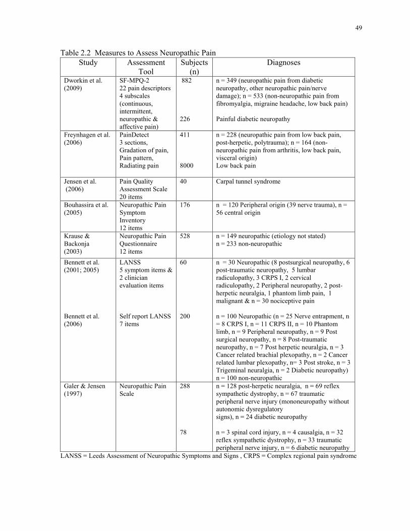
49
Table 2.2 Measures to Assess Neuropathic Pain
Study Assessment Tool
Subjects (n)
Diagnoses
Dworkin et al. (2009)
SF-MPQ-2 22 pain descriptors 4 subscales (continuous, intermittent, neuropathic & affective pain)
882 226
n = 349 (neuropathic pain from diabetic neuropathy, other neuropathic pain/nerve damage); n = 533 (non-neuropathic pain from fibromyalgia, migraine headache, low back pain) Painful diabetic neuropathy
Freynhagen et al. (2006)
PainDetect 3 sections, Gradation of pain, Pain pattern, Radiating pain
411 8000
n = 228 (neuropathic pain from low back pain, post-herpetic, polytrauma); n = 164 (non-neuropathic pain from arthritis, low back pain, visceral origin) Low back pain
Jensen et al. (2006)
Pain Quality Assessment Scale 20 items
40 Carpal tunnel syndrome
Bouhassira et al. (2005)
Neuropathic Pain Symptom Inventory 12 items
176
n = 120 Peripheral origin (39 nerve trauma), n = 56 central origin
Krause & Backonja (2003)
Neuropathic Pain Questionnaire 12 items
528 n = 149 neuropathic (etiology not stated) n = 233 non-neuropathic
Bennett et al. (2001; 2005) Bennett et al. (2006)
LANSS 5 symptom items & 2 clinician evaluation items Self report LANSS 7 items
60 200
n = 30 Neuropathic (8 postsurgical neuropathy, 6 post-traumatic neuropathy, 5 lumbar radiculopathy, 3 CRPS I, 2 cervical radiculopathy, 2 Peripheral neuropathy, 2 post-herpetic neuralgia, 1 phantom limb pain, 1 malignant & n = 30 nociceptive pain n = 100 Neuropathic (n = 25 Nerve entrapment, n = 8 CRPS I, n = 11 CRPS II, n = 10 Phantom limb, n = 9 Peripheral neuropathy, n = 9 Post surgical neuropathy, n = 8 Post-traumatic neuropathy, n = 7 Post herpetic neuralgia, n = 3 Cancer related brachial plexopathy, n = 2 Cancer related lumbar plexopathy, n= 3 Post stroke, n = 3 Trigeminal neuralgia, n = 2 Diabetic neuropathy) n = 100 non-neuropathic
Galer & Jensen (1997)
Neuropathic Pain Scale
288 78
n = 128 post-herpetic neuralgia, n = 69 reflex sympathetic dystrophy, n = 67 traumatic peripheral nerve injury (mononeuropathy without autonomic dysregulatory signs), n = 24 diabetic neuropathy n = 3 spinal cord injury, n = 4 causalgia, n = 32 reflex sympathetic dystrophy, n = 33 traumatic peripheral nerve injury, n = 6 diabetic neuropathy
LANSS = Leeds Assessment of Neuropathic Symptoms and Signs , CRPS = Complex regional pain syndrome
50
None of the questionnaires, outlined in Table 2.2, have been specifically validated in patients
with traumatic nerve injuries. In general, the SF-MPQ is the most widely used measure to
assess the qualities and intensity of pain. These measurement scales provide valuable
information regarding the frequency, intensity and quality of pain but do not assess the
impact of the pain on the patient. Depending on the type of pain and other associated factors
(injury, personal and environmental), the impact of the pain may vary between individuals.
2.5.4 Cold Sensitivity
Cold sensitivity or intolerance has been defined as an abnormal response (pain, discomfort,
sensory disturbance, stiffness and/or color changes) to cold exposure following a traumatic
injury (Campbell et al., 1998). Cold sensitivity or cold intolerance has been associated with
poor outcome following traumatic upper extremity injuries (Campbell et al., 1998; Collins et
al., 1996; Craigen et al., 1999; Graham et al., 2008; Irwin et al., 1997; Koman, Slone, Smith,
Ruch, & Poehling, 1998; Lithell et al., 1998; Povlsen et al., 1995b; Ruijs et al., 2007;
Stokvis, Ruijs, van Neck, & Coert, 2009). Cold sensitivity and cold intolerance are two terms
that are commonly used to describe these abnormal symptoms with exposure to cold. These
terms have been used interchangeably in the literature which has added to the confusion in
the field. Campbell and Kay (1998) propose the term “Trauma Induced Cold Associated
Symptoms” to describe this condition. In my dissertation, I have chosen to use the term cold
sensitivity except in reference to publications in which the authors have used the term cold
intolerance.
51
The symptoms associated with cold sensitivity are often associated with poor outcome
following traumatic peripheral nerve injuries and have been frequently reported in patients
with replantation injuries and digital amputations (Backman et al., 1993; Campbell et al.,
1998; Carlsson, Cederlund, Holmberg, & Lundborg, 2003; Collins et al., 1996; Craigen et al.,
1999; Dabernig et al., 2006; Graham et al., 2008; Irwin et al., 1997; Lithell et al., 1998;
McCabe et al., 1991; Nylander et al., 1987; Ruijs et al., 2006; Ruijs et al., 2007). Poor long
term outcome has been associated with persistence of cold sensitivity in patients with hand
injuries, nerve injuries and replantation injuries (Campbell et al., 1998; Collins et al., 1996;
Nancarrow et al., 1996; Povlsen et al., 1995a; Povlsen et al., 1995b). Campbell and Kay
(1998) evaluated patients following hand injuries and reported that 73% of 176 patients
reported cold-related symptoms and most were associated with pain. In the study by Graham
and Schofield (2008), patients who were more than 2 years following hand injury were
contacted for evaluation. The response rate for trauma patients was 34%. Cold intolerance
was reported by 90% of the patients with traumatic injuries and only 9% of these patients
reported an improvement in their symptoms over time. In patients following hand trauma,
Nijhuis et al. (2009) reported 38% of patients had abnormal cold intolerance and more severe
trauma was associated with more severe cold intolerance. Nancarrow et al. (1996) assessed
long term cold intolerance in 65 patients with hand injuries. Of these, 69% indicated the
presence of cold intolerance and 5 years after injury, 97% continued to report these
symptoms. In patients who were at least 5 years following nerve injury, Collins et al. (1996)
reported that 76% of patients had cold sensitivity and 87% of these patients had moderate or
severe symptoms. No difference in reported cold sensitivity was found between patients who
reported normal hand sensation compared to those with abnormal sensation. Dabernig et al.
52
(2006) used the DASH to evaluate patients after digital replantation (mean follow-up time of
5 years). The mean DASH score in this group of patients was 11, which indicated a relatively
low level of disability, but 87% of patients reported cold intolerance. In a group of patients
with neuropathic pain from a variety of etiologies which included traumatic nerve injuries,
the most intense pain was reported as occurring from cold provocation (Meyer-Rosberg et
al., 2001b). These studies provide evidence of the negative relationship between cold
sensitivity and outcome. Identification of patients with these symptoms may provide the
opportunity for appropriate treatment of cold sensitivity.
2.5.5 Measures of Cold Sensitivity
A variety of self-report and physician/health care professional-administered assessment tools
are available to evaluate cold sensitivity; including physical assessments, verbal rating scales
and patient reported questionnaires (Carlsson et al., 2008; Collins et al., 1996; Craigen et al.,
1999; Graham et al., 2008; Irwin et al., 1997; McCabe et al., 1991; Ruijs et al., 2006;
Traynor et al., 2008). Objective tests which involve cold immersion and measurement of
rewarming or arterial pressures are valid measures of the vascular status, but these types of
objective assessments do not provide information regarding the pain and/or cold symptoms
experienced by the patient (Backman et al., 1993; Nylander et al., 1987; Povlsen et al.,
1995a; Povlsen et al., 1995b; Traynor et al., 2008). Questionnaires such as the Cold
Sensitivity Scale (CSS) introduced by McCabe et al. (1991) and the Cold Intolerance
Symptom Scale (CISS) (Irwin et al., 1997; Ruijs et al., 2006; Ruijs et al., 2007) introduced
by Irwin et al. (1997) utilize patient report to rank symptoms on a numeric scale.
53
The McCabe CSS was developed from items reported by 35 patients who had cold exposure
symptoms following a hand injury (McCabe et al., 1991). The authors reduced these items to
four items to assess the severity of cold sensitivity and added three work related questions for
the work exposure score. The CSS was then completed by 80 control subjects and 20 patients
with nerve injury. Validity of the scale was determined by a significantly higher score in the
nerve injured patients compared to the control subjects. However, the authors did not assess
the validity of the specific tasks within the questionnaires by asking the subjects to perform
each task. Test-retest reliability of the CSS was established in a group of 35 patients (r =
0.92) and for the work exposure subscale (r = 0.94). The CSS questionnaire was used to
prospectively evaluate 123 patients with hand trauma injuries (Craigen et al., 1999). In this
series of patients, the significant single predictor of long term cold sensitivity was traumatic
bony injury. In contrast to other studies, the presence of cold sensitivity was not related to
smoking or amputation injuries (Irwin et al., 1997). Collins et al. (1996) used the CSS
questionnaire to evaluate cold sensitivity in patients following an upper extremity nerve
injury and reported substantial cold sensitivity in these patients. The mean CSS score was
137 and there was a significant relationship between the CSS and patient subjective rating of
cold sensitivity. The CSS is a reliable measure and possesses face validity.
The CISS includes the questions from the CSS in addition to specific questions regarding the
frequency of symptoms and impact on daily activities (Irwin et al., 1997). A scoring system
was developed for the CISS and the questionnaire was tested on 331 patients with cold
intolerance and 67 subjects with no symptoms of cold intolerance. Cold intolerance was more
likely to occur in patients who were smokers and who had crush or avulsion injuries.
Complete median or ulnar nerve division and a concomitant blood vessel injury were
54
associated with more severe cold intolerance. The CISS was found to have good test-retest
reliability (r = .9) in patients who were assessed six months following the first assessment.
Ruijs et al. (2006) administered the CISS to 68 healthy subjects and found that neither age
nor gender was significantly associated with cold intolerance. Based upon these healthy
subjects, the authors recommended that a score of 30 (95% confidence interval from the
mean CISS score 12.9) be used as the threshold to identify patients with cold intolerance.
However this criterion was not based upon the assessment of subjects with cold sensitivity.
Carlsson et al. (2008) evaluated the reliability and validity of a Swedish translation of the
CISS and the CSS. Test retest reliability was established in 100 patients and all of the ICCs
were greater than 0.85. Validity was evaluated by comparison of the CISS score to the SF-36,
the DASH, subjective patient assessment and VAS. Moderate correlation with the CISS was
reported with correlation coefficients ranging from 0.64 (SF-36 bodily pain) to 0.81 (VAS
cold intolerance).
CISS scores were evaluated by Ruijs et al. (2009) who examined digital rewarming patterns
in 12 patients with median and ulnar nerve injuries and 12 control subjects following
exposure to a cold water stress test. The patients with better rewarming patterns had lower
CISS scores and better recovery of sensation. Stokvis et al. (2009) reported CISS scores
before and after surgery for treatment of an upper extremity neuroma. The mean preoperative
CISS score was 54 and there were 30 patients (91%) who had scores greater than 30 which
indicated a level of abnormal cold intolerance. Postoperatively, the mean CISS score was 52
which was not significantly different from the preoperative CISS scores. The CISS scores
were significantly correlated with VAS pain intensity (r = 0.443, p = 0.01) and the DASH
scores (r = 0.438, p = 0.01). Carlsson et al. (2008) evaluated disability and cold intolerance in
55
patients following hand injury and reported moderate correlation between the DASH and
cold sensitivity (CSS, r = 0.67 and CISS r = 0.73). The CISS is a reliable measure which has
been shown to be a valid measure of cold sensitivity in patients with nerve injury.
To evaluate the assessment of cold sensitivity, Traynor and MacDermid (2008) compared
physical assessment with cold immersion and a patient reported questionnaire, the CISS, in
healthy subjects. Each subject completed the CISS and underwent cold immersion of one
hand (12 degrees Celsius for 5 minutes with temperature recovery measured for 20 minutes).
The authors reported strong test-retest reliability with the subjective questionnaires and the
physical assessment. There were no significant correlations between the patient reported
questionnaires and the objective physical assessments. These assessments were performed in
healthy subjects and not patients who have cold sensitivity. The responses may differ in those
patients with hand trauma or nerve injury compared to normal uninjured subjects.
Taken together, the literature indicates that cold sensitivity is associated with poor outcome
following hand trauma and injury to the upper extremity peripheral nerves. Many studies
have utilized the CISS and/or the CSS to assess cold sensitivity and comparison of patient
and/or surgeon subjective reports of outcome. Few studies have utilized valid and reliable
measures to compare disability and cold sensitivity following traumatic upper extremity
injuries.
2.6 Disablement Frameworks
The concept of disability as defined by Nagi (1965) refers to a physical or mental limitation
within a social context. The medical model considers disability as a characteristic of the
56
individual which is a direct result of the injury or disease, whereas a social model considers
disability as a result of a social problem not the individual. In terms of nerve injury,
disablement models may be useful to define or delineate the consequences of the nerve injury
on the individual and within the context of society. The biopsychosocial model of
disablement has integrated both the medical and social frameworks of disability and has
integrated the biological, personal and social perspectives within one model (Jette, 2006).
Nagi (1965) described a pathway of active pathology, impairment, functional limitation and
disability. Active pathology may be caused by an etiology that disrupts the normal cellular
processes. In the case of an injury to a motor nerve, it may represent muscle denervation.
Impairment refers to an abnormality of the tissue, organ or body system, and in the case of
nerve injury, this may be the resultant loss of muscle strength with an injury to a motor nerve
or numbness with injury to a sensory nerve. Impairments may occur due to the initial
pathology, or secondarily, due to limitations as a result of the initial pathology, such as
deconditioning of the surrounding muscles from disuse in patients with nerve injury.
Functional limitations are restrictions in basic physical performance, such as limitations in
raising the arm overhead. In Nagi’s model (1965), disability represents the discrepancy
between an individual’s capability and the specific demands of their physical and social
environment. Disability would then translate into the inability to play a sport, such as
baseball, that requires the capability to perform overhead arm activity involved in throwing
and catching a ball. Verbrugge and Jette (1994) expanded the Nagi model to include factors
which may influence the disablement pathway including predisposing risk factors (such as
medical comorbidities, sociodemographics, lifestyle), intra-individual factors (such as
lifestyle, activity or behavioural changes, coping skills, attitudes) and extra-individual factors
57
that are external to the individual (such as physical and social environment, external
supports, medical care). These additional factors may affect the interrelationships between
the factors within the disablement pathway.
The WHO introduced and subsequently modified the International Classification of
Impairments, Disabilities and Handicap (ICF) model which comprises 3 main concepts
(impairments, disabilities, handicaps) related to disease and health conditions (World Health
Organization, 2001). The ICF incorporates the positive and negative influences on the health
condition and factors which may influence that health condition. Functioning is considered at
three levels: the body part, the entire person and the entire person in the context of their
environment. Each level contains 3 domains of function related to body function and
structures, activities and participation (Figure 2.1).
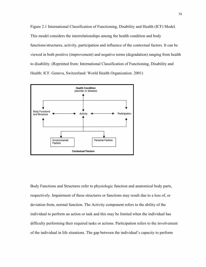
58
Figure 2.1 International Classification of Functioning, Disability and Health (ICF) Model.
This model considers the interrelationships among the health condition and body
functions/structures, activity, participation and influence of the contextual factors. It can be
viewed in both positive (improvement) and negative terms (degradation) ranging from health
to disability. (Reprinted from: International Classification of Functioning, Disability and
Health: ICF. Geneva, Switzerland: World Health Organization. 2001)
Body Functions and Structures refer to physiologic function and anatomical body parts,
respectively. Impairment of these structures or functions may result due to a loss of, or
deviation from, normal function. The Activity component refers to the ability of the
individual to perform an action or task and this may be limited when the individual has
difficulty performing their required tasks or actions. Participation refers to the involvement
of the individual in life situations. The gap between the individual’s capacity to perform
59
actions and their environment, including personal factors reflects performance. The
Contextual factors which include environmental and personal factors include the individual’s
environment in which they live and other factors such as age, gender, medical co-morbidities,
coping styles, other psychosocial issues (Jette & Keysor, 2003; World Health Organization,
2001). The activity and participation may be termed positively or negatively and it is the
negative aspect which reflects decline and disability.
Although the models by the ICF, Nagi and Verbrugge and Jette, differ in theory and
structure, these models provide a framework to conceptualize the disablement process and its
components, to consider the individual at many levels and specify the factors that may be
interrelated. Consideration of factors beyond physical impairment such as sensory loss or
muscle weakness which occur following nerve injury provides a broader perspective to
assess the factors which influence functional loss and disability. The ICF model has been
described for use in patients with musculoskeletal pathologies such as arthritis and distal
radius fractures which represent different types of pathologies ranging from an ongoing
inflammatory process to a traumatic injury and thus different pathologies would have
different expected outcomes over the course of the disease and/or recovery process (Harris,
MacDermid, & Roth, 2005; Jette et al., 2003; Stucki & Cieza, 2004).
Dixon, Johnston, McQueen & Court-Brown (2008) evaluated the ability of the DASH to
operationalize the ICF. The authors included 24 reviewers to allocate the items of the DASH
into the various ICF constructs. They found that five items were related to impairment, 19 to
activity, three to participation and seven were related to activity and participation restrictions.
Harris et al. (2005) reviewed the ICF framework in relation to distal radius fracture. Patient
reported questionnaires for the wrist and health status were included in the evaluation and the
60
authors reported that the ICF model provided a useful framework for evaluation beyond
physical impairment in patients following distal radius fracture.
The diversity of the ICF model lends itself to application in patients following nerve injury.
The consideration of factors beyond physical impairment is essential for the assessment of
the health effects and disability following nerve injury. The inclusion of contextual (personal
and environmental) factors have been lacking in the assessment of patients following nerve
injury and the influence of these factors on outcome.
2.7 Psychosocial Factors
The emphasis in outcome studies regarding patients following peripheral nerve injury has
been on the physical impairments related to motor and sensory recovery. Less importance has
been directed towards the influence of psychosocial factors on outcome, particularly the
association between psychosocial factors and pain and disability. Jacquet et al. (2002)
evaluated the psychological impact of nerve injuries in the forearm and the impact on
functional outcome and return to work. Using the Impact of Event Scale, the authors reported
higher psychological stress in patients with multiple nerve injuries. Functional outcome and
return to work were associated with higher levels of psychological stress. Argoff (2007)
reports the importance of comorbid conditions, such as depression, anxiety and sleep
disturbance which may negatively affect response to treatment of patients with neuropathic
pain, although in this review of the literature multiple etiologies of neuropathic pain were
included. Meyer-Rosberg et al. (2001b; 2001a) found sleep disturbance and other aspects of
quality of life were impaired in their patients with neuropathic pain and this in turn affected
61
their participation in social roles and work. Patients with traumatic nerve injuries were
included in the studies by Meyer-Rosberg et al. (2001b; 2001a) as were other etiologies
which resulted in neuropathic pain. Marchie and Mahoney (2008) reported an association
between perceived social support and disability as measured by the QuickDASH in patients
following carpal tunnel release. The negative aspects of outcome, lower rates of return to
work and higher levels of psychological stress have been associated with chronic nerve
injury and neuropathic pain (Argoff, 2007; Meyer-Rosberg et al., 2001b; Meyer-Rosberg et
al., 2001a; Turk, Audette, Levy, Mackey, & Stanos, 2010). The importance of other
psychological constructs, such as pain catastrophizing, depression and post-traumatic stress
symptoms has been demonstrated through their association with higher levels of pain and
poorer outcome. The relevant literature is reviewed in the following section.
2.7.1 Pain Catastrophizing
Pain catastrophizing refers to negative thinking regarding the experience of pain. Those
individuals who catastrophize feel overwhelmed by pain, ruminate about it and/or feel
helpless in the face of pain (Sullivan, Bishop, & Pivik, 1995; Sullivan, Lynch, & Clark,
2005). Catastrophizing about pain has been shown to negatively influence pain-related
outcomes and to contribute to increased pain responses, more emotional distress and
increased pain behaviour. Studies that have evaluated patients with acute postoperative pain
and more chronic musculoskeletal conditions, such as osteoarthritis and fibromyalgia, show
that pain catastrophizing is associated with poor outcome such as higher reported levels of
pain and decreased functional outcome (Buenaver, Edwards, & Haythornthwaite, 2007;
Edwards et al., 2005; Edwards et al., 2006; Edwards, Haythornthwaite, Smith, Klick, & Katz,
62
2009; Gracely et al., 2004; Lozano Calderon et al., 2008a; Lozano Calderon et al., 2008b;
Niekel, Lindenhovius, Watson, Vranceanu, & Ring, 2009; Pavlin, Sullivan, Freund, &
Roesen, 2005; Picavet, Vlaeyen, & Schouten, 2002; Riddle, Wade, Jiranek, & Kong, 2010;
Severeijns, Vlaeyen, van den Hout, & Weber, 2001; Sommer et al., 2010; Sullivan et al.,
2005).
Patient reported questionnaires such as the Pain Catastrophizing Scale (PCS) are designed to
assess the degree of pain castrophizing (Sullivan et al., 1995; Sullivan et al., 2005). The PCS
was designed to assess pain catastrophizing with an overall total score and three subscales
(rumination, magnification and helplessness). The validity and reliability have been
established for the PCS (Chibnall & Tait, 2005; Osman et al., 1997; Osman et al., 2000; Van
Damme, Crombez, Bijttebier, Goubert, & Van Houdenhove, 2002). Osman et al. (1997)
evaluated the factor structure in 288 students and showed that the three factor model relating
to each subscale was the best fit. The factor structure of the PCS was also evaluated as a
function of race and workers’ compensation in patients with low back pain (Chibnall et al.,
2005). In these patients, a two factor structure including helplessness and magnification was
the best fit for the data. Van Damme et al. (2002) evaluated the PCS in Dutch speaking
patients with low back pain and fibromyalgia and in pain-free individuals. They found that
the data were best represented in a three factor model which confirmed the original factor
structure of the scale: rumination, magnification and helplessness.
Pain catastrophizing has been associated with acute pain following surgery (Pavlin et al.,
2005; Riddle et al., 2010; Sommer et al., 2010). Sommer et al. (2010) evaluated the
predictors of acute postoperative pain in patients undergoing a variety of elective surgical
procedures including surgeries to the upper extremity. The significant predictors of acute
63
postoperative pain were pain catastrophizing, preoperative pain, expected pain and fear of
surgery. Pavlin et al. (2005) evaluated the influence of catastrophizing on postoperative pain
in patients undergoing anterior cruciate ligament repair and higher levels of acute
postoperative pain were associated with pain catastrophizing. Similarly, Riddle et al. (2010)
reported pain castrophizing as a predictor of higher pain scores in patients following knee
arthroplasty.
In patients with chronic conditions and chronic pain, catastrophizing had been negatively
associated with outcome including higher pain intensity and pain disability (Severeijns et al.,
2001; Sullivan et al., 2005). Sullivan et al. (2005) evaluated the specific facets of pain
catastrophizing in patients with neuropathic pain. The authors included 80 patients with
diabetic neuropathy, herpetic neuralgia, postsurgical and post-trauma. The authors reported
significant associations between pain catastrophizing and pain intensity and pain disability.
They also found a strong association between pain intensity and the PCS helplessness
subscale. Severeijns et al. (2001) evaluated the relationship between pain intensity, disability
and pain catastrophizing in a group of patients with chronic pain from a variety of etiologies.
Higher levels of pain intensity, disability and psychological distress were reported in patients
who catastrophized. However, the authors emphasized that strong associations were found
but the direction of causality could not be determined with a cross sectional design. In a
review article, Edwards et al. (2006) found that pain catastrophizing was an important factor
in patients with rheumatologic disorders. The authors report a relationship between pain
catastrophizing and higher levels of pain intensity, depression and disability. In a study of
patients with scleroderma, social environments such as lower education levels were found to
be associated with pain catastrophizing (Edwards et al., 2005). However, the relationship
64
between education level and pain intensity was not significant after controlling for pain
catastrophizing and symptoms of depression. Using brain imaging techniques, Gracely et al.
(2004) reported a relationship between pain catastrophizing and increased activation of brain
structures which were associated with pain processing and the attentional and emotional
aspects in patients with fibromyalgia.
Pain catastrophizing has been associated with negative outcomes in patients with chronic
pain, low back pain and acute knee surgery. Turner, Jensen & Romano (2000) evaluated
patients from a pain treatment program who had chronic pain from a variety of etiologies.
After controlling for age, sex and pain intensity, pain castrophizing and beliefs were
independent predictors of depression but were not predictors of physical disability. Pain
catastrophizing has also been shown to be associated with pain intensity and outcome
following surgery. Edwards et al. (2009) evaluated patients who underwent total knee
arthroplasty using a longitudinal study design to assess patients from baseline to 12 months
following surgery. Pain catastrophizing did not change over the course of the study in
contrast to decreases in pain intensity and symptoms of depression. Catastrophizing was a
predictor of pain severity when depression scores were not included in the model. In patients
with low back pain, Picavet et al. (2002) reported an association between pain
catastrophizing and fear of movement. Buenaver et al. (2007) evaluated the association
between pain catastrophizing, pain outcomes and social responses in patients with low back
pain and upper and lower limb pain from a variety of etiologies. Pain catastrophizing was
correlated with pain intensity, severity and interference. The authors concluded that
perceived social responses did influence the relationship between pain outcomes and pain
catastrophizing. Cano, Leonard & Franz (2005) introduced an addition to the PCS to include
65
the consideration of the influence of catastrophizing of significant others such as spouse,
partner, friends or family. The authors assessed undergraduate students from an urban
university and reported an association with pain catastrophizing between patients and their
partners. Higher levels of pain catastrophizing in significant others was associated with
higher levels of pain intensity, depression symptoms and other psychological distress.
In patients with upper extremity pathologies, previous studies have evaluated the association
between outcome and psychosocial factors including pain catastrophizing and depression
(Lozano Calderon et al., 2008a; Lozano Calderon et al., 2008b; Niekel et al., 2009). In
patients following carpal tunnel release, Lozano Calderon et al. (2008a) evaluated the
relationship between disability, patient satisfaction and psychosocial factors. The authors
reported a significant negative correlation between the DASH scores and patient satisfaction.
The presence of pain catastrophizing and depressive symptoms were significant predictors of
disability. In patients with a variety of hand pathologies, Niekel et al. (2009) evaluated the
relationship between DASH scores and psychosocial measures including symptoms of pain
catastrophizing, pain anxiety and depression. There was a small correlation between DASH
scores and pain catastrophizing and a moderate correlation between the DASH scores and
symptoms of depression and anxiety. In the regression analysis, pain catastrophizing and
depression symptoms were predictors of the DASH.
These studies highlight the importance of pain castrophizing in patients with musculoskeletal
pathologies and the potential relationship that may exist between pain catastrophizing and
disability in patients with upper extremity nerve injury. The role of pain catastrophizing and
outcome has yet to be evaluated in patients following traumatic upper extremity nerve injury.
66
2.7.2 Depression Symptoms
Chronic pain and disability in patients with upper extremity pathologies have been associated
with symptoms of depression and these symptoms may contribute to poor outcome (Lozano
Calderon et al., 2008a; Lozano Calderon et al., 2008b; Ring et al., 2006). In patients with a
variety of upper extremity pathologies including carpal tunnel syndrome, trigger finger, distal
radius fracture, elbow pain and de Quervain’s tenosynovitis, Ring et al. (2006) reported
strong correlations between the DASH score and symptoms of depression as measured by the
Center for Epidemiologic Studies-Depression Scale. Similarly, in patients with
trapeziometacarpal joint arthrosis, DASH scores were associated with symptoms of
depression and pain catastrophizing (Lozano Calderon et al., 2008b). As a single predictor,
the depression scores accounted for 50% of the variance. Patient satisfaction and DASH
scores after carpal tunnel release were also associated with depression symptoms (Lozano
Calderon et al., 2008a).
Patient report questionnaires such as the Hospital Anxiety and Depression Scale (HADS)
may be used to identify symptoms of anxiety and depression in patients with milder
symptoms (Zigmond & Snaith, 1983). The HADS is composed of 14 items which can be
summarized by the anxiety and depression subscales and was shown to be more sensitive in
individuals with milder symptoms of anxiety and depression (Zigmond et al., 1983). Good
validity and reliability have been reported for the HADS (Aben, Verhey, Lousberg, Lodder,
& Honig, 2002; Bjelland, Dahl, Haug, & Neckelmann, 2002; Hann, Winter, & Jacobsen,
1999; Lisspers, Nygren, & Oderman, 1997; Mykletun, Stordal, & Dahl, 2001; Quintana et
al., 2003). With data based from the general population in Norway, Mykletun et al. (2001)
67
evaluated the factor structure of the HADS. The authors reported satisfactory internal
consistency with both subscales and showed that the items loaded onto 2 factors although
two of the items did not appear to be unique to either anxiety or depression. The factor
structure was assessed in patients from a musculoskeletal pain clinic (Pallant & Bailey,
2005). The authors report a two factor structure with items loading on anxiety and depression
although similar to the results reported by Mykletun et al. (2001), one item did not load onto
the anxiety factor. Mykletun et al. (2001) reported high levels of anxiety and depression as
measured by the HADS in patients from a musculoskeletal pain clinic. The mean value for
the HADS anxiety was 9.26 and for the HADS depression was 8.14. The authors found these
values in patients with musculoskeletal pain were higher compared to other reports of anxiety
and depression scores from patients with breast cancer, end stage renal disease, and chronic
pulmonary disease (Mykletun et al., 2001). Overall, the HADS has been shown to be useful
in the assessment of symptoms of anxiety and depression in patients with non-psychiatric
diagnoses.
2.7.3 Post-traumatic Stress Symptoms
Anxiety related to injury may be exhibited by patients following a traumatic injury. Post-
traumatic stress disorder (PTSD) is defined as an anxiety disorder which is related to a
traumatic event and this disorder is a classification within the Diagnostic and Statistical
Manual of Mental Disorders - 4th edition (DMS-IV) of the American Psychiatric Association
(2000). PTSD may include avoidance of stimuli related to the traumatic event, emotional
numbing of general responsiveness, hyperarousal and/or re-experiencing the traumatic event
in thoughts and dreams (American Psychiatric Association, 2000; Breslau, 2002). PTSD is
68
commonly associated with military trauma but as Breslau outlines, PTSD associated with
non-military trauma is more common compared to military trauma cases (Breslau, 2002). In
the general population, PTSD is reported in 7-12% of the population and the prevalence is
reportedly higher in survivors of previous trauma (Asmundson et al., 2002). In a study by
Breslau et al. (1998) which included an urban sample, traumatic events including serious
accidents, were more prevalent in men, younger age and those in minority groups. An
increased risk of developing PTSD has been associated with childhood trauma and
psychiatric history of the individual and/or family (Brewin, Andrews, & Valentine, 2000).
Individuals with PTSD may also be at risk for other associated disorders such as depression,
anxiety and substance abuse (Breslau, 2002). PTSD and chronic pain often occur together
and may share a mutual dependence (Asmundson et al., 2002; Asmundson et al., 2008). In
patients with PTSD, there have been higher reports of pain and more health related problems
and similarly, there have been reports of higher levels of PTSD in patients with chronic pain
(Asmundson et al., 2002; Asmundson et al., 2008). As outlined by these authors, certain
characteristics of chronic pain may maintain or exacerbate the symptoms of PTSD and vice
versa some components of PTSD may maintain or exacerbate the symptoms related to
chronic pain. Post-traumatic stress symptoms may occur as a subthreshold level and not meet
the criteria of the diagnosis of PTSD. This cluster of post-traumatic stress symptoms may be
distressing to the patient and may compromise recovery. Reports of low levels of post-
traumatic stress symptoms have been reported in patients with traumatic hand injuries
(Opsteegh et al., 2009; Opsteegh et al., 2010). Symptoms of post-traumatic stress were
associated higher levels of pain and lower rates of return work. Although PTSD has been
associated with other types of non-military trauma and with chronic pain, post-traumatic
69
stress symptoms or anxiety are not typically considered with traumatic peripheral nerve
injury nor are these symptoms frequently assessed. Post-traumatic stress symptoms have not
been previously assessed in patients following upper extremity nerve injury using valid,
reliable measures. If present, these symptoms may be associated with higher levels of pain
and poorer outcome.
The Post-traumatic Stress Disorder Checklist - Civilian version (PCL-C) is a self report
questionnaire designed to assess symptoms of post-traumatic stress (Weathers, Litz, Huska,
& Keane, 1994; Blanchard, Jones-Alexander, Buckley, & Forneris, 1996). Good reliability
and validity have been reported with this measure (Blanchard et al., 1996; Ruggiero, Del
Ben, Scotti, & Rabalais, 2003). Blanchard et al. (1996) evaluated individuals following a
motor vehicle accident or sexual assault. The authors found high internal consistency for the
PCL-C (Cronbach’s alpha 0.9) and strong correlation (r = 0.9) with another measure of post-
traumatic stress, the Clinician Administered PTSD Scale. Ruggiero et al. (2003) reported
good reliability (test-retest, internal consistency) and good validity when using the PCL-C in
a group of college students. Using exploratory factor analysis, Pagé, Kleiman, Asmundson &
Katz (2009) evaluated the factor structure of the PCL-C in patients who were undergoing
major surgery. The authors reported in the pain-free patients two factors (re-
experiencing/avoidance and emotional/numbing/hyperarousal) were the best fit of the data
and in patients with pain, one factor accounted for 51.1% of the variance. In patients
following motor vehicle accidents or sexual assault, Blanchard et al. (1996) reported a mean
PCL-C score of 45.8 + 16.1. Similarly, in patients undergoing major surgery, Pagé et al.
(2009) reported a mean PCL-C score of 45 + 20.6 in pain-free patients and 46.7 + 9.7 in
patients with pain. However, there has been lack of agreement regarding the criterion score
70
for the diagnosis of PTSD. This criterion value reportedly varies between 44, 45 and 50 and
appears to be different in patients with different etiologies (Ruggiero et al., 2003). In the
assessment of patients with traumatic nerve injury, post-traumatic stress is rarely considered
and questionnaires such as the PCL-C are not reported.
Concurrent symptoms of both chronic pain and post-traumatic stress symptoms are not
uncommon following trauma. Pain may be related to musculoskeletal origin or nerve related.
While there are many reports of pain following nerve injury, assessment of anxiety or post-
traumatic stress symptoms in patients with nerve injury are lacking. Aspects of both chronic
pain and post-traumatic stress may share a mutual dependence to maintain the high
comorbidity between the post-traumatic stress symptoms and pain in patients with nerve
injury.
2.8 Summary
The above review indicates that there is wide variability in the reports of outcome in patients
following upper extremity nerve injury, and standardized methods of assessment have not
been universally accepted. Assessment of physical impairment is the most frequently
reported outcome in the literature. Many patients sustain what appears to be a similar injury,
yet outcomes vary in terms of recovery of physical impairment, pain and disability. In terms
of outcome, the biomedical factors which affect nerve regeneration and reinnervation of the
motor and sensory end organ targets have been examined. More recently, the influence of
psychosocial factors has been considered in the outcome of patients with musculoskeletal
disorders but the role of these factors in the experience of pain and disability has received

71
very little empirical attention in patients with upper extremity peripheral nerve injuries. The
relationships among the various biomedical and psychosocial factors outlined above have yet
to be evaluated in patients with traumatic peripheral nerve injury.
72
Chapter 3 Objectives & Hypotheses
3 Main Objective
The main objective of my dissertation is to evaluate the biomedical and psychosocial factors
associated with pain and disability in patients following traumatic upper extremity nerve
injuries. This main objective has been translated into specific questions and hypotheses as
articulated in the following two sections.
3.1 Specific Questions
1. Do patients following peripheral nerve injury have high levels of disability?
2. Is pain present in patients following peripheral nerve injury?
3. What is the relationship between pain and disability?
4. Do peripheral nerve surgeons routinely assess pain in patients following traumatic nerve
injuries?
5. Is cold sensitivity associated with higher levels of pain and disability following peripheral
nerve injury?
6. Are biomedical factors such as gender, age and level of injury associated with pain and
disability in patients following peripheral nerve injury?
7. Are psychosocial factors such as depression, anxiety and pain catastrophizing associated
with pain and disability in patients following peripheral nerve injury?
73
3.2 Hypotheses
The specific hypotheses for each study are outlined. The data analyses are described in detail
in the Methods of each study (Chapters 5, 6 and 7).
Study 1 (Chapter 5), Evaluation of Pain Measurement Practices and Opinions of Peripheral
Nerve Surgeons
Specific hypothesis:
1. Most peripheral nerve surgeons do not routinely assess pain.
2. When pain is assessed, pain intensity rating scales are most frequently used.
Study 2 (Chapter 6), Patient Reported Outcome Following Peripheral Nerve Injury
Specific hypotheses:
1. High levels of disability will be present.
2. High levels of pain will be present.
3. Patients with brachial plexus lesions will report more pain and disability.
4. Pain will be associated with greater disability.
74
Study 3 (Chapter 7), Biomedical and Psychosocial Factors Associated with Pain and
Disability after Peripheral Nerve Injury
Specific hypotheses:
1. High levels of disability will be present.
2. High levels of pain will be present.
3. Pain will be associated with greater disability.
4. Cold sensitivity will be associated with more pain and disability.
5. Patients with brachial plexus lesions will report more pain and disability.
6. Symptoms of depression, anxiety and pain catastrophizing will be associated with greater
disability.
75
Chapter 4 Materials and Methods
4 Materials and Methods
The specific materials and methods for each of the studies are described in the chapters
related to each individual study (Chapters 5, 6 and 7).
4.1 Research Ethics Board Approval
Studies 1, 2 and 3 (Chapters 5, 6 and 7) were approved by the Research Ethics Boards of the
following institutions; University Health Network, University of Toronto and York
University (Appendix A). Study 3 (Chapter 7) included patients from the Division of Plastic
& Reconstructive Surgery, Washington University School of Medicine, St. Louis, Missouri
and this study was also approved by the Human Studies Committee at Washington
University School of Medicine in St. Louis (Appendix A). The approved consent forms for
Study 3 (Chapter 7) are included in Appendix B.
76
Chapter 5 Study 1
The literature reviewed in the introduction of this dissertation and recently published review
(Novak, C. B. & Katz, J. Neuropathic Pain in Patients with Upper Extremity Nerve Injury.
Physiotherapy Canada (2010) 62:190-201.) revealed that most outcome studies of patients
following nerve injury report motor and sensory impairments but fail to assess other relevant,
non-biomedical factors that have been shown to be associated with pain and recovery in other
patient samples. Few outcome studies even assess for the presence or absence of pain or pain
relief, and those that do report pain have used verbal or numeric rating scales of pain
intensity, leaving out other important dimensions of pain (e.g., affective, evaluative, quality,
frequency, duration). The purpose of Study 1 was to ascertain the extent to which peripheral
nerve surgeons assess for pain in their patients and to examine their opinions about pain in
patients with upper extremity nerve injury.
Study 1, Evaluation of Pain Measurement Practices and Opinions of Peripheral Nerve
Surgeons, surveyed the practices and opinions of peripheral nerve surgeons regarding pain
assessment in patients following upper extremity nerve injury. This study was published in
Hand 2009; 4(4):344–349, and permission to reproduce this manuscript has been obtained
from the publisher. This study was presented at the American Society for Peripheral Nerve,
Annual Meeting, Maui, Hawaii, January 9-11, 2009.
77
5 Evaluation of Pain Measurement Practices and Opinions of Peripheral Nerve Surgeons
Reprinted with permission from Hand (Springer Publishers):
Novak, C. B., Anastakis, D. J., Beaton, D. E. & Katz, J. (2009). Evaluation of Pain
Measurement Practices and Opinions of Peripheral Nerve Surgeons. Hand, 4, 344-349.
5.1 Abstract
Purpose: The purpose of this study was to evaluate the opinions and practices of peripheral
nerve surgeons regarding assessment and treatment of pain in patients following nerve injury.
Methods: Surgeons with expertise in upper extremity peripheral nerve injuries and members
of an international peripheral nerve society were sent an introductory letter and electronic
survey by email (n = 146). Seventy members responded to the survey (48%) and 59 surgeons
completed the survey.
Results: For patients referred for motor or sensory dysfunction, 31 surgeons (52%) indicated
that they always formally assess pain. In patients referred for pain, 44 surgeons (75%)
quantitatively assess pain using a verbal scale (n = 24) or verbal numeric scale (n = 36). The
most frequent factors considered very important in the development of chronic neuropathic
pain were psychosocial factors (64%), mechanism of injury (59%), workers’
compensation/litigation (54%) and iatrogenic injury (48%). In patients more than six months
following injury, surgeons frequently see: cold sensitivity (54%), decreased motor function
78
(42%), paraesthesia/numbness (41%), fear of returning to work (22%), neuropathic pain
(20%) and emotional/psychological distress (17%).
Conclusion: Only 52% of surgeons who responded to the survey always evaluate pain in
patients referred for motor/sensory dysfunction. Pain assessment most frequently includes
verbal patient response and assessment of psychosocial factors is rarely included.
Predominately patient related factors were considered important in the development of
chronic neuropathic pain.
5.2 Introduction
Pain can negatively affect outcome and health related quality of life (Cocito et al., 2006). In
patients following nerve injury, neuropathic pain can contribute to significant disability and
has been reported following peripheral nerve injury, particularly in patients with brachial
plexus injuries (Bentolila et al., 1999; Bertelli et al., 2008a; Cocito et al., 2006; Htut et al.,
2006; Jensen et al., 2007; Samii et al., 2001). Many outcome studies of nerve injured patients
present motor and sensory functional outcomes but do not report information regarding the
absence or presence of pain (Aberg et al., 2007; Bertelli et al., 2007; Chuang et al., 1992;
Chuang et al., 1995; Chuang et al., 1993; Jaquet et al., 2001; Leechavengvongs et al., 1998;
Leechavengvongs, Witoonchart, Uerpairojkit, & Thuvasethakul, 2003; Mackinnon et al.,
2005; Novak et al., 2003; Tung et al., 2003). In the surgical literature, many studies that
report pain in nerve injured patients generally use unidimensional measures (verbal ratings or
visual analog scales of pain intensity) and do not include assessment of the psychosocial
79
factors that may be associated with pain (Bentolila et al., 1999; Bertelli et al., 2008a; Htut et
al., 2006; Kato et al., 2006; Kim et al., 2003; Nath et al., 2006; Samii et al., 2001).
The purpose of this study was to evaluate peripheral nerve surgeons’ opinions and practices
regarding pain assessment and treatment in patients following nerve injury.
5.3 Methods
A survey was developed to include questions related to the assessment and treatment of pain
in patients following nerve injury. A physical therapist, hand surgeon, psychologist and
clinical epidemiologist developed the preliminary questionnaire. To examine the survey
questions for content, clinical relevance, clarity, flow and wording, three different surgeons
who operate on patients with upper extremity nerve injuries reviewed the questionnaire.
Based on their feedback, the survey was modified and pilot tested on three different surgeons
to ensure that the questions were understandable and clinically relevant.
The opinions of surgeons with expertise in the treatment of patients with upper extremity
peripheral nerve injuries were sought for this study. Following approval by our institutional
and university Research Ethics Boards, the active surgeons from an international peripheral
nerve society who had a registered email address were invited to participate (n = 146). An
introductory letter and anonymous electronic survey were sent by email. The non-
respondents were sent two subsequent reminders by email at two weeks following the initial
email and then three weeks following the first reminder. In total, 70 members responded to
the survey (48%): after first mailing, n = 39, after second mailing, n = 19, after third mailing,
n = 16. There were 6 surgeons who declined to participate and 5 who indicated that they do
80
not operate on patients with nerve injuries. In total, the sample consisted of 59 surgeons who
completed the questionnaire.
There were 49 men and 10 women with a mean age of 50 years (standard deviation (sd) 9
years) and mean years in practice of 16 (sd 10 years). The majority of the surgeons were
from plastic surgery (n = 37) followed by orthopedic surgery (n = 12), neurosurgery (n = 8),
general surgery (n = 1) and one respondent indicated both plastic and orthopedic surgery.
Thirty-nine surgeons (66%) reported that they operate on patients with brachial plexus
injuries.
5.3.1 Data Analysis
Data analyses were performed using Statistical Package for the Social Sciences (version 15.0
for Windows, SPSS Inc., Chicago, IL, USA). Using chi-squared analyses for the categorical
data and t-tests for the continuous data, comparisons were made of the demographics and
survey responses between: 1) those surgeons who operate on brachial plexus injuries and
those that do not operate on brachial plexus injuries. 2) those surgeons who quantitatively
assess pain and those that do not. 3) those surgeons who “always” assess pain in patients with
motor or sensory dysfunction and those that do not. 4) those surgeons who responded after
the first mailing compared to the late responders (surgeons that responded to the subsequent
mailings).
81
5.4 Results
Comparisons between the surgeons who responded to the survey early (after the first email)
or late (to subsequent emails) revealed no significant differences in the demographics; male
vs. female (p = .736), age (p = .483), surgical specialty (p = .588), years in practice (p = .787)
or surgeons who operate on brachial plexus patients compared to those that do not (p = .409).
5.4.1 Questionnaire Responses to Pain Assessment and Treatment
In patients referred for pain resulting from a nerve injury, 44 surgeons (75%) indicated that
they “quantitatively assess pain” using a verbal scale (n = 24), verbal numeric scale (n = 36),
visual analog scale (n = 14) or pain questionnaire (n = 7). The specific “pain questionnaires”
identified were the McGill pain questionnaire, the Disabilities of the Arm, Shoulder and
Hand (DASH), the SF-36 and pain questionnaires developed by the surgeon. There were no
significant differences in the demographics between surgeons who do or do not quantitatively
assess pain (male vs. female, p = .306; age, p = .174; years in practice, p = .281; surgical
specialty, p = .346; early vs. late mailing response, p = .796). There was a lower frequency of
pain assessment in patients referred for motor or sensory dysfunction resulting from nerve
injury; 31 surgeons (52%) reported that they “always formally assess pain” in these patients.
In this survey, neuropathic pain was defined as “pain resulting from a nerve injury”. When
asked “when neuropathic pain becomes chronic?”, six months was the most frequent
response (n = 23), however the responses ranged from three days to one year (less than six
months, n = 23; greater than six months, n = 9; no answer, n = 4). The factors that were
considered very important in the development of chronic neuropathic pain included
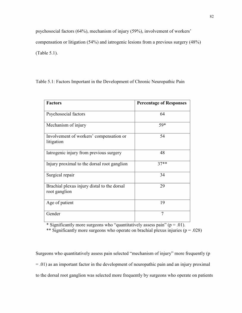
82
psychosocial factors (64%), mechanism of injury (59%), involvement of workers’
compensation or litigation (54%) and iatrogenic lesions from a previous surgery (48%)
(Table 5.1).
Table 5.1: Factors Important in the Development of Chronic Neuropathic Pain
Factors
Percentage of Responses
Psychosocial factors
64
Mechanism of injury
59*
Involvement of workers’ compensation or litigation
54
Iatrogenic injury from previous surgery
48
Injury proximal to the dorsal root ganglion
37**
Surgical repair
34
Brachial plexus injury distal to the dorsal root ganglion
29
Age of patient
19
Gender
7
* Significantly more surgeons who “quantitatively assess pain” (p = .01). ** Significantly more surgeons who operate on brachial plexus injuries (p = .028)
Surgeons who quantitatively assess pain selected “mechanism of injury” more frequently (p
= .01) as an important factor in the development of neuropathic pain and an injury proximal
to the dorsal root ganglion was selected more frequently by surgeons who operate on patients
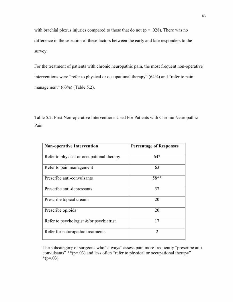
83
with brachial plexus injuries compared to those that do not (p = .028). There was no
difference in the selection of these factors between the early and late responders to the
survey.
For the treatment of patients with chronic neuropathic pain, the most frequent non-operative
interventions were “refer to physical or occupational therapy” (64%) and “refer to pain
management” (63%) (Table 5.2).
Table 5.2: First Non-operative Interventions Used For Patients with Chronic Neuropathic
Pain
Non-operative Intervention
Percentage of Responses
Refer to physical or occupational therapy
64*
Refer to pain management
63
Prescribe anti-convulsants
58**
Prescribe anti-depressants
37
Prescribe topical creams
20
Prescribe opioids
20
Refer to psychologist &/or psychiatrist
17
Refer for naturopathic treatments
2
The subcategory of surgeons who “always” assess pain more frequently “prescribe anti-convulsants” **(p=.03) and less often “refer to physical or occupational therapy” *(p=.03).
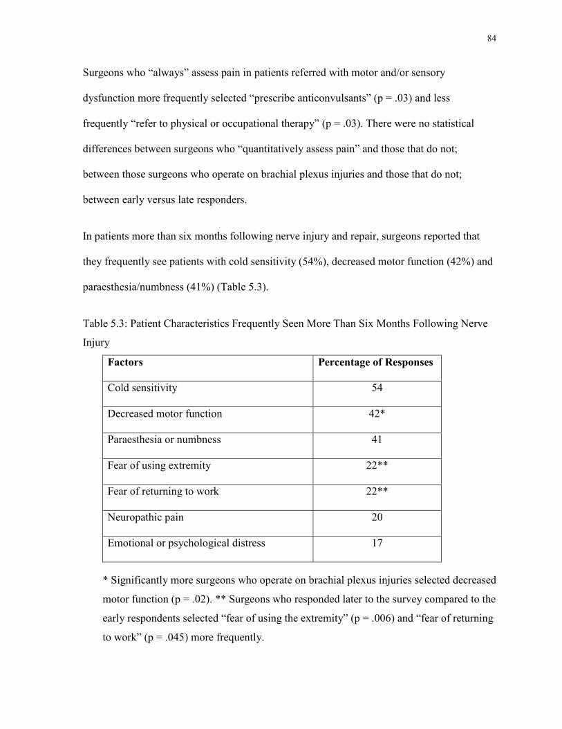
84
Surgeons who “always” assess pain in patients referred with motor and/or sensory
dysfunction more frequently selected “prescribe anticonvulsants” (p = .03) and less
frequently “refer to physical or occupational therapy” (p = .03). There were no statistical
differences between surgeons who “quantitatively assess pain” and those that do not;
between those surgeons who operate on brachial plexus injuries and those that do not;
between early versus late responders.
In patients more than six months following nerve injury and repair, surgeons reported that
they frequently see patients with cold sensitivity (54%), decreased motor function (42%) and
paraesthesia/numbness (41%) (Table 5.3).
Table 5.3: Patient Characteristics Frequently Seen More Than Six Months Following Nerve
Injury
Factors
Percentage of Responses
Cold sensitivity
54
Decreased motor function
42*
Paraesthesia or numbness
41
Fear of using extremity
22**
Fear of returning to work
22**
Neuropathic pain
20
Emotional or psychological distress
17
* Significantly more surgeons who operate on brachial plexus injuries selected decreased
motor function (p = .02). ** Surgeons who responded later to the survey compared to the
early respondents selected “fear of using the extremity” (p = .006) and “fear of returning
to work” (p = .045) more frequently.
85
Surgeons who operate on patients with brachial plexus injuries did select “decreased motor
function” more frequently compared to surgeons who do not operate on the brachial plexus
(p = .024). Surgeons who responded later to the survey compared to the early respondents
selected “fear of using the extremity” (p = .006) and “fear of returning to work” (p = .045)
more frequently. There were no statistical differences between those surgeons who
quantitatively assess pain and those that do not.
5.5 Discussion
Neuropathic pain is defined by the International Association for the Study of Pain as pain
resulting from dysfunction or a lesion in the peripheral or central nervous system (Cruccu et
al., 2004; Merskey et al., 1994). Chronic neuropathic pain is usually defined as pain after six
months from onset and in our survey, many surgeons (39%) defined the onset of “chronic” at
six months following nerve injury. However, there were more surgeons who defined it by
periods of time more than or less than six months following injury which points to
inconsistency in the definition of “chronic neuropathic pain” among peripheral nerve
surgeons. In the surgeons’ general comments submitted in the survey, “chronic neuropathic
pain” was often attributed to neuropathic pain, which was uncontrolled and non-responsive to
treatment.
Neuropathic pain and cold sensitivity are frequently reported as contributing to poor outcome
following nerve injury (Berman et al., 1998; Bruxelle et al., 1988; Collins et al., 1996;
Craigen et al., 1999; Freedlander, 1986; Graham et al., 2008; Htut et al., 2006; Irwin et al.,
1997; Kato et al., 2006). However few outcome studies provide details regarding these

86
patient symptoms and most studies of nerve injured patients report only motor and sensory
function. Graham and Schofield (2008) reported the prevalence of cold intolerance symptoms
in patients greater than two years following hand injuries. The questionnaire was returned by
25% of patients and cold intolerance was reported by most of these patients (90% in trauma
cases). Only 9% of patients with cold intolerance reported an improvement over time. Collins
et al. (1996) evaluated 85 patients who were at least five years following nerve injury and
76% reported cold intolerance, which did not appear to decrease over time. In our survey,
54% of surgeons reported that six months following nerve injury, cold sensitivity was a
frequent symptom. However, there was no indication of a specific outcome measure used to
quantify the degree of pain or disability related to cold sensitivity. Many of the respondents
to our survey indicated that referral to pain management and/or physical or occupational
therapy is one of the first non-operative interventions utilized for patients with neuropathic
pain. Therefore, many of the surgeons may assume that these facets of outcome regarding
pain and disability are assessed and addressed by other sub-specialties.
Many questionnaires, which evaluate pain, disability, cold sensitivity and psychosocial
factors, are time consuming to complete and analyze and therefore may be difficult to include
in clinical practice. The European Federation of Neurological Societies recently outlined
their neuropathic pain assessment guidelines (Cruccu et al., 2004). Visual analog scales,
numerical rating scales or verbal rating scales, which are simple to use, can provide a
quantitative assessment of pain intensity and may be used to provide a baseline assessment
(Cruccu et al., 2004). Others have advocated assessment of pain intensity in addition to
sensory descriptors, temporal variation and functional impact (Gilron et al., 2006). Based
upon simple assessment, patients who indicate the presence of neuropathic pain can be
87
referred to pain specialists for more in depth evaluation and prompt treatment of the pain and
associated sequelae.
The response rate for our survey was 48%. To minimize the likelihood of non-response bias
and to increase the generalizability, a higher response rate is desirable but when compared to
the response rate for physician questionnaires, this rate is similar to those previously reported
(Asch, Jedrziewski, & Christakis, 1997; Braithwaite, Emery, de Lusignan, & Sutton, 2003;
Cull, O'Connor, Sharp, & Tang, 2005; Cummings, Savitz, & Konrad, 2001; Drummond,
Sharp, Carsin, Kelleher, & Comber, 2008; Kellerman & Herold, 2001). Many of the studies
that have evaluated physician survey response rates used postal mail surveys and in a study
comparing electronic and postal mail surveys, there was a shorter response time with
electronic surveys but no difference in response rate or data quality (Akl, Maroun, Klocke,
Montori, & Schunemann, 2005). We utilized several of the methods recommended to
increase the response rate, which included a short questionnaire, personalized email, survey
began with the demographic data and each subsequent email to non-respondents had a link to
the questionnaire (Akl et al., 2005; Braithwaite et al., 2003; Drummond et al., 2008;
Kellerman et al., 2001). Monetary and non-monetary incentives have been used to increase
response rates and in a recent systematic review, VanGeest, Johnson & Welch (2007) found
that monetary incentives improved the response rate. In our study, no incentives were given
to complete the survey.
The most significant limitation associated with a low response rate is the presence of non-
response bias. The comparison of early and late respondents to the survey has been
introduced as one method to address this issue. Previous studies have suggested that
respondents to later mailings may by classified as “proxies” for the non-respondents and
88
these late responders may represent qualities similar to the non-respondents (Kellerman et al.,
2001; Sobal & Ferentz, 1989). The validity of this conclusion has been challenged. Blalock
and Dial (1990) hypothesized that these similarities may not be present in the unevaluated
responses or demographics. In our survey, there were no differences in the demographics
between the early and late responders but the late responders did more frequently report
patients with “fear of using the extremity” and “fear of returning to work”. There were no
differences in the factors considered important in the development of chronic neuropathic
pain.
Leslie (1972) proposed that individuals with a group identity share similar traits concerning
attitudes and opinions towards group issues and therefore response bias is less likely in
studies that use a homogeneous group. Peripheral nerve surgeons represent a relatively
homogeneous group regarding knowledge and training compared to other physicians or the
general population. Therefore the differences between respondents and non-respondents in
surveys involving peripheral nerve surgeons may have less significance in the attitude
towards pain following nerve injury. However, the relatively low response rate in our study
does limit the assumptions that one can make regarding the practices and beliefs of the non-
respondents. Based on the surgeons who did complete our survey and the results reported in
outcome studies, there appears to be many surgeons who do not quantitatively assess pain or
the associated factors in patients following nerve injury and yet many recognize the presence
of cold sensitivity, pain and psychological sequelae following nerve injury.
Significant disability, related to cold sensitivity and/or neuropathic pain, may result
following nerve injury and early treatment is advocated (Ahmed-Labib et al., 2007; Birch,
Bonney, & Wynn Parry, 1998; Carlsson et al., 2003; Carlsson et al., 2008; Collins et al.,
89
1996; Craigen et al., 1999; Htut et al., 2006; Irwin et al., 1997; Kato et al., 2006). However,
in our study, few surgeons reported using valid, reliable questionnaires to assess pain, cold
sensitivity or disability following nerve injury and in those patients not specifically referred
for a pain problem only 52% of surgeons “formally assess pain”. Therefore these symptoms
with respect to frequency, intensity and disability may be under represented in the reports of
outcome in patients following nerve injury. Early recognition of neuropathic pain and prompt
intervention may provide the opportunity for more efficacious treatment.
90
Chapter 6 Study 2
This study evaluated patient reported pain and disability and the factors associated with
disability in patients following an upper extremity traumatic nerve injury. I hypothesized that
in these patients high levels of disability would be reported and that pain would be a
significant predictor of disability. Study 2, Patient Reported Outcome Following Peripheral
Nerve Injury, was published in the Journal of Hand Surgery 2009; 34A(2):281-287 and
permission to reproduce this manuscript was granted by the publisher. This study was
presented at the American Association for Hand Surgery, Annual Meeting, Maui, Hawaii,
January 7-10, 2009 and the Canadian Pain Society, Annual Meeting, Quebec City, Quebec,
May 27-30, 2009.
91
6 Patient Reported Outcome Following Peripheral Nerve Injury
Reprinted with permission from Journal of Hand Surgery (Elsevier Limited):
Novak, C. B., Anastakis, D. J., Beaton, D. E. & Katz, J. (2009). Patient Reported Outcome
Following Peripheral Nerve Injury. Journal of Hand Surgery, 34A, 281-287.
6.1 Abstract
Purpose: This study evaluated patient reported outcome and the factors associated with
disability following an upper extremity traumatic nerve injury. We hypothesized that patients
at least 6 months following injury would report significant disability and that pain would be
the strongest predictor of the DASH.
Methods: Following Research Ethics Board approval, the medical charts were reviewed of
patients with these inclusion criteria: adults; presenting to a nerve surgeon; six months or
greater after traumatic nerve injury. Patients completed the DASH and the SF-36 as a routine
part of the initial evaluation. These data were reviewed retrospectively to determine the
predictors of the DASH score.
Results: There were 84 patients (mean age 38 years, sd 14 yrs), which included brachial
plexus (n = 27) and peripheral nerve (n = 57) injuries. The mean time following injury was
38 months (sd 47). For all SF-36 domains, the mean values of the nerve injured patients were
significantly lower than the normative data (p < 0.001), indicating a lower health status. The
92
mean DASH score was 52 (sd 22)/100. Significantly more disability was associated with
more SF-36 bodily pain (r = -.59, p < .0001) and with brachial plexus injuries (p = 0.023). In
the final regression model, SF-36 bodily pain (beta = -.475, p < 0.001), age (beta = .360, p =
0.014) and nerve injured (beta = -4.804, p = 0.023) were significant predictors of the DASH
(R2 = 44.5%). SF-36 bodily pain (beta = -.523, p < 0.001) accounted for 35% of the variance.
Conclusion: Substantial long term disability (high DASH scores) was found in patients
following nerve injury that was predicted by higher pain, older age and brachial plexus
injury. Further investigation of this pain and the associated factors may provide the
opportunity for improved health related quality of life.
6.2 Introduction
Outcome following peripheral nerve injury frequently includes only measurement of nerve
impairment such as sensory and motor function (Chuang et al., 1992; Chuang et al., 1995;
Chuang et al., 1993; Leechavengvongs et al., 1998; Leechavengvongs et al., 2003;
Mackinnon et al., 2005; Nakamichi & Tachibana, 1998; Novak & Mackinnon, 2002a; Novak
& Mackinnon, 2002b; Novak et al., 2003; Novak & Mackinnon, 2004; Oberlin et al., 1994;
Richardson, 1997; Samardzic, Grujicic, & Antunovic, 1992; Tung et al., 2003). Patient
reported outcome and disability using valid and reliable measurement tools are rarely
included in published reports and few studies have evaluated functional outcome or health
related quality of life following peripheral nerve injury (Ahmed-Labib et al., 2007; Choi et
al., 1997; Kitajima et al., 2006).
93
Chronic neuropathic pain which may occur following a peripheral nerve injury is often
severe and directly impacts patient outcome and previous studies have reported the presence
of severe chronic neuropathic pain in patients with brachial plexus injuries (Berman et al.,
1998; Bertelli et al., 2008a; Bruxelle et al., 1988; Htut et al., 2006; Kato et al., 2006). Studies
of patients with elbow and wrist injuries have identified pain as a significant contributor to
upper extremity disability (Doornberg et al., 2005; Souer et al., 2008). Our clinical
impression is that the presence of chronic neuropathic pain following peripheral nerve injury
is a dominant predictor of disability and poor functional outcome.
The objective of this study was to evaluate patient reported outcome and the factors
associated with disability in patients following an upper extremity traumatic nerve injury. We
hypothesized that in select patients who sought a medical assessment at least six months
following an upper extremity nerve injury, high levels of disability as measured by the
DASH would be reported and that pain as assessed by the SF-36 would be a significant
predictor of disability.
6.3 Material & Methods
This study used an existing clinical database from a single surgeon to identify eligible
patients that were seen in a specialty clinic for patients with nerve related diagnoses from
January 1997 to March 2007. A chart review of the medical records was then performed. The
database included the records of 791 patients with nerve related diagnoses (such as nerve
compression, nerve injury, tumors, congenital anomalies) to the upper and lower extremities.
For this study, the inclusion criteria included patients with an upper extremity traumatic
94
peripheral nerve injury and who, at their initial consult visit, were at least six months
following injury. Six months was chosen as the lower threshold to allow us to look at longer
term outcomes in patients who sought medical assessment at least six months following
nerve injury. Following approval from the institutional and university Research Ethics
Boards, the database was reviewed to obtain the names of patients who met the inclusion
criteria. The medical charts were then reviewed. At the initial patient consult, each patient
was asked to complete two questionnaires, the Disabilities of the Arm, Shoulder, and Hand
(DASH) and the SF-36. Patients who were unable to understand the questionnaires due to
language barriers and did not have a translator, did not complete the questionnaires and were
excluded. Demographic data, the DASH and the SF-36 from the initial consult visit were
extracted from the chart.
There were 65 men and 19 women with a mean age of 39 years (standard deviation (sd) 14
years). The mean + sd time from injury to initial consult was 38 + 47 months (range 6 to 239
months). The dominant hand was involved in 44 cases and workers’ compensation or
litigation was involved in 20 cases. All cases involved traumatic nerve injury and the most
frequent site of injury was the brachial plexus (n = 27). The other nerves injured included the
median (n = 18), ulnar (n = 16), median and ulnar (n = 5), radial (n = 8), ulnar and radial (n =
1), accessory (n = 2), axillary (n = 3), long thoracic (n = 3) and suprascapular (n = 1).
6.3.1 Measures
The SF-36, Version 2, is a generic health status questionnaire with established validity and
reliability (Brazier et al., 1992; Garratt et al., 1993; Jenkinson, Wright, & Coulter, 1994;
95
McHorney et al., 1993; Ware et al., 1992). There are eight domains (physical functioning,
role-physical, bodily pain, general health, vitality, social functioning, role-emotional, mental
health), which can be reported separately or a normalized composite score for the physical
and mental components can be calculated. A higher score indicates a better health status.
The DASH is a 30-item self-reported questionnaire to assess disease specific upper extremity
outcome and disability. It was developed to measure physical function and symptoms in
patients with upper limb musculoskeletal disorders and good reliability and validity have
been reported (Beaton et al., 2001; Dias et al., 2008; Gummesson et al., 2003; Hudak et al.,
1996; SooHoo et al., 2008). Each item is ranked on a 5-point Likert scale ranging from “no
difficulty” to “unable”. A composite score is calculated from the completed responses and
the score cannot be calculated if more than three items are missing. A higher DASH score
reflects a greater degree of disability.
6.3.2 Data Analysis
Data were entered in a spreadsheet and the statistical analyses were performed with
Statistical Package for the Social Sciences (version 15.0 for Windows, SPSS Inc., Chicago,
IL, USA). Means and standard deviations were calculated for continuous data which were
tested for normality and collinearity before analysis. Frequency counts were calculated for
categorical data. One-sample t-tests were used to compare the SF-36 scores from the present
sample with the published Canadian normative values (Hopman et al., 2000) on the eight
domains and the physical and mental component scores. Non-parametric Spearman
correlations were used to evaluate the association between the DASH and the SF-36 bodily
96
pain and the time since injury. T-tests were used to compare the DASH scores between the
following independent variables; workers’ compensation or litigation involvement status (yes
vs. no), dominant hand affected (yes vs. no) and gender (male vs. female). A one-way
ANOVA was used to compare the DASH scores of patients with brachial plexus injuries,
single shoulder nerve injuries and distal nerve injuries (median, ulnar and radial). If a
significant F-test (main effect) was found (p < .05), paired linear contrasts between each of
the pairs was completed.
Multiple linear regression using manual backward elimination was used to evaluate the
variables that predicted outcome as measured by the DASH (Harrell, 2001). The initial
model included the following seven independent variables; gender, involvement of workers’
compensation or litigation, dominant hand injured, time since injury, age, nerve injured and
SF-36 bodily pain. Collinearity between the variables was checked by the appropriate
correlation coefficient. Correlations greater than 0.8 were deemed to be collinear and one of
the pair was eliminated from the model based on clinical sensibility. Subsequent models were
derived by manual backward elimination using a criterion p-value associated with the beta
coefficient of 0.1 or greater to remove a variable. The variable with the largest p-value was
eliminated from the model first and the subsequent regression model was fitted with the
remaining variables. This process was repeated until only variables with a p-value less than
0.1 remained in the final model. To investigate the influence of each variable included in the
final model, a forward entry multiple regression was used in which the variable with the
smallest p-value was entered first followed by the next until all significant variables had been
entered.
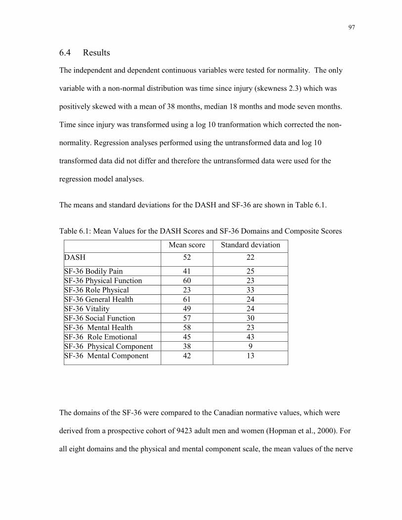
97
6.4 Results
The independent and dependent continuous variables were tested for normality. The only
variable with a non-normal distribution was time since injury (skewness 2.3) which was
positively skewed with a mean of 38 months, median 18 months and mode seven months.
Time since injury was transformed using a log 10 tranformation which corrected the non-
normality. Regression analyses performed using the untransformed data and log 10
transformed data did not differ and therefore the untransformed data were used for the
regression model analyses.
The means and standard deviations for the DASH and SF-36 are shown in Table 6.1.
Table 6.1: Mean Values for the DASH Scores and SF-36 Domains and Composite Scores
Mean score Standard deviation
DASH 52 22
SF-36 Bodily Pain 41 25
SF-36 Physical Function 60 23
SF-36 Role Physical 23 33
SF-36 General Health 61 24
SF-36 Vitality 49 24
SF-36 Social Function 57 30
SF-36 Mental Health 58 23
SF-36 Role Emotional 45 43
SF-36 Physical Component 38 9
SF-36 Mental Component 42 13
The domains of the SF-36 were compared to the Canadian normative values, which were
derived from a prospective cohort of 9423 adult men and women (Hopman et al., 2000). For
all eight domains and the physical and mental component scale, the mean values of the nerve
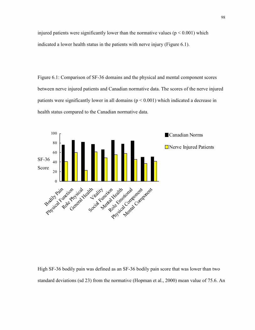
98
injured patients were significantly lower than the normative values (p < 0.001) which
indicated a lower health status in the patients with nerve injury (Figure 6.1).
Figure 6.1: Comparison of SF-36 domains and the physical and mental component scores
between nerve injured patients and Canadian normative data. The scores of the nerve injured
patients were significantly lower in all domains (p < 0.001) which indicated a decrease in
health status compared to the Canadian normative data.
0
20
40
60
80
100
Bodily
Pain
Physical Function
Role Phy
sical
General Health
Vitality
Social Fu
nctio
n
Mental H
ealth
Role Emotional
Physical Com
ponent
Mental Com
ponent
Canadian Norms
Nerve Injured Patients
High SF-36 bodily pain was defined as an SF-36 bodily pain score that was lower than two
standard deviations (sd 23) from the normative (Hopman et al., 2000) mean value of 75.6. An
SF-36
Score
99
SF-36 bodily pain score less than 30 was considered different from the normative data and
based on this criterion, there were 27 nerve injured patients with more bodily pain.
6.4.1 Univariate Variables Associated with DASH
There was a significant negative correlation between the DASH score and the SF-36 bodily
pain score (r = -.602, p < .0001) indicating a higher level of disability in those patients with
more pain as measured by the SF-36 and no correlational relationship was found between the
DASH and time since injury (r = -.009, p = .844). There were no signficant differences in
DASH scores between patients with or without workers’ compensation or litigation (p =
0.37), with or without the dominant hand affected (p = 0.22) and between males and females
(p = 0.095). The one-way ANOVA comparing the DASH score between nerve groups was
significant (p = .048) and the planned contrasts indicated that DASH scores were
significantly higher (p = 0.023) in patients with brachial plexus injuries (60 + 20) compared
to patients with more distal nerve injuries (48 + 23) and single shoulder nerve injuries (46 +
18) (Figure 6.2).
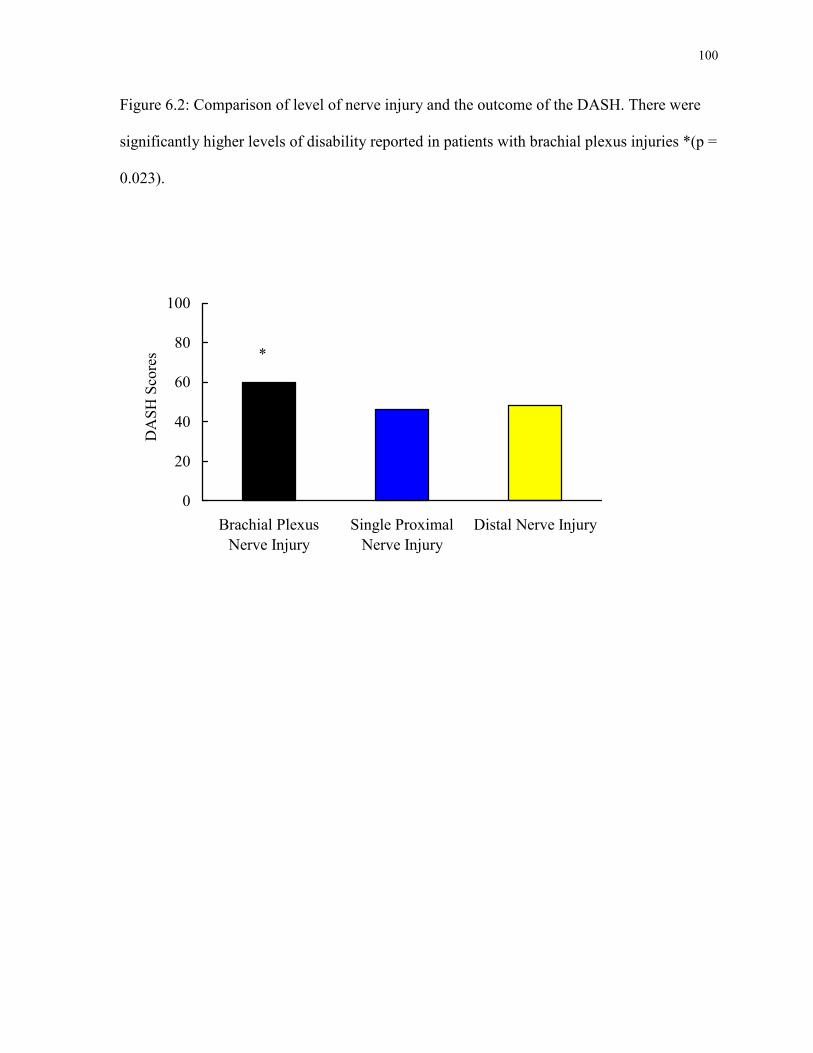
100
Figure 6.2: Comparison of level of nerve injury and the outcome of the DASH. There were
significantly higher levels of disability reported in patients with brachial plexus injuries *(p =
0.023).
0
20
40
60
80
100
Brachial Plexus
Nerve Injury
Single Proximal
Nerve Injury
Distal Nerve Injury
DA
SH
Sco
res
*
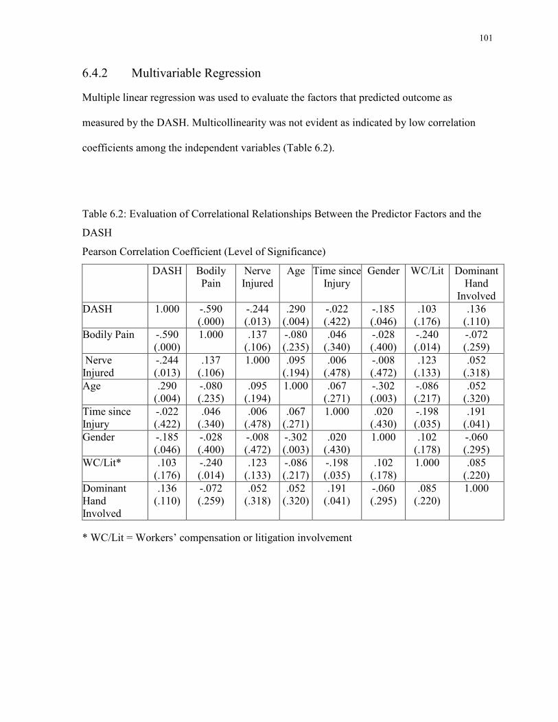
101
6.4.2 Multivariable Regression
Multiple linear regression was used to evaluate the factors that predicted outcome as
measured by the DASH. Multicollinearity was not evident as indicated by low correlation
coefficients among the independent variables (Table 6.2).
Table 6.2: Evaluation of Correlational Relationships Between the Predictor Factors and the
DASH
Pearson Correlation Coefficient (Level of Significance)
DASH Bodily Pain
Nerve Injured
Age Time since Injury
Gender WC/Lit Dominant Hand
Involved
DASH 1.000 -.590 (.000)
-.244 (.013)
.290 (.004)
-.022 (.422)
-.185 (.046)
.103 (.176)
.136 (.110)
Bodily Pain
-.590 (.000)
1.000 .137 (.106)
-.080 (.235)
.046 (.340)
-.028 (.400)
-.240 (.014)
-.072 (.259)
Nerve Injured
-.244 (.013)
.137 (.106)
1.000 .095 (.194)
.006 (.478)
-.008 (.472)
.123 (.133)
.052 (.318)
Age
.290 (.004)
-.080 (.235)
.095 (.194)
1.000 .067 (.271)
-.302 (.003)
-.086 (.217)
.052 (.320)
Time since Injury
-.022 (.422)
.046 (.340)
.006 (.478)
.067 (.271)
1.000 .020 (.430)
-.198 (.035)
.191 (.041)
Gender
-.185 (.046)
-.028 (.400)
-.008 (.472)
-.302 (.003)
.020 (.430)
1.000 .102 (.178)
-.060 (.295)
WC/Lit*
.103 (.176)
-.240 (.014)
.123 (.133)
-.086 (.217)
-.198 (.035)
.102 (.178)
1.000 .085 (.220)
Dominant Hand Involved
.136 (.110)
-.072 (.259)
.052 (.318)
.052 (.320)
.191 (.041)
-.060 (.295)
.085 (.220)
1.000
* WC/Lit = Workers’ compensation or litigation involvement
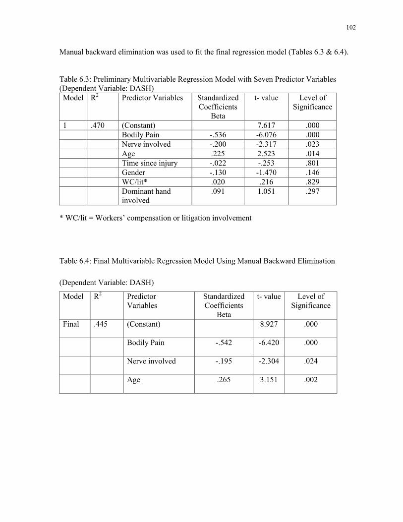
102
Manual backward elimination was used to fit the final regression model (Tables 6.3 & 6.4).
Table 6.3: Preliminary Multivariable Regression Model with Seven Predictor Variables (Dependent Variable: DASH)
Model
R2 Predictor Variables Standardized Coefficients
Beta
t- value Level of Significance
1 .470 (Constant) 7.617 .000
Bodily Pain -.536 -6.076 .000
Nerve involved -.200 -2.317 .023
Age .225 2.523 .014
Time since injury -.022 -.253 .801
Gender -.130 -1.470 .146
WC/lit* .020 .216 .829
Dominant hand involved
.091 1.051 .297
* WC/lit = Workers’ compensation or litigation involvement
Table 6.4: Final Multivariable Regression Model Using Manual Backward Elimination
(Dependent Variable: DASH)
Model
R2 Predictor Variables
Standardized Coefficients
Beta
t- value Level of Significance
Final
.445 (Constant) 8.927 .000
Bodily Pain -.542 -6.420 .000
Nerve involved -.195 -2.304 .024
Age .265 3.151 .002
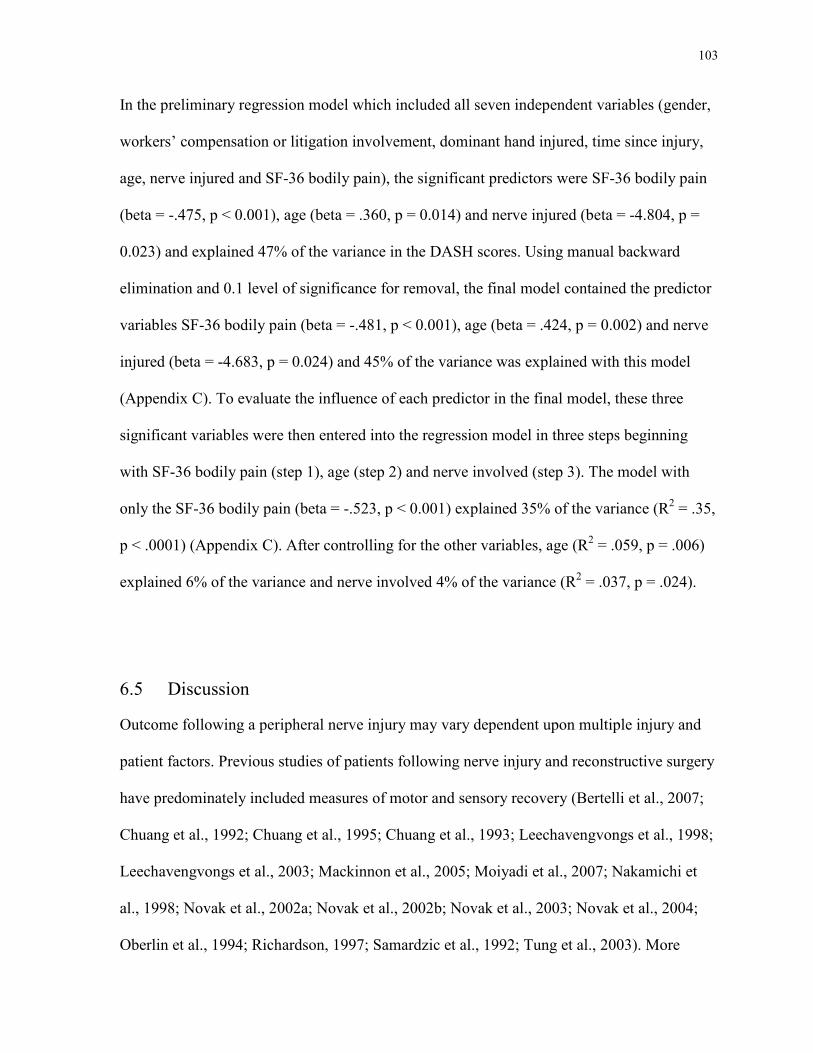
103
In the preliminary regression model which included all seven independent variables (gender,
workers’ compensation or litigation involvement, dominant hand injured, time since injury,
age, nerve injured and SF-36 bodily pain), the significant predictors were SF-36 bodily pain
(beta = -.475, p < 0.001), age (beta = .360, p = 0.014) and nerve injured (beta = -4.804, p =
0.023) and explained 47% of the variance in the DASH scores. Using manual backward
elimination and 0.1 level of significance for removal, the final model contained the predictor
variables SF-36 bodily pain (beta = -.481, p < 0.001), age (beta = .424, p = 0.002) and nerve
injured (beta = -4.683, p = 0.024) and 45% of the variance was explained with this model
(Appendix C). To evaluate the influence of each predictor in the final model, these three
significant variables were then entered into the regression model in three steps beginning
with SF-36 bodily pain (step 1), age (step 2) and nerve involved (step 3). The model with
only the SF-36 bodily pain (beta = -.523, p < 0.001) explained 35% of the variance (R2 = .35,
p < .0001) (Appendix C). After controlling for the other variables, age (R2 = .059, p = .006)
explained 6% of the variance and nerve involved 4% of the variance (R2 = .037, p = .024).
6.5 Discussion
Outcome following a peripheral nerve injury may vary dependent upon multiple injury and
patient factors. Previous studies of patients following nerve injury and reconstructive surgery
have predominately included measures of motor and sensory recovery (Bertelli et al., 2007;
Chuang et al., 1992; Chuang et al., 1995; Chuang et al., 1993; Leechavengvongs et al., 1998;
Leechavengvongs et al., 2003; Mackinnon et al., 2005; Moiyadi et al., 2007; Nakamichi et
al., 1998; Novak et al., 2002a; Novak et al., 2002b; Novak et al., 2003; Novak et al., 2004;
Oberlin et al., 1994; Richardson, 1997; Samardzic et al., 1992; Tung et al., 2003). More
104
recently, the trends in outcomes research following nerve injury have been directed towards
more functional results and patient reported outcomes (Ahmed-Labib et al., 2007; Choi et al.,
1997; Kitajima et al., 2006). High levels of disability and decreased health status in patients
following brachial plexus injury and reconstructive surgery have been reported (Ahmed-
Labib et al., 2007; Kitajima et al., 2006). In our select patients, who were at least six months
following nerve injury and sought medical assessment, there was a high level of disability
and a significantly lower health status in all SF-36 domains.
In our present study, the high level of disability as measured by the DASH was predicted by
SF-36 bodily pain, older age and brachial plexus injuries. The final regression model showed
SF-36 bodily pain to be a strong predictor of disability and accounted for 35% of the
explained variance. Chronic neuropathic pain has been associated with a poor outcome in
patients following nerve injury (Berman et al., 1998; Htut et al., 2006; Kitajima et al., 2006;
Cocito et al., 2006). Choi et al. (1997) reported a moderately high quality of life in patients
with brachial plexus injuries, although 75% of the patients had significant pain and 38%
continued to use pain medications. Higher pain intensity has been associated with brachial
plexus patients who did not undergo surgical repair, had more roots avulsed and had a long
delay between injury and surgery (Htut et al., 2006; Kato et al., 2006). Previous studies have
established an association between injury and patient demographic variables and outcome
and our study used multivariable regression analysis to evaluate the predictors of disability.
In our study, the SF-36 bodily pain score was the most significant predictor of disability,
however only 44.5% of the variance was explained by the injury and demographic variables
included in our analysis. Psychosocial factors such as depression, pain catastrophizing,
anxiety have been associated with outcome or satisfaction in patients with carpal tunnel
105
syndrome, distal radius fractures and elbow fractures (Doornberg et al., 2005; Lozano
Calderon et al., 2008a; Souer et al., 2008). The unexplained variance in our study may be
associated with other biomedical or psychosocial factors, which remain to be identified.
This retrospective study only included patients who were at least six months following nerve
injury and who had been referred to a specialty nerve clinic. There are patients following
nerve injury who do not have chronic neuropathic pain or who have low levels of pain and
are managing well (low disability) but there are others who have significant neuropathic pain
and disability. We chose to look at patients who were at least six months following nerve
injury because these are likely the patients who do have a higher level of chronic neuropathic
pain and associated disability. While we do not know what percentage of nerve injured
patients this group would comprise, our clinical impression is that these patients are the most
difficult to manage. In this preliminary investigation, we have shown that patients who seek
medical assessment at least six months following nerve injury report a significant level of
disability and this is in part associated with chronic neuropathic pain. However, in our
regression model, 55.5% of the variance was unexplained and this variance may be related to
other biomedical and psychosocial factors.
Further investigation into chronic neuropathic pain following peripheral nerve injury may
provide a better understanding of the underlying mechanisms, relevant biomedical and
psychosocial factors associated with pain and disability and, ultimately, more effective
treatments and improved health related quality of life.
106
Chapter 7 Study 3
In Study 2 (Chapter 6), substantial disability was reported by patients with upper extremity
nerve injury and the significant variables associated with the DASH were pain, older age and
brachial plexus nerve injury. A cross sectional study of prospectively collected data provided
the opportunity to confirm these findings and to evaluate the association between the DASH
and additional biomedical and psychosocial factors in patients with long standing nerve
injury. This study was partially funded by a Research Grant Award from the American
Association of Hand Surgery. This study has been submitted for publication and it is
presently under peer review. This study was presented at the Canadian Physiotherapy
Association annual meeting, St. John’s, Newfoundland, July 24, 2010 and will be the
American Society for Surgery of the Hand annual meeting, Boston, Massachusetts, October
9, 2010.
107
7 Biomedical and Psychosocial Factors Associated with Pain and Disability after Peripheral Nerve Injury
Novak, C. B., Beaton, D. E., Anastakis, D. J., Mackinnon, S. E. & Katz, J. (2010).
Biomedical and Psychosocial Factors Associated with Pain and Disability after Peripheral
Nerve Injury. Submitted for publication and presently under peer review.
7.1 Abstract
Background: The purpose of this study was to evaluate the biomedical and psychosocial
factors associated with pain and disability more than six months following upper extremity
nerve injury.
Methods: This cross sectional study included patients between six months and 15 years
following an upper extremity nerve injury. Assessment included a series of patient self-report
questionnaires. Disabilities of the Arm, Shoulder and Hand (DASH) scores were compared
using independent samples t-tests, analysis of variance or correlations. Multivariable linear
regression was used to evaluate the predictors of the DASH scores.
Results: The sample included 158 patients with a mean age of 41 + 16 years and median time
from injury was 14 months (range 6-167). DASH scores were significantly higher in patients
with workers’ compensation or litigation (p = 0.02), brachial plexus nerve injuries (p <
0.001) and in those unemployed (p < 0.001). There was a significant positive correlation
between DASH scores and pain intensity (r = .51, p < 0.001). In the multivariable regression
108
analysis for the predictors of the DASH, the final model explained 52.7% of the variance
with the following predictors; pain intensity (Beta = .230, p = 0.006), brachial plexus nerve
injury (Beta = -.220, p = 0.000), time since injury (Beta = -.198, p = 0.002), pain
catastrophizing score (Beta = .192, p = 0.025), age (Beta = .187, p = 0.002), work status
(Beta = .179, p = 0.008), cold sensitivity (Beta = .171, p = 0.015), depression score (Beta =
.133, p = 0.066), workers’ compensation or litigation (Beta = .116, p = 0.049) and gender
(Beta = -.104, p = 0.09).
Conclusions: Patients with peripheral nerve injury reported substantial disability, pain and
cold sensitivity. Disability as measured by the DASH score was predicted by brachial plexus
nerve injury, older age, pain intensity, work status, time since injury, cold sensitivity and
pain catastrophizing.
7.2 Introduction
Recovery following upper extremity peripheral nerve injury is variable and may result in
ongoing significant morbidity. Outcome studies of patients following upper extremity nerve
injury frequently include measures of physical impairment such as range of motion, strength
and sensation (Bertelli et al., 2007; Chuang et al., 1995; Chuang et al., 1993; Haninec et al.,
2007; Htut et al., 2006; Htut et al., 2007; Mackinnon et al., 2005; Merrell et al., 2001; Nath,
Lyons, & Bietz, 2007; Novak & Katz, 2010; Terzis et al., 2006; Tung et al., 2003). Measures
which evaluate the impact of the physical impairment on the patient and validated measures
of disability are not commonly included in the surgical literature.
109
Although few studies have evaluated disability after peripheral nerve injury, the evidence
points to high levels of disability in these patients (Ahmed-Labib et al., 2007; Bengtson et al.,
2008; Novak et al., 2009b). Disability following nerve injury may be related to biomedical
factors including motor or sensory dysfunction, pain, and cold sensitivity. However,
psychosocial factors may also be associated with disability in patients with nerve injury.
Previous studies have shown evidence of an association between psychosocial factors (such
as anxiety, depression and pain catastrophizing) and pain intensity, patient satisfaction and
disability in patients with distal radius fractures, osteoarthritis and nerve pathologies (Bailey
et al., 2009; Lozano Calderon et al., 2008a; Lozano Calderon et al., 2008b; Souer et al.,
2008). The relative contribution to, and association of the biomedical and psychosocial
factors with, reported disability has yet to be determined in patients with peripheral nerve
injury. Identification of the factors that are associated with functional outcome and disability
will assist in the development of more comprehensive treatment strategies for patients with
nerve injury.
The main objective of this cross sectional study was to evaluate the biomedical and
psychosocial factors associated with disability and pain in patients following upper extremity
nerve injury. We hypothesized that; 1) patients with peripheral nerve injury would present
with high levels of disability; 2) pain would be significantly associated with disability; 3)
patients with brachial plexus lesions would report higher levels of pain and disability than
those with more distal nerve injuries; 4) cold sensitivity would be associated with increased
pain and greater disability; 5) higher levels of psychosocial distress (depression, post-
traumatic stress and pain catastrophizing) would be associated with more disability.
110
7.3 Methods
This cross sectional study was approved by our institutional and university Research Ethics
Boards.
7.3.1 Subjects
Adult patients were included in the study if they were between six months and 15 years
following traumatic upper extremity peripheral nerve injury. The lower criterion of six
months was chosen to include patients who would be classified as having chronic pain
following a peripheral nerve injury. In this sample, all patients were adults at the time of
injury. Those patients with a previous upper motor neuron lesion, an amputation, a self-
inflicted injury or who were unable to understand the questionnaires were excluded from the
study. Between September 2007 and August 2009, patients were recruited when the study
coordinator was present at the clinic (University of Toronto Hand Program, Toronto, Ontario,
Canada and the Division of Plastic & Reconstructive Surgery, Washington University School
of Medicine, St. Louis, Missouri, USA). One hundred and sixty-four patients were invited to
participate in the study (six patients declined); 158 patients participated including 77 patients
from the University of Toronto Hand Program, Toronto, Ontario, Canada and 81 from the
Division of Plastic & Reconstructive Surgery, Washington University School of Medicine,
St. Louis, Missouri, USA.
111
7.3.2 Testing Protocol
Following signed informed consent, all testing was completed at one visit (approximately 30
minutes). The assessment included a series of questionnaires; Cold Intolerance Severity Scale
(CISS), Short Form McGill Pain Questionnaire (SF-MPQ), Disabilities of the Arm, Shoulder
and Hand (DASH) questionnaire, Hospital Anxiety and Depression Scale (HADS), Post-
traumatic Stress Disorder Checklist - Civilian version (PCL-C), Pain Catastrophizing Scale
(PCS), co-morbidity index. The order of questionnaire administration was randomly
determined for each patient. The questionnaires were numbered and computer software was
used to generate a randomization schedule for the administration order of the questionnaires
for each patient.
7.3.2.1 Disabilities of the Arm, Shoulder and Hand (DASH) Questionnaire
The DASH was designed to assess disability in patients with upper extremity
musculoskeletal disorders (Beaton et al., 2001; Hudak et al., 1996). There are 30 items
related to symptoms and physical function, which are ranked on a five-point Likert scale and
the DASH score is calculated from the responses. A higher DASH score indicates greater
level of disability. As recommended by the questionnaire developers, missing items were
replaced with the mean value of all subjects for that item (Beaton et al., 2001). As well, the
developers state that the total score should not be calculated (i.e., estimated) if more than
three items are missing; however, in the present study no patient had more than two missing
responses. Good validity and reliability have been reported for the DASH (Beaton et al.,
2001; Gummesson et al., 2003; Hudak et al., 1996; SooHoo et al., 2008).
112
7.3.2.2 Short Form McGill Pain Questionnaire (SF-MPQ)
The McGill Pain Questionnaire (MPQ) was developed to assess quantitative and qualitative
measures of the pain experience (Melzack, 1975). The SF-MPQ consists of 15 adjectives
from the sensory (n = 11) and affective (n = 4) categories of the original MPQ (Melzack,
1987). Each descriptor is rated on a four-point rating scale and a pain rating index is
calculated. Pain intensity was assessed on a 10 cm visual analog scale (VAS) with the
anchors “no pain” and “worst possible pain”. Good validity and reliability have been shown
for the SF-MPQ (Grafton et al., 2005; Melzack, 1987).
7.3.2.3 Cold Intolerance Symptom Scale (CISS)
The CISS was designed to evaluate the sensitivity of exposure to cold with questions
regarding the frequency, duration, severity and impact of cold sensitivity (Irwin et al., 1997;
Ruijs et al., 2006). The scores for each item are summed to give a total score ranging from 0
to 100; the higher the score, the greater the cold sensitivity. There is evidence of good
validity and reliability for the CISS in patients following upper extremity trauma (Carlsson et
al., 2008; Irwin et al., 1997; Ruijs et al., 2006). The CISS includes questions regarding cold
sensitivity while performing certain physical tasks. Some patients with peripheral nerve
injury are physically unable to perform these tasks and therefore are unable to answer these
questions. A validated method to control for missing data in the CISS scoring has not been
published. Therefore we performed a sensitivity analysis using four different methods to
replace the missing values (mean, median, linear trend, linear interpolation). There were no
113
statistical differences between the mean values calculated with each method and strong
correlation coefficients (r = .99, p < 0.001) were found between all methods. Therefore we
chose to impute missing data for the CISS using linear trend regression analysis for each item
(Norman & Streiner, 2000).
7.3.2.4 Hospital Anxiety and Depression Scale (HADS)
The HADS was used to measure symptoms of anxiety and depression (Zigmond et al., 1983).
In total, there are 14 items in the HADS, which include seven items for anxiety and seven
items for depression. Each item is ranked by the patient on a scale from 0 to 3 and the sum
score is calculated for each subscale. A higher score indicates a higher degree of anxiety or
depressive symptoms. The validity and reliability of the HADS has been established (Aben et
al., 2002; Bjelland et al., 2002; Hann et al., 1999; Lisspers et al., 1997; Mykletun et al., 2001;
Quintana et al., 2003).
7.3.2.5 Post-traumatic Stress Disorder Checklist – Civilian Version (PCL-C)
The PCL-C is a 17-item questionnaire designed to evaluate post-traumatic stress symptoms.
The patient indicates on a scale of 1 (not at all) to 5 (extremely) how much they are bothered
by each item. A higher score indicates more symptoms of post-traumatic stress. Validity,
internal consistency and reliability have been shown for this measure of post-traumatic stress
symptoms (Blanchard et al., 1996; Pagé, Kleiman, Asmundson, & Katz, 2009; Ruggiero et
al., 2003).
114
7.3.2.6 Pain Catastrophizing Scale (PCS)
The PCS was designed to measure the degree of exaggerated negative thinking relative to the
pain experience (Sullivan et al., 2005). This questionnaire includes 13 items and patients are
asked to indicate the degree on a scale from 0 (not at all) to 4 (all the time) that they feel each
thought or feeling when they experience pain (Sullivan et al., 1995). There are three
subscales (rumination, magnification, helplessness) and a total score which may be
calculated. A higher score indicates a higher degree of pain catastrophizing. Validity and
reliability have been established for the PCS (Chibnall et al., 2005; Gracely et al., 2004;
Osman et al., 1997; Osman et al., 2000; Van Damme et al., 2002).
7.3.2.7 Co-morbidity Index
The co-morbidity index is a patient reported questionnaire to assess medical co-morbidities.
The validity of this self-reported co-morbidity questionnaire has been established with
comparison to the review of medical charts (Sangha, Stucki, Liang, Fossel, & Katz, 2003).
The patient is asked to indicate if a particular medical condition is present, being treated and
limits activities and each medical condition may score up to 3 points (score range 0 to 45). A
higher co-morbidity score indicates more medical co-morbidities.
7.3.3 Statistical Methods
For continuous data, means and standard deviations or medians and ranges were calculated
and for categorical data, frequency counts were calculated. Continuous data were tested for
normality and collinearity before analysis. The DASH scores were compared using unpaired
115
t-tests for the following independent variables; gender (male vs. female), workers’
compensation or litigation involvement (yes vs. no), dominant limb affected (yes vs. no),
marital status (married or living with partner vs. single, widowed, separated, divorced),
education (college or higher vs. high school graduate or lower), geographic location (Toronto
vs. St. Louis). Correlation coefficients were used to evaluate the association between DASH
scores and age and time since injury. A one-way analysis of variance (ANOVA) was used to
compare DASH scores and nerve injured (brachial plexus nerve injuries, single shoulder
nerve injuries defined as a single nerve injury to the shoulder region (axillary, suprascapular,
long thoracic), distal nerve injuries defined as an injury to the median, ulnar, radial or digital
nerves), to compare the DASH scores and work status (working, homemaker/student/retired,
unemployed) and to compare the DASH scores between income groups. A significant (p <
.05) F-test (main effect) was followed up with Tukey’s post hoc analysis.
To evaluate the variables that were associated with disability as measured by the DASH
(dependent variable), multivariable linear regression with manual backward elimination was
used (Harrell, 2001). Based upon the univariate statistical analysis, the preliminary regression
model included the predictor variables with a p-value of 0.2 or smaller (Hosmer &
Lemeshow, 1989). Collinearity between the predictors that were entered into the
multivariable linear regression was assessed with the appropriate correlation coefficient and
if correlations were greater than 0.8, one of the pair was eliminated from the model based on
the authors’ clinical experience. Regression models were derived by manual backward
elimination using a beta coefficient criterion p-value of 0.1 or greater to remove a variable.
The predictor variable with the largest p-value was eliminated and the subsequent regression
116
model was fitted with the remaining variables. The final model included only those
predictors with a beta coefficient p-value less than 0.1.
Based on the regression analysis for the final model, a total of 158 patients provided
sufficient power (0.8) for this analysis. This was based upon one main dependent outcome
variable (DASH) and 10 patients per predictor variable (restricting the number of variables in
the model to fewer than 16) (Norman et al., 2000).
7.4 Results
Patient demographics are presented in Table 7.1. There were 158 patients (53 women, 105
men) included in this study. The mean + standard deviation (sd) age was 41 + 16 years. The
time from injury to recruitment was positively skewed (median 14 months; range 6 to 167
months). The dominant hand was involved in 95 cases. The level of injury in the upper
extremity was the brachial plexus in 61 cases, single shoulder nerve in 14 cases (axillary (6),
suprascapular (3), long thoracic (5)) and distal nerve in 83 cases (median (17), ulnar (19),
radial (28), digital (8), median & ulnar (10), median & radial (1)). Workers’ compensation or
litigation was involved in 50 cases and there was no difference between nerve injured groups
(p = .875). At the time of the study, 81 patients reported working full or part-time, 21 were
retired, students or homemakers and 56 patients were unemployed (including those on
disability or workers’ compensation).
There were 94 patients who reported being married or living with a partner; 64 were single,
separated, divorced or widowed. There were 106 patients who reported having college or
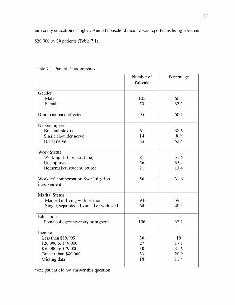
117
university education or higher. Annual household income was reported as being less than
$20,000 by 30 patients (Table 7.1).
Table 7.1 Patient Demographics
Number of Patients
Percentage
Gender Male Female
105 53
66.5 33.5
Dominant hand affected
95
60.1
Nerves Injured Brachial plexus Single shoulder nerve Distal nerve
61 14 83
38.6 8.9 52.5
Work Status Working (full or part time) Unemployed Homemaker, student, retired
81 56 21
51.6 35.4 13.4
Workers’ compensation &/or litigation involvement
50 31.6
Marital Status Married or living with partner Single, separated, divorced or widowed
94 64
59.5 40.5
Education Some college/university or higher*
106
67.1
Income Less than $19,999 $20,000 to $49,000 $50,000 to $79,000 Greater than $80,000 Missing data
30 27 50 33 18
19
17.1 31.6 20.9 11.4
*one patient did not answer this question
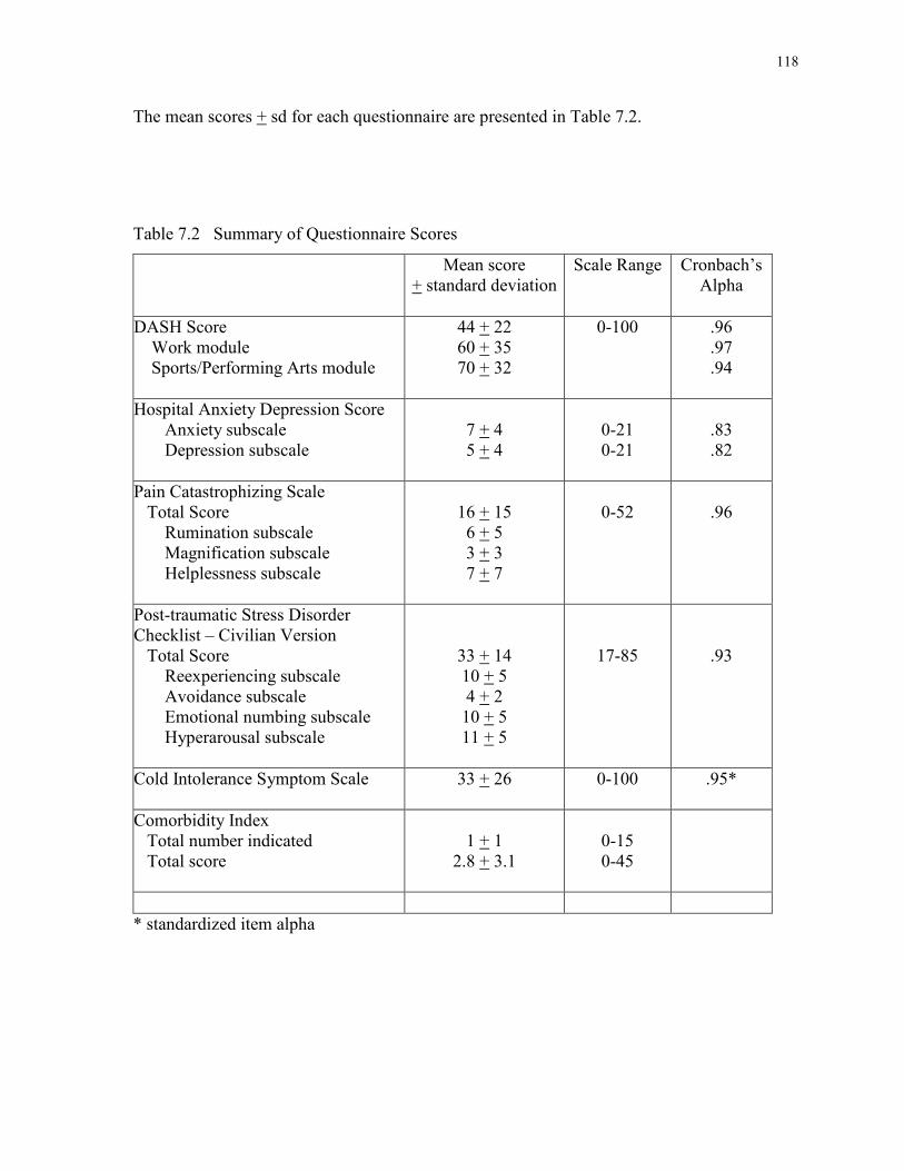
118
The mean scores + sd for each questionnaire are presented in Table 7.2.
Table 7.2 Summary of Questionnaire Scores
Mean score + standard deviation
Scale Range Cronbach’s Alpha
DASH Score Work module Sports/Performing Arts module
44 + 22 60 + 35 70 + 32
0-100 .96 .97 .94
Hospital Anxiety Depression Score Anxiety subscale Depression subscale
7 + 4 5 + 4
0-21 0-21
.83 .82
Pain Catastrophizing Scale Total Score Rumination subscale Magnification subscale Helplessness subscale
16 + 15 6 + 5 3 + 3 7 + 7
0-52
.96
Post-traumatic Stress Disorder Checklist – Civilian Version Total Score Reexperiencing subscale Avoidance subscale Emotional numbing subscale Hyperarousal subscale
33 + 14 10 + 5 4 + 2 10 + 5 11 + 5
17-85
.93
Cold Intolerance Symptom Scale
33 + 26
0-100 .95*
Comorbidity Index Total number indicated Total score
1 + 1
2.8 + 3.1
0-15 0-45
* standardized item alpha
119
The mean DASH score was 44 + 22 indicating a substantial level of disability. Eighty seven
patients (55%) scored higher than 39.46 on the DASH, a score that corresponds to two sds
above the mean normative value reported by Hunsaker et al (2002). A significant positive
association was found between brachial plexus injury and a high DASH score (p < 0.001).
The mean VAS pain intensity score was 4.2 + 3.0 and the mean total SF-MPQ Pain Rating
Index was 13.0 + 10.8 (sensory 10.5 + 8.3, affective 2.5 + 3.1). The mean CISS score was 33
+ 26 indicating a high level of cold sensitivity. There were 81 patients (51%) who were
classified with abnormal cold sensitivity (CISS score greater than 30, per Ruijs et al (2006)).
In patients with upper extremity nerve injury, excellent internal consistency was found for
the DASH (Cronbach’s alpha = .96), PCS (Cronbach’s alpha = .96), PCL-C (Cronbach’s
alpha = .93) and the CISS (standardized item alpha = .95) and good internal consistency for
the HADS anxiety and depression subscales (Cronbach’s alpha = .83 and .82, respectively).
7.4.1 Univariate Statistical Analyses
Patients receiving workers’ compensation or engaged in litigation had significantly higher
DASH scores than those who were not (p = 0.02) (Table 7.3). Statistically significant
differences in the DASH scores were not found for the other independent variables; gender (p
= 0.06), dominant limb injured (p = 0.30), marital status (p = 0.65), education level (p =
0.06) or geographic location (p = 0.23). The one-way ANOVA comparing the DASH score
among nerve injury groups was statistically significant (p < 0.05). Post hoc analysis revealed
that DASH scores in patients with brachial plexus injuries were significantly higher than in
patients with distal nerve injuries (p < 0.001). The one-way ANOVA comparing DASH
scores between work status groups was statistically significant (p < 0.05). Post hoc analysis

120
revealed that the DASH scores in patients who were unemployed were significantly higher
than those working (p < 0.001). There was no significant difference in DASH scores as a
function of level of income (p = 0.284).
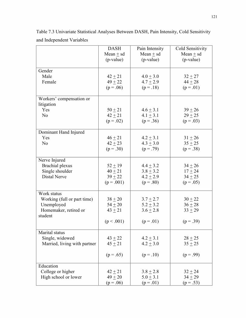
121
Table 7.3 Univariate Statistical Analyses Between DASH, Pain Intensity, Cold Sensitivity
and Independent Variables
DASH Mean + sd (p-value)
Pain Intensity Mean + sd (p-value)
Cold Sensitivity Mean + sd (p-value)
Gender Male Female
42 + 21 49 + 22 (p = .06)
4.0 + 3.0 4.7 + 2.9 (p = .18)
32 + 27 44 + 28 (p = .01)
Workers’ compensation or litigation Yes No
50 + 21 42 + 21 (p = .02)
4.6 + 3.1 4.1 + 3.1 (p = .36)
39 + 26 29 + 25 (p = .03)
Dominant Hand Injured Yes No
46 + 21 42 + 23 (p = .30)
4.2 + 3.1 4.3 + 3.0 (p = .79)
31 + 26 35 + 25 (p = .38)
Nerve Injured Brachial plexus Single shoulder Distal Nerve
52 + 19 40 + 21 39 + 22
(p = .001)
4.4 + 3.2 3.8 + 3.2 4.2 + 2.9 (p = .80)
34 + 26 17 + 24 34 + 25 (p = .05)
Work status Working (full or part time) Unemployed Homemaker, retired or student
38 + 20 54 + 20 43 + 21
(p < .001)
3.7 + 2.7 5.2 + 3.2 3.6 + 2.8
(p = .01)
30 + 22 36 + 28 33 + 29
(p = .39)
Marital status Single, widowed Married, living with partner
43 + 22 45 + 21
(p = .65)
4.2 + 3.1 4.2 + 3.0
(p = .10)
28 + 25 35 + 25
(p = .99)
Education College or higher High school or lower
42 + 21 49 + 20 (p = .06)
3.8 + 2.8 5.0 + 3.1 (p = .01)
32 + 24 34 + 29 (p = .53)
122
DASH scores and VAS pain intensity scores were positively correlated (r = .51, p < 0.001).
There was a weak positive correlation between DASH scores and age (r = .16, p = 0.02).
VAS pain intensity was strongly positively correlated with the CISS score (r = .50, p <
0.001), the PCS total score (r = .66, p < 0.001) and the PCS subscales; rumination (r = .66, p
< 0.001), magnification (r = .57, p < 0.001) helplessness (r = .65, p < 0.001). There was a
moderate positive correlation between VAS pain intensity scores and PCL total score (r =
.47, p < 0.001), HADS anxiety score (r = .32, p < 0.001) and HADS depression score (r =
.41, p < 0.001) (Table 7.4).
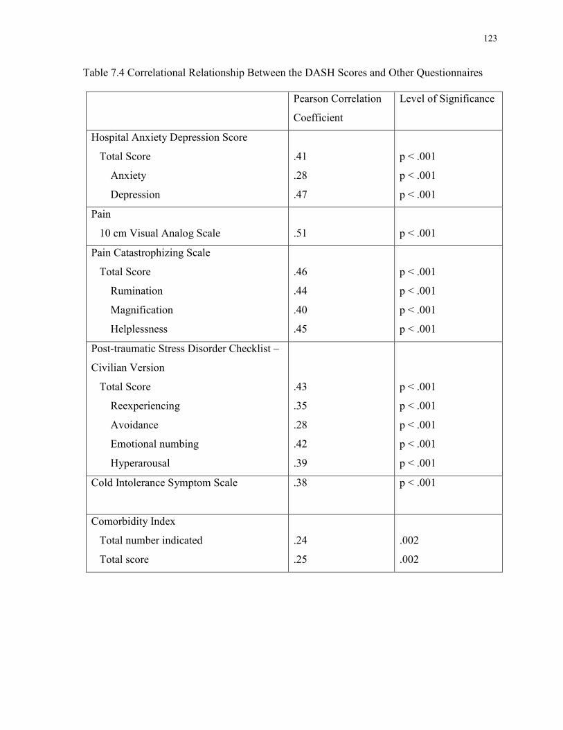
123
Table 7.4 Correlational Relationship Between the DASH Scores and Other Questionnaires
Pearson Correlation
Coefficient
Level of Significance
Hospital Anxiety Depression Score
Total Score
Anxiety
Depression
.41
.28
.47
p < .001
p < .001
p < .001
Pain
10 cm Visual Analog Scale
.51
p < .001
Pain Catastrophizing Scale
Total Score
Rumination
Magnification
Helplessness
.46
.44
.40
.45
p < .001
p < .001
p < .001
p < .001
Post-traumatic Stress Disorder Checklist –
Civilian Version
Total Score
Reexperiencing
Avoidance
Emotional numbing
Hyperarousal
.43
.35
.28
.42
.39
p < .001
p < .001
p < .001
p < .001
p < .001
Cold Intolerance Symptom Scale
.38 p < .001
Comorbidity Index
Total number indicated
Total score
.24
.25
.002
.002
124
7.4.2 Multivariable Regression Analysis
Multivariable linear regression with manual backward elimination was used to evaluate the
factors that statistically predicted disability as measured by the DASH. Our criterion for
multicollinearity was r = 0.8 and the correlation coefficient between the HADS anxiety score
and the PCL-C was r = 0.76. To control for the potential multicollinearity between these two
closely related variables, the HADS anxiety score was not included in the regression model.
Both the HADS anxiety and PCL-C assess anxiety related symptoms and this likely accounts
for the high correlation coefficient. Because this study assessed patients following a
traumatic nerve injury and the PCL-C was designed to assess symptoms of post-traumatic
stress, we chose to include the PCL-C in the regression model and not the HADS anxiety
score as the former measure is more closely related to the nature of the injury than is the
latter. The correlation coefficients between the variables in the regression analysis are
presented in Table 7.5.
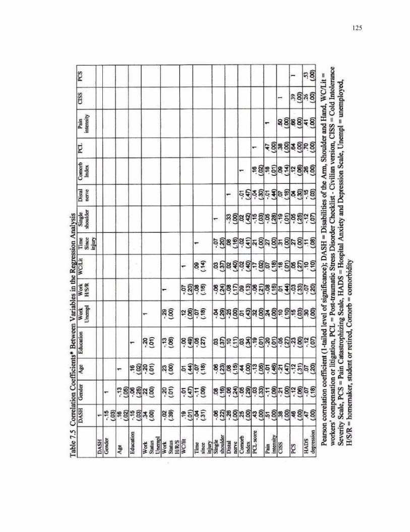
125
126
The distribution of time from injury was positively skewed (skewness = 2.8). The non-
normality was corrected using a log10 transformation and these transformed data were used
in the regression analysis (Norman et al., 2000). Based upon the univariate statistical
analyses (p-value less than 0.2), the preliminary regression model included the following
independent variables; gender, age, education, work status, time since injury, workers’
compensation or litigation involvement, nerve injured, comorbidity index, HADS depression
score, PCL-C score, VAS pain intensity, CISS score, PCS total score. Using manual
backward elimination and 0.1 level of significance for removal (Table 7.6), the final model
accounted for 52.7% of the variance and contained 10 predictor variables (Table 7.7); pain
intensity (Beta = .230, p = 0.006), nerve injured (Beta = -.220, p < 0.001), time since injury
(Beta = -.198, p = 0.002), pain catastrophizing score (Beta = .192, p = 0.025), age (Beta =
.187, p = 0.002), work status (Beta = .179, p = 0.008), cold sensitivity (Beta = .171, p =
0.015), depression score (Beta = .133, p = 0.066), workers’ compensation or litigation (Beta
= .116, p = 0.049) and gender (Beta = -.104, p = 0.09).
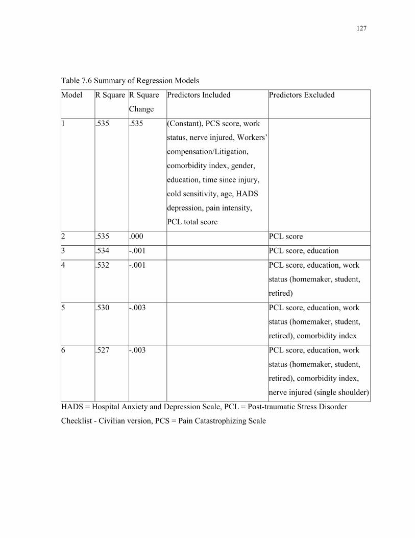
127
Table 7.6 Summary of Regression Models
Model R Square R Square
Change
Predictors Included Predictors Excluded
1 .535 .535 (Constant), PCS score, work
status, nerve injured, Workers’
compensation/Litigation,
comorbidity index, gender,
education, time since injury,
cold sensitivity, age, HADS
depression, pain intensity,
PCL total score
2 .535 .000 PCL score
3 .534 -.001 PCL score, education
4 .532 -.001 PCL score, education, work
status (homemaker, student,
retired)
5 .530 -.003 PCL score, education, work
status (homemaker, student,
retired), comorbidity index
6 .527 -.003 PCL score, education, work
status (homemaker, student,
retired), comorbidity index,
nerve injured (single shoulder)
HADS = Hospital Anxiety and Depression Scale, PCL = Post-traumatic Stress Disorder
Checklist - Civilian version, PCS = Pain Catastrophizing Scale
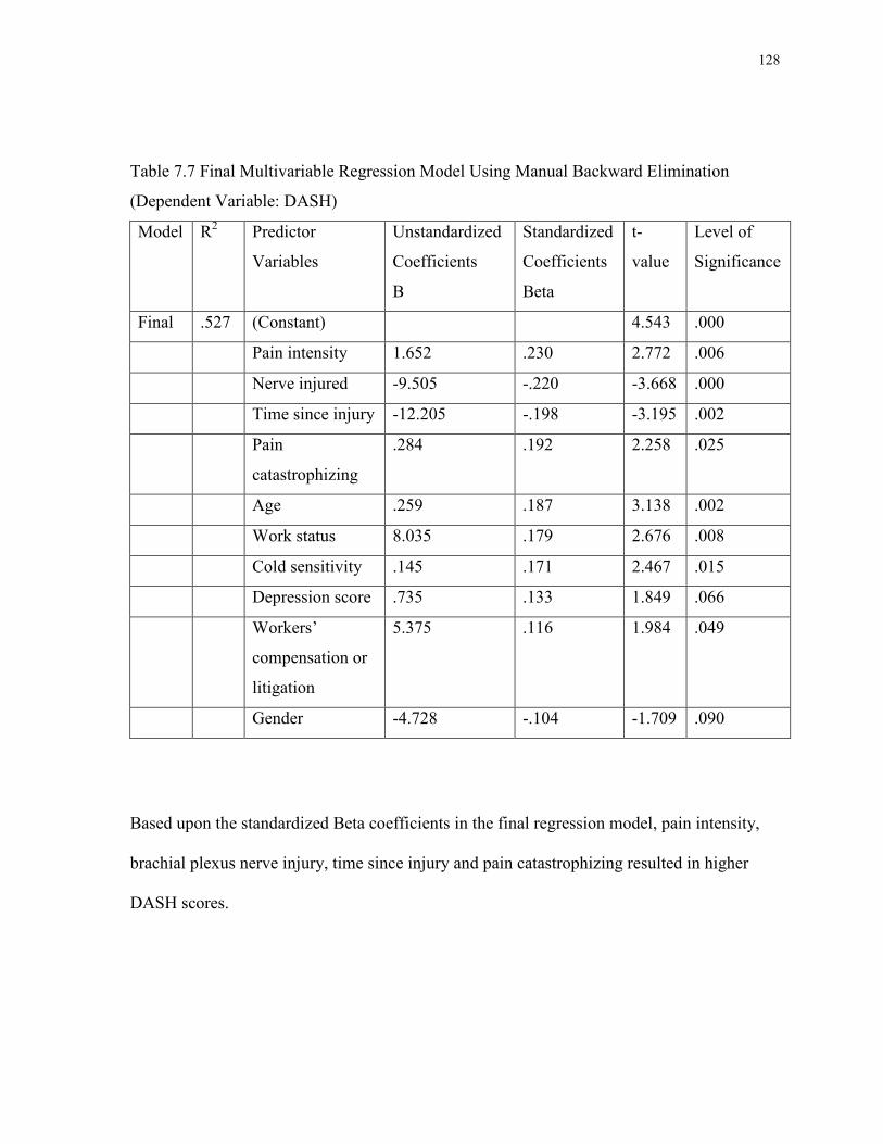
128
Table 7.7 Final Multivariable Regression Model Using Manual Backward Elimination
(Dependent Variable: DASH)
Model
R2 Predictor
Variables
Unstandardized
Coefficients
B
Standardized
Coefficients
Beta
t-
value
Level of
Significance
Final .527 (Constant) 4.543 .000
Pain intensity 1.652 .230 2.772 .006
Nerve injured -9.505 -.220 -3.668 .000
Time since injury -12.205 -.198 -3.195 .002
Pain
catastrophizing
.284 .192 2.258 .025
Age .259 .187 3.138 .002
Work status 8.035 .179 2.676 .008
Cold sensitivity .145 .171 2.467 .015
Depression score .735 .133 1.849 .066
Workers’
compensation or
litigation
5.375 .116 1.984 .049
Gender -4.728 -.104 -1.709 .090
Based upon the standardized Beta coefficients in the final regression model, pain intensity,
brachial plexus nerve injury, time since injury and pain catastrophizing resulted in higher
DASH scores.
129
7.5 Discussion
The results of the present study indicate that patients with a peripheral nerve injury reported
substantial disability which was predicted by a combination of biomedical and psychosocial
factors (pain intensity, brachial plexus nerve injury, pain catastrophizing, older age, work
status, time since injury and cold sensitivity). Previous studies of patients following traumatic
peripheral nerve injuries tend to report only measures of physical impairment (Bengtson et
al., 2008; Novak et al., 2010). Assessments of motor and sensory recovery provide data
regarding nerve remyelination and/or regeneration and reinnervation of the motor fibres
and/or sensory end organs but do not provide insight into the overall impact of the injury on
the patient. More recently studies have recognized the importance of patient self report
questionnaires, disability and health related quality of life (Ahmed-Labib et al., 2007; Bailey
et al., 2009; Bengtson et al., 2008; Choi et al., 1997; Novak et al., 2009b).
The World Health Organization (WHO) and others have presented biopsychosocial models
for disability and function which link together the biomedical, personal and social
perspectives (Jette, 2006; Verbrugge et al., 1994; World Health Organization, 2001; Jette et
al., 2005). The WHO has proposed the International Classification of Function, Disability
and Health (ICF) model and within this framework, body structures and functions
(physiological function), activity and participation are considered in the context of life
domains in addition to the impact of environmental and personal factors (World Health
Organization, 2001; Jette, 2009). In terms of nerve injury, this model captures the interaction
between the condition after injury (physical impairment and activity performance) and the
personal and environmental factors.
130
Disability is considered a limitation in tasks, activities or performance of daily activities and
in the case of nerve injury, due to physical impairment. The DASH was designed to assess
disability in patients with upper extremity musculoskeletal disorders based upon physical
function and symptoms (Beaton et al., 2001; Gummesson et al., 2003; Hudak et al., 1996;
SooHoo et al., 2008). Few studies have reported disability as measured by the DASH in
patients with peripheral nerve injury (Ahmed-Labib et al., 2007; Novak et al., 2009b; Topel
et al., 2009). In a previous retrospective chart review of patients with upper extremity nerve
injury, we found substantial disability as measured by the DASH which was associated with
pain, older age and brachial plexus nerve injuries (Novak et al., 2009b). The results of the
present study confirm these initial findings, showing that high levels of disability were
reported by patients with an upper extremity nerve injury. Other studies have reported high
levels of disability in patients with brachial plexus injuries (Ahmed-Labib et al., 2007;
Davidson, 2004). Using the DASH and the SF-36, Ahmed-Labib et al. (2007) reported a
correlation between poor functional outcome and root avulsion injuries and delayed surgical
repair. Topel et al. (2009) evaluated 33 patients with upper extremity arterial injuries and
reported more severe functional deficits in patients with a concomitant peripheral nerve
injury although no statistically significant differences were noted. In the present study, we
found high levels of disability in patients with an upper extremity nerve injury and a
significant predictor of higher disability scores was brachial plexus nerve injury.
Poor outcome associated with neuropathic pain following traumatic nerve injuries has been
reported (Campbell & Meyer, 2006; Dworkin et al., 2007; Htut et al., 2006; Kato et al., 2006;
Novak et al., 2009b). Brachial plexus nerve injury is often associated with more severe pain
and is a significant predictor of disability. Choi et al. (1997) evaluated a group of patients
131
following brachial plexus injury and surgery. These patients reported a moderately high
general quality of life, although 75% reported significant pain. In a sample of patients with
nerve compression and nerve injury, Bailey et al. (2009) reported moderate pain which was
associated with lower quality of life and higher depression scores. In patients with brachial
plexus nerve injury, Htut et al. (2006) found that surgical repair was associated with pain
relief and pain intensity was highest among patients who did not undergo surgical repair.
Kato et al. (2006) investigated the impact of operative delay on pain relief in 148 patients
with brachial plexus injuries. Significant positive correlations were observed between pain
intensity and number of avulsed nerve roots and a longer interval between injury and surgery.
In the present study, we did not examine root avulsions specifically but did find that patients
with brachial plexus injuries had more pain and disability compared to patients with more
distal nerve injuries. Pain intensity was a significant predictor of disability and was also
strongly correlated with cold sensitivity. In our previous retrospective chart review study,
more bodily pain, as assessed by the SF-36, was reported in patients with an upper extremity
nerve injury (Novak et al., 2009b). Pain intensity has also been associated with disability in
other musculoskeletal disorders. Souer et al. (2008) evaluated patients following distal radius
fracture and pain was a significant positive predictor of the DASH scores. Patients with intra-
articular elbow fractures were evaluated using physician based rating scales and patient
reported questionnaires (DASH, SF-36) (Doornberg et al., 2005). The predictors of the
DASH included pain and range of motion and as a single predictor pain explained 36% of the
variance in the regression model. In another study following elbow trauma, Lindenhovius et
al. (2008) reported that the strongest predictor of the DASH was pain and accounted for 41%
of the variance. Consistent with these findings, the results of the present study of patients
132
with a traumatic peripheral nerve injury, show that pain intensity is a significant predictor of
disability (Beta = .230, p = 0.006). These data highlight the importance of the relationship
between pain intensity and patient reported disability and emphasize the need for good long
term pain management and a comprehensive treatment approach to these patients.
Pain may also be associated with cold sensitivity; symptoms of pain, stiffness, sensory
disturbance and color changes with exposure to cold (Graham et al., 2008; Novak et al.,
2009a; Vaksvik et al., 2009). When present, cold sensitivity is often associated with high
morbidity and poor outcome (Backman et al., 1993; Campbell et al., 1998; Collins et al.,
1996; Craigen et al., 1999; Dabernig et al., 2006; Graham et al., 2008; Irwin et al., 1997;
McCabe et al., 1991; Nancarrow et al., 1996; Nylander et al., 1987; Povlsen et al., 1995a;
Povlsen et al., 1995b; Ruijs et al., 2006; Ruijs et al., 2007; Stokvis et al., 2009; Vaksvik et
al., 2009). The present study found high levels of cold sensitivity and disability in patients
with upper extremity nerve injury and disability was predicted by both pain and cold
sensitivity. Dabernig et al. (2006) reported a relatively low mean DASH score (11/100) in
patients after digit replantation, although 87% of patients reported cold intolerance.
Following traumatic hand injury, many studies have reported long standing and unresolved
cold sensitivity with high levels of pain (Campbell et al., 1998; Collins et al., 1996; Graham
et al., 2008; Meyer-Rosberg et al., 2001b; Nancarrow et al., 1996; Stokvis et al., 2009). Cold
sensitivity remains a substantial challenge following trauma and nerve injury particularly for
patients who reside or work in cold environments.
Historically, in the surgical literature, biomedical factors associated with outcome have been
emphasized over other potentially important variables. More recently the importance of the
relationship between psychosocial factors and outcome has been recognized. In the present
133
sample of patients with an upper extremity nerve injury, depression scores were moderately
positively correlated with DASH scores and pain catastrophizing was a significant positive
predictor of the DASH (Beta = .192, p = .025). Previous studies have evaluated the
relationship between psychosocial factors such as pain catastrophizing and depression in
patients with various hand pathologies, including trapeziometacarpal arthrosis and after
carpal tunnel release (Lozano Calderon et al., 2008a; Lozano Calderon et al., 2008b; Niekel
et al., 2009). Lozano Calderon et al. (2008a) reported a significant negative correlation
between the DASH scores and patient satisfaction in patients after carpal tunnel release and
pain catatrophizing and depressive symptoms were predictors of disability. Niekel et al.
(2009) evaluated the relationship between DASH scores and psychosocial measures
including pain catastrophizing, pain anxiety and symptoms of depression in patients with a
variety of hand pathologies. The authors found a small positive correlation between DASH
scores and pain catastrophizing and a moderate positive correlation between the DASH
scores and symptoms of depression and anxiety. Both pain catastrophizing and depression
symptoms were predictors of higher DASH scores.
Pain catastrophizing is a maladaptive cognitive style that involves negative thinking
regarding the pain experience (Sullivan et al., 1995; Sullivan et al., 2005). Patients who
catastrophize feel overwhelmed by pain, ruminate about it, and feel helpless in the face of
pain. In a group of patients with chronic pain from a variety of etiologies, Severeijns et al.
(2001) reported higher pain intensity, disability and psychological distress in patients who
catastrophized. In the present study, patients following traumatic nerve injury exhibited high
levels of pain castrophizing and disability. However, disability is a multidimensional
construct, which is influenced by physical impairment, activity and participation and also by
134
environmental and personal factors and as such requires a multi-disciplinary treatment
approach (Jette, 2009; World Health Organization, 2001).
The limitations of this study include the unique patient sample, cross sectional study design,
inclusion of nerve injuries six months after injury, and possibility of low statistical power for
detecting certain relationships. In this investigation, we evaluated patients who were at least
six months following a traumatic peripheral nerve injury. This presents a unique group of
patients who continue to seek medical or surgical assessment and treatment and may not be
representative of all patients following nerve injury. There are patients following nerve injury
who have complete recovery and no apparent morbidity. However, patients who remain with
physical impairments, pain and disability are often the most difficult to treat, overwhelm the
available resources and have persistent, diminished health related quality of life. As well,
inclusion of patients with nerve injuries who have not achieved full recovery may have
biased the results in the direction of finding more pain and greater disability in these patients.
The primary aim of this study was to evaluate the predictors of the DASH using a
multivariable regression model and a sample size of 158 was sufficient to support the
inclusion of the all of the factors in the preliminary regression model. The t-tests comparing
DASH scores on the dependent variables were performed, in part, to select the factors to be
included in the regression model and may not have had sufficient power to detect a
significant difference in all cases. Finally, given the cross sectional design used in the present
study, conclusions about the direction of causality cannot be made. The present study
provides the preliminary data for a comprehensive longitudinal study to evaluate the specific
risk factors for persistent pain and disability following nerve injury and also to describe the
nature of treatment and the relationship between measures of impairment and disability.
135
In summary, patients with a peripheral nerve injury reported substantial disability and pain.
Disability, as measured by the DASH, was predicted by a combination of biomedical and
psychosocial factors including pain intensity, brachial plexus nerve injury, pain
catastrophizing, older age, work status, time since injury and cold sensitivity. Future
investigation regarding efficacious treatments of the factors that are associated with disability
and chronic neuropathic pain will hopefully help to improve health related quality of life in
patients with traumatic nerve injury.
136
Chapter 8 General Discussion
8 General Discussion
This general discussion is organized into the following specific sections relating the findings
of the three studies of my dissertation to the existing literature on: disability, neuropathic
pain, cold sensitivity, psychosocial factors followed by limitations and directions for future
research. My dissertation is the first to evaluate the association between biomedical and
psychosocial factors and disability in patients with traumatic upper extremity nerve injury.
The inclusion of both biomedical and psychosocial factors in the regression models and
patients with a wide variety of traumatic upper extremity nerve injuries added to the strength
of my dissertation. Studies 2 and 3 (Chapters 6 and 7) found high levels of disability and
neuropathic pain in patients with chronic nerve injury. In Study 3 (Chapter 7), disability as
measured by the DASH was predicted by biomedical and psychosocial factors including pain
intensity, nerve injured, time since injury, pain catastrophizing score, age, work status, cold
sensitivity, depression score, workers’ compensation or litigation and gender. This final
regression model accounted for 52.7% of the variance.
Many of the previous studies which have evaluated patient outcome following traumatic
peripheral nerve injuries reported only measures of physical impairment and even when
using these measures, there are large variations in the types of assessments and the standards
applied (Bengtson et al., 2008). While reports of motor and sensory recovery provide insight
regarding neural regeneration and reinnervation of the motor fibres and/or sensory end
organs, these outcome measures do not indicate the functional impact of these impairments
137
on the patient. More recent studies have recognized the importance of patient self report of
domains such as disability and health related quality of life in the assessment of outcome
following nerve injury (Ahmed-Labib et al., 2007; Bailey et al., 2009; Bengtson et al., 2008;
Choi et al., 1997; Kitajima et al., 2006). Ahmed-Labib et al. (2007) reported significantly
higher DASH and SF-36 physical domain scores in patients who underwent reconstructive
surgery for a brachial plexus nerve injury. Kitajama et al. (2006) reported significantly lower
health status (SF-36 scores) in patients with a brachial plexus nerve injury who underwent
surgery compared to the Japanese normative data. Using a telephone survey with questions
adapted from the US General Social Survey, Choi et al. (1997) reported moderately high
levels of quality of life and overall life satisfaction in patients following brachial plexus
nerve surgery. In a sample of patients with nerve compression or nerve injury, Bailey et al.
(2009) reported lower quality of life compared to normative data and overall quality of life
was predicted by participation in activity and depressive symptoms. The results of Study 3
(Chapter 7) of the present dissertation are in general agreement with the above literature. In
Study 3, high levels of disability were reported by patients following traumatic nerve injury
and disability was associated with factors other than physical impairment. Motor and sensory
recovery following nerve injury may directly impact physical impairments however
assessment of this physical impairment on the patient’s life domains and activities will more
accurately reflect the morbidity associated with upper extremity peripheral nerve injury. In
Study 3, high levels of disability were reported by patients following traumatic nerve injury
and disability was associated with factors beyond physical impairment.
138
8.1 Disability
Disability is a multidimensional construct and as defined by Nagi (1965), disability
represents a mental or physical limitation within a social context. The biopsychosocial model
for disability provides a framework for the integration of the biological, personal and social
perspectives (Jette, 2006; Verbrugge et al., 1994). The ICF model, which was developed by
the WHO, includes the consideration of contextual factors in activity and participation as key
elements within the framework (World Health Organization, 2001). In the surgical literature
regarding patients following nerve injury, the studies that have evaluated outcome do not
frequently include the assessment of the impact of the injury on the patient or other types of
patient reported outcome. However, in those studies that have included functional outcomes,
the DASH is the most commonly used questionnaire to assess disability in patients with
upper extremity injury. In Study 3 (Chapter 7), high levels of disability as measured by the
DASH were reported in patients who were at least 6 months following nerve injury.
Multivariable regression analysis indicated that the DASH scores were associated with pain
intensity, nerve injured, time since injury, pain catastrophizing score, age, work status, cold
sensitivity, scores on the depression questionnaire, workers’ compensation or litigation and
gender.
The DASH is a self report questionnaire. Based upon physical functioning and symptoms, it
was designed to assess disability in patients with upper extremity musculoskeletal disorders
(Beaton et al., 2001; Hudak et al., 1996). Good validity, reliability and responsiveness have
been reported for the DASH in a variety of upper extremity musculoskeletal pathologies
(Beaton et al., 2001; Gummesson et al., 2003; Hudak et al., 1996; SooHoo et al., 2008). To
date, studies have not evaluated the internal consistency or validity of the DASH in a sample
139
of patients with nerve injury. Indeed, there are very few outcome studies of patients with
peripheral nerve injury which have reported disability (Ahmed-Labib et al., 2007; Davidson,
2004; Novak et al., 2010; Topel et al., 2009). In Study 3 (Chapter 7), the DASH showed high
internal consistency which supports the use of the DASH in patients with traumatic upper
extremity nerve injury. In my retrospective chart review (Study 2, Chapter 6) and prospective
study of patients with chronic upper extremity nerve injury (Study 3, Chapter 7), high levels
of disability as measured by the DASH were reported with mean values of 52 + 22 and 44 +
22, respectively, in a normal distribution. In both studies, higher DASH scores were
associated with higher pain intensity, older age and brachial plexus nerve injuries. Other
studies have also reported high levels of disability in patients with brachial plexus injuries
(Ahmed-Labib et al., 2007; Davidson, 2004; Topel et al., 2009). Using the DASH and the
SF-36, Ahmed-Labib et al. (2007) reported poor functional outcome which was correlated
with root avulsion injuries and delayed surgical repair. Davidson (2004) evaluated patients
with a variety of upper extremity pathologies which included amputation injuries and
brachial plexus nerve injuries and reported higher DASH scores in patients with brachial
plexus nerve injury. In a study of patients following traumatic arterial injuries, Topel et al.
(2009) reported more severe functional deficits in those patients with arterial and
concomitant nerve injuries although in this study there were no statistical differences
reported. In patients with median or ulnar nerve injuries, Vordemvenne, Langer, Ochman,
Raschke & Schult (2007) reported an overall mean DASH score of 22. In patients with radial
nerve palsy following humeral fractures, Ekholm et al. (2008) reported relatively low DASH
scores and this likely represents the high number of patients who had recovery of radial nerve
function. In my dissertation studies (Chapters 6 & 7), high levels of disability in patients with
140
chronic upper extremity nerve injury were found and consistent with the existing literature
(Ahmed-Labib et al., 2007), a significant factor associated with higher disability scores was
brachial plexus nerve injury. In my studies, I recruited patients who were at least six months
following upper extremity nerve injury and who had sought medical assessment. These
patients represent a wide variation of recovery and many patients particularly those with
brachial plexus nerve injuries had not attained maximal recovery.
The DASH questionnaire contains two additional modules regarding work and recreation
(sports/performing arts). Each separate module contains four questions regarding the impact
of the upper extremity pathology on the patient’s work or sports/performing arts (such as
athletes and musicians). In accordance with the ICF model (World Health Organization,
2001), these separate subsections stress the importance of activity and participation in the
context of disability and provide the opportunity to assess these parameters. Activity is
incorporated in the items of the DASH and participation restriction may be captured in the
work and sports/performing arts modules. In my studies (Chapters 6 & 7), patients with long
standing nerve injury had substantial disability reported in the work and recreation modules
of the DASH. This restriction in participation of life roles may impact the relations of the
individual in these social roles. Disability as related to activity and participation may be
present in patients with long standing traumatic nerve injury and identification of these
limitations may be useful in recognizing the negative impact on these patients.
141
8.2 Neuropathic Pain
My dissertation demonstrated a high prevalence of neuropathic pain in patients who sought
medical assessment and/or treatment following an upper extremity nerve injury. In Study 3
(Chapter 7), patients with long standing nerve injury reported high levels of pain intensity
and this was significantly associated with disability. As a single predictor in a regression
model, pain intensity accounted for 26% of the variance. In Study 2 (Chapter 6), significantly
more pain, as assessed by the SF-36 bodily pain domain, was reported in patients with long
standing nerve injury compared to the published Canadian normative values. Chronic
neuropathic pain is frequently associated with poor outcome independent of the originating
etiology (Bennett, 2003; Campbell et al., 2006; Dworkin et al., 2007). Bailey et al. (2009)
reported moderate pain in a group of patients with nerve compression or nerve injury and
pain was strongly correlated with a lower quality of life and higher scores of depression. In
the regression model, participation in activity and depression scores were associated with
overall quality of life although pain was not retained in the final regression model. Following
traumatic nerve injury, severe neuropathic pain has been reported particularly in patients with
brachial plexus nerve injuries (Bertelli et al., 2008a; Choi et al., 1997; Htut et al., 2007; Kato
et al., 2006). In a group of patients following brachial plexus reconstructive surgery, Choi et
al. (1997) found 75% of patients reported “significant pain”. Htut et al. (2006) found that
surgical repair following brachial plexus nerve injury was associated with pain relief and that
pain intensity was highest among patients who did not have surgery; consistent with theory
of nerve repair. Kato et al. (2006) reported significant positive correlations between pain
intensity and nerve root avulsion in patients with brachial plexus nerve injuries. In Studies 2
and 3 (Chapters 6 and 7), nerve root avulsion was not specifically evaluated but patients with
142
brachial plexus injuries had significantly higher levels of disability compared to more distal
nerve injuries. The mean pain intensity was 4.4 + 3.2 and there was no statistically significant
difference in pain intensity between brachial plexus and distal nerve injuries. Pain intensity
was a significant predictor of disability and it was also strongly correlated with cold
sensitivity. Consistent with previous reports in the literature, the results of my dissertation
indicate that pain intensity is strongly related to reported disability in patients with traumatic
upper extremity nerve injury.
Pain has also been identified as a significant predictor of disability in other musculoskeletal
pathologies (Doornberg et al., 2005; Lindenhovius et al., 2008; Souer et al., 2008). In
patients following distal radius fracture, Souer et al. (2008) reported that pain was a
significant positive predictor of the DASH scores. In patients with intra-articular elbow
fractures, the predictors of the DASH included pain and range of motion and as a single
predictor in the regression model, pain explained 36% of the variance (Doornberg et al.,
2005). In a study following elbow trauma, Lindenhovius et al. (2008) reported that the
strongest predictor of the DASH was pain which explained 41% of the variance. These
studies highlight the importance of the relationship between pain intensity and higher levels
of disability in patients with upper extremity pathologies and the need for comprehensive
multidisciplinary pain management approaches.
In Study 1 (Chapter 5) of my dissertation, many nerve surgeons reported that they do not
evaluate pain in patients with a nerve injury who were not specifically referred for pain. As
well, among patients with nerve injury, few surgeons reported using valid, reliable
questionnaires to assess pain, cold sensitivity or disability following nerve injury. Therefore
patient symptoms related to pain with respect to frequency, intensity and disability may be
143
under represented in the reports of outcome following nerve injury. The results of Study 1
(Chapter 5) are consistent with the review of the literature in which assessment of pain is less
frequently reported in outcome studies of patients after nerve injury compared to recovery
related to sensory and/or motor function. When pain was evaluated, pain intensity was most
frequently reported and the inclusion of measures to evaluate the impact of the pain on the
patient were rare (Atherton et al., 2008; Bentolila et al., 1999; Berman et al., 1998; Bertelli et
al., 2008a; Chen et al., 2006; Emery et al., 1997; Evans et al., 1994; Geertzen et al., 2000;
Hazari et al., 2004; Kanpolat et al., 2008; Kim et al., 2001a; Lefaucheur et al., 2004;
Mackinnon et al., 1987; Rath et al., 1997; Ricardo, 2005; Saitoh et al., 2003; Samii et al.,
2001; Sindou et al., 2005; Sood et al., 1998; Thomas et al., 1994; Waikakul et al., 1999).
To evaluate the unique qualities of neuropathic pain, a modification of the SF-MPQ was
introduced by Dworkin et al. (2009) which includes pain descriptors that are specific to
neuropathic pain. In the original SF-MPQ each descriptor was ranked on a scale from none,
mild, moderate or severe. However, in the modification of the SF-MPQ, each pain descriptor
is ranked on a scale from zero to ten. The exploratory factor analysis resulted in a three factor
solution and these three sub-scales were identified as continuous pain descriptors (6 items),
intermittent pain descriptors (6 items) and neuropathic pain descriptors (4 items). A fourth
subscale included the affective descriptors (4 items) from the SF-MPQ. Good validity and
reliability for this modification of the SF-MPQ were shown in a group of patients with
various chronic pain diagnoses (Dworkin et al., 2009). This modification of the SF-MPQ
may assist in the assessment of neuropathic pain following nerve injury, although this
questionnaire has not been validated in patients with traumatic nerve injury. Utilization of
questionnaires to differentiate neuropathic pain from nociceptive pain may assist in
144
identifying patients with neuropathic pain that may be more amenable to treatment strategies
specific for neuropathic pain.
8.3 Cold Sensitivity
Pain following nerve injury may be constant, intermittent and/or associated with exposure to
cold temperatures. The results of Study 3 (Chapter 7) showed substantial cold sensitivity
among patients with long standing nerve injury and cold sensitivity was significantly
associated with higher disability. Sensitivity to cold is a common sequela of upper extremity
trauma that is accompanied by one or more of the following symptoms: pain, sensory
disturbance, stiffness and colour changes (Graham et al., 2008; Vaksvik et al., 2009). When
cold sensitivity is present, it is often associated with high morbidity and poor outcome
(Backman et al., 1993; Campbell et al., 1998; Collins et al., 1996; Craigen et al., 1999;
Dabernig et al., 2006; Graham et al., 2008; Irwin et al., 1997; McCabe et al., 1991;
Nancarrow et al., 1996; Nylander et al., 1987; Povlsen et al., 1995a; Povlsen et al., 1995b;
Ruijs et al., 2006; Ruijs et al., 2007; Stokvis et al., 2009; Vaksvik et al., 2009). As seen in
Study 3 (Chapter 7), cold sensitivity may contribute to higher levels of disability which is
consistent with the poor outcome previously reported in the literature. Interventions to
minimize cold sensitivity in the patients with ongoing symptoms may positively impact
outcome and decrease disability following nerve injury.
In the surgical literature, patient self report questionnaires are more frequently used to assess
cold sensitivity compared to objective measures of vascular status. The CISS and CSS are the
two most frequently used questionnaires to assess cold sensitivity (Irwin et al., 1997;
145
McCabe et al., 1991). These types of questionnaires provide the opportunity to assess the
severity of cold sensitivity and the impact of the symptoms with cold exposure on the patient
and their activities. In Study 3 (Chapter 7), the CISS questionnaire showed good internal
consistency (standardized item alpha = .95) in patients with nerve injuries at various levels in
the upper extremity. Using the criteria established by Ruijs et al. (2006), in my study
(Chapter 7), there were 81 patients (51%) who were classified as having abnormal cold
sensitivity. The mean CISS was 33 + 26 which is consistent with reports in the literature of
patients with upper extremity nerve injury (Carlsson et al., 2008; Irwin et al., 1997; Ruijs et
al., 2007). In the study by Dabernig et al. (2006), 87% of patients reported cold intolerance
following digit replantation, however, the mean DASH score was relatively low at 11/100.
This is in contrast to the high mean DASH score (44 + 22) found in my study (Study 3,
Chapter 7) in patients with nerve injury. The lower scores reported by Dabernig et al. (2006)
may reflect the capacity of patients to better adapt following digital replantation compared to
traumatic upper extremity nerve injuries or a weakness of the DASH in capturing subtle
disabilities following digit replantation. Meyer-Rosberg et al. (2001b) reported that cold
evoked pain was ranked as the most intense pain in patients with neuropathic pain. Campbell
and Kay (1998) found that 73% of patients with traumatic hand injuries reported cold related
symptoms and most complaints were pain related. The unresolved continuing symptoms of
cold sensitivity with exposure to cold temperatures have been reported in many patients
following traumatic upper extremity symptoms. Graham and Schofield (2008) reported cold
intolerance in 90% of patients with traumatic injuries and less than 10% of these patients
reported an improvement in their symptoms over time. Nancarrow et al. (1996) reported 69%
of patients with hand injuries had continuing symptoms of cold intolerance and 97%
146
remained with symptoms five years following injury. Collins et al. (1996) reported 76% of
patients following nerve injury reported continued cold intolerance more than five years
following trauma. Cold sensitivity remains a cause of morbidity following trauma and nerve
injury and this is particularly problematic for patients who reside and/or work in cold
environments. The high rate of cold sensitivity found in my study (Chapter 7) is consistent
with the reports in the literature of high levels of cold sensitivity following traumatic injuries.
There was a positive correlation between cold sensitivity scores and time since injury which
is consistent with the literature reports of unresolved symptoms following traumatic injury.
Cold sensitivity is related to higher levels of pain intensity and in my study (Chapter 7) was
associated with high levels of disability in patients with long standing nerve injury. Strategies
to treat this cold sensitivity may help to minimize the morbidity and disability associated
with upper extremity traumatic nerve injury.
8.4 Psychosocial Factors
In my study (Study 3, Chapter 7), high levels of anxiety, depression and pain catastrophizing
symptoms were reported in patients with traumatic upper extremity nerve injuries. In the
final regression model, pain catastrophizing and depression scores were associated with
disability, as measured by the DASH. Post-traumatic stress and anxiety related to the injury
was assessed using the PCL-C but this variable was not retained in the final regression
model. There is wide variability in patient outcome which is reported following upper
extremity nerve injury. Many patients sustain what appears to be a similar injury and
outcome may vary in terms of physical impairment, pain and disability. The mechanisms by
which this occurs are unclear and it may be related to the complex relationships among
147
multiple biomedical and psychosocial factors. Review of the surgical literature has revealed
an emphasis on the biomedical factors associated with outcome following nerve injury
compared to other potentially important variables, such as personal or psychosocial factors.
However, more recently the importance of the influence of psychosocial factors on outcome
in patients with upper extremity pathologies has been more reported (Bailey et al., 2009;
Lozano Calderon et al., 2008a; Lozano Calderon et al., 2008b; Niekel et al., 2009; Ring et al.,
2006; Souer et al., 2008; Vranceanu, Barsky, & Ring, 2009). My dissertation is the first to
show the association between disability and psychosocial factors, particularly symptoms of
depression, pain catastrophizing, in a sample of patients with long standing upper extremity
nerve injury.
8.4.1 Pain Catastrophizing
Pain catastrophizing involves exaggerated negative thinking regarding the experience of pain
(Sullivan et al., 1995; Sullivan et al., 2001; Sullivan et al., 2005). Individuals who
catastrophize feel overwhelmed by pain, feel helpless in the face of pain and ruminate about
the pain. The PCS was designed to assess the degree of pain castrophizing and comprises
three subscales (rumination, magnification, helplessness) (Sullivan et al., 1995). In my study
of patients with long standing upper extremity nerve injury (Chapter 7), high internal
consistency was found with the PCS. Strong correlations were found between pain intensity
and the PCS total score and the PCS subscales; rumination, magnification and helplessness.
Pain catastrophizing was a significant predictor of disability as measured by the DASH
scores.
148
In patients following traumatic injury who exhibit high levels of pain castrophizing, high
levels of disability have also been reported. In a group of patients with chronic pain resulting
from different etiologies, Severeijns et al. (2001) reported higher levels of pain intensity,
disability and psychological distress in patients who catastrophize. Strong associations
among these factors were reported but no conclusions were presented regarding the direction
of causality. Pain castastrophizing has also been reported in patients with a variety of hand
diagnoses. In a group of patients with different hand pathologies, Niekel et al. (2009)
evaluated the relationship between DASH scores and psychosocial measures (symptoms of
depression, pain catastrophizing and pain anxiety) and reported a weak correlation between
DASH scores and pain catastrophizing. In patients who were attending a pain treatment
program, pain catastrophizing was significantly associated with depression scores but not
disability as assessed by the Roland Scale of disability (Turner, Jensen, & Romano, 2000).
Karels et al. (2007) evaluated patients in a physical therapy practice with complaints of
shoulder and neck pain and reported that persistent complaints were significantly associated
with pain catastrophizing.
The studies which have reported relationships between pain catastrophizing and pain
intensity and disability in patients with upper extremity pathologies have been cross-sectional
study designs and/or the evaluation of catastrophizing has been assessed post-injury (Karels
et al., 2007; Niekel et al., 2009; Severeijns et al., 2001; Turner et al., 2000). As such, it is not
possible to establish causality to pain catastrophizing. In a longitudinal study, Sullivan et al.
(2002) evaluated catastrophizing as a predictor of activity intolerance in a group of uninjured
individuals. After controlling for pain and negative mood, pain catastrophizing was found to
be a significant predictor of reduced activity. However as outlined by the authors, young
149
healthy volunteers were included in this study and this may introduce a selection bias. The
relationships may differ in patients with pain related pathologies and higher pain intensities
and warrants further investigation.
8.4.2 Symptoms of Depression
In my study (Chapter 7) of patients with chronic upper extremity nerve injury, depression
scores were moderately correlated with the DASH scores and the HADS score was a factor
in the final regression model. Validity, reliability and internal consistency have been
presented for the English version of the HADS, in addition to several language translations
(Bjelland et al., 2002; Golden, Conroy, & O'Dwyer, 2007; Herrmann, 1997; Lisspers et al.,
1997; Mykletun et al., 2001; Pallant et al., 2005; Quintana et al., 2003; Spinhoven et al.,
1997; Zigmond et al., 1983). Previous studies which evaluated the internal consistency of the
HADS reported Cronbach’s alpha coefficients which ranged from .68 to .93 for the anxiety
subscale and from .67 to .90 for the depression subscale (Bjelland et al., 2002). The internal
consistency of the HADS has not been previously assessed in a sample of patients with nerve
injury. In Study 3 (Chapter 7), good internal consistency was found in the anxiety and
depression subscales in patients with traumatic upper extremity nerve injury supporting the
use of this measure in these patients.
The relationships between psychosocial factors including depression have been reported in
patients with various hand pathologies, such as trapeziometacarpal arthrosis and after carpal
tunnel release (Bailey et al., 2009; Lozano Calderon et al., 2008a; Lozano Calderon et al.,
2008b; Niekel et al., 2009; Ring et al., 2006). In patients following carpal tunnel release,
150
Lozano Calderon et al. (2008a) reported a significant negative correlation between the DASH
scores and patient satisfaction, which indicated that higher levels of disability were
associated with higher levels of dissatisfaction. This study also reported that the symptoms of
depression and pain catatrophizing were predictors of disability, as measured by the DASH.
In a group of patients with various hand pathologies, Niekel et al. (2009) reported a moderate
positive correlation between the DASH scores and symptoms of depression and anxiety; in
addition pain catastrophizing and depression symptoms were predictors of the DASH.
Similarly in patients with trapeziometacarpal arthrosis, symptoms of depression and pain
catastrophizing were associated with disability as measured by the DASH (Lozano Calderon
et al., 2008b). In patients with a variety of hand pathologies, Ring el al. (2006) reported a
moderate correlation between the DASH scores and symptoms of depression. Similar to
other studies, depression was reported to be a significant predictor of the DASH. Bailey et al.
(2009) reported high levels of depressive symptoms (as measured by the Center for
Epidemiological Studies Depression Scale) and 39% of these patients had scores which may
be indicative of clinical depression. The final regression model explained 61% of the
variance and quality of life was predicted by depression scores and participation in activity.
In Study 3 (Chapter 7), pain catastrophizing and pain intensity were significantly associated
with disability in patients who were at least six months following nerve injury. The reasons
for this relationship cannot be determined with this cross sectional study design. Previous
studies have concluded that those patients who catastrophize report higher levels of pain
intensity. However, it may be that patients with high levels of pain intensity catastrophize
because they have intense pain rather than pain catastrophizing resulting in high pain
intensity. These complex relationships between pain intensity, pain catastrophizing,
151
depression and disability warrant further investigation to assist in appropriate assessment and
treatment of these patients following traumatic nerve injury.
8.4.3 Post-traumatic Stress Symptoms
Patients with chronic nerve injury resulting from trauma do show evidence of post-traumatic
stress. In Study 3 (Chapter 7), there were moderate levels of post-traumatic stress symptoms
(mean PCL-C score of 33 + 14) in patients who were at least 6 months following traumatic
upper extremity nerve injury. There was a moderate correlation found between pain intensity
and the PCL-C score and the DASH and the PCL-C score. The PCL-C score was not retained
in the final regression model. Other studies of surgical patients and trauma victims have
reported higher levels of post-traumatic stress symptoms compared to our findings in patients
with traumatic nerve injury (Blanchard et al., 1996; Pagé et al., 2009).
In a meta-analysis of the risk factors associated with post-traumatic stress, Brewin et al.
(2000) highlight the differences between military and civilian trauma and the importance of
the relationship between post-traumatic stress symptoms and the responses following trauma.
Post-traumatic stress symptoms have been associated with increased difficulties with
physical and mental health (Asmundson et al., 2002). These symptoms have been related to
three clusters; 1) reexperiencing the event, 2) avoidance and emotional numbing, 3)
hyperarousal. An increased reporting of pain has also been associated with post-traumatic
stress symptoms. Although the level of post-traumatic stress symptoms following hand
trauma injury was relatively low in a study by Opsteegh et al. (2010), the authors found
152
higher levels of pain in patients with more symptoms of post-traumatic stress, as assessed by
the Self-Rating Scale PTSD.
With evidence of post-traumatic stress in patients with chronic nerve injury, more indepth
investigation in the relationships of these factors and the role of these symptoms in disability
and pain is necessary. High levels of chronic pain and post-traumatic stress symptoms in
patients following traumatic nerve injury may suggest an overlap of symptoms and as such
certain patients may be more likely to develop these disorders as has been documented
following other traumatic events (Asmundson et al., 2002; Asmundson et al., 2008; Sharp &
Harvey, 2001). Sharp and Harvey (2001) hypothesized that post-traumatic stress, pain,
distress and disability may affect and be affected by attentional biases, anxiety sensitivity,
trauma reminders, symptoms of depression, reduced activity levels, anxiety and pain
perception and limitation of adaptation. The level of post-traumatic stress symptoms found in
patients following nerve injury was not as high as those reported with other non-military
traumatic events. However, these symptoms were higher than reported in the general
population and merit further investigation, particularly regarding the influence that these
symptoms may have on other factors such as reported pain intensity, symptoms of
depression, participation in activity and disability. Also the interrelationships between pain
catastrophizing, depression and post-traumatic stress may contribute to prolonged disability.
8.5 Strengths & Limitations
There are several strengths and limitations to the present series of studies. My dissertation is
the first to examine the biomedical and psychosocial factors associated with pain and
153
disability after peripheral nerve injury. This dissertation includes the opinions of peripheral
nerve surgeons regarding pain assessment and outlines the limitations in the assessment and
reporting of neuropathic pain including the associated factors in medical practice and in the
literature. In Studies 2 and 3 (Chapters 6 and 7), the levels of pain and disability were
evaluated in patients following traumatic upper extremity nerve injury and the factors
associated with disability were investigated. With evidence of high levels of disability and
pain in patients with long standing nerve injury, the inclusion of biomedical and psychosocial
factors in the regression models and patients with a wide variety of traumatic upper extremity
nerve injuries added to the strength of Study 3 (Chapter 7).
Five limitations associated with the studies in the present dissertation are outlined; unique
patient sample, inclusion of nerve injuries six months after injury, cross sectional study
design, possibility of low statistical power for detecting certain relationships and low
response rate to the surgeon survey. These limitations are described in the following
paragraphs.
In Studies 2 and 3 (Chapters 6 and 7), patients who were at least six months following nerve
injury and who had sought medical assessment and/or treatment were included. These
patients who continue to seek medical or surgical assessment and treatment may represent
unique characteristics that are coincident with ongoing need for care, either physical or
psychosocial, and these factors may influence the relationships that were observed.
Generalization of these results to all patients with nerve injury may be limited. Many patients
following nerve injury have complete recovery with no residual morbidity. However, as
shown in our studies and in other outcome studies, there are patients following traumatic
injury who remain with physical impairment, pain and disability. It is these patients who

154
often present with treatment challenges, overwhelm the available resources and have
persistent, diminished health related quality of life.
In Studies 2 and 3 (Chapters 6 and 7), patients who were at least six months following
traumatic upper extremity nerve injury were recruited and these patients reported high levels
of disability and pain. The primary aim of these studies was to evaluate the factors associated
with pain and disability after nerve injury and therefore patients with a variety of levels of
nerve injury and time since injury were included in these studies. However, inclusion of
patients with nerve injuries who have not achieved full recovery may have biased the results
in the direction of finding more pain and greater disability in these patients.
In Studies 2 and 3 (Chapters 6 and 7), strong relationships among the biomedical and
psychosocial factors and disability were found in patients with chronic nerve injury. A cross
sectional study design was utilized and one cannot conclude the direction of the relationships
or causality. Based upon the selection criteria in the univariate analyses, the power associated
with the sample size and the original hypotheses, a limited number of variables were
included in the multivariable regression analyses. There may be other variables which are
associated with disability and pain following peripheral nerve injury which were not included
in these studies, may be important in the context of disability and merit investigation in
future studies.
In Studies 2 and 3 (Chapters 6 and 7), the main aim of these studies was to evaluate the
factors associated with the DASH and this was investigated using a multivariable regression
model. Sample sizes of 84 in Study 2 (Chapter 6) and 158 in Study 3 (Chapter 7) were
sufficient to support the inclusion of all of the factors which were included in the preliminary
155
regression models. The univariate statistical analyses to compare the DASH scores on the
dependent variables were performed, in part, to select the factors to be included in the
regression model and may not have had sufficient power to detect a significant difference in
all cases.
In Study 1 (Chapter 5), 48% of the invited surgeons responded to the survey and this
introduces the possibility of response bias. However, peripheral nerve surgeons represent a
relatively homogeneous group regarding knowledge and training and therefore may share
similar attitudes and opinions towards assessment of pain. Although these differences in
opinions between respondents and non-respondents may be small, the relatively low response
rate does limit the generalizations regarding the practices and beliefs of all surgeons.
8.6 Future Directions
High levels of disability and chronic neuropathic pain were found in patients with
longstanding nerve injury. Disability as measured by the DASH was predicted by multiple
biomedical and psychosocial factors emphasizing the importance of consideration of these
factors which are associated with patient outcome. In Study 3 (Chapter 7), the final
regression model accounted for 52.7% of the variance and therefore 47.3% of the variance
remains unexplained. While physical impairment associated with motor and/or sensory
impairment may account for some of this variance, other biomedical and psychosocial factors
may be associated with disability. Other studies which evaluated the psychosocial factors
after trauma have documented associations between pain catastrophizing, depression, post-
156
traumatic stress, pain intensity and disability. The interaction of the factors may also be
important in patients with nerve injury and merits future investigation.
The influence of pain in outcome and disability has been documented and when assessed by
peripheral nerve surgeons, pain intensity is most frequently reported. This dissertation
emphasizes the importance of the impact of injury and resultant physical impairments on the
patient and these constructs are not captured by the assessment of pain intensity. Cold
sensitivity was frequently reported and it was a significant predictor of disability.
Longitudinal studies to evaluate the treatment strategies to address the morbidity associated
with cold sensitivity would be very beneficial to patients following traumatic nerve injury,
particularly for those who reside or work in cold climates.
Symptoms of depression and pain catastrophizing were associated with greater disability.
The mechanisms by which these psychosocial factors interact and the inter-relationships
between pain catastrophizing, depression and post-traumatic stress in patients following
traumatic nerve injury require further investigation. A longitudinal, prospective study of the
risk factors for disability would permit the identification of which factors occur first and then
which factors may be modified with appropriate treatment. Following the identification of
the specific psychosocial factors which are most important, investigations regarding
efficacious treatments could be implemented to determine the best methods and approaches
to treatment.
In Studies 2 and 3 (Chapters 6 and 7), patients with chronic upper extremity peripheral nerve
injury reported high levels of disability and pain. Disability, which was measured by the
DASH, was predicted by biomedical and psychosocial factors which included brachial plexus
157
nerve injury, older age, pain, work status, time since injury, cold sensitivity and pain
catastrophizing. Disability is a multidimensional construct. Previous studies have identified
numerous biomedical and psychosocial factors which are associated with disability following
traumatic injury and as such suggests that a multi-facet treatment approach is required. This
dissertation provides evidence of the relationships between the biomedical and psychosocial
factors which are associated with pain and disability in patients with traumatic upper
extremity nerve injury. Future investigation regarding efficacious treatments of these factors
that are associated with disability and chronic neuropathic pain will assist to improve health
related quality of life in patients with traumatic nerve injury. A comprehensive longitudinal
study to evaluate the specific risk factors for persistent pain and disability following nerve
injury will help to provide further data to assist these patients achieve optimal outcome.
Morbidity resulting from long term nerve injury may have a clinically significant impact that
is associated with modifiable factors which have not been previously addressed. My
dissertation has contributed to a new understanding of outcome following nerve injury and
might lead to new interventions to minimize disability and improve health related quality of
life in these patients.
158
References
Aben, I., Verhey, F., Lousberg, R., Lodder, J., & Honig, A. (2002). Validity of the
Beck Depression Inventory, Hospital Anxiety and Depression Scale, SCL-90, and Hamilton
Depression Rating Scale as screening instruments for depression in stroke patients.
Psychosomatics, 43, 386-393.
Aberg, M., Ljungberg, C., Edin, E., Jenmalm, P., Millqvist, H., Nordh, E. et al.
(2007). Considerations in evaluating new treatment alternatives following peripheral nerve
injuries: A prospective clinical study of methods used to investigate sensory, motor and
functional recovery. Journal of Plastic, Reconstructive & Aesthetic Surgery, 60, 103-113.
Ahmed-Labib, M., Golan, J. D., & Jacques, L. (2007). Functional outcome of brachial
plexus reconstruction after trauma. Neurosurgery, 61, 1016-1023.
Akl, E. A., Maroun, N., Klocke, R. A., Montori, V., & Schunemann, H. J. (2005).
Electronic mail was not better than postal mail for surveying residents and faculty. Journal of
Clinical Epidemiology, 58, 425-429.
Altintas, A. A., Altintas, M. A., Gazyakan, E., Gohla, T., Germann, G., & Sauerbier,
M. (2009). Long-term results and the Disabilities of the Arm, Shoulder, and Hand score
analysis after modified Brooks and d'Aubigne tendon transfer for radial nerve palsy. Journal
of Hand Surgery, 34A, 474-478.
159
American Psychiatric Association (2000). Diagnostic and Statistical Manual of
Mental Disorders. (revised 4th edition) Washington, D.C.: American Psychiatric
Association.
Argoff, C. E. (2007). The coexistence of neuropathic pain, sleep and psychiatric
disorders. Clinical Journal of Pain, 23, 15-22.
Asch, D. A., Jedrziewski, M. K., & Christakis, N. A. (1997). Response rates to mail
surveys published in medical journals. Journal of Clinical Epidemiology, 50, 1129-1136.
Asmundson, G. J. G., Coons, M. J., Taylor, S., & Katz, J. (2002). PTSD and the
experience of pain: Research and clinical implications of shared vulnerability and mutual
maintenance models. Canadian Journal of Psychiatry, 10, 903-937.
Asmundson, G. J. G. & Katz, J. (2008). Understanding pain and posttraumatic stress
disorder comorbidity: Do pathological responses to trauma alter the perception of pain? Pain,
138, 247-249.
Atherton, D. D., Fabre, J., Anand, P., & Elliot, D. (2008). Relocation of painful
neuromas in Zone III of the hand and forearm. Journal of Hand Surgery, 33E, 155-162.
Backman, C., Nystrom, A., Backman, C., & Bjerle, P. (1993). Arterial spasticity and
cold intolerance in relation to time after digital replantation. Journal of Hand Surgery, 18B,
551-555.
160
Bailey, R., Vaskutas, V., Fox, I., Baum, C. M., & Mackinnon, S. E. (2009). Effect of
upper extremity nerve damage on activity participation, pain, depression and quality of life.
Journal of Hand Surgery, 34A, 1682-1688.
Battiston, B. & Lanzetta, M. (1999). Reconstruction of high ulnar nerve lesions by
distal double median to ulnar nerve transfer. Journal of Hand Surgery, 24A, 1185-1191.
Beaton, D. E., Katz, J. N., Fossel, A. H., Wright, J. G., Tarasuk, V., & Bombardier,
C. (2001). Measuring the whole or the parts? Validity, reliability and responsiveness of the
Disabilities of the Arm, Shoulder and Hand outcome measure in different regions of the
upper extremity. Journal of Hand Therapy, 14, 128-146.
Beaton, D. E., Wright, J. G., Katz, J. N., & Upper Extremity Collaborative Group
(2005). Development of the QuickDASH: Comparison of three item-reduction approaches.
Journal of Bone and Joint Surgery, 87-A, 1038-1046.
Beghi, E., Kurland, L. T., Mulder, D. W., & Nicolosi, A. (1985). Brachial plexus
neuropathy in the population of Rochester, Minnesota, 1970-1981. Annals of Neurology, 18,
320-323.
Bengtson, K. A., Spinner, R. J., Bishop, A. T., Kaufman, K. R., Coleman-Wood, K.,
Kircher, M. F. et al. (2008). Measuring outcomes in adult brachial plexus reconstruction.
Hand Clinics, 24, 401-415.
Bennett, G. J. (2003). Neuropathic pain: A crisis of definition? Anesthesia &
Analgesia, 96, 619-620.
161
Bennett, M. I. (2001). The LANSS Pain Scale: the Leeds assessment of neuropathic
symptoms and signs. Pain, 92, 147-157.
Bennett, M. I., Attal, N., Backonja, M. M., Baron, R., Bouhassira, D., Freynhagen, R.
et al. (2007). Using screening tools to identify neuropathic pain. Pain, 127, 199-203.
Bennett, M. I., Smith, B. H., Torrance, N., & Lee, A. J. (2006). Can pain be more or
less neuropathic? Comparison of symptom assessment tools with ratings of certainty by
clinicians. Pain, 122, 289-294.
Bennett, M. I., Smith, B. H., Torrance, N., & Potter, J. (2005). The S-LANSS score
for identifying pain of predominately neuropathic origin: Validation for use in clinical and
postal research. Journal of Pain, 6, 149-158.
Bentolila, V., Nizard, R., Bizot, P., & Sedel, L. (1999). Complete traumatic brachial
plexus palsy. Treatment and outcome after repair. Journal of Bone and Joint Surgery, 81-A,
20-28.
Berman, J. S., Anand, P., Chen, L., Taggart, M., & Birch, R. (1996). Pain relief from
preganglionic injury to the brachial plexus by late intercostal nerve transfer. Journal of Bone
and Joint Surgery, 78B, 759-760.
Berman, J. S., Birch, R., & Anand, P. (1998). Pain following human brachial plexus
injury with spinal cord root avulsion and the effect of surgery. Pain, 75, 199-207.
162
Berman, J. S., Symonds, C., & Birch, R. (2004). Efficacy of two cannabis based
medicinal extracts for relief of central neuropathic pain from brachial plexus avulsion:
Results of a randomised controlled trial. Pain, 112, 299-306.
Bertelli, J. A. & Ghizoni, M. F. (2007). Transfer of the accessory nerve to the
suprascapular nerve in brachial plexus reconstruction. Journal of Hand Surgery, 32A, 989-
998.
Bertelli, J. A. & Ghizoni, M. F. (2008a). Pain after avulsion injuries and complete
palsy of the brachial plexus: The possible role of nonavulsed roots in pain generation.
Neurosurgery, 62, 1104-1114.
Bertelli, J. A. & Ghizoni, M. F. (2008b). Results of grafting the anterior and posterior
divisions of the upper trunk in complete palsies of the brachial plexus. Journal of Hand
Surgery, 33A, 1529-1540.
Birch, R., Bonney, G., & Wynn Parry, C. B. (1998). Surgical Disorders of the
Peripheral Nerves. Edinburgh: Churchill Livingstone.
Bjelland, I., Dahl, A., Haug, T. T., & Neckelmann, D. (2002). The validity of the
Hospital Anxiety and Depression Scale: An updated literature review. Journal of
Psychosomatic Research, 52, 69-77.
Blalock, R. R. & Dial, T. H. (1990). Assessing sample representativeness in surveys
of physicians. Evaluation & the Health Professions, 13, 364-366.
163
Blanchard, E. B., Jones-Alexander, J., Buckley, T., & Forneris, C. (1996).
Psychometric properties of the PTSD Checklist (PCL). Behaviour Research and Therapy, 34,
669-673.
Bouhassira, D., Attal, N., Alchaar, H., Boureau, F., Brochet, B., Bruxelle, J. et al.
(2005). Comparison of pain syndromes associated with nervous or somatic lesions and
development of a new Neuropathic Pain Diagnostic Questionnaire. Pain, 114, 29-36.
Braithwaite, D., Emery, J., de Lusignan, S., & Sutton, S. (2003). Using the Internet to
conduct surveys of health professionals: A valid alternative? Family Practice, 20, 545-551.
Brazier, J. E., Harper, R., Jones, N. M., O'Cathain, A., Thomas, K. J., Usherwood, T.
et al. (1992). Validating the SF-36 health survey questionnaire: New outcome measure for
primary care. British Medical Journal, 305, 160-164.
Breslau, N. (2002). Epidemiologic studies of trauma, posttraumatic stress disorder
and other psychiatric disorders. Canadian Journal of Psychiatry, 47, 923-929.
Breslau, N., Kessler, R. C., Chilcoat, H. D., Schultz, L. R., Davis, G. C., & Andreski,
P. (1998). Trauma and posttraumatic stress disorder in the community: The 1996 Detroit area
survey of trauma. Archives of General Psychiatry, 55, 626-632.
Brewin, C. R., Andrews, B., & Valentine, J. D. (2000). Meta-analysis of risk factors
for posttraumatic stress disorder in trauma-exposed adults. Journal of Consulting and
Clinical Psychology, 68, 748-766.
164
Bruxelle, J., Travers, V., & Thiebaut, J. B. (1988). Occurrence and treatment of pain
after brachial plexus injury. Clinical Orthopaedics and Related Research, 237, 87-95.
Bruyns, C. N. P., Jaquet, J.-B., Schreuders, T. A. R., Kalmijn, S., Kuypers, P. D. L.,
& Hovius, S. E. R. (2003). Predictors for return to work in patients with median and ulnar
nerve injuries. Journal of Hand Surgery, 28A, 28-34.
Buenaver, L. F., Edwards, R. R., & Haythornthwaite, J. A. (2007). Pain-related
catastrophizing and perceived social responses: Inter-relationships in the context of chronic
pain. Pain, 127, 234-242.
Bushnell, B. D., McWilliams, A. D., Whitener, G. B., & Messer, T. M. (2008). Early
clinical experience with collagen nerve tubes in digital nerve repair. Journal of Hand
Surgery, 33A, 1080-1087.
Campbell, D. A. & Kay, S. P. (1998). What is cold intolerance? Journal of Hand
Surgery, 23B, 3-5.
Campbell, J. N. & Meyer, R. A. (2006). Mechanisms of neuropathic pain. Neuron,
52, 77-92.
Cano, A., Leonard, M. T., & Franz, A. (2005). The significant other version of the
Pain Catastrophizing Scale (PCS-S): Preliminary validation. Pain, 119, 26-37.
Cardenas-Mejia, A., O'Boyle, C., Chen, K.-T., & Chuang, D. C.-C. (2007).
Evaluation of single, double and triple-nerve transfers for shoulder abduction in 90 patients
165
with supraclavicular brachial plexus injury. Plastic & Reconstructive Surgery, 122, 1470-
1478.
Carlsson, I., Cederlund, R., Hoglund, P., Lundborg, G., & Rosen, B. (2008). Hand
injuries and cold sensitivity: Reliability and validity of cold sensitivity questionnaires.
Disability and Rehabilitation, 30, 1920-1928.
Carlsson, I., Cederlund, R., Holmberg, J., & Lundborg, G. (2003). Behavioural
treatment of post-traumatic and vibration-induced digital cold sensitivity. Scandinavian
Journal of Plastic & Reconstructive Surgery & Hand Surgery, 37, 371-378.
Chen, H. J., Lu, K., & Yeh, M.-C. (2003). Combined dorsal root entry zone lesions
and neural reconstruction for early rehabilitation of brachial plexus avulsion injury. Acta
Neurochirurgica, Suppl 87, 95-97.
Chen, H. J. & Tu, Y. K. (2006). Long term follow-up results of dorsal root entry zone
lesions for intractable pain after brachial plexus avulsion injuries. Acta Neurochirurgica, 99,
73-75.
Chibnall, J. T. & Tait, R. C. (2005). Confirmatory factor analysis of the Pain
Catastrophizing Scale in African American and Caucasian workers' compensation claimants
with low back injuries. Pain, 113, 369-375.
Choi, P. D., Novak, C. B., Mackinnon, S. E., & Kline, D. G. (1997). Quality of life
and functional outcome following brachial plexus injury. Journal of Hand Surgery, 22A,
605-612.
166
Chuang, D. C.-C., Lee, G. W., Hashem, F., & Wei, F.-C. (1995). Restoration of
shoulder abduction by nerve transfer in avulsed brachial plexus injury: Evaluation of 99
patients with various nerve transfers. Plastic & Reconstructive Surgery, 96, 122-128.
Chuang, D. C.-C., Yeh, M.-C., & Wei, F.-C. (1992). Intercostal nerve transfer of the
musculocutaneous nerve in avulsed brachial plexus injuries: Evaluation of 66 patients.
Journal of Hand Surgery, 17A, 822-828.
Chuang, D. C., Epstein, M. D., Yeh, M. C., & Wei, F.-C. (1993). Functional
restoration of elbow flexion in brachial plexus injuries: Results in 167 patients. Journal of
Hand Surgery, 18A, 285-291.
Cocito, D., Paolasso, I., Tavella, A., Poglio, F., Ciaramitaro, P., Scarmozzino, A. et
al. (2006). Pain affects the quality of life of neuropathic patients. Neurological Sciences, 27,
155-160.
Collins, E. D., Novak, C. B., Mackinnon, S. E., & Weisenborn, S. A. (1996). Long
term follow-up of cold intolerance after nerve injury. Journal of Hand Surgery, 21A, 1078-
1085.
Craigen, M., Kleinert, J. M., Crain, G. M., & McCabe, S. J. (1999). Patient and injury
characteristics in the development of cold sensitivity of the hand: A prospective cohort study.
Journal of Hand Surgery, 24A, 8-15.
Cruccu, G., Anand, P., Attal, N., Garcia-Larrea, L., Haanpaa, M., Jorum, E. et al.
(2004). EFNS guidelines on neuropathic pain assessment. European Journal of Neurology,
11, 153-162.
167
Cull, W. L., O'Connor, K. G., Sharp, S., & Tang, S. S. (2005). Response rates and
response bias for 50 surveys of pediatricians. Health Services Research, 40, 213-226.
Cummings, S. M., Savitz, L. A., & Konrad, T. R. (2001). Reported response rates to
mailed physician questionnaires. Health Services Research, 35, 1347-1355.
Dabernig, J., Hart, A. M., Schwabegger, A. H., Dabernig, W., & Harpf, C. (2006).
Evaluation of outcome of replanted digits using the DASH score: Review of 38 patients.
International Journal of Surgery, 4, 30-36.
Davidson, J. (2004). A comparison of upper limb amputees and patients with upper
limb injuries using the Disability of the Arm, Shoulder and Hand (DASH). Disability and
Rehabilitation, 26, 917-923.
Dellon, A. L. & Mackinnon, S. E. (1986). Treatment of the painful neuroma by
neuroma resection and muscle implantation. Plastic & Reconstructive Surgery, 77, 427-438.
Dias, J. J., Bhowal, B., Wildin, C. J., & Thompson, J. R. (2001). Assessing the
outcome of disorders of the hand. Is the patient evaluation measure reliable, valid, responsive
and without bias? Journal of Bone and Joint Surgery, 83-B, 235-240.
Dias, J. J. & Garcia-Elias, M. (2006). Hand injury costs. Injury, 37, 1071-1077.
Dias, J. J., Rajan, R. A., & Thompson, J. R. (2008). Which questionnaire is best: The
reliability, validity and ease of use of the patient evaluation measure, the Disabilities of the
Arm, Shoulder and Hand and the Michigan hand outcome measure. Journal of Hand
Surgery, 33E, 9-17.
168
Dixon, D., Johnston, M., McQueen, M., & Court-Brown, C. (2008). The Disabilities
of the Arm, Shoulder and Hand Questionnaire (DASH) can measure the impairment, activity
limitations and participation restriction constructs from the International Classification of
Functioning, Disability and Health (ICF). BMC Musculoskeletal Disorders, 9, 1-6.
Doi, K., Muramatsu, K., Hattori, Y., Otsuka, K., Tan, S. H., Nanada, V. et al. (2000).
Restoration of prehension with the double free muscle technique following complete
avulsion of the brachial plexus. Indications and long term results. Journal of Bone and Joint
Surgery, 82A, 652-666.
Doornberg, J. N., Ring, D., Fabian, L. M., Malhotra, L., Zurakowski, D., & Jupiter, J.
B. (2005). Pain dominates measurements of elbow function and health status. Journal of
Bone and Joint Surgery, 87A, 1725-1731.
Drummond, F. J., Sharp, L., Carsin, A.-E., Kelleher, T., & Comber, H. (2008).
Questionnaire order significantly increased response to a postal survey sent to primary care
physicians. Journal of Clinical Epidemiology, 61, 177-185.
Dworkin, R. H., Jensen, M. P., Gammaitoni, A. R., Olaleye, D. O., & Galer, B. S.
(2007). Symptom profiles differ in patients with neuropathic versus non-neuropathic pain.
Journal of Pain, 8, 118-126.
Dworkin, R. H., Turk, D. C., Revicki, D. A., Harding, G., Coyne, K. S., Peirce-
Sandner, S. et al. (2009). Development and initial validation of an expanded and revised
version of the Short-form McGill Pain Questionnaire (SF-MPQ-2). Pain, 144, 35-42.
169
Edwards, R. R., Bingham, C. O., Bathon, J., & Haythornthwaite, J. A. (2006).
Catastrophizing and pain in arthritis, fibromyalgia and other rheumatic diseases. Arthritis &
Rheumatism, 55, 325-332.
Edwards, R. R., Goble, L., Kwan, A., Kudel, I., Mcguire, L., Heinberg, L. et al.
(2005). Catastrophizing, pain, and social adjustment in scleroderma: Relationships with
educational level. Clinical Journal of Pain, 22, 639-646.
Edwards, R. R., Haythornthwaite, J. A., Smith, M. T., Klick, B., & Katz, J. N. (2009).
Catastrophizing and depressive symptoms as prospective predictors of outcomes following
total knee replacement. Pain Research & Management, 14, 307-311.
Eggers, I. M. & Mennen, U. (2001). The evaluation of function of the flail upper limb
classification system: Its application to unilateral brachial plexus injuries. Journal of Hand
Surgery, 26A, 68-76.
Ekholm, R., Adami, J., Tidermark, J., Hansson, K., Tornkvist, H., & Ponzer, S.
(2006). Fractures of the shaft of the humerus: An epidemiological study of 401 fractures.
Journal of Bone and Joint Surgery, 88-B, 1469-1473.
Ekholm, R., Ponzer, S., Tornkvist, H., Adami, J., & Tidermark, J. (2008). Primary
radial nerve palsy in patients with acute humeral shaft fractures. Journal of Orthopaedic
Trauma, 22, 408-414.
Emery, E., Blondet, E., Mertens, P., & Sindou, M. P. (1997). Microsurgical
DREZotomy for pain due to brachial plexus avulsion: Long-term results in a series of 37
patients. Stereotactic & Functional Neurosurgery, 68, 155-160.
170
Evans, G. R. & Dellon, A. L. (1994). Implantation of the palmar cutaneous branch of
the median nerve into the pronator quadratus for treatment of painful neuroma. Journal of
Hand Surgery, 19A, 203-206.
Ferraresi, S., Garozzo, D., & Buffatti, P. (2004). Reinnervation of biceps in C5-7
brachial plexus avulsion injuries: Results after distal bypass surgery. Neurosurgical Focus,
16, 1-4.
Freedlander, E. (1986). The relationships between cold intolerance and cutaneous
blood flow in digital replantation patients. Journal of Hand Surgery, 11B, 15-19.
Freynhagen, R., Baron, R., Gockel, U., & Tolle, T. R. (2006). PainDetect: A new
screening questionnaire to identify neuropathic components in patients with back pain.
Current Medical Research and Opinion, 22, 1911-1920.
Fu, S. Y. & Gordon, T. (1995). Contributing factors to poor functional recovery after
delayed nerve repair: Prolonged denervation. Journal of Neuroscience, 15, 3886-3895.
Galer, B. S. & Jensen, M. P. (1997). Development and preliminary validation of a
pain measure specific to neuropathic pain: The Neuropathic Pain Scale. Neurology, 48, 332-
338.
Garratt, A. M., Ruta, D. A., Abdalla, M. I., Buckingham, J. K., & Russell, I. T.
(1993). The SF36 health survey questionnaire: an outcome measure suitable for routine use
within the NHS? British Medical Journal, 306, 1440-1444.
171
Gay, R. E., Amadio, P. C., & Johnson, J. C. (2003). Comparative responsiveness of
the Disabilities of the Arm, Shoulder and Hand, the Carpal Tunnel Questionnaire, and the
SF-36 to clinical change after carpal tunnel release. Journal of Hand Surgery, 28A, 250-254.
Geertzen, J. H. B., Groothoff, J. W., Nicolai, J. P., & Rietman, J. S. (2000). Brachial
plexus neuropathy. Journal of Hand Surgery, 25B, 461-464.
Gilron, I., Bailey, C. M., Dongsheng, T., Holden, R. R., Weaver, D. F., & Houlden,
R. L. (2005). Morphine, gabapentin, or their combination for neuropathic pain. New England
Journal of Medicine, 352, 1324-1334.
Gilron, I., Watson, P. N., Cahill, C. M., & Moulin, D. E. (2006). Neuropathic pain: A
practical guide for the clinician. Canadian Medical Association Journal, 175, 265-275.
Golden, J., Conroy, R. M., & O'Dwyer, A. M. (2007). Reliability and validity of the
Hospital Anxiety and Depression Scale and the Beck Depression Inventory in detecting
depression in persons with hepatitis C. Journal of Affective Disorders, 100, 265-269.
Gousheh, J. & Arastreh, E. (2009). Upper limb functional restoration in old and
complete brachial plexus paralysis. Journal of Hand Surgery, 35E, 16-22.
Gracely, R. H., Geisser, M. E., Giesecke, T., Grant, M. A. B., Petzke, F., & Williams,
D. A. (2004). Pain catastrophizing and neural responses to pain among persons with
fibromyalgia. Brain, 127, 843.
Grafton, K. V., Foster, N. E., & Wright, C. C. (2005). Test-retest reliability of the
short-form McGill Pain Questionnaire. Clinical Journal of Pain, 21, 73-82.
172
Graham, B. & Schofield, M. (2008). Self-reported symptoms of cold intolerance in
workers with injuries of the hand. Hand, 3, 203-209.
Gu, Y. D. (2007). Contralateral C7 root transfer over the last 20 years in China.
Chinese Medical Journal, 120, 1123-1126.
Gummesson, C., Atroshi, I., & Ekdahl, C. (2003). The Disabilities of the Arm,
Shoulder and Hand (DASH) outcome questionnaire: Longitudinal construct validity and
measuring self-rated health change after surgery. BMC Musculoskeletal Disorders, 4, 11-16.
Gummesson, C., Ward, M. M., & Atroshi, I. (2006). The shortened Disabilities of the
Arm, Shoulder and Hand questionnaire (QuickDASH): Validity and reliability based on the
responses within the full-length DASH. BMC Musculoskeletal Disorders, 7, 1-7.
Gustorff, B., Dorner, T., Likar, R., Grisold, W., Lawrence, K., Schwarz, F. et al.
(2008). Prevalence of self-reported neuropathic pain and impact on quality of life: A
prospective representative survey. Acta Anaesthesiologica Scandinavica, 52, 132-136.
Haninec, P., Samal, F., Tomas, R., Houstava, L., & Dubovy, P. (2007). Direct repair
(nerve grafting), neurotization, and end-to-side neurorrhaphy in the treatment of brachial
plexus injury. Journal of Neurosurgery, 106, 391-399.
Hann, D., Winter, K., & Jacobsen, P. (1999). Measurement of depressive symptoms
in cancer patients: Evaluation of the Center For Epidemiology Studies Depression Scale.
Journal of Psychosomatic Research, 46, 437-443.
173
Harrell, F. E. Jr. (2001). Regression Modeling Strategies. With Applications to Linear
Models, Logistic Regression and Survival Analysis. New York: Springer.
Harris, J. E., MacDermid, J. C., & Roth, J. H. (2005). The International Classification
of Functioning as an explanatory model of health after distal radius fracture: A cohort study.
Health and Quality of Life Outcomes, 3, 1-9.
Hattori, Y., Doi, K., Sakamoto, S., & Yukata, K. (2009). Sensory recovery of the
hand with intercostal nerve transfer following complete avulsion of the brachial plexus.
Plastic & Reconstructive Surgery, 123, 276-283.
Hayes, C., Browne, S., Lantry, G., & Burstal, R. (2002). Neuropathic pain in the
acute pain service: A prospective study. Acute Pain, 4, 45-48.
Hazari, A. & Elliot, D. (2004). Treatment of end-neuromas, neuromas-in-continuity
and scarred nerves of the digits by proximal relocation. Journal of Hand Surgery, 29B, 338-
350.
Herrmann, C. (1997). International experiences with The Hospital Anxiety And
Depression Scale - A review of validation data and clinical results. Journal of Psychosomatic
Research, 42, 17-41.
Hopman, W. M., Towheed, T., Anastassiades, T., Tenenhouse, A., Poliquin, S.,
Berger, C. et al. (2000). Canadian normative data for the SF-36 health survey. Canadian
Medical Association Journal, 163, 265-271.
174
Horng, Y.-S., Lin, M.-C., Feng, C.-T., Huang, C.-H., Wu, H.-C., & Wang, J.-D.
(2010). Responsiveness of the Michigan Hand Outcomes Questionnaire and the Disabilities
of the Arm, Shoulder, and Hand Questionnaire in patients with hand injury. Journal of Hand
Surgery, 35A, 430-436.
Hosmer, D. W. & Lemeshow, S. (1989). Applied Logistic Regression. New York:
Wiley.
Hsu, S. P. C., Shih, Y.-H., Huang, M.-C., Chuang, T.-Y., Huang, W.-C., Wu, H.-M.
et al. (2004). Repair of multiple cervical root avulsion with sural nerve graft. Injury, 35, 896-
907.
Htut, M., Misra, P., Anand, P., Birch, R., & Carlstedt, T. (2006). Pain phenomena and
sensory recovery following brachial plexus injury and surgical repairs. Journal of Hand
Surgery, 31B, 596-605.
Htut, M., Misra, V. P., Anand, P., Birch, R., & Carlstedt, T. (2007). Motor recovery
and the breathing arm after brachial plexus surgical repairs, including re-implantation of
avulsed spinal roots into the spinal cord. Journal of Hand Surgery, 32E, 170-178.
Hudak, P. L., Amadio, P. C., & Bombardier, C. (1996). Development of an upper
extremity outcome measure: The DASH (disabilities of the arm, shoulder and hand).
American Journal of Industrial Medicine, 29, 602-608.
Hunsaker, F. G., Cioffi, D. A., Amadio, P. C., & Caughlin, B. (2002). The American
Academy of Orthopaedic Surgeons outcomes instruments. Journal of Bone and Joint
Surgery, 84-A, 208-215.
175
Irwin, M. S., Gilbert, S. E. A., Terenghi, G., Smith, R. W., & Green, C. J. (1997).
Cold intolerance following peripheral nerve injury. Natural history and factors predicting
severity of symptoms. Journal of Hand Surgery, 22B, 308-316.
Jaquet, J.-B., Kalmijn, S., Kuypers, P. D. L., Hofman, A., Passchier, J., & Hovius, S.
E. R. (2002). Early psychological stress after forearm nerve injuries: A predictor for long-
term functional outcome and return to productivity. Annals of Plastic Surgery, 49, 82-90.
Jaquet, J.-B., Luijsterburg, A. J. M., Kalmijn, S., Kuypers, P. D. L., Hofman, A., &
Hovius, S. E. R. (2001). Median, ulnar, and combined median-ulnar nerve injuries:
Functional outcome and return to productivity. Journal of Trauma, 51, 687-692.
Jenkinson, C., Wright, L., & Coulter, A. (1994). Criterion validity and reliability of
the SF-36 in a population sample. Quality of Life Research, 3, 7-12.
Jensen, M. P. (2004). Chronic pain studies of the lidocaine patch 5% using the
Neuropathic Pain Scale. Current Medical Research and Opinion, 20 Suppl, S1-S4.
Jensen, M. P., Chodroff, M. J., & Dworkin, R. H. (2007). The impact of neuropathic
pain on health-related quality of life: Review and implications. Neurology, 68, 1178-1182.
Jensen, M. P., Dworkin, R. H., Gammaitoni, A. R., Olaleye, D. O., Oleka, N., &
Galer, B. S. (2005). Assessment of pain quality in chronic neuropathic and nociceptive pain
clinical trials with the Neuropathic Pain Scale. Journal of Pain, 6, 98-106.
176
Jensen, M. P., Gammaitoni, A. R., Olaleye, D. O., Oleka, N., Nalamachu, S. R., &
Galer, B. S. (2006). The Pain Quality Assessment Scale: Assessment of pain quality in carpal
tunnel syndrome. Journal of Pain, 7, 823-832.
Jette, A. M. (2006). Toward a common language for function, disability, and health.
Physical Therapy, 86, 726-734.
Jette, A. M. (2009). Toward a common language of disablement. Journal of
Gerontology: Medical Sciences, 64A, 1165-1168.
Jette, A. M. & Haley, S. M. (2005). Contemporary measurement techniques for
rehabilitation outcomes assessment. Journal of Rehabilitation Medicine, 37, 339-345.
Jette, A. M. & Keysor, J. J. (2003). Disability models: Implications for arthritis
exercise and physical activity limitations. Arthritis & Rheumatism, 49, 114-120.
Jivan, S., Kumar, N., Wiberg, M., & Kay, S. P. (2009). The influence of pre-surgical
delay on functional outcome after reconstruction of brachial plexus injuries. Journal of
Plastic, Reconstructive & Aesthetic Surgery, 62, 472-479.
Kanpolat, Y., Tuna, H., Bozkurt, M., & Elhan, A. H. (2008). Spinal and nucleus
caudalis dorsal root entry zone operations for chronic pain. Neurosurgery, 62, ONS235-
ONS244.
Karels, C. H., Bierma-Zeinstra, S. M. A., Burdorf, A., Verhagen, A. P., Nauta, A. P.,
& Koes, B. W. (2007). Social and psychological factors influenced the course of arm, neck
and shoulder complaints. Journal of Clinical Epidemiology, 60, 839-848.
177
Kato, H., Minami, A., Kobayashi, M., Takahara, M., & Ogino, T. (1998). Functional
results of low median and ulnar nerve repair with intraneural fascicular dissection and
electrical fascicular orientation. Journal of Hand Surgery, 23A, 471-482.
Kato, N., Htut, M., Taggart, M., Carlstedt, T., & Birch, R. (2006). The effects of
operative delay on the relief of neuropathic pain after injury to the brachial plexus. Journal of
Bone and Joint Surgery, 88B, 756-759.
Katz, J., Finnerup, N. B., & Dworkin, R. H. (2008). Clinical trial outcome in
neuropathic pain. Relationship to study characteristics. Neurology, 70, 263-272.
Katz, J. & Melzack, R. (1999). Measurement of pain. Surgical Clinics of North
America, 79, 231-252.
Kellerman, S. E. & Herold, J. (2001). Physician response to surveys. American
Journal of Preventive Medicine, 20, 61-67.
Kim, D. H., Cho, Y.-J., Tiel, R. L., & Kline, D. G. (2003). Outcomes of surgery in
1019 brachial plexus lesions treated at Louisiana State University Health Sciences Center.
Journal of Neurosurgery, 98, 1005-1016.
Kim, D. H., Han, K., Tiel, R. L., Murovic, J. A., & Kline, D. G. (2003). Surgical
outcomes of 654 ulnar nerve lesions. Journal of Neurosurgery, 98, 993-1004.
Kim, D. H., Kam, A. C., Chandika, P., Tiel, R. L., & Kline, D. G. (2001a). Surgical
management and outcome in patients with radial nerve lesions. Journal of Neurosurgery, 95,
573-583.
178
Kim, D. H., Kam, A. C., Chandika, P., Tiel, R. L., & Kline, D. G. (2001b). Surgical
management and outcomes in patients with median nerve lesions. Journal of Neurosurgery,
95, 584-594.
Kitajima, I., Doi, K., Hattori, Y., Takka, S., & Estrella, E. (2006). Evaluation of
quality of life in brachial plexus injury patients after reconstructive surgery. Hand Surgery,
11, 103-107.
Kobayashi, J., Mackinnon, S. E., Watanabe, O., Ball, D., Gu, X. M., Hunter, D. A. et
al. (1997). The effect of duration of muscle denervation on functional recovery in the rat
model. Muscle & Nerve, 20, 858-866.
Koman, L. A., Slone, S. A., Smith, B. P., Ruch, D. S., & Poehling, G. G. (1998).
Significance of cold intolerance in upper extremity disorders. Journal of the Southern
Orthopaedic Association, 7, 192-197.
Krause, S. J. & Backonja, M. M. (2003). Development of a Neuropathic Pain
Questionnaire. Clinical Journal of Pain, 19, 306-314.
Leechavengvongs, S., Witoonchart, K., Uerpairojkit, C., & Thuvasethakul, P. (2003).
Nerve transfer to deltoid muscle using the nerve to the long head of the triceps, part II: A
report of 7 cases. Journal of Hand Surgery, 28A, 633-638.
Leechavengvongs, S., Witoonchart, K., Uerpairojkit, C., Thuvasethakul, P., &
Ketmalasiri, W. (1998). Nerve transfer to biceps muscle using a part of the ulnar nerve in
brachial plexus injury (upper arm type): A report of 32 cases. Journal of Hand Surgery, 23A,
711-716.
179
Leeuw, M., Goossens, M. E., Linton, S. J., Crombez, G., Boersma, K., & Vlaeyen, J.
W. (2007). The fear-avoidance model of musculoskeletal pain: Current state of scientific
evidence. Journal of Behavioral Medicine, 30, 77-94.
Lefaucheur, J. P., Drouet, X., Cunin, P., Bruckert, R., Lepetit, H., Dreange, A. et al.
(2009). Motor cortex stimulation for the treatment of refractory peripheral neuropathic pain.
Brain, 132, 1463-1471.
Lefaucheur, J. P., Drouet, X., Menard-Lefaucheur, I., Zerah, F., Bendib, B., Cesaro,
P. et al. (2004). Neurogenic pain relief by repetitive transcranial magnetic cortical
stimulation depends on the origin and the site of pain. Journal of Neurology, Neurosurgery &
Psychiatry, 75, 612-616.
Leslie, L. L. (1972). Are high response rates essential to valid surveys? Social
Science & Medicine, 1, 323-334.
Lien, S. C., Cederna, P. S., & Kuzon, W. M. (2008). Optimizing skeletal muscle
reinnervation with nerve transfer. Hand Clinics, 24, 445-454.
Lindenhovius, A. L. C., Buijze, G. A., Kloen, P., & Ring, D. (2008). Correspondence
between perceived disability and objective physical impairment after elbow trauma. Journal
of Bone and Joint Surgery, 90, 2090-2097.
Lisspers, J., Nygren, A., & Oderman, A. (1997). Hospital Anxiety and Depression
Scale (HAD): Some psychometric data for a Swedish sample. Acta Psychiatrica
Scandinavica, 96, 281-286.
180
Lithell, M., Backman, C., & Nystrom, A. (1998). Cold intolerance is not more
common or disabling after digital replantation than after other treatment of compound digital
injuries. Annals of Plastic Surgery, 40, 256-259.
Loeser, J. D. & Treede, R.-D. (2008). The Kyoto protocol of IASP Basic Pain
Terminology. Pain, 134, 473-477.
Lozano Calderon, S. A., Paiva, A., & Ring, D. (2008a). Patient satisfaction after open
carpal tunnel release correlates with depression. Journal of Hand Surgery, 33A, 303-307.
Lozano Calderon, S. A., Souer, J. S., Jupiter, J. B., & Ring, D. (2008b). Psychological
differences between patients that elect operative or nonoperative treatment for
trapeziometacarpal joint arthrosis. Hand, 3, 271-275.
Lundborg, G. & Rosen, B. (2007). Review. Hand function after nerve repair. Acta
Physiologica Scandinavica, 189, 207-217.
Lynch, M. E., Clark, A. J., & Sawynok, J. (2003). Intravenous adenosine alleviates
neuropathic pain: a double blind placebo controlled crossover trial using an enriched
enrolment design. Pain, 103, 111-117.
MacDermid, J. C., Richards, R. S., Donner, A., Bellany, N., & Roth, J. H. (2000).
Responsiveness of the Short Form-36, Disability of the Arm, Shoulder and Hand
Questionnaire, Patient Rated Wrist Evaluation, and physical impairment measurements in
evaluating recovery after a distal radius fracture. Journal of Hand Surgery, 25A, 330-340.
181
MacDermid, J. C. & Tottenham, V. (2004). Responsiveness of the Disability of the
Arm, Shoulder and Hand (DASH) and Patient-Rated Wrist/Hand Evaluation (PRWHE) in
evaluation change after hand therapy. Journal of Hand Therapy, 17, 18-23.
Mackinnon, S. E. & Dellon, A. L. (1987). Results of treatment of recurrent
dorsoradial wrist neuromas. Annals of Plastic Surgery, 19, 54-61.
Mackinnon, S. E. & Dellon, A. L. (1988). Surgery of the Peripheral Nerve. New
York: Thieme Medical Publishers.
Mackinnon, S. E., Novak, C. B., Myckatyn, T. M., & Tung, T. H. (2005). Results of
reinnervation of the biceps and brachialis muscles with a double fascicular transfer for elbow
flexion. Journal of Hand Surgery, 30A, 978-985.
Marchie, A. & Mahoney, J. (2008). Are there any correlations between social and
hand functions in patients with carpal tunnel release surgery postoperatively? Canadian
Journal of Plastic Surgery, 16, 216-220.
Marx, R. G., Bombardier, C., Hogg-Johnson, S. A., & Wright, J. G. (1999).
Clinimetric and psychometric strategies for development of a health measurement scale.
Journal of Clinical Epidemiology, 52, 105-111.
Mavrogenis, A. F., Spyridonos, S. G., Antonopoulos, D., Soucacos, P. N., &
Papagelopoulos, P. J. (2009). Effect of sensory re-education after low median nerve complete
transection and repair. Journal of Hand Surgery, 34A, 1210-1215.
182
McAllister, R. M. R., Gilbert, S. E. A., Calder, J. S., & Smith, P. J. (1996). The
epidemiology and management of upper limb peripheral nerve injuries in modern practice.
Journal of Hand Surgery, 21B, 4-13.
McCabe, S. J., Mizgala, C., & Glickman, L. (1991). The measurement of cold
sensitivity of the hand. Journal of Hand Surgery, 16A, 1037-1040.
McHorney, C. A. & Raczek, A. E. (1993). The MOS 36-item short-form health
survey (SF-36): II. Psychometric and clinical tests of validity in measuring physical and
mental health constructs. Medical Care, 31, 247-263.
McMillan, C. R. & Binhammer, P. A. (2009). Which outcome measure is the best?
Evaluating responsiveness of the Disabilities of the Arm, Shoulder and Hand questionnaire,
the Michigan Hand Questionnaire and the Patient-Specific Functional Scale following hand
and wrist surgery. Hand, 4, 311-318.
Medical Research Council of the U.K. (1976). Aids to the Examination of the
Peripheral Nervous System. Palo Alto: Pentagon House.
Meek, M. F., Coert, J. H., & Wong, K. H. (2003). Recovery of touch after median
nerve lesion and subsequent repair. Microsurgery, 23, 2-5.
Meiners, P. M., Coert, J. H., Robinson, P. H., & Meek, M. F. (2005). Impairment and
employment issues after nerve repair in the hand and forearm. Disability and Rehabilitation,
27, 617-623.
183
Melzack, R. (1975). The McGill Pain Questionnaire: Major properties and scoring
methods. Pain, 1, 277-299.
Melzack, R. (1987). The short-form McGill Pain Questionnaire. Pain, 30, 191-197.
Melzack, R. & Katz, J. (2006). Assessment of pain in adult patients. In S.B.McMahon
& M. Koltzenburg (Eds.), Wall & Melzack's Textbook of Pain (5th edition ed., pp. 291-304).
New York: Churchill-Livingstone.
Merrell, G. A., Barrie, K. A., Katz, D. L., & Wolfe, S. W. (2001). Results of nerve
transfer techniques for restoration of shoulder and elbow function in the context of a meta-
analysis of the English literature. Journal of Hand Surgery, 26A, 303-314.
Merskey, H. & Bogduk, N. (1994). Classification of Chronic Pain. (Second ed.)
Seattle: IASP Press.
Meyer-Rosberg, K., Burckhardt, C. S., Huizar, K., Kvarnstrom, A., Nordfors, L.-O.,
& Kistofferson, A. (2001a). A comparison of the SF-36 and Nottingham Health Profile in
patients with chronic neuropathic pain. European Journal of Pain, 5, 391-403.
Meyer-Rosberg, K., Kvarnstrom, A., Kinnman, E., Gordh, T., Nordfors, L.-O., &
Kistofferson, A. (2001b). Peripheral neuropathic pain - a multidimensional burden for
patients. European Journal of Pain, 5, 379-389.
Midha, R. (1997). Epidemiology of brachial plexus injuries in a multitrauma
population. Neurosurgery, 40, 1182-1189.
184
Mitz, V., Meriaux, J. L., & Vilain, R. (1984). Functional sequelae after ulnar nerve
repair. Study of forty-nine cases. Annales de Chirurgie de la Main, 3, 193-205.
Moiyadi, A. V., Devi, B. I., & Nair, K. P. S. (2007). Brachial plexus injuries:
Outcome following neurotization with intercostal nerve. Journal of Neurosurgery, 107, 308-
313.
Mykletun, A., Stordal, E., & Dahl, A. (2001). Hospital, Anxiety and Depression
(HAD) Scale: Factor structure, item analysis and internal consistency in a large population.
British Journal of Psychiatry, 179, 540-544.
Nagi, S. Z. (1965). Some conceptual issues in disability and rehabilitation. In
M.B.Sussman (Ed.), Sociology and Rehabilitation (pp. 100-113). Washington, DC: American
Sociological Association.
Nakamichi, K.-I. & Tachibana, S. (1998). Iatrogenic injury of the spinal accessory
nerve: Results of repair. Journal of Bone and Joint Surgery, 80A, 1616-1621.
Nancarrow, J. D., Rai, S. A., Sterne, G. D., & Thomas, A. K. (1996). The natural
history of cold intolerance of the hand. Injury, 27, 607-611.
Nath, R. K., Lyons, A. B., & Bietz, G. (2006). Physiological and clinical advantages
of median nerve fascicle transfer to the musculocutaneous nerve following brachial plexus
root avulsion injury. Journal of Neurosurgery, 105, 830-834.
185
Nath, R. K., Lyons, A. B., & Bietz, G. (2007). Microneurolysis and decompression of
long thoracic nerve injury are effective in reversing scapular winging: Long-term results in
50 cases. BMC Musculoskeletal Disorders, 8, 1-7.
Nicholson, B. & Verma, S. (2004). Comorbidities in chronic neuropathic pain. Pain
Medicine, 5, S9-S27.
Niekel, M. C., Lindenhovius, A. L. C., Watson, J. B., Vranceanu, A. M., & Ring, D.
(2009). Correlation of DASH and QuickDASH with measures of psychological distress.
Journal of Hand Surgery, 34A, 1499-1505.
Nijhuis, T. H. J., Smits, E. S., Jaquet, J. B., Van Oosterom, F. J. T., Selles, R. W., &
Hovius, S. E. R. (2009). Prevalence and severity of cold intolerance in patients after hand
fracture. Journal of Hand Surgery Eur Vol, 35, 306-311.
Noble, J., Munro, C. A., Prasad, V., & Midha, R. (1998). Analysis of upper and lower
extremity peripheral nerve injuries in a population of patients with multiple injuries. Journal
of Trauma, Injury, Infection, and Critical Care, 45, 116-122.
Norman, G. R. & Streiner, D. L. (2000). Biostatistics The Bare Essentials. (2nd ed.)
Hamilton: B.C. Decker Inc.
Novak, C. B., Anastakis, D. J., Beaton, D. E., & Katz, J. (2009a). Evaluation of pain
measurement practices and opinions of peripheral nerve surgeons. Hand, 4, 344-349.
Novak, C. B., Anastakis, D. J., Beaton, D. E., & Katz, J. (2009b). Patient reported
outcome following peripheral nerve injury. Journal of Hand Surgery, 34A, 281-287.
186
Novak, C. B. & Katz, J. (2010). Neuropathic pain in patients with upper extremity
nerve injury. Physiotherapy Canada, 62, 190-201.
Novak, C. B., Kelly, L., & Mackinnon, S. E. (1992). Sensory recovery after median
nerve grafting. Journal of Hand Surgery, 17A, 59-68.
Novak, C. B. & Mackinnon, S. E. (2002a). Patient outcome after surgical
management of an accessory nerve injury. Otolaryngology - Head and Neck Surgery, 127,
221-224.
Novak, C. B. & Mackinnon, S. E. (2002b). Surgical treatment of a long thoracic
nerve injury. Annals of Thoracic Surgery, 73, 1643-1645.
Novak, C. B. & Mackinnon, S. E. (2004). Treatment of a proximal accessory nerve
injury with a nerve transfer. Laryngoscope, 114, 1482-1484.
Novak, C. B., Mackinnon, S. E., & Tung, T. H. (2003). Patient outcome following a
thoracodorsal to musculocutaneous nerve transfer for reconstruction of elbow flexion. British
Journal of Plastic Surgery, 55, 416-419.
Nylander, G., Nylander, E., & Lassvik, C. (1987). Cold sensitivity after replantation
in relation to arterial circulation and vasoregulation. Journal of Hand Surgery, 12B, 78-81.
Oberlin, C., Beal, D., Leechavengvongs, S., Salon, A., Dauge, M. C., & Sarcy, J. J.
(1994). Nerve transfer to biceps muscle using a part of ulnar nerve for C5-C6 avulsion of the
brachial plexus: Anatomical study and report of four cases. Journal of Hand Surgery, 19A,
232-237.
187
Opsteegh, L., Reinders-Messelink, H. A., Groothoff, J. W., Postema, K., Dijkstra, P.
U., & van der Sluis, C. K. (2010). Symptoms of acute posttraumatic stress disorder in
patients with acute hand injuries. Journal of Hand Surgery, 35A, 961-967.
Opsteegh, L., Reinders-Messelink, H. A., Schollier, D., Groothoff, J. W., Postema,
K., Dijkstra, P. U. et al. (2009). Determinants of return to work in patients with hand
disorders and hand injuries. Journal of Occupational Rehabilitation, 19, 245-255.
Osman, A., Barrios, F. X., Gituerrez, P. M., Kopper, B. A., Merrifield, T., &
Grittmann, L. (2000). The Pain Catastrophizing Scale: Further psychometric evaluation with
adult samples. Journal of Behavioral Medicine, 23, 351-365.
Osman, A., Barrios, F. X., Kopper, B. A., Hauptmann, W., Jones, J., & O'Neill, E.
(1997). Factor structure, reliability, and validity of the Pain Catastrophizing Scale. Journal of
Behavioral Medicine, 20, 589-605.
Pagé, G. M., Kleiman, V., Asmundson, G. J. G., & Katz, J. (2009). Structure of
posttraumatic stress disorder symptoms in pain and pain-free patients scheduled for major
surgery. Journal of Pain, 10, 984-991.
Pallant, J. F. & Bailey, C. M. (2005). Assessment of the structure of the Hospital
Anxiety and Depression Scale in musculoskeletal patients. Health and Quality of Life
Outcomes, 3, 1-9.
Pavlin, D. J., Sullivan, M. J. L., Freund, P. R., & Roesen, K. (2005). Catastrophizing:
A risk factor for postsurgical pain. Clinical Journal of Pain, 21, 83-90.
188
Picavet, H. S. J., Vlaeyen, J. W. S., & Schouten, J. S. A. G. (2002). Pain
catastrophizing and kinesiophobia: Predictors of chronic low back pain. American Journal of
Epidemiology, 156, 1028-1034.
Polatkan, S., Orhun, E., Polatkan, O., Nuzumlali, E., & Bayri, O. (1998). Evaluation
of the improvement of sensibility after primary median nerve repair at the wrist.
Microsurgery, 18, 192-196.
Poole, K. & Mason, H. (2005). Disability in the upper extremity and quality of life in
hand-arm vibration syndrome. Disability and Rehabilitation, 27, 1373-1380.
Povlsen, B., Nylander, G., & Nylander, E. (1995a). Cold-induced vasospasm after
digital replantation does not improve with time. A 12-year prospective study. Journal of
Hand Surgery, 20B, 237-239.
Povlsen, B., Nylander, G., & Nylander, E. (1995b). Natural history of digital
replantation: A 12-year prospective study. Microsurgery, 16, 138-140.
Quintana, J. M., Padierna, A., Esteban, C., Arostegui, I., Bilbao, A., & Ruiz, I.
(2003). Evaluation of the psychometric characteristics of the Spanish version of the Hospital
Anxiety and Depression Scale. Acta Psychiatrica Scandinavica, 107, 216-221.
Rath, S. A., Seitz, K., Soliman, N., Khamba, J. F., Antoniadis, G., & Richter, H. P.
(1997). DREZ coagulations for deafferentation pain related to spinal and peripheral nerve
lesions: Indications and results of 79 consecutive procedures. Stereotactic & Functional
Neurosurgery, 68, 161-167.
189
Ricardo, M. (2005). Surgical treatment of brachial plexus injuries in adults.
International Orthopaedics, 29, 351-354.
Richardson, P. M. (1997). Recovery of biceps function after delayed repair for
brachial plexus injury. Journal of Trauma, Injury, Infection, and Critical Care, 42, 791-792.
Riddle, D. L., Wade, J. B., Jiranek, W. A., & Kong, X. (2010). Preoperative pain
catastrophizing predicts pain outcome after knee arthroscopy. Clinical Orthopaedics and
Related Research, 468, 798-806.
Riess, K. P., Cogbill, T. H., Patel, N. Y., Lambert, P. J., & Mathiason, M. A. (2007).
Brachial plexus injury: Long-term functional outcome is determined by associated
scapulothoracic dissociation. Journal of Trauma, 63, 1021-1025.
Ring, D., Kadzielski, J., Fabian, L., Zurakowski, D., Malhotra, L. R., & Jupiter, J. B.
(2006). Self-reported upper extremity health status correlates with depression. Journal of
Bone and Joint Surgery, 88A, 1983-1988.
Roganovic, Z. (2004). Missile-caused ulnar nerve injuries: Outcomes of 128 repairs.
Neurosurgery, 55, 1120-1129.
Roganovic, Z. (2005). Missile-caused median nerve injuries: Results of 81 repairs.
Surgical Neurology, 63, 410-419.
Roganovic, Z. & Petkovic, S. (2004). Missile severances of the radial nerve. Results
of 131 repairs. Acta Neurochirurgica, 146, 1185-1192.
190
Rorabeck, C. H. (1980). The management of the flail upper extremity in brachial
plexus injuries. Journal of Trauma, Injury, Infection, and Critical Care, 20, 491-493.
Rosberg, H. E., Carlsson, K. S., & Dahlin, L. B. (2005a). Prospective study of
patients with injuries to the hand and forearm: Costs, function and general health.
Scandinavian Journal of Plastic & Reconstructive Surgery & Hand Surgery, 39, 360-369.
Rosberg, H. E., Carlsson, K. S., Hojgard, S., Lindgren, B., Lundborg, G., & Dahlin,
L. B. (2005b). Injury to the human median and ulnar nerves in the forearm - analysis of costs
for treatment and rehabilitation of 69 patients in southern Sweden. Journal of Hand Surgery,
30B, 35-39.
Rosen, B., Dahlin, L. B., & Lundborg, G. (2000a). Assessment of functional outcome
after nerve repair in a longitudinal cohort. Scandinavian Journal of Plastic & Reconstructive
Surgery & Hand Surgery, 34, 71-78.
Rosen, B. & Lundborg, G. (2000b). A model instrument for the documentation of
outcome after nerve repair. Journal of Hand Surgery, 25A, 535-543.
Ruggiero, K. J., Del Ben, K., Scotti, J. R., & Rabalais, A. E. (2003). Psychometric
properties of the PTSD Checklist - civilian version. Journal of Traumatic Stress, 16, 495-
502.
Ruijs, A. C. J., Jaquet, J.-B., Brandsma, M., Daanen, H. A., & Hovius, S. E. R.
(2008). Application of infrared thermography for the analysis of rewarming in patients with
cold intolerance. Scandinavian Journal of Plastic & Reconstructive Surgery & Hand
Surgery, 42, 206-210.
191
Ruijs, A. C. J., Jaquet, J.-B., Daanen, H. A. M., & Hovius, S. E. R. (2006). Cold
intolerance of the hand measured by the CISS questionnaire in the normative study
population. Journal of Hand Surgery, 31B, 533-536.
Ruijs, A. C. J., Jaquet, J.-B., Kalmijn, S., Giele, H., & Hovius, S. E. R. (2005).
Median and ulnar nerve injuries: A meta-analysis of predictors of motor and sensory
recovery after modern microsurgical nerve repair. Plastic & Reconstructive Surgery, 116,
484-494.
Ruijs, A. C. J., Jaquet, J.-B., Van Riel, W. G., Daanen, H. A. M., & Hovius, S. E. R.
(2007). Cold intolerance following median and ulnar nerve injuries: Prognosis and
predictors. Journal of Hand Surgery, 32E, 434-439.
Ruijs, A. C. J., Niehof, S. P., Selles, R. W., Jaquet, J. B., Daanen, H. A. M., &
Hovius, S. E. R. (2009). Digital rewarming patterns after median and ulnar nerve injury.
Journal of Hand Surgery, 34A, 54-64.
Saitoh, Y., Kato, A., Ninomiya, H., Baba, T., Shibata, M., Mashimo, T. et al. (2003).
Primary motor cortex stimulation within the central sulcus for treating deafferentation pain.
Acta Neurochirurgica, Suppl 87, 149-152.
Samardzic, M., Grujicic, D., & Antunovic, V. (1992). Nerve transfer in brachial
plexus traction injuries. Journal of Neurosurgery, 76, 191-197.
Samii, M., Bear-Henney, S., Ludeman, W., Tatagiba, M., & Blomer, U. (2001).
Treatment of refractory pain after brachial plexus avulsion with dorsal root entry zone
lesions. Neurosurgery, 48, 1269-1277.
192
Sangha, O., Stucki, G., Liang, M. H., Fossel, A. H., & Katz, J. N. (2003). The self-
administered comorbidity questionnaire: A new method to assess comorbidity for clinical
and health services research. Arthritis & Rheumatism, 49, 156-163.
Seddon, H. J. (1943). Three types of nerve injury. Brain, 66, 237-288.
Severeijns, R., Vlaeyen, J. W. S., van den Hout, M. A., & Weber, W. E. J. (2001).
Pain catastrophizing predicts pain intensity, disability and psychological distress independent
of the level of physical impairment. Clinical Journal of Pain, 17, 165-172.
Shao, Y. C., Harwood, P., Grotz, M. R. W., Limb, D., & Giannoudis, P. V. (2005).
Radial nerve palsy associated with fractures of the shaft of the humerus. Journal of Bone and
Joint Surgery, 87B, 1647-1652.
Sharp, T. J. & Harvey, A. G. (2001). Chronic pain and posttraumatic stress disorder:
Mutual maintenance. Clinical Psychology Review, 21, 857-877.
Shergill, G., Bonney, G., Munshi, P., & Birch, R. (2001). The radial and posterior
interosseous nerves. Results of 260 repairs. Journal of Bone and Joint Surgery, 83-B, 646-
649.
Sindou, M. P., Blondet, E., Emery, E., & Mertens, P. (2005). Microsurgical lesioning
in the dorsal root entry zone for pain due to brachial plexus avulsion: A prospective series of
55 patients. Journal of Neurosurgery, 102, 1018-1028.
Sobal, J. & Ferentz, K. S. (1989). Comparing physicians' responses to the first and
second mailings of a questionnaire. Evaluation & the Health Professions, 12, 329-339.

193
Sommer, M., de Rijke, J. M., van Kleef, M., Kessels, A. G. H., Peters, M. L., Geurts,
J. W. et al. (2010). Predictors of acute postoperative pain after elective surgery. Clinical
Journal of Pain, 26, 87-94.
Sood, M. K. & Elliot, D. (1998). Treatment of painful neuromas of the hand and wrist
by relocation into the pronator quadratus muscle. Journal of Hand Surgery, 23B, 214-219.
SooHoo, N. F., McDonald, A. P., Seiler, J. G., & McGillivary, G. R. (2008).
Evaluation of construct validity of the DASH questionnaire by correlation to the SF-36.
Journal of Hand Surgery, 27A, 537-541.
Souer, J. S., Lozano Calderon, S. A., & Ring, D. (2008). Predictors of wrist function
and health status after operative treatment of fractures of the distal radius. Journal of Hand
Surgery, 33A, 157-163.
Spinhoven, P. H., Ormel, J., Sloekers, P. P. A., Kempen, G. I. J. M., Speckens, A. E.
M., & Van Hemert, A. M. (1997). A validation study of the Hospital Anxiety and Depression
Scale (HADS) in different groups of Dutch subjects. Psychological Medicine, 27, 363-370.
Stokvis, A., Ruijs, A. C. J., van Neck, J. W., & Coert, J. H. (2009). Cold intolerance
in surgically treated neuroma patients: A prospective follow-up study. Journal of Hand
Surgery, 34A, 1689-1695.
Stucki, G. & Cieza, A. (2004). The International Classification of Functioning,
Disability and Health (ICF) Core Sets for rheumatoid arthritis: A way to specify functioning.
Annals of the Rheumatic Diseases, 63 Suppl 2, ii40-ii45.
194
Sullivan, M. J. L., Bishop, S. R., & Pivik, J. (1995). The pain catastrophizing scale:
Development and validation. Psychological Assessment, 7, 524-532.
Sullivan, M. J. L., Lynch, M. E., & Clark, A. J. (2005). Dimensions of catastrophic
thinking associated with pain experience and disability in patients with neuropathic pain
conditions. Pain, 113, 310-315.
Sullivan, M. J. L., Rodgers, W. M., Wilson, P. M., Bell, G. J., Murray, T. C., &
Fraser, S. N. (2002). An experimental investigation of the relation between catastrophizing
and activity intolerance. Pain, 100, 47-53.
Sullivan, M. J. L., Thorn, B., Haythornthwaite, J. A., Keefe, F., Martin, M., Bradley,
L. A. et al. (2001). Theoretical perspectives on the relation between catastrophizing and pain.
Clinical Journal of Pain, 17, 52-64.
Sunderland, S. (1978). Nerve and Nerve Injuries. Edinburgh: Churchill Livingstone.
Suzuki, K., Doi, K., Hattori, Y., & Pagsaligan, J. M. (2007). Long-term results of
spinal accessory nerve transfer to the suprascapular nerve in upper-type paralysis of brachial
plexus injury. Journal of Reconstructive Microsurgery, 23, 295-299.
Taylor, K. S., Anastakis, D. J., & Davis, K. D. (2009). Cutting your nerve changes
your brain. Brain, 132, 3122-3133.
Terzis, J. K. & Kokkalis, Z. T. (2008). Outcomes of secondary reconstruction of ulnar
nerve lesions: Our experience. Plastic & Reconstructive Surgery, 122, 1100-1110.
195
Terzis, J. K. & Kostas, I. (2006). Suprascapular nerve reconstruction in 118 cases of
adult posttraumatic brachial plexus. Plastic & Reconstructive Surgery, 117, 613-629.
Terzis, J. K., Vekris, M. D., & Soucacos, P. N. (1999). Outcomes of brachial plexus
reconstruction in 204 patients with devastating paralysis. Plastic & Reconstructive Surgery,
104, 1221-1240.
Thomas, D. G. & Kitchen, N. D. (1994). Long-term follow up of dorsal root entry
zone lesions in brachial plexus avulsion. Journal of Neurology, Neurosurgery & Psychiatry,
57, 737-738.
Tonkin, M. A., Eckersley, J. R. T., & Gschwind, C. R. (1996). The surgical treatment
of brachial plexus injuries. Australian & New Zealand Journal of Surgery, 66, 29-33.
Topel, I., Pfister, K., Moser, A., Stehr, A., Steinbauer, M., Prantl, L. et al. (2009).
Clinical outcome and quality of life after upper extremity arterial trauma. Annals of Vascular
Surgery, 23, 317-323.
Torrance, N., Smith, B. H., Bennett, M. I., & Lee, A. J. (2006). The epidemiology of
chronic pain of predominately neuropathic origin. Results from a general population survey.
Journal of Pain, 7, 281-289.
Toth, C., Lander, J., & Wiebe, S. (2009). The prevalence and impact of chronic pain
with neuropathic pain symptoms in the general population. Pain Medicine, 10, 918-929.
Traynor, R. & MacDermid, J. C. (2008). Immersion in cold-water evaluation (ICE)
and self-reported cold intolerance are reliable but unrelated measures. Hand, 3, 212-219.
196
Tung, T. H., Novak, C. B., & Mackinnon, S. E. (2003). Nerve transfers to the biceps
and brachialis branches to improve elbow flexion strength after brachial plexus injuries.
Journal of Neurosurgery, 98, 313-318.
Turk, D. C., Audette, J., Levy, R. M., Mackey, S. C., & Stanos, S. (2010).
Assessment and treatment of psychosocial comorbidities in patients with neuropathic pain.
Mayo Clinical Proceedings, 85, S42-S50.
Turner, J. A., Jensen, M. P., & Romano, J. M. (2000). Do beliefs, coping and
catastrophizing independently predict functioning in patients with chronic pain? Pain, 85,
115-125.
Vaksvik, T., Hetland, K., Rokkum, M., & Holm, I. (2009). Cold hypersensitivity 6 to
10 years after replantation of revascularisation of fingers: Consequences for work and leisure
activities. Journal of Hand Surgery, 34E, 12-17.
Van Damme, S., Crombez, G., Bijttebier, P., Goubert, L., & Van Houdenhove, B.
(2002). A confirmatory factor analysis of the Pain Catastrophizing Scale: Invariant factor
structure across clinical and non-clinical populations. Pain, 96, 319-324.
VanGeest, J. B., Johnson, T. P., & Welch, V. L. (2007). Methodologies for improving
response rates in surveys of physicians: A systematic review. Evaluation & the Health
Professions, 30, 303-321.
Verbrugge, L. M. & Jette, A. M. (1994). The disablement process. Social Science &
Medicine, 38, 1-14.
197
Vordemvenne, T., Langer, M., Ochman, S., Raschke, M., & Schult, M. (2007). Long-
term results after primary microsurgical repair of ulnar and median nerve injuries. A
comparison of common score systems. Clinical Neurology and Neurosurgery, 109, 263-271.
Vranceanu, A. M., Barsky, A., & Ring, D. (2009). Psychosocial aspects of disabling
musculoskeletal pain. Journal of Bone and Joint Surgery, 91, 2014-2018.
Waikakul, S., Wongtragul, S., & Vanadurongwan, V. (1999). Restoration of elbow
flexion in brachial plexus avulsion injury: Comparing spinal accessory nerve transfer with
intercostal nerve transfer. Journal of Hand Surgery, 24A, 571-577.
Ware, J. E. & Sherbourne, C. D. (1992). The MOS 36-item short-form health survey
(SF-36). I. Conceptual framework and item selection. Medical Care, 30, 473-483.
Weathers, F. W., Litz, B. T., Huska, J. A., & Keane, T. M. (1994). PTSD Checklist -
Civilian version. Boston: National Center for PTSD, Behavioral Science Division.
Wong, J. Y. P., Fung, B. K. K., Cuh, M. M. L., & Chan, R. K. Y. (2007). The use of
Disabilities of the Arm, Shoulder and Hand questionnaire in rehabilitation after acute
traumatic hand injuries. Journal of Hand Therapy, 20, 49-56.
Wong, K. H., Coert, J. H., Robinson, P. H., & Meek, M. F. (2006). Comparison of
assessment tools to score recovery of function after repair of traumatic lesions of the median
nerve. Scandinavian Journal of Plastic & Reconstructive Surgery & Hand Surgery, 40, 219-
224.
198
World Health Organization (2001). ICF: International Classification of Functioning,
Disability and Health. Geneva: World Health Organization.
Zheng, Z.-Y., Hou, C.-L., Gu, Y.-D., Shi, Q.-L., & Guan, S.-B. (2008). Repair of
brachial plexus lower trunk injury by transferring brachialis muscle branch of
musculocutaneous nerve: anatomic feasibility and clinical trials. Chinese Medical Journal,
121, 99-104.
Zigmond, A. S. & Snaith, R. P. (1983). The Hospital Anxiety Depression Scale. Acta
Psychiatrica Scandinavica, 67, 361-370.
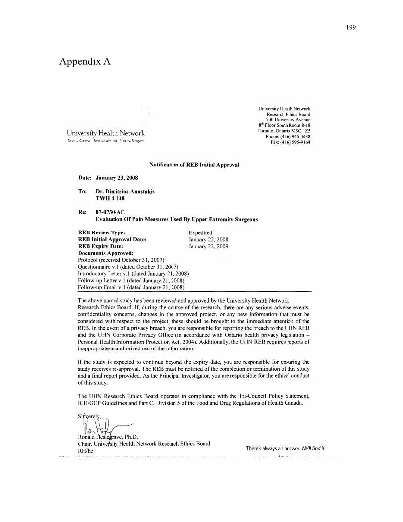
199
Appendix A
200
201
202
203
204
205
206
207
Appendix B
208
209
210
211
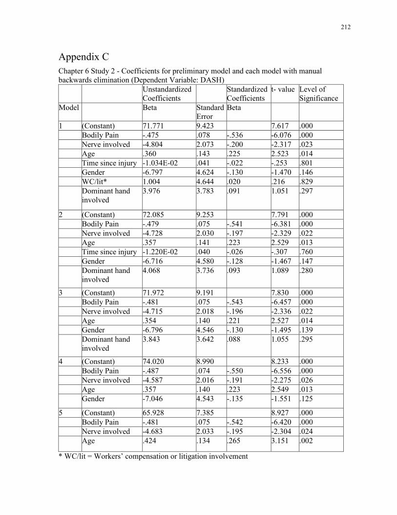
212
Appendix C
Chapter 6 Study 2 - Coefficients for preliminary model and each model with manual backwards elimination (Dependent Variable: DASH)
Unstandardized Coefficients
Standardized Coefficients
t- value Level of Significance
Model Beta Standard Error
Beta
1 (Constant) 71.771 9.423 7.617 .000
Bodily Pain -.475 .078 -.536 -6.076 .000
Nerve involved -4.804 2.073 -.200 -2.317 .023
Age .360 .143 .225 2.523 .014
Time since injury -1.034E-02 .041 -.022 -.253 .801
Gender -6.797 4.624 -.130 -1.470 .146
WC/lit* 1.004 4.644 .020 .216 .829
Dominant hand involved
3.976 3.783 .091 1.051 .297
2 (Constant) 72.085 9.253 7.791 .000
Bodily Pain -.479 .075 -.541 -6.381 .000
Nerve involved -4.728 2.030 -.197 -2.329 .022
Age .357 .141 .223 2.529 .013
Time since injury -1.220E-02 .040 -.026 -.307 .760
Gender -6.716 4.580 -.128 -1.467 .147
Dominant hand involved
4.068 3.736 .093 1.089 .280
3 (Constant) 71.972 9.191 7.830 .000
Bodily Pain -.481 .075 -.543 -6.457 .000
Nerve involved -4.715 2.018 -.196 -2.336 .022
Age .354 .140 .221 2.527 .014
Gender -6.796 4.546 -.130 -1.495 .139
Dominant hand involved
3.843 3.642 .088 1.055 .295
4 (Constant) 74.020 8.990 8.233 .000
Bodily Pain -.487 .074 -.550 -6.556 .000
Nerve involved -4.587 2.016 -.191 -2.275 .026
Age .357 .140 .223 2.549 .013
Gender -7.046 4.543 -.135 -1.551 .125
5 (Constant) 65.928 7.385 8.927 .000
Bodily Pain -.481 .075 -.542 -6.420 .000
Nerve involved -4.683 2.033 -.195 -2.304 .024
Age .424 .134 .265 3.151 .002
* WC/lit = Workers’ compensation or litigation involvement
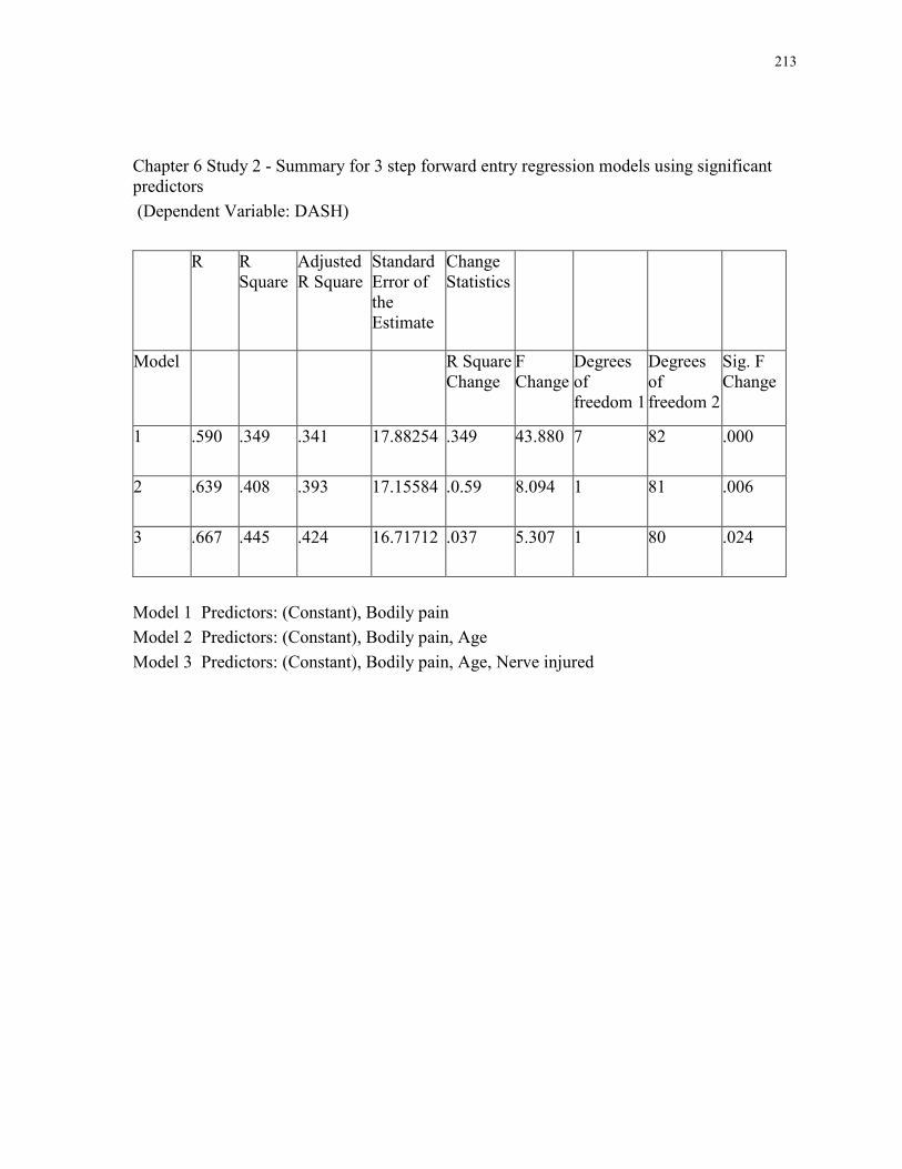
213
Chapter 6 Study 2 - Summary for 3 step forward entry regression models using significant predictors
(Dependent Variable: DASH)
R R Square
Adjusted R Square
Standard Error of the Estimate
Change Statistics
Model
R Square Change
F Change
Degrees of freedom 1
Degrees of freedom 2
Sig. F Change
1
.590 .349 .341 17.88254 .349 43.880 7 82 .000
2
.639 .408 .393 17.15584 .0.59 8.094 1 81 .006
3
.667 .445 .424 16.71712 .037 5.307 1 80 .024
Model 1 Predictors: (Constant), Bodily pain
Model 2 Predictors: (Constant), Bodily pain, Age
Model 3 Predictors: (Constant), Bodily pain, Age, Nerve injured
214
Copyright Acknowledgements
The following manuscripts, which are located in Chapters 2, 5, 6 and 7 of this dissertation,
have been published or are under peer review and permission to reproduce the published
manuscripts was granted for use in this dissertation:
Novak, C. B. & Katz, J. (2010). Neuropathic Pain in Patients with Upper Extremity Nerve
Injury. Physiotherapy Canada, 62, 190-201.
Novak, C. B., Anastakis, D. J., Beaton, D. E. & Katz, J. (2009). Evaluation of Pain
Measurement Practices and Opinions of Peripheral Nerve Surgeons. Hand, 4, 344-349.
Novak, C. B., Anastakis, D. J., Beaton, D. E. & Katz, J. (2009). Patient Reported Outcome
Following Peripheral Nerve Injury. Journal of Hand Surgery, 34A, 281-287.
Novak, C. B., Beaton, D. E., Anastakis, D. J., Mackinnon, S. E. & Katz, J. (2010).
Biomedical and Psychosocial Factors Associated with Pain and Disability after Peripheral
Nerve Injury. Submitted for publication and presently under peer review.