Embed Size (px)
Citation preview

Biomechanics of Locomotion
Biomechanics of Locomotion
D. Gordon E. Robertson, PhD, FCSB
Biomechanics, Laboratory,
School of Human Kinetics,
University of Ottawa, Ottawa, Canada
D. Gordon E. Robertson, PhD, FCSB
Biomechanics, Laboratory,
School of Human Kinetics,
University of Ottawa, Ottawa, Canada

Quantitative Domains
• Temporal– phases (stance/swing) and events (foot-
strike, toe-off), stride rate
• Electromyography– muscle activation patterns
• Kinematic (motion description)– stride length, velocity, ranges of motion,
acceleration
• Kinetic (causes of motion)– ground reaction forces, pressure patterns,
joint forces, moments of force, work, energy and power

Temporal Analysis
• Stride time (s)
• Stride rate = 1/time (/s)
• Stride cadence = 120 × rate (b/min)
• Instrumentation– Photocells and timers
– Videography (1 frame = 1/30 second)
– Metronome

Donovan Bailey sets world record (9.835) despite slowest reaction time (0.174) of finalists

Electromyography
Delsys electrodes Mega system
Noraxon systemBortec system

EMG of normal walking
gait initiation
rectus femoris
vastus lateralis
tibialis anterior
gastrocnemius
biceps femoris
heel switch
strides

EMG of normal walking
rectus femoris
vastus lateralis
tibialis anterior
gastrocnemius
biceps femoris
heel switch
rectus femoris contracts twice per cycle, once in early stance and once in late stance

EMG of normal walking
rectus femoris
vastus lateralis
tibialis anterior
gastrocnemius
biceps femoris
heel switch
biceps femoris has one longer contraction in late swing and early stance, synchronous with one burst of rectus femoris

EMG of normal walking
rectus femoris
vastus lateralis
tibialis anterior
gastrocnemius
biceps femoris
heel switch
tibialis anterior has two bursts of activity one in mid-swing and one during early stance. It is very active at initiation.

EMG of normal walking
rectus femoris
vastus lateralis
tibialis anterior
gastrocnemius
biceps femoris
heel switch
gastrocnemius has one long contraction throughout stance.
It is asynchronous with tibialis anterior.

Kinematic Analysis
• Linear position– Ruler, tape measure, optical
• Linear velocity– radar gun, photo-optical timer
• Linear acceleration– Accelerometry, videography
miniature accelerometers
radar gun

Motion Capture
• Cinefilm, video or infrared video
• Subject is filmed and locations of joint centres are digitized
Panasonic videocamera
Basler charge-coupled device (CCD) camera
Vicon infra-red camera
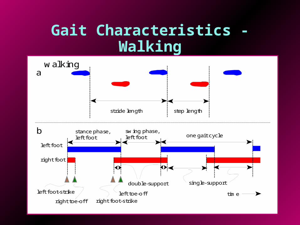
Gait Characteristics - Walking
stride length step length
left foot
swing phase,left foot
right foot
stance phase,left foot
single-support
left toe-off
one gait cycle
time
double-supportleft foot-strike
right foot-strikeright toe-off
a
b
walking

Gait Characteristics – Running/Sprinting
stride length step length
left foot
swing phase,left foot
right foot
stance phase,left foot
left toe-off
one gait cycle
timeleft foot-strike right foot-strikeright toe-off
a
b
running/sprinting
flight phase

Motion Capture(e.g., SIMI or Vicon)
3D motiondata
EMG data
F-Scandata
Force platformdata
Videodata

Passive Infrared Motion Capture (e.g., Vicon or M.A.C.)
Infrared video cameras
Kistler force platforms
M.A.C.system

Active Infrared Motion Capture
• NDI’s Optotrak
Infrared video cameras
Infrared emitting diodes

Gait and Movement Analysis Laboratory
• Motion capture system for marker trajectories
• Force platforms for ground reactions
• Electromyography for muscle activity
• Pressure mapping systems for in-shoe pressure patterns

3D Geometric Model(Visual3D)
from stick-figures to geometrical solids of revolution with known inertial properties
from markers to joint centres and stick-figure of body

Kinetic Analysis
Causes of motion
• Forces and moments of force
• Work, energy and power
• Impulse and momentum
• Inverse Dynamics derives forces and moments from kinematics and body segment parameters (mass, centre of gravity, and moment of inertia)

Normal Walking Example
• Female subject
• Speed was 1.77 m/s (fast)
• IFS = ipsilateral foot-strike
• ITO = ipsilateral toe-off
• CFS = contralateral foot-strike
• CTO = contralateral toe-off

Results
0.0 0.2 0.4 0.6 0.8 1.0 1.2Time (s)
-200
-100
0
100
-100
0
100
-10
0
10
P
ow
er
(W)
Mo
me
nt
(N.m
)
A
ng
. V
el.
(ra
d/s
)
Trial: 2SFN3Ang. velocityMomentPower
CFS ITO IFS CTO CFS ITO
Dorsiflexion
Plantar flexion
Dorsiflexors
Plantar flexors
Concentric
Eccentric
• Angular velocity tells whether joint is flexing or extending
• Moment of force records whether flexors or extensors are performing work
• Power quantifies whether work done was positive or negative

Ankle angular velocity, moment of force and power
• Dorsiflexors produce dorsiflexion during swing
• Plantar flexors control dorsiflexion
• Large burst of power by plantar flexors for push-off 0.0 0.2 0.4 0.6 0.8 1.0 1.2
Time (s)
-200
-100
0
100
-100
0
100
-10
0
10
P
ow
er
(W)
Mo
me
nt
(N.m
)
A
ng
. V
el.
(ra
d/s
)
Trial: 2SFN3Ang. velocityMomentPower
CFS ITO IFS CTO CFS ITO
Dorsiflexion
Plantar flexion
Dorsiflexors
Plantar flexors
Concentric
Eccentric

Knee angular velocity, moment of force and power
• Negative work by knee extensors to control flexion at push-off
• another to cushion weight-acceptance
• Negative work by knee flexors to control knee extension prior to foot-strike
0.0 0.2 0.4 0.6 0.8 1.0 1.2Time (s)
-200
-100
0
100
-100
0
100
-10
0
10
P
ow
er
(W)
M
om
en
t (N
.m)
A
ng
. V
el.
(ra
d/s
)
Trial: 2SFN3Ang. velocityMomentPower
CFS ITO IFS CTO CFS ITO
Extension
Flexion
Extensors
Flexors
Concentric
Eccentric

Hip angular velocity, moment of force and power
0.0 0.2 0.4 0.6 0.8 1.0 1.2Time (s)
-200
-100
0
100
-100
0
100
-10
0
10
P
ow
er
(W)
Mo
me
nt
(N.m
)
A
ng
. V
el.
(ra
d/s
)
Trial: 2SFN3Ang. velocityMomentPower
CFS ITO IFS CTO CFS ITO
Flexion
Extension
Flexors
Extensors
Concentric
Eccentric
• Positive work by hip flexors to swing thigh & flex knee
• Positive work by hip extensors to extend hip in early stance
• Negative work by hip flexors to control extension

Solid-Ankle, Cushioned Heel (SACH) Prostheses

0.0 0.2 0.4 0.6 0.8 1.0 1.2 1.4
Time (s)
-200.
-100.
0.
100.
-100.
0.
100.
-10.
0.
10.
Po
we
r (W
)
Mo
me
nt
(N.m
)
An
gu
lar
ve
l. (
/s)
Ankle angular velocity, moment of force and power of SACH foot prosthesis
• No power produced during push-off
Trial: WB24MH-SAng. velocityNet momentPower
ITO IFS CTO CFS ITO
Dorsiflexing
Plantar flexing
Dorsiflexor
Plantar flexor
Concentric
Eccentric
• Power dissipation during weight acceptance and push-off

FlexFoot Prostheses(energy-storing)
Recent models
Original model

Ankle angular velocity, moment of force and power of FlexFoot prosthesis
• Some energy returned during push-off
0.0 0.2 0.4 0.6 0.8 1.0 1.2
Time (s)
-500.
-250.
0.
250.
-100.
0.
100.
-10.
0.
10.
Po
we
r (W
)
M
om
en
t (N
.m)
A
ng
ula
r v
el.
(/s
)
Trial: WB13MH-FAng. velocityNet momentPower
ITO IFS CTO CFS ITO
Dorsiflexing
Plantar flexing
Dorsiflexor
Plantar flexor
Concentric
Eccentric

Above-knee Prostheses

Running Prostheses