Embed Size (px)
Citation preview
Review article | Published 19 July 2012, doi:10.4414/smw.2012.13583
Cite this as: Swiss Med Wkly. 2012;142:w13583
Biomechanics and pathomechanismsof osteoarthritis
Christian Egloff, Thomas Hügle, Victor Valderrabano
Orthopaedic Department, University Hospital, University of Basel, Switzerland
Summary
Today, the most frequent chronic musculoskeletal disorderand the leading cause of disability in the elderly is os-teoarthritis (OA). Approximately 43 million people in theUnited States and 15% of the world population are affec-ted. Due to demographic changes, the incidence of OA israpidly increasing, leading to an ascending socioeconom-ical and personal burden. Despite the exact cause of OAremains unknown, the pathogenic role of biomechanicaldysfunction in OA is well established. For weight-bearingjoints altered loading mechanisms, increased mechanicalforces and changed biomechanics are significant contrib-uting factors for initiation and progression of OA. Thus,OA is a disease of the whole joint, including muscles,tendons, ligaments, synovium and bone. This review fo-cuses on the influence of biomechanics on the pathogenesisand progression of OA. We notably illustrate the patho-logical bioreactivity of soft tissues, subchondral bone andjoint inflammation. Procedures, conservative or surgical,which actively alter the biomechanics of the lower limb,are promising strategies to treat symptoms as well as to in-fluence disease progression in OA.
Key words: osteoarthritis; OA; biomechanics;malalignment; joint loading; synovitis
Introduction
Osteoarthritis (OA) is one of the most common causes ofdisability in the world. Epidemiological studies estimatearound 43 million affected patients in the United Statesalone and about 15% of the world population [1, 2]. Theincidence is estimated to 100,000 new cases per year [3].The risk of mobility impairments caused by knee OA aloneis greater than due to any other medical condition in peopleover 65 [4]. It leads to social, psychological and econom-ical burdens in patients with substantial financial conse-quences [5]. In France the cumulated health costs resultingfrom OA almost doubled within 10 years (from1993–2003) [1]. A further increase has to be expected dueto the increasing prevalence of obesity and ascending lifeexpectancy of our population [6].
The role of biomechanics in the development and progres-sion of OA, especially of the lower limb has become integ-ral in current knowledge of this disease.In this review we focus on the influence of different bio-mechanical factors on the pathogenesis and progression ofOA, underlining the pathological bioreactivity of soft tis-sues, subchondral bone and subsequent joint inflammation.We discuss current conservative and surgical interventionstrategies aimed to reduce biomechanical loads and therebypreventing or slowing down the vicious cycle of OA. Todate our knowledge is still limited concerning pathobio-mechanical loads and its impact on joint homeostasis lead-ing to destructive OA. We concentrate our perspective onthe lower extremity; as the hip, knee and ankle are thejoints which are highly weight bearing and the most com-monly affected sites by OA [3].
Pathobiomechanics of osteoarthritis
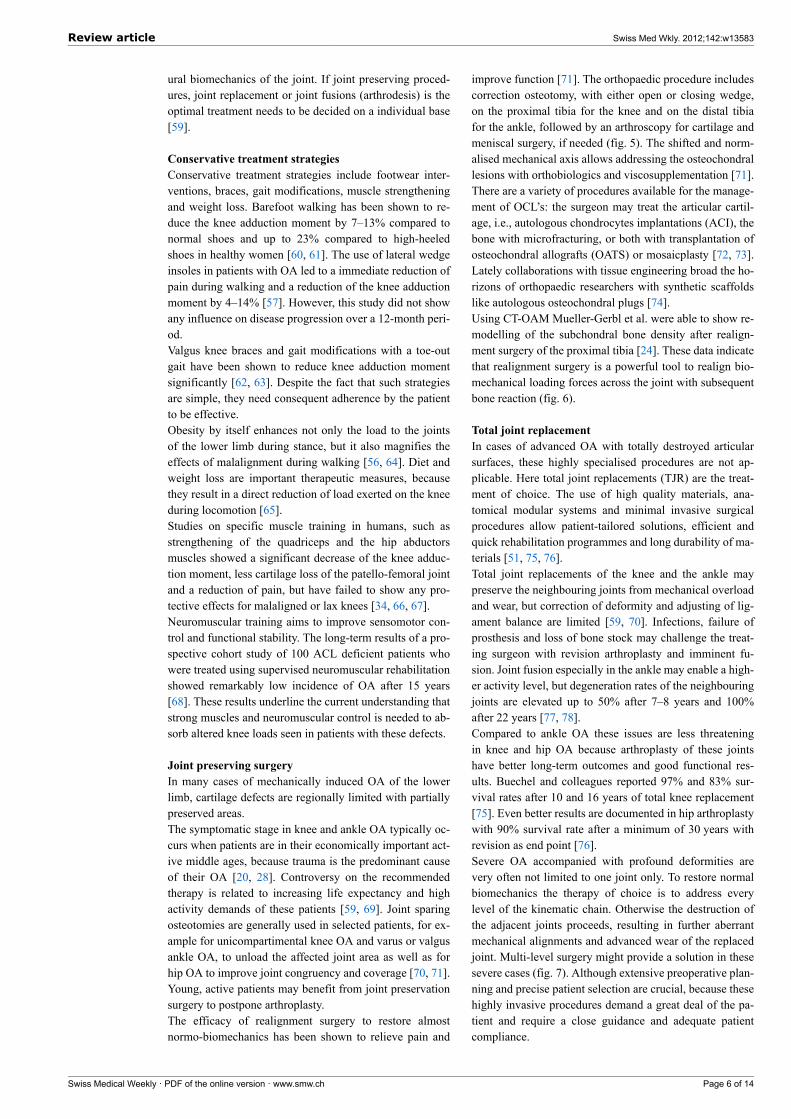
OA is regarded as a whole joint disease with a multi-factorial etiology, including increased mechanical stress,ligament derangements, cartilage degradation, subchondralbone changes and muscular impairments (fig. 1). Further-more, secondary synovial inflammation plays a role in OA,notably in the early stage [7]. Several studies pointed outthe importance of mechanical factors in the destructive cas-cade of this disease [8–10].Epidemiological studies of the last decades tried to definerisk factors such as age, genetic predisposition, obesity,joint congruency, increased mechanical stress and greaterbone density [11]. OA may evolve as a consequence afteran antecedent incidence, such as intraarticular fractures andligament lesions, systemic diseases like rheumatoid arthrit-is, hemochromatosis, haemophilia, post infectious arthritisor osteochondrosis dissecans, or as a result of a congenitalor developmental anatomic abnormality [10].OA occurs when the dynamic steady state between de-structive forces and repair mechanisms destabilises thejoint homeostasis [12, 13]. This imbalance is thought to bethe driving force in this progressive disease and may pro-duce pain and disability, although many patients with obvi-ous radiographic findings don’t complain of any symptomsrelated to OA [14]. OA is most common in weight bearing
Swiss Medical Weekly · PDF of the online version · www.smw.ch Page 1 of 14
joints such as the hips, knees and the ankle but it can occurin any synovial joint of the body.
Bone and cartilageOA of the hips and knees are mostly the result of a slowand degenerative process [15], whereas clinical treatmentsstudies of ankle OA showed a close coherence with a pre-cedent trauma and are therefore classified as post-traumaticosteoarthritis [10, 16, 17]. This phenomenon is thought tobe caused by the different and unique anatomical and bio-mechanical tissue characteristics of the hip, knee and ankle.Anatomically the contact areas are larger in the hip andthe knee compared to the ankle (at 500-N load: ankle, 350mm2; hip, 1100 mm2; knee, 1,120 mm2) [18–20]. There-fore, the ankle is exposed to higher loading pressures permm2. Interestingly, the histological images of ankle cartil-age show a higher proteoglycan density, lower matrix de-gradation and higher compressive stiffness [21].Biomechanically the knee joint bears higher shear forcesthan the hip or ankle joint as it incorporates sliding, rotatingand rolling motions during movements [21]. These tissueproperties combined with the different kinematics may bean explanation why the ankle joint is more resistant to de-generative OA than the hip or knee joint.The role of bone density in OA is currently debated. Typ-ically, a reduced bone density of the subchondral bone isobserved in the early stages of OA [22]. However, at a laterstage, subchondral bone sclerosis and higher bone dens-ity are seen radiologically. Subchondral osteosclerosis as-sessed by bone-densitometry (BMD) showed that subchon-dral BMD indeed might predict cartilage defect develop-ment [23]. Functional computed tomography like CT-os-teoabsorptiometry (CT-OAM) also enabled visualization ofthe load-dependent reaction of subchondral bone mineral-isation density in advanced OA [24]. The newly organ-ised subchondral bone also contains new vessels and nervefibers, which are likely involved in OA pathogenesis and
Figure 1
Schematic illustration of OA as a “whole joint disease” withstructures involved in OA pathology. Muscle atrophy can cause OA,but is also seen as a consequence of OA, e.g., pain-induced.Synovitis with secretion of proinflammatory cytokines into the jointspace correlates with pain and radiological progression. Cartilagedegradation is the hallmark of OA. Subchondral bone pathology isalso observed in OA, ultimately leading to osteosclerosis.
pain sensation, respectively [25]. Cartilage and bone re-ceive and dissipate contact loads associated with move-ments and weight bearing, and are therefore continuouslychallenged biomechanically. Recent data support the viewthat cartilage and bone do communicate over the calcifiedtissue barrier through molecular crosstalk. This interactionis crucial for the subchondral bone–articular cartilage unitand seems to show specific changes in the development ofOA [25–27]. Currently it is not clear whether subchondralbone changes occur as cause or consequence of cartilagedamage.
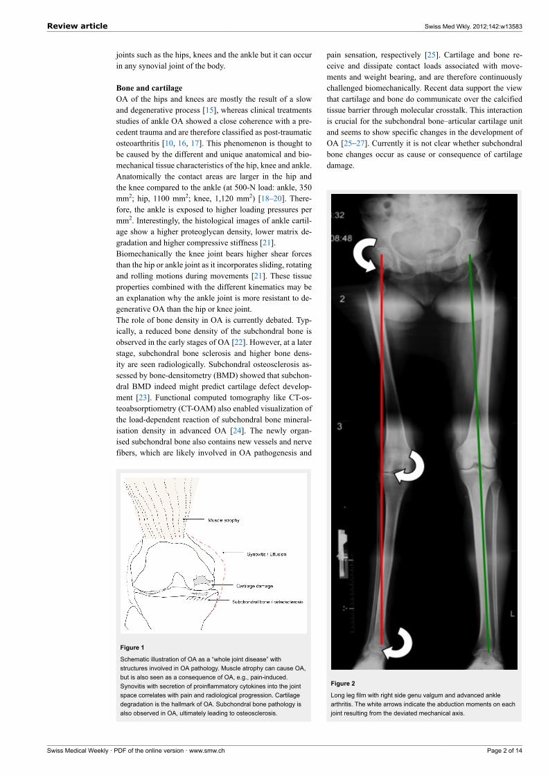
Figure 2
Long leg film with right side genu valgum and advanced anklearthritis. The white arrows indicate the abduction moments on eachjoint resulting from the deviated mechanical axis.
Review article Swiss Med Wkly. 2012;142:w13583
Swiss Medical Weekly · PDF of the online version · www.smw.ch Page 2 of 14
Pathobiomechanics: the fractureAlthough it is virtually impossible to establish a specifictime of disease onset in degenerative OA, in secondary OApatients recall a precedent injury taking into responsibilityfor disease initiation. For example, an intraarticular frac-ture implicates an incongruence of the joint line followedby axis deviation and altered load distribution across thejoint. Recurrent ankle sprains or ankle fractures are veryoften consistent with osteochondral lesions (OCL). Duringarthroscopy, Taga and colleagues found chondral lesionsof 89% in 9 patients with acute ankle injuries and 95% in22 patients with chronic ankle injuries. [28]. Hintermannet al. reported in their investigation on 148 patients that66% of those with lateral ankle instability had cartilage le-sions after one or more ankle sprains, as did 98% of thosewith instability on the medial side [29]. Following recur-rent unphysiological loading of an instable joint, the shear-ing forces cause chondrocyte deformation up to irrevers-ible damage and lead to chondrocyte apoptosis [30]. Theyaffect the biomechanical protection (gliding and shock ab-sorption) of the joint and the contact pressures around theOCL rise combined with pain and a local inflammatory re-action [9, 31]. These stimuli let the lesion size grow, andthe initial small and local damage turns into a joint-wideOA.
Pathobiomechanics: ligament Instability and muscleweaknessBesides the initial cartilage damage, i.e. through injury, thecontribution of ligaments, muscles and nerves to the mech-anical environment have become central to pathobiomech-anical investigation. Chronic ligament laxity is frequentlyobserved in ankle OA [17]. It has been postulated that in-stability of the talus leads to a pathologic range of trans-lation and rotation, which increase the shear forces on thecartilage surface. Furthermore chronic ligament instability,on the medial or lateral side of the ankle, enhances theprogressive varus or valgus malalignment of the hindfoot,
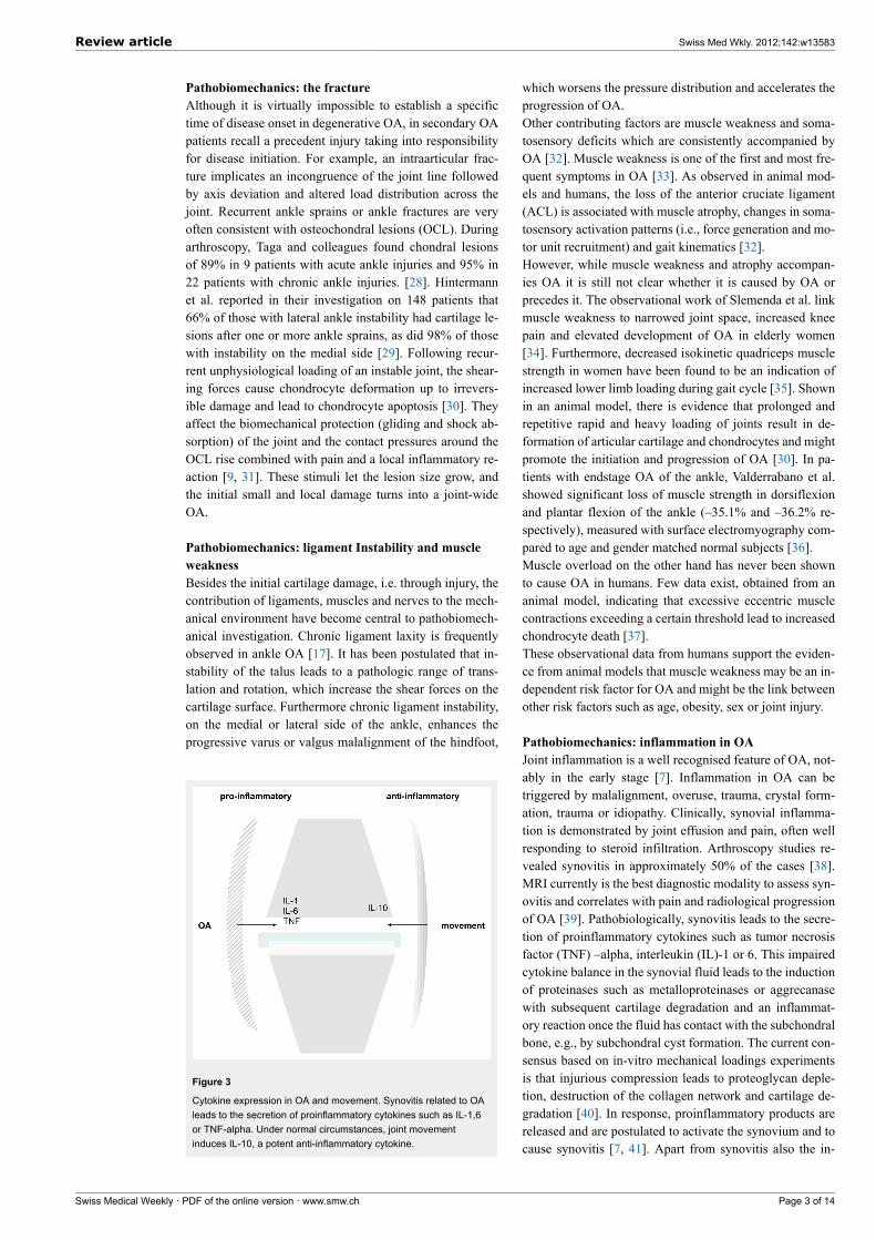
Figure 3
Cytokine expression in OA and movement. Synovitis related to OAleads to the secretion of proinflammatory cytokines such as IL-1,6or TNF-alpha. Under normal circumstances, joint movementinduces IL-10, a potent anti-inflammatory cytokine.
which worsens the pressure distribution and accelerates theprogression of OA.Other contributing factors are muscle weakness and soma-tosensory deficits which are consistently accompanied byOA [32]. Muscle weakness is one of the first and most fre-quent symptoms in OA [33]. As observed in animal mod-els and humans, the loss of the anterior cruciate ligament(ACL) is associated with muscle atrophy, changes in soma-tosensory activation patterns (i.e., force generation and mo-tor unit recruitment) and gait kinematics [32].However, while muscle weakness and atrophy accompan-ies OA it is still not clear whether it is caused by OA orprecedes it. The observational work of Slemenda et al. linkmuscle weakness to narrowed joint space, increased kneepain and elevated development of OA in elderly women[34]. Furthermore, decreased isokinetic quadriceps musclestrength in women have been found to be an indication ofincreased lower limb loading during gait cycle [35]. Shownin an animal model, there is evidence that prolonged andrepetitive rapid and heavy loading of joints result in de-formation of articular cartilage and chondrocytes and mightpromote the initiation and progression of OA [30]. In pa-tients with endstage OA of the ankle, Valderrabano et al.showed significant loss of muscle strength in dorsiflexionand plantar flexion of the ankle (–35.1% and –36.2% re-spectively), measured with surface electromyography com-pared to age and gender matched normal subjects [36].Muscle overload on the other hand has never been shownto cause OA in humans. Few data exist, obtained from ananimal model, indicating that excessive eccentric musclecontractions exceeding a certain threshold lead to increasedchondrocyte death [37].These observational data from humans support the eviden-ce from animal models that muscle weakness may be an in-dependent risk factor for OA and might be the link betweenother risk factors such as age, obesity, sex or joint injury.
Pathobiomechanics: inflammation in OAJoint inflammation is a well recognised feature of OA, not-ably in the early stage [7]. Inflammation in OA can betriggered by malalignment, overuse, trauma, crystal form-ation, trauma or idiopathy. Clinically, synovial inflamma-tion is demonstrated by joint effusion and pain, often wellresponding to steroid infiltration. Arthroscopy studies re-vealed synovitis in approximately 50% of the cases [38].MRI currently is the best diagnostic modality to assess syn-ovitis and correlates with pain and radiological progressionof OA [39]. Pathobiologically, synovitis leads to the secre-tion of proinflammatory cytokines such as tumor necrosisfactor (TNF) –alpha, interleukin (IL)-1 or 6. This impairedcytokine balance in the synovial fluid leads to the inductionof proteinases such as metalloproteinases or aggrecanasewith subsequent cartilage degradation and an inflammat-ory reaction once the fluid has contact with the subchondralbone, e.g., by subchondral cyst formation. The current con-sensus based on in-vitro mechanical loadings experimentsis that injurious compression leads to proteoglycan deple-tion, destruction of the collagen network and cartilage de-gradation [40]. In response, proinflammatory products arereleased and are postulated to activate the synovium and tocause synovitis [7, 41]. Apart from synovitis also the in-
Review article Swiss Med Wkly. 2012;142:w13583
Swiss Medical Weekly · PDF of the online version · www.smw.ch Page 3 of 14
traarticular fat body secretes profibrotic cytokines such asIL-6 [42]. Interestingly, movement of a joint induces theexpression of IL-10, which is a potent anti-inflammatorycytokine [43] (fig. 3). This indicates that not only the in-duction of inflammation, but also the lack of resolution ofinflammation might be of importance in OA. Possibly animpaired biomechanical process of a joint will lack thisanti-inflammatory reaction in form of IL-10 expression.
The role of mechanics and thekinematic chain
The concept of biomechanics includes the assembly of thestructural reaction of joint tissues to mechanical stimuli.This interplay has gained much attention in the current un-derstanding of pathogenesis of OA [9, 12]. A joint rep-resents the connective unit between two bones or func-tional segments. Joints connected serially act as a kin-ematic chain. This construct allows motion and simultan-eously provides stability, congruency and shock absorp-tion. Alignments, adduction moments and muscle balan-cing are the key determinants for optimal load reductionand distribution as well as to guarantee painless gliding:hence unphysiological loading patterns on one joint mayinfluence the adjacent levels as well.All joints are exposed to biomechanical loading but mostscientific data result from the lower extremity, especiallyfrom the knee joint, because it is the most commonly af-fected joint by OA and it has been extensively examinedby joint kinematic testing, gait analysis and other investig-ations [44–46].Adequate mechanical loading provides the essential stim-ulus to maintain physiological joint homeostasis, whereasexcessive mechanical stress as well as unloading the jointis crucial for the disease onset and progression [31, 47].Over the last two decades it has been shown that alteredjoint biomechanics of the knee, such as loss of cruciate lig-aments, removal of menisci, posttraumatic cartilage dam-age, changes in bone alignments, unloading through cast-ing and overloading through intense exercise may causedisease initiation and progression of cartilage degradation[8, 9, 12].
Mechanical axisThe mechanical axis or alignment of the lower extremity isdefined as a line drawn from the centre of the femoral headto the centre of the talus. The hip-knee-ankle alignment isbest assessed with long leg films (fig. 2). This line passesclose to the centre of the tibial head between the eminentiatibiae, approximately 1° in varus (neutral alignment 0–2°)[48]. Therefore, the medial compartment of the knee sus-tains 60–70% of the load [46, 48]. This physiologically im-balanced dispensation may be a predisposing factor to me-dially accentuated tibio-femoral OA. A varus-aligned kneeis described as “bow-legged” and a valgus-aligned knee isdescribed as “knocked-knee". This valgus or varus align-ment have been described to influence the load distributionacross the articular joint surface [49]. This asymmetry re-duces the area of load bearing and amplifies the resultantload to the remaining joint surface. The deviation of themechanical axis does strongly correlate with radiograph-
ic joint space narrowing, subchondral cyst formation, bonesclerosis, and functional decline in OA [49].
Dynamic loadsStatic loads have a significant impact for OA patho-physiology, but they do not reflect loads generated duringlocomotion. Movements, angulation and normal gait pro-duce dynamic loads on joints up to five times body weightin the ankle and up to three times in the knee relative tostanding [50]. Especially shearing forces are greatest dur-ing gait and ambulation. Therefore, it is most important tocalculate these forces in order to assess dynamic loadingduring physiologic activity and its influence on OA. Dy-namic loading can be measured using intraarticular pres-sure devices, but they are less established in humans, asdirect interventions to control loading patterns are difficultto perform. However, as observed in animal models, mala-lignment leads to excessive dynamic loads followed by car-tilage destruction and progressive OA [8, 9].Indirect methods include gait analysis that is noninvasiveand easily reproducible in humans and are therefore widelyaccepted [46, 51]. During gait cycles video cameras andground reaction force plates summarise pressure and move-ments and transform these data to external “moments” re-lative to internal joint loads. In mechanics a “moment” isthe tendency of force to twist or rotate an object. It is val-ued mathematically as the product of the force and the mo-ment (lever) arm [52]. In varus alignment this “adductionmoment” (AdM) represents a varus torque on the knee jointand is determined by the ground reaction force (GRF, forcegenerated by the foot touching the ground) and by the dis-tance of the GRF vector from the center of the knee joint. Ifany of these parameters alter, the AdM is gravely affected.For example, a genu varus has a mechanical axis, whichruns medial to the joint centre; therefore the lever arm in-creases resulting in an AdM of greater magnitude. Yang etal. showed in a recent study in humans that maximum val-ues of stress and strain on articular cartilage of the knee arein line with the peak adduction moment during the stancephase of gait. At the early and late stance phase (heel striketo foot flat and toe off, respectively) a valgus moment ispresent together with maximum compressive stress on thelateral knee compartment. During mid stance phase (i.e.,25–75% of gait cycle) a varus moment occurs to the knee inline with maximum compressive stress on the medial kneecompartment [53]. Moreover, they showed that a preexist-ing varus alignment of the knee increase the compressivestresses compared to subjects with normal or valgus kneealignment. In the only longitudinal study, Miyazaki et al.found that for every one unit increase in the peak AdM,there was a 6.5-fold increase in the risk of medial compart-ment disease progression on X-ray in a cohort of 74 pa-tients [54]. Moreover, several authors found that the AdMis directly associated with radiographic findings, cartilageloss and pain [49, 55].The AdM has become a widely accepted marker to measurejoint loading. At the same time its limitations are given asit only reflects the load at a specific time of gait. Differ-ences during gait, walking speed and stance phase are notrepresented. This comes into consideration because obeseand older patients with OA walk slower and have longer
Review article Swiss Med Wkly. 2012;142:w13583
Swiss Medical Weekly · PDF of the online version · www.smw.ch Page 4 of 14
stance phases, so their total time weight bearing is longerthan normal [51, 56]. Up to now the identification of dis-tinct prognostic factors for progression of OA of the lowerlimb has been difficult with the strongest evidence beingfor static malalignment [49]. However, even though AdMhas been shown to predict medial knee OA, a recent studyby the same group showed that reducing the AdM by con-servative means even after one year, did not provide anystructural or symptomatic benefits compared to a untreatedcontrol group [57]. These findings arouse the suspicion thatAdM is an important determinant but not the only one inthe multifactorial disease of OA.As mentioned before, the contribution of muscles in loadabsorption and distribution throughout the kinematic chainof the lower limb play an integral role. Various analysesof gait have found that the quadriceps, hamstrings and thegastrocnemius muscles are able to produce an internal val-gus moment and provide stabilisation against external ad-duction moments [53, 58]. In patients with medial OA andan varus alignment these muscle show altered activity leveland is probably an attempt to unload the medial knee com-partment [53]. This underlines the complexity to calculatethe total loads running across a joint and the resultant effecton the articular surfaces.
Osteoarthritis treatment options
To date a medical cure for OA, a “restitutio ad integrum”,does not exist, nor do disease modifying OA drugs(DMOADs) comparable to disease modifying anti-rheum-
Figure 4
Treatment algorhythm for OA patients.
Figure 5
Patient before and after realignment surgery with an open wedgeosteotomy of the proximal tibia.
atic drugs (DMARs). Treating patients suffering from OAconsists of mainly treating symptoms, providing joint sta-bility and trying to postpone end stage OA. Notwithstand-ing, inflammation in OA is a more and more recognisedtarget for therapies in form of biologics. Within the varietyof treatment options, surgical interventions should onlytake place if conservative strategies have failed to reliefsymptoms (fig. 4). The aim is to restore ligament balan-cing, to realign mechanical axes and to re-establish the nat-
Figure 6
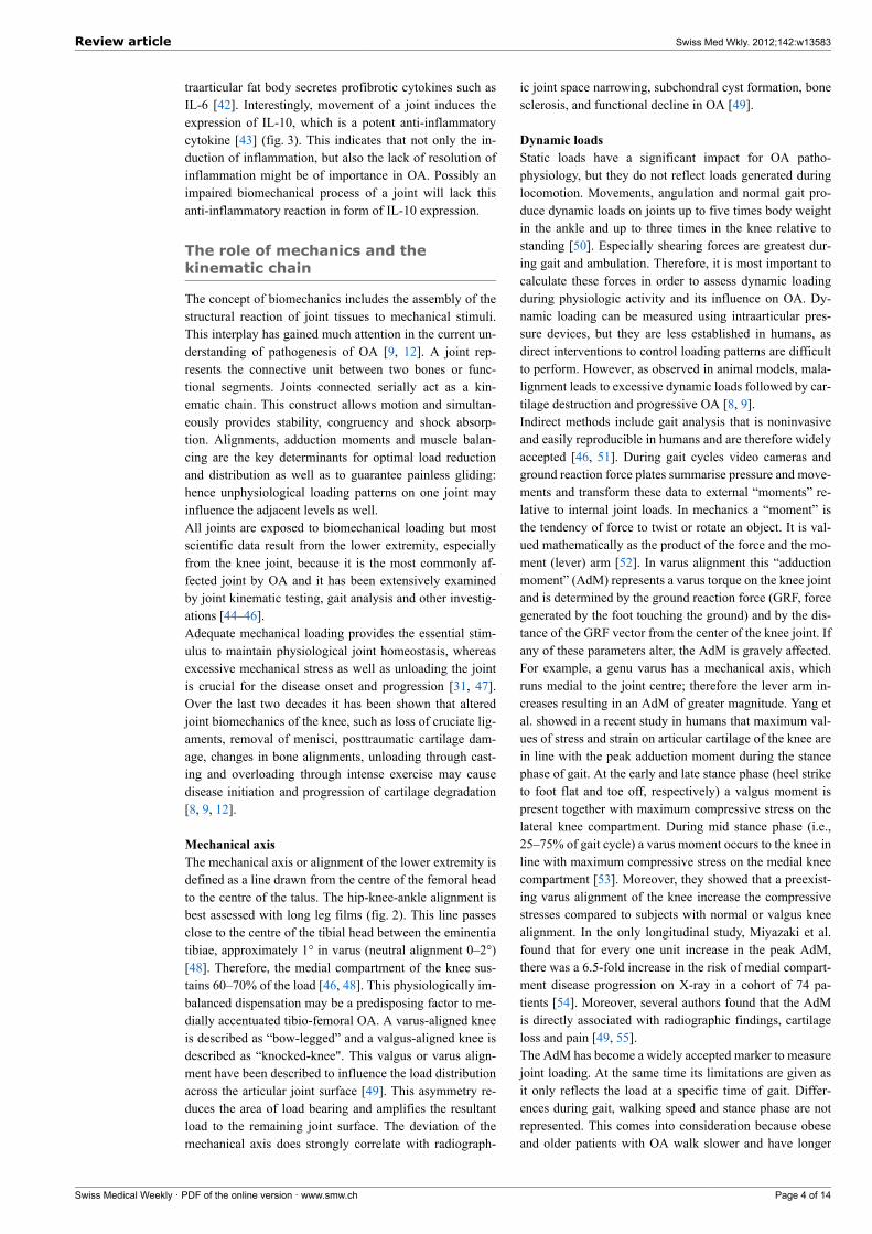
CT-OAM before and 2 years after valgisation osteotomy of theproximal tibia. Areas with high subchondral bone density arecoloured in red and yellow (© Madry H, van Dijk CN, Mueller-GerblM. The basic science of the subchondral bone. Knee Surg SportsTraumatol Arthrosc. 2010;18(4):419–33. Reprinted with kindpermission).
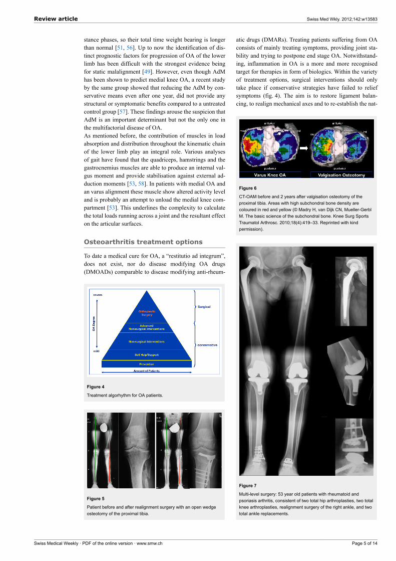
Figure 7
Multi-level surgery: 53 year old patients with rheumatoid andpsoriasis arthritis, consistent of two total hip arthroplasties, two totalknee arthroplasties, realignment surgery of the right ankle, and twototal ankle replacements.
Review article Swiss Med Wkly. 2012;142:w13583
Swiss Medical Weekly · PDF of the online version · www.smw.ch Page 5 of 14
ural biomechanics of the joint. If joint preserving proced-ures, joint replacement or joint fusions (arthrodesis) is theoptimal treatment needs to be decided on a individual base[59].
Conservative treatment strategiesConservative treatment strategies include footwear inter-ventions, braces, gait modifications, muscle strengtheningand weight loss. Barefoot walking has been shown to re-duce the knee adduction moment by 7–13% compared tonormal shoes and up to 23% compared to high-heeledshoes in healthy women [60, 61]. The use of lateral wedgeinsoles in patients with OA led to a immediate reduction ofpain during walking and a reduction of the knee adductionmoment by 4–14% [57]. However, this study did not showany influence on disease progression over a 12-month peri-od.Valgus knee braces and gait modifications with a toe-outgait have been shown to reduce knee adduction momentsignificantly [62, 63]. Despite the fact that such strategiesare simple, they need consequent adherence by the patientto be effective.Obesity by itself enhances not only the load to the jointsof the lower limb during stance, but it also magnifies theeffects of malalignment during walking [56, 64]. Diet andweight loss are important therapeutic measures, becausethey result in a direct reduction of load exerted on the kneeduring locomotion [65].Studies on specific muscle training in humans, such asstrengthening of the quadriceps and the hip abductorsmuscles showed a significant decrease of the knee adduc-tion moment, less cartilage loss of the patello-femoral jointand a reduction of pain, but have failed to show any pro-tective effects for malaligned or lax knees [34, 66, 67].Neuromuscular training aims to improve sensomotor con-trol and functional stability. The long-term results of a pro-spective cohort study of 100 ACL deficient patients whowere treated using supervised neuromuscular rehabilitationshowed remarkably low incidence of OA after 15 years[68]. These results underline the current understanding thatstrong muscles and neuromuscular control is needed to ab-sorb altered knee loads seen in patients with these defects.
Joint preserving surgeryIn many cases of mechanically induced OA of the lowerlimb, cartilage defects are regionally limited with partiallypreserved areas.The symptomatic stage in knee and ankle OA typically oc-curs when patients are in their economically important act-ive middle ages, because trauma is the predominant causeof their OA [20, 28]. Controversy on the recommendedtherapy is related to increasing life expectancy and highactivity demands of these patients [59, 69]. Joint sparingosteotomies are generally used in selected patients, for ex-ample for unicompartimental knee OA and varus or valgusankle OA, to unload the affected joint area as well as forhip OA to improve joint congruency and coverage [70, 71].Young, active patients may benefit from joint preservationsurgery to postpone arthroplasty.The efficacy of realignment surgery to restore almostnormo-biomechanics has been shown to relieve pain and
improve function [71]. The orthopaedic procedure includescorrection osteotomy, with either open or closing wedge,on the proximal tibia for the knee and on the distal tibiafor the ankle, followed by an arthroscopy for cartilage andmeniscal surgery, if needed (fig. 5). The shifted and norm-alised mechanical axis allows addressing the osteochondrallesions with orthobiologics and viscosupplementation [71].There are a variety of procedures available for the manage-ment of OCL’s: the surgeon may treat the articular cartil-age, i.e., autologous chondrocytes implantations (ACI), thebone with microfracturing, or both with transplantation ofosteochondral allografts (OATS) or mosaicplasty [72, 73].Lately collaborations with tissue engineering broad the ho-rizons of orthopaedic researchers with synthetic scaffoldslike autologous osteochondral plugs [74].Using CT-OAM Mueller-Gerbl et al. were able to show re-modelling of the subchondral bone density after realign-ment surgery of the proximal tibia [24]. These data indicatethat realignment surgery is a powerful tool to realign bio-mechanical loading forces across the joint with subsequentbone reaction (fig. 6).
Total joint replacementIn cases of advanced OA with totally destroyed articularsurfaces, these highly specialised procedures are not ap-plicable. Here total joint replacements (TJR) are the treat-ment of choice. The use of high quality materials, ana-tomical modular systems and minimal invasive surgicalprocedures allow patient-tailored solutions, efficient andquick rehabilitation programmes and long durability of ma-terials [51, 75, 76].Total joint replacements of the knee and the ankle maypreserve the neighbouring joints from mechanical overloadand wear, but correction of deformity and adjusting of lig-ament balance are limited [59, 70]. Infections, failure ofprosthesis and loss of bone stock may challenge the treat-ing surgeon with revision arthroplasty and imminent fu-sion. Joint fusion especially in the ankle may enable a high-er activity level, but degeneration rates of the neighbouringjoints are elevated up to 50% after 7–8 years and 100%after 22 years [77, 78].Compared to ankle OA these issues are less threateningin knee and hip OA because arthroplasty of these jointshave better long-term outcomes and good functional res-ults. Buechel and colleagues reported 97% and 83% sur-vival rates after 10 and 16 years of total knee replacement[75]. Even better results are documented in hip arthroplastywith 90% survival rate after a minimum of 30 years withrevision as end point [76].Severe OA accompanied with profound deformities arevery often not limited to one joint only. To restore normalbiomechanics the therapy of choice is to address everylevel of the kinematic chain. Otherwise the destruction ofthe adjacent joints proceeds, resulting in further aberrantmechanical alignments and advanced wear of the replacedjoint. Multi-level surgery might provide a solution in thesesevere cases (fig. 7). Although extensive preoperative plan-ning and precise patient selection are crucial, because thesehighly invasive procedures demand a great deal of the pa-tient and require a close guidance and adequate patientcompliance.
Review article Swiss Med Wkly. 2012;142:w13583
Swiss Medical Weekly · PDF of the online version · www.smw.ch Page 6 of 14
Conclusion
OA and biomechanics are inescapably linked together.However, the contribution of biomechanical factors to aeti-ology, pathogenesis and to disease progression require fur-ther research in order to reduce the enormous socioeco-nomic and personal impact of this disease. To this end,modern treatment pathways including the collaboration ofbasic sciences e.g., tissue engineering and pathology, dia-gnostics, biomarker research, conservative treatmentstrategies and orthopaedic surgery are necessary to guaran-tee an optimal, individually adapted treatment plan with re-spect to joint biomechanics and biomechanical reactivity.
Funding / potential competing interests: No financial supportand no other potential conflict of interest relevant to this articlewere reported.
Correspondence: Professor Victor Valderrabano, PhD,
Department of Orthopaedic Surgery, University Hospital,
University of Basel, Spitalstrasse 21, CH-4051 Basel,
Switzerland, vvalderrabano[at]uhbs.ch
Literature
1 Solignac M. COART France 2003 report on new socioeconomic dataon osteoarthritis in France. Presse Med. 2004;33(9 Pt 2):S4–6.
2 Lawrence RC, Felson DT, Helmick CG, Arnold LM, Choi H, Deyo RA,et al. Estimates of the prevalence of arthritis and other rheumatic condi-tions in the United States. Part II. Arthritis Rheum. 2008;58(1):26–35.
3 (CDC) CfDCaP. Prevalence and Impact of chronic joint symptoms –seven states. MMWR – Morb Mortal Wkly Rep. 1998;47:345–51.
4 Guccione AA, Felson DT, Anderson JJ, Anthony JM, Zhang Y, WilsonPW, et al. The effects of specific medical conditions on the functionallimitations of elders in the Framingham Study. Am J Public Health.1994;84(3):351–8.
5 Gupta S, Hawker GA, Laporte A, Croxford R, Coyte PC. The economicburden of disabling hip and knee osteoarthritis (OA) from the perspect-ive of individuals living with this condition. Rheumatology (Oxford).2005;44(12):1531–7.
6 Arias E. United States life tables, 2002. Natl Vital Stat Rep.2004;53(6):1–38.
7 Sellam J, Berenbaum F. The role of synovitis in pathophysiology andclinical symptoms of osteoarthritis. Nat Rev Rheumatol.2010;6(11):625–35.
8 Herzog W, Adams ME, Matyas JR, Brooks JG. Hindlimb loading, mor-phology and biochemistry of articular cartilage in the ACL-deficient catknee. Osteoarthritis Cartilage. 1993;1(4):243–51.
9 Sun HB. Mechanical loading, cartilage degradation, and arthritis. AnnN Y Acad Sci. 2010;1211:37–50.
10 Valderrabano V, Horisberger M, Russell I, Dougall H, HintermannB. Etiology of ankle osteoarthritis. Clin Orthop Relat Res.2009;467(7):1800–6.
11 World Health Organization. The Bone and Joint Decade. ht-tp://www.boneandjointdecade.org. 2001.
12 Helminen HJ, Saamanen AM, Jurvelin J, Kiviranta I, Parkkinen JJ,Lammi MJ, et al. The effect of loading on articular cartilage. Duodecim.1992;108(12):1097–107.
13 Eyre DR. Collagens and cartilage matrix homeostasis. Clin OrthopRelat Res. 2004;(427 Suppl):S118–22.
14 Bagge E, Bjelle A, Eden S, Svanborg A. Osteoarthritis in the elderly:clinical and radiological findings in 79 and 85 year olds. Ann RheumDis. 1991;50(8):535–9.
15 Gunther KP, Sturmer T, Sauerland S, Zeissig I, Sun Y, Kessler S, et al.Prevalence of generalised osteoarthritis in patients with advanced hip
and knee osteoarthritis: the Ulm Osteoarthritis Study. Ann Rheum Dis.1998;57(12):717–23.
16 Thomas RH, Daniels TR. Ankle arthritis. J Bone Joint Surg Am.2003;85-A(5):923–36.
17 Valderrabano V, Hintermann B, Horisberger M, Fung TS. Ligamentousposttraumatic ankle osteoarthritis. Am J Sports Med.2006;34(4):612–20.
18 Brown TD, Shaw DT. In vitro contact stress distributions in the naturalhuman hip. J Biomech. 1983;16(6):373–84.
19 Kimizuka M, Kurosawa H, Fukubayashi T. Load-bearing pattern of theankle joint. Contact area and pressure distribution. Arch Orthop TraumaSurg. 1980;96(1):45–9.
20 Ihn JC, Kim SJ, Park IH. In vitro study of contact area and pressuredistribution in the human knee after partial and total meniscectomy. IntOrthop. 1993;17(4):214–8.
21 Treppo S, Koepp H, Quan EC, Cole AA, Kuettner KE, Grodzinsky AJ.Comparison of biomechanical and biochemical properties of cartilagefrom human knee and ankle pairs. J Orthop Res. 2000;18(5):739–48.
22 Intema F, Hazewinkel HA, Gouwens D, Bijlsma JW, Weinans H, Lafe-ber FP, et al. In early OA, thinning of the subchondral plate is directlyrelated to cartilage damage: results from a canine ACLT-meniscectomymodel. Osteoarthritis Cartilage. 2010;18(5):691–8.
23 Dore D, Quinn S, Ding C, Winzenberg T, Cicuttini F, Jones G. Subchon-dral bone and cartilage damage: a prospective study in older adults. Ar-thritis Rheum. 2010;62(7):1967–73.
24 Madry H, van Dijk CN, Mueller-Gerbl M. The basic science of thesubchondral bone. Knee Surg Sports Traumatol Arthrosc.2010;18(4):419–33.
25 Walsh DA, McWilliams DF, Turley MJ, Dixon MR, Franses RE, MappPI, et al. Angiogenesis and nerve growth factor at the osteochondraljunction in rheumatoid arthritis and osteoarthritis. Rheumatology (Ox-ford). 2010;49(10):1852–61.
26 Goldring MB, Goldring SR. Articular cartilage and subchondral bonein the pathogenesis of osteoarthritis. Ann N Y Acad Sci.2010;1192:230–7.
27 Sanchez C, Pesesse L, Gabay O, Delcour JP, Msika P, Baudouin C, et al.Regulation of subchondral bone osteoblast metabolism by cyclic com-pression. Arthritis Rheum. 2011.
28 Taga I, Shino K, Inoue M, Nakata K, Maeda A. Articular cartilage le-sions in ankles with lateral ligament injury. An arthroscopic study. AmJ Sports Med. 1993;21(1):120–6; discussion 6–7.
29 Hintermann B, Boss A, Schafer D. Arthroscopic findings in patientswith chronic ankle instability. Am J Sports Med. 2002;30(3):402–9.
30 Abusara Z, Seerattan R, Leumann A, Thompson R, Herzog W. A novelmethod for determining articular cartilage chondrocyte mechanics invivo. J Biomech. 2011;44(5):930–4.
31 Sah RL, Kim YJ, Doong JY, Grodzinsky AJ, Plaas AH, Sandy JD.Biosynthetic response of cartilage explants to dynamic compression. JOrthop Res. 1989;7(5):619–36.
32 Herzog W, Longino D. The role of muscles in joint degeneration andosteoarthritis. J Biomech. 2007;40 Suppl 1:S54–63.
33 Palmieri-Smith RM, Thomas AC, Karvonen-Gutierrez C, Sowers MF.Isometric quadriceps strength in women with mild, moderate, andsevere knee osteoarthritis. Am J Phys Med Rehabil. 2010;89(7):541–8.
34 Slemenda C, Heilman DK, Brandt KD, Katz BP, Mazzuca SA, Braun-stein EM, et al. Reduced quadriceps strength relative to body weight:a risk factor for knee osteoarthritis in women? Arthritis Rheum.1998;41(11):1951–9.
35 Mikesky AE, Meyer A, Thompson KL. Relationship between quadri-ceps strength and rate of loading during gait in women. J Orthop Res.2000;18(2):171–5.
36 Valderrabano V, von Tscharner V, Nigg BM, Hintermann B, GoepfertB, Fung TS, et al. Lower leg muscle atrophy in ankle osteoarthritis. JOrthop Res. 2006;24(12):2159–69.
37 Horisberger M, Fortuna R, Leonard TR, Valderrabano V, Herzog W.The influence of cyclic concentric and eccentric submaximal muscleloading on cell viability in the rabbit knee joint. Clin Biomech (Bristol,Avon). 2011.
Review article Swiss Med Wkly. 2012;142:w13583
Swiss Medical Weekly · PDF of the online version · www.smw.ch Page 7 of 14
38 Ayral X, Pickering EH, Woodworth TG, Mackillop N, Dougados M.Synovitis: a potential predictive factor of structural progression of me-dial tibiofemoral knee osteoarthritis – results of a 1 year longitudinalarthroscopic study in 422 patients. Osteoarthritis Cartilage.2005;13(5):361–7.
39 Krasnokutsky S, Belitskaya-Levy I, Bencardino J, Samuels J, Attur M,Regatte R, et al. Quantitative magnetic resonance imaging evidence ofsynovial proliferation is associated with radiographic severity of kneeosteoarthritis. Arthritis Rheum. 2011;63(10):2983–91.
40 Oliviero F, Ramonda R, Punzi L. New horizons in osteoarthritis. SwissMed Wkly. 2010;140:w13098.
41 Goldring MB, Otero M, Plumb DA, Dragomir C, Favero M, El HachemK, et al. Roles of inflammatory and anabolic cytokines in cartilagemetabolism: signals and multiple effectors converge upon MMP-13regulation in osteoarthritis. Eur Cell Mater. 2011;21:202–20.
42 Klein-Wieringa IR, Kloppenburg M, Bastiaansen-Jenniskens YM,Yusuf E, Kwekkeboom JC, El-Bannoudi H, et al. The infrapatellar fatpad of patients with osteoarthritis has an inflammatory phenotype. AnnRheum Dis. 2011;70(5):851–7.
43 Helmark IC, Mikkelsen UR, Borglum J, Rothe A, Petersen MC, Ander-sen O, et al. Exercise increases interleukin-10 levels both intraarticu-larly and peri-synovially in patients with knee osteoarthritis: a random-ized controlled trial. Arthritis Res Ther. 2010;12(4):R126.
44 Jackson BD, Wluka AE, Teichtahl AJ, Morris ME, Cicuttini FM.Reviewing knee osteoarthritis – a biomechanical perspective. J Sci MedSport. 2004;7(3):347–57.
45 Hassan BS, Doherty SA, Mockett S, Doherty M. Effect of pain reduc-tion on postural sway, proprioception, and quadriceps strength in sub-jects with knee osteoarthritis. Ann Rheum Dis. 2002;61(5):422–8.
46 Andriacchi TP. Dynamics of knee malalignment. Orthop Clin NorthAm. 1994;25(3):395–403.
47 Torzilli PA, Grigiene R, Huang C, Friedman SM, Doty SB, BoskeyAL, et al. Characterization of cartilage metabolic response to staticand dynamic stress using a mechanical explant test system. J Biomech.1997;30(1):1–9.
48 Cooke TD, Sled EA, Scudamore RA. Frontal plane knee alignment: acall for standardized measurement. J Rheumatol. 2007;34(9):1796–801.
49 Tanamas S, Hanna FS, Cicuttini FM, Wluka AE, Berry P, UrquhartDM. Does knee malalignment increase the risk of development and pro-gression of knee osteoarthritis? A systematic review. Arthritis Rheum.2009;61(4):459–67.
50 Harrington IJ. A bioengineering analysis of force actions at the knee innormal and pathological gait. Biomed Eng. 1976;11(5):167–72.
51 Valderrabano V, Nigg BM, von Tscharner V, Stefanyshyn DJ, GoepfertB, Hintermann B. Gait analysis in ankle osteoarthritis and total anklereplacement. Clin Biomech (Bristol, Avon). 2007;22(8):894–904.
52 Roberts A. Statics and Dynamics with a Background in Mathematic-s2003.
53 Yang NH, Nayeb-Hashemi H, Canavan PK, Vaziri A. Effect of frontalplane tibiofemoral angle on the stress and strain at the knee cartilageduring the stance phase of gait. J Orthop Res. 2010;28(12):1539–47.
54 Miyazaki T, Wada M, Kawahara H, Sato M, Baba H, Shimada S. Dy-namic load at baseline can predict radiographic disease progressionin medial compartment knee osteoarthritis. Ann Rheum Dis.2002;61(7):617–22.
55 Bennell KL, Bowles KA, Wang Y, Cicuttini F, Davies-Tuck M, HinmanRS. Higher dynamic medial knee load predicts greater cartilage lossover 12 months in medial knee osteoarthritis. Ann Rheum Dis.2011;70(10):1770–4.
56 Sharma L, Lou C, Cahue S, Dunlop DD. The mechanism of the effect ofobesity in knee osteoarthritis: the mediating role of malalignment. Ar-thritis Rheum. 2000;43(3):568–75.
57 Bennell KL, Bowles KA, Payne C, Cicuttini F, Williamson E, ForbesA, et al. Lateral wedge insoles for medial knee osteoarthritis: 12 monthrandomised controlled trial. BMJ. 2011;342:d2912.
58 Shelburne KB, Torry MR, Pandy MG. Contributions of muscles, liga-ments, and the ground-reaction force to tibiofemoral joint loading dur-ing normal gait. J Orthop Res. 2006;24(10):1983–90.
59 Valderrabano V, Pagenstert G, Horisberger M, Knupp M, HintermannB. Sports and recreation activity of ankle arthritis patients before andafter total ankle replacement. Am J Sports Med. 2006;34(6):993–9.
60 Kerrigan DC, Lelas JL, Karvosky ME. Women’s shoes and knee os-teoarthritis. Lancet. 2001;357(9262):1097–8.
61 Shakoor N, Block JA. Walking barefoot decreases loading on the lowerextremity joints in knee osteoarthritis. Arthritis Rheum.2006;54(9):2923–7.
62 Self BP, Greenwald RM, Pflaster DS. A biomechanical analysis of amedial unloading brace for osteoarthritis in the knee. Arthritis CareRes. 2000;13(4):191–7.
63 Jenkyn TR, Hunt MA, Jones IC, Giffin JR, Birmingham TB. Toe-outgait in patients with knee osteoarthritis partially transforms externalknee adduction moment into flexion moment during early stance phaseof gait: a tri-planar kinetic mechanism. J Biomech. 2008;41(2):276–83.
64 Felson DT, Anderson JJ, Naimark A, Walker AM, Meenan RF. Obesityand knee osteoarthritis. The Framingham Study. Ann Intern Med.1988;109(1):18–24.
65 Messier SP, Gutekunst DJ, Davis C, DeVita P. Weight loss reducesknee-joint loads in overweight and obese older adults with knee os-teoarthritis. Arthritis Rheum. 2005;52(7):2026–32.
66 Amin S, Baker K, Niu J, Clancy M, Goggins J, Guermazi A, et al.Quadriceps strength and the risk of cartilage loss and symptom progres-sion in knee osteoarthritis. Arthritis Rheum. 2009;60(1):189–98.
67 Thorp LE, Wimmer MA, Foucher KC, Sumner DR, Shakoor N, BlockJA. The biomechanical effects of focused muscle training on medialknee loads in OA of the knee: a pilot, proof of concept study. J Muscu-loskelet Neuronal Interact. 2010;10(2):166–73.
68 Ageberg E, Pettersson A, Friden T. 15-year follow-up of neuromuscularfunction in patients with unilateral nonreconstructed anterior cruciateligament injury initially treated with rehabilitation and activity modi-fication: a longitudinal prospective study. Am J Sports Med.2007;35(12):2109–17.
69 Hardeman F, Londers J, Favril A, Witvrouw E, Bellemans J, Victor J.Predisposing factors which are relevant for the clinical outcome afterrevision total knee arthroplasty. Knee Surg Sports Traumatol Arthrosc.2011.
70 Paley D, Pfeil J. Principles of deformity correction around the knee.Orthopade. 2000;29(1):18–38.
71 Pagenstert GI, Hintermann B, Barg A, Leumann A, Valderrabano V. Re-alignment surgery as alternative treatment of varus and valgus ankle os-teoarthritis. Clin Orthop Relat Res. 2007;462:156–68.
72 Valderrabano V, Leumann A, Rasch H, Egelhof T, Hintermann B,Pagenstert G. Knee-to-ankle mosaicplasty for the treatment of osteo-chondral lesions of the ankle joint. Am J Sports Med. 2009;37(Suppl1):105S–11S.
73 Bedi A, Feeley BT, Williams RJ, 3rd. Management of articular cartilagedefects of the knee. J Bone Joint Surg Am. 2010;92(4):994–1009.
74 Scotti C, Wirz D, Wolf F, Schaefer DJ, Burgin V, Daniels AU, et al.Engineering human cell-based, functionally integrated osteochondralgrafts by biological bonding of engineered cartilage tissues to bonyscaffolds. Biomaterials. 2010;31(8):2252–9.
75 Buechel FF, Sr. Long-term followup after mobile-bearing total knee re-placement. Clin Orthop Relat Res. 2002(404):40–50.
76 Callaghan JJ, Templeton JE, Liu SS, Pedersen DR, Goetz DD, SullivanPM, et al. Results of Charnley total hip arthroplasty at a minimum ofthirty years. A concise follow-up of a previous report. J Bone Joint SurgAm. 2004;86-A(4):690–5.
77 Coester LM, Saltzman CL, Leupold J, Pontarelli W. Long-term resultsfollowing ankle arthrodesis for post-traumatic arthritis. J Bone JointSurg Am. 2001;83-A(2):219–28.
78 Takakura Y, Tanaka Y, Sugimoto K, Akiyama K, Tamai S. Long-termresults of arthrodesis for osteoarthritis of the ankle. Clin Orthop RelatRes. 1999(361):178–85.
Review article Swiss Med Wkly. 2012;142:w13583
Swiss Medical Weekly · PDF of the online version · www.smw.ch Page 8 of 14
Figures (large format)
Figure 1
Schematic illustration of OA as a “whole joint disease” with structures involved in OA pathology. Muscle atrophy can cause OA, but is also seenas a consequence of OA, e.g., pain-induced. Synovitis with secretion of proinflammatory cytokines into the joint space correlates with pain andradiological progression. Cartilage degradation is the hallmark of OA. Subchondral bone pathology is also observed in OA, ultimately leading toosteosclerosis.
Review article Swiss Med Wkly. 2012;142:w13583
Swiss Medical Weekly · PDF of the online version · www.smw.ch Page 9 of 14
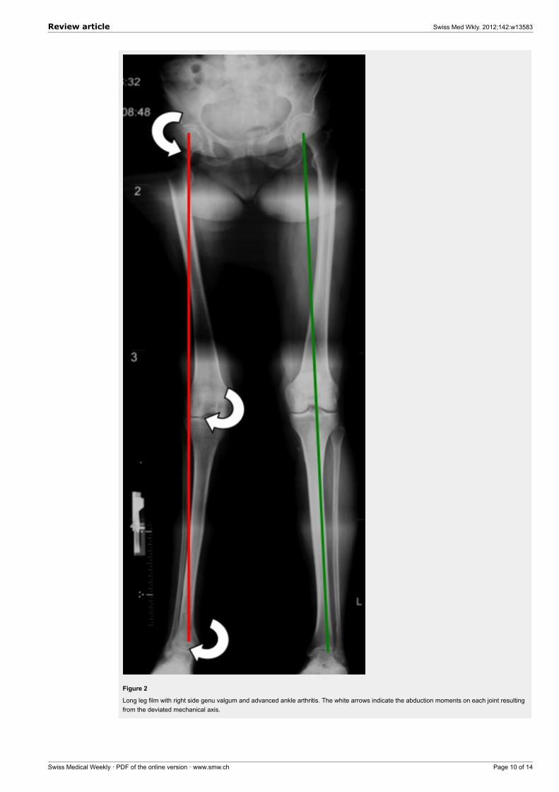
Figure 2
Long leg film with right side genu valgum and advanced ankle arthritis. The white arrows indicate the abduction moments on each joint resultingfrom the deviated mechanical axis.
Review article Swiss Med Wkly. 2012;142:w13583
Swiss Medical Weekly · PDF of the online version · www.smw.ch Page 10 of 14
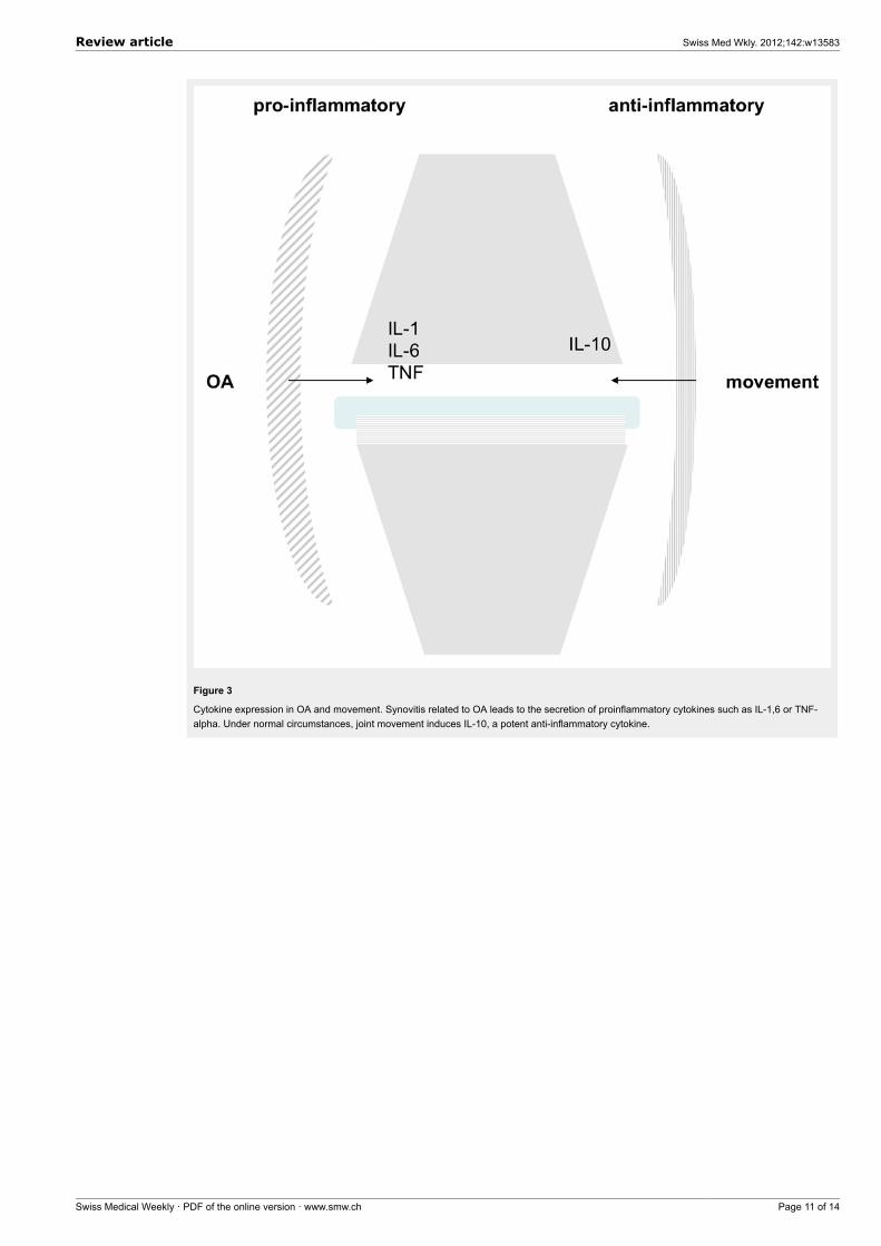
Figure 3
Cytokine expression in OA and movement. Synovitis related to OA leads to the secretion of proinflammatory cytokines such as IL-1,6 or TNF-alpha. Under normal circumstances, joint movement induces IL-10, a potent anti-inflammatory cytokine.
Review article Swiss Med Wkly. 2012;142:w13583
Swiss Medical Weekly · PDF of the online version · www.smw.ch Page 11 of 14
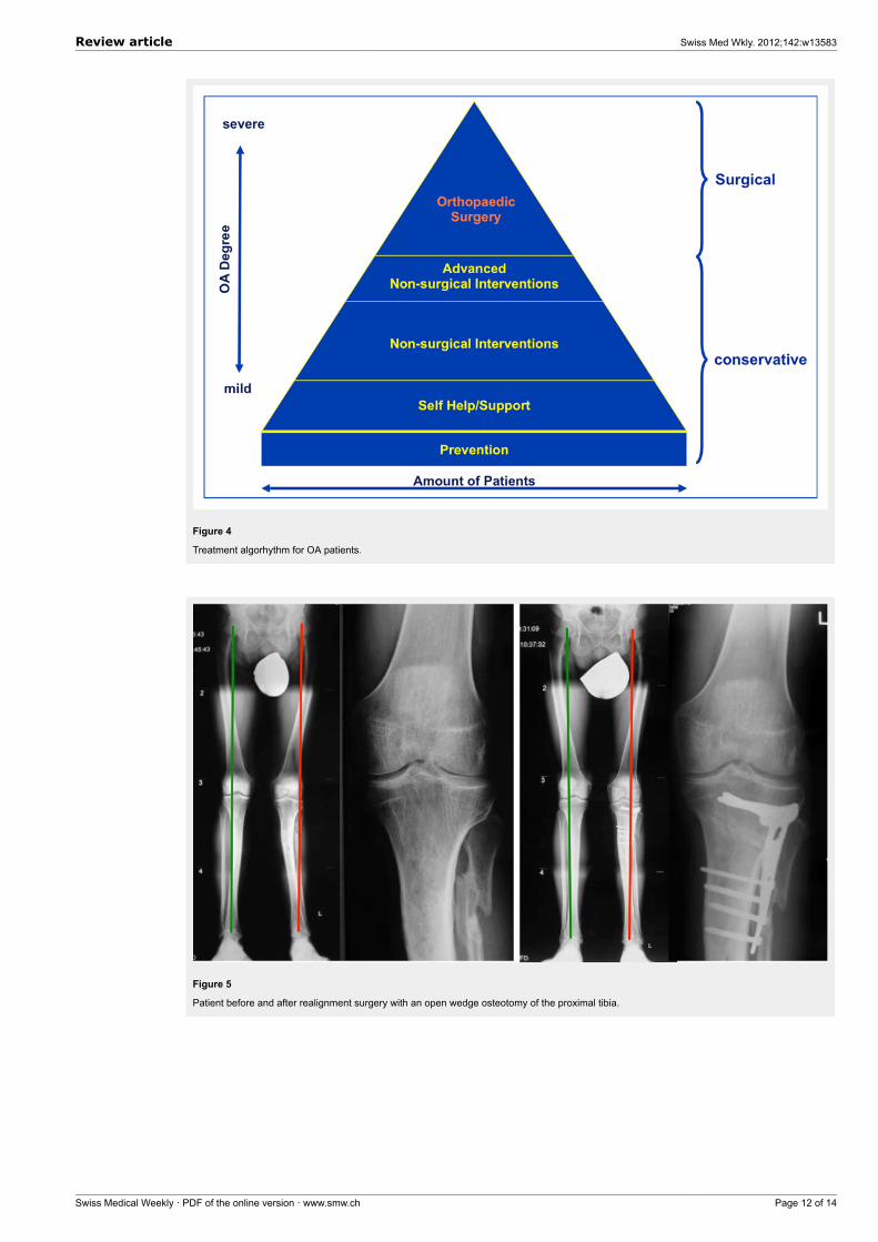
Figure 4
Treatment algorhythm for OA patients.
Figure 5
Patient before and after realignment surgery with an open wedge osteotomy of the proximal tibia.
Review article Swiss Med Wkly. 2012;142:w13583
Swiss Medical Weekly · PDF of the online version · www.smw.ch Page 12 of 14
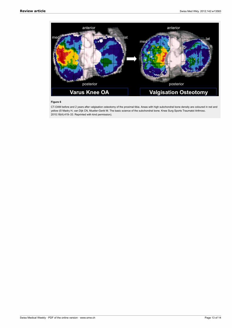
Figure 6
CT-OAM before and 2 years after valgisation osteotomy of the proximal tibia. Areas with high subchondral bone density are coloured in red andyellow (© Madry H, van Dijk CN, Mueller-Gerbl M. The basic science of the subchondral bone. Knee Surg Sports Traumatol Arthrosc.2010;18(4):419–33. Reprinted with kind permission).
Review article Swiss Med Wkly. 2012;142:w13583
Swiss Medical Weekly · PDF of the online version · www.smw.ch Page 13 of 14
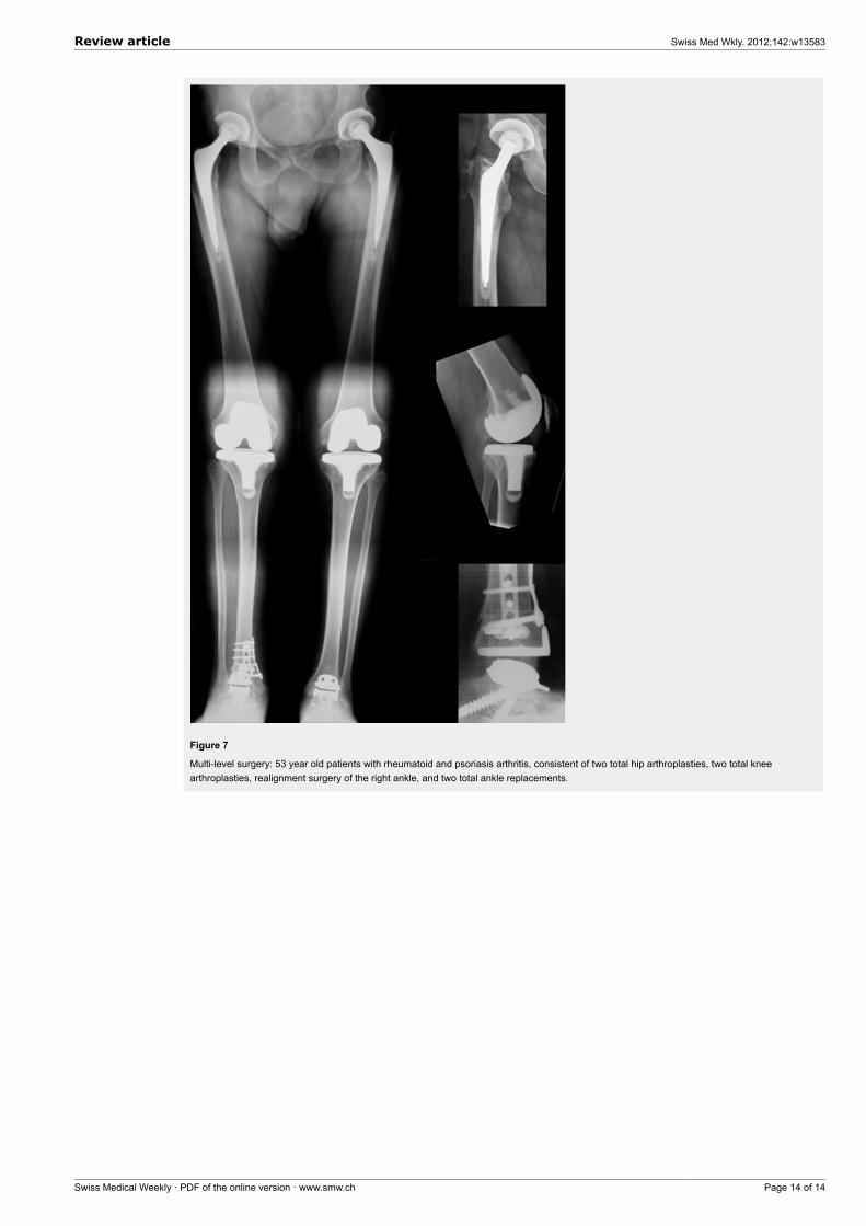
Figure 7
Multi-level surgery: 53 year old patients with rheumatoid and psoriasis arthritis, consistent of two total hip arthroplasties, two total kneearthroplasties, realignment surgery of the right ankle, and two total ankle replacements.
Review article Swiss Med Wkly. 2012;142:w13583
Swiss Medical Weekly · PDF of the online version · www.smw.ch Page 14 of 14


![Osteoarthritis and Articular Cartilage: Biomechanics …file.scirp.org/pdf/AAR_2014082914024425.pdf · R. Marks 299 conditions [5], and light and electron microscopic studies of articular](https://img.pdfslide.us/doc/110x75/5b1c5cb47f8b9a2d258fb3f6/osteoarthritis-and-articular-cartilage-biomechanics-filescirporgpdfaar-.jpg)