Embed Size (px)
DESCRIPTION
NSCA Study guide
Citation preview
ESSENTIALS OF STRENGTH TRAINING AND CONDITIONING MULTIMEDIA SYMPOSIUM
Presentation 3:
Biomechanical Principles
by: David Potach, MS, PT, CSCS,*D; NSCA-CPT,*D
NSCA Certification Commission®
3333 Landmark Circle ● Lincoln, NE 68504 ● 402-476-6669 ● 888-746-2378 ● [email protected] ● www.nsca-cc.org

Biomechanical Principles Presentation 3
©2006 NSCA Certification Commission Page 2
MULTIMEDIA SYMPOSIUM OVERVIEW This multimedia symposium was developed for the specific purpose of providing individuals who sit for the Certified Strength and Conditioning Specialist® (CSCS®) certification exam with a review of facts, concepts, and theories that are relevant to strength training and conditioning. You are encouraged to simultaneously listen and watch the symposium video presentation, view the PowerPoint® slide show, follow along in the presentation outline, and add your own notes in the spaces within the outline (more paper may be necessary). To maximize the value of the multimedia symposium when preparing for the CSCS exam, you may find it helpful to first study the Essentials of Strength Training and Conditioning (2nd edition) text. Further, candidates who perform well on the CSCS exam typically have considerable practical experience in strength training and conditioning athletes (e.g., designing programs, teaching proper exercise technique, performing testing sessions) and a strong academic background in the exercise sciences (i.e., anatomy, physiology, biomechanics, etc.). For additional suggestions for preparing for the CSCS exam, go to www.nsca-cc.org. NOTICE: • Although this presentation was recorded live and then professionally edited for scope and length, there are some room sounds, voice fluctuations, abrupt video “cuts” and piecing of video clips, and content variations. Every possible effort was made to minimize these irregularities. • All of the content of the multimedia symposium is protected by copyright. No part of the multi-media symposium may be reproduced or transmitted (in part or in full) by any means or in any form, electronic or mechanical, including digital copying or recording, or via any information retrieval system, including internet and intranet communications, without permission in writing from the NSCA Certification Commission. • While comprehension of the information provided by the multimedia symposium should certainly increase the likelihood of passing the CSCS certification exam, it does not guarantee a successful performance. The questions on the CSCS exam are developed from numerous resources in addition to the multimedia symposium.
Biomechanical Principles Presentation 3
©2006 NSCA Certification Commission Page 3
BIOMECHANICS TERMINOLOGY I. Functional Anatomy
II. Kinesiology
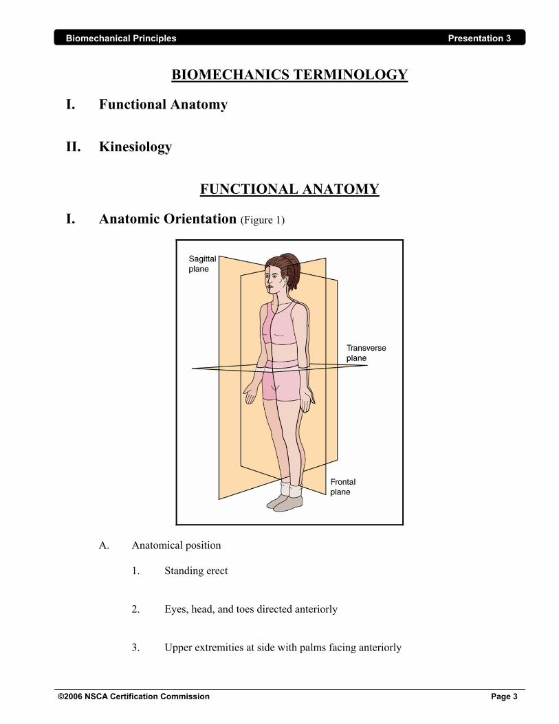
FUNCTIONAL ANATOMY I. Anatomic Orientation (Figure 1)
A. Anatomical position
1. Standing erect
2. Eyes, head, and toes directed anteriorly
3. Upper extremities at side with palms facing anteriorly
Biomechanical Principles Presentation 3
©2006 NSCA Certification Commission Page 4
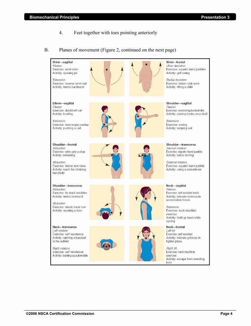
4. Feet together with toes pointing anteriorly
B. Planes of movement (Figure 2, continued on the next page)
Biomechanical Principles Presentation 3
©2006 NSCA Certification Commission Page 5
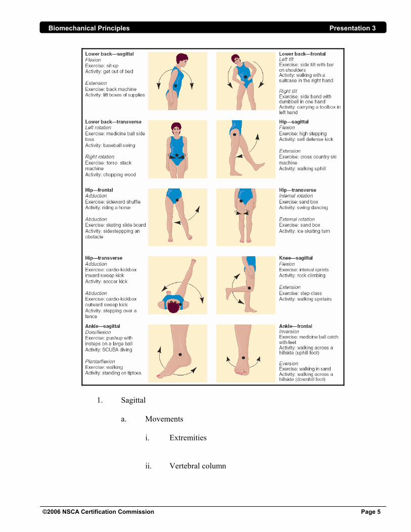
1. Sagittal
a. Movements
i. Extremities ii. Vertebral column
Biomechanical Principles Presentation 3
©2006 NSCA Certification Commission Page 6
2. Frontal
a. Movements i. Extremities ii. Vertebral column 3. Transverse/horizontal a. Movements i. Extremities ii. Vertebral column
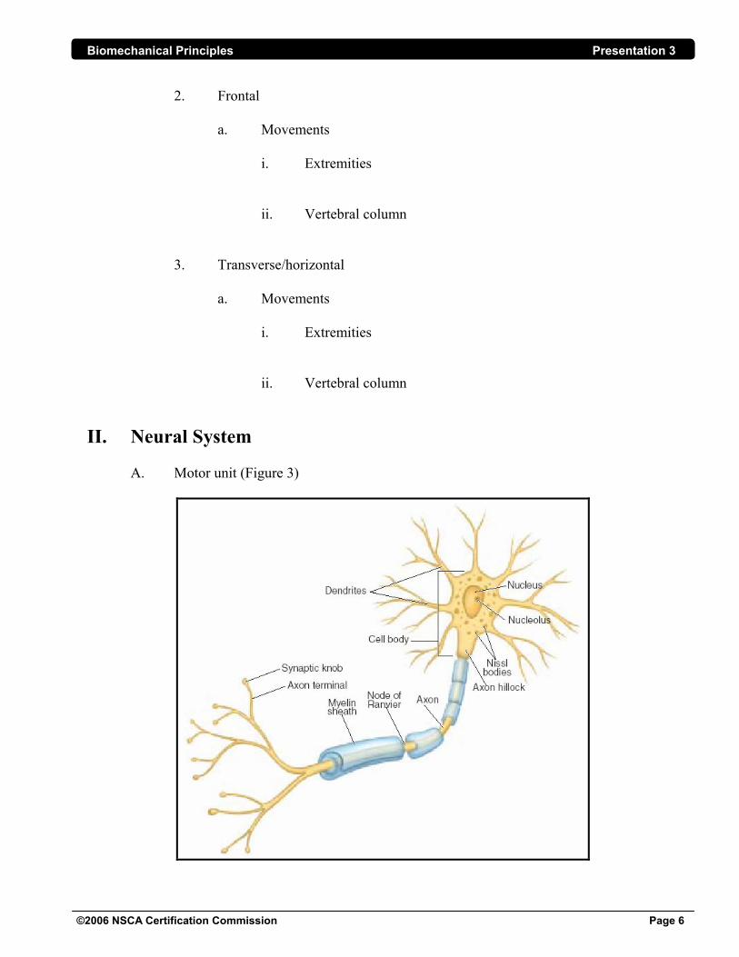
II. Neural System A. Motor unit (Figure 3)
Biomechanical Principles Presentation 3
©2006 NSCA Certification Commission Page 7
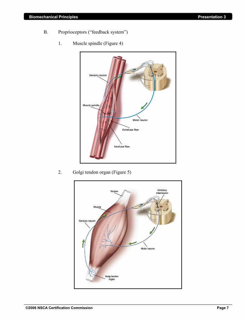
B. Proprioceptors (“feedback system”) 1. Muscle spindle (Figure 4)
2. Golgi tendon organ (Figure 5)
Biomechanical Principles Presentation 3
©2006 NSCA Certification Commission Page 8
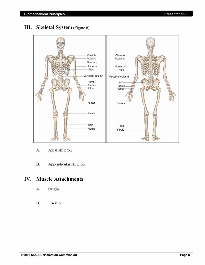
III. Skeletal System (Figure 6)
A. Axial skeleton
B. Appendicular skeleton
IV. Muscle Attachments A. Origin B. Insertion
Biomechanical Principles Presentation 3
©2006 NSCA Certification Commission Page 9
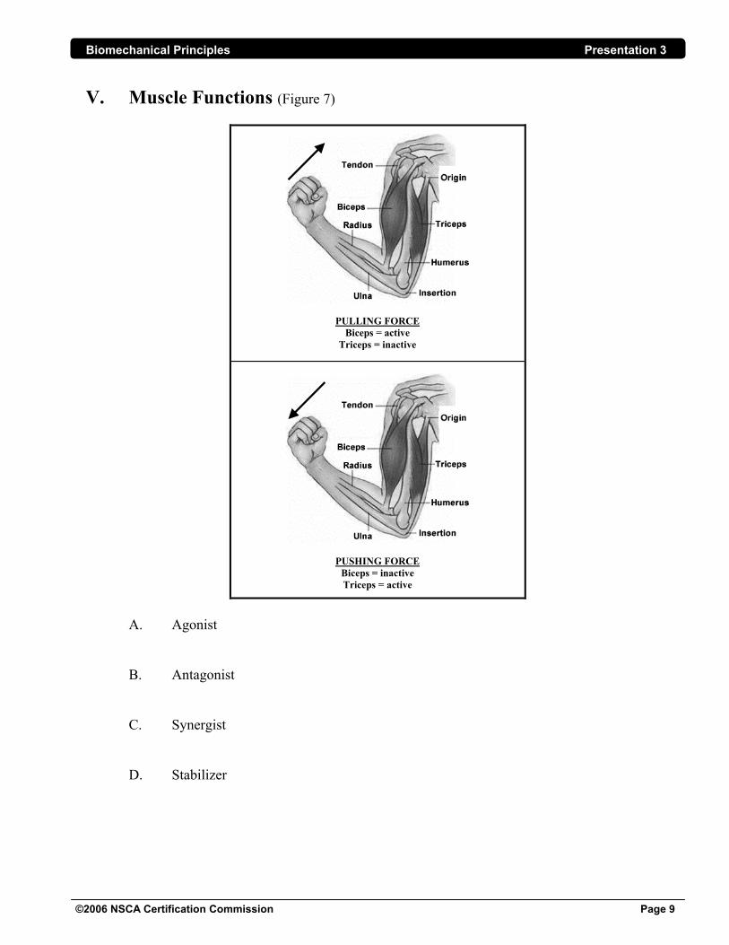
V. Muscle Functions (Figure 7)
PULLING FORCE Biceps = active
Triceps = inactive
PUSHING FORCE Biceps = inactive Triceps = active
A. Agonist B. Antagonist C. Synergist D. Stabilizer
Biomechanical Principles Presentation 3
©2006 NSCA Certification Commission Page 10
VI. Functional Movement A. Triplanar movement 1. Functional movement does not occur in only one plane of motion
2. Exercise and sports movements require motion in multiple planes
B. Muscle function
1. Muscles do not function in isolation 3. Several muscles function to produce complementary movements to complete a
given task
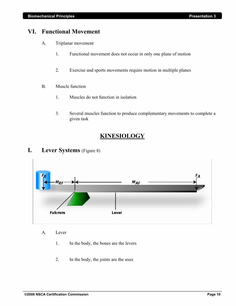
KINESIOLOGY I. Lever Systems (Figure 8)
A. Lever 1. In the body, the bones are the levers 2. In the body, the joints are the axes
Biomechanical Principles Presentation 3
©2006 NSCA Certification Commission Page 11
B. Three forces of a lever 1. Axis (fulcrum) 2. Effort (moving) force a. Tends to create the desired movement; in the body the muscle activity is the effort force b. Effort arm: the perpendicular distance from the axis to the effort force 3. Resistive force a. Tends to oppose the desired movement; the weight of a body part or an external weight is the resistive force b. Resistance arm: the perpendicular distance from the axis to the resistive force
C. Lever classification
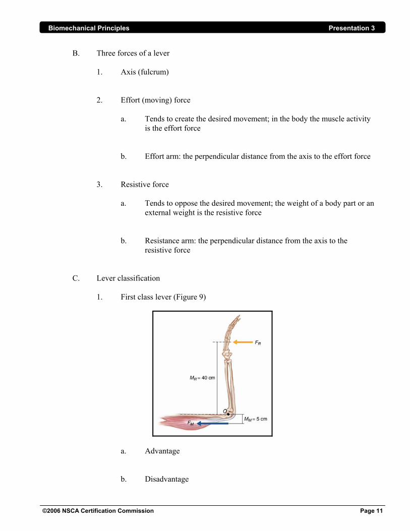
1. First class lever (Figure 9)
a. Advantage b. Disadvantage
Biomechanical Principles Presentation 3
©2006 NSCA Certification Commission Page 12
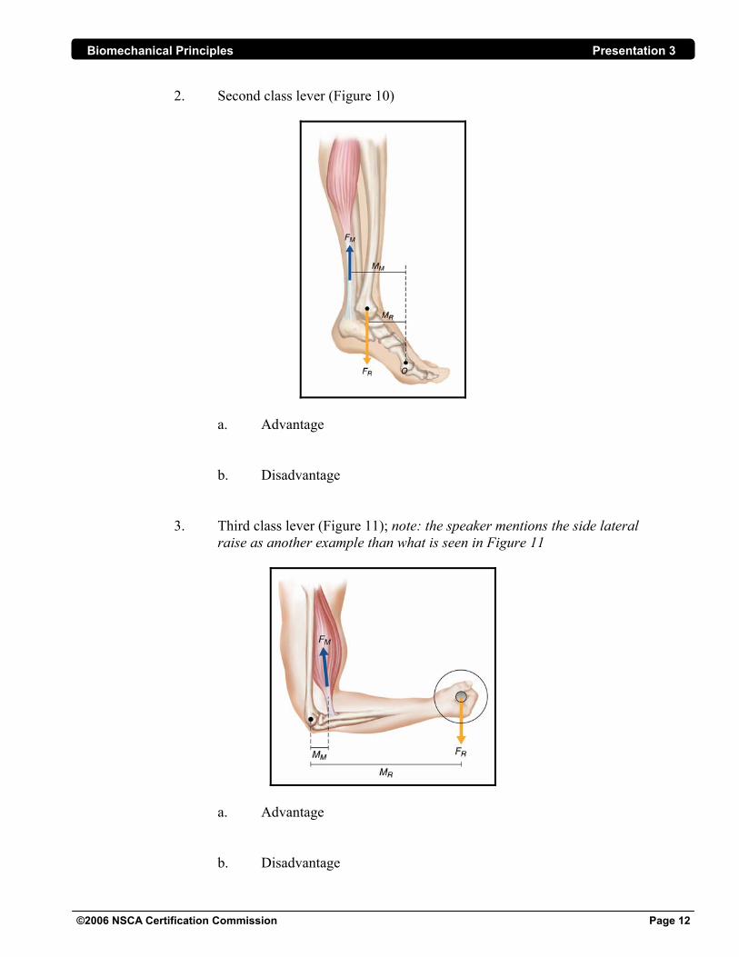
2. Second class lever (Figure 10)
a. Advantage
b. Disadvantage 3. Third class lever (Figure 11); note: the speaker mentions the side lateral raise as another example than what is seen in Figure 11
a. Advantage b. Disadvantage
Biomechanical Principles Presentation 3
©2006 NSCA Certification Commission Page 13
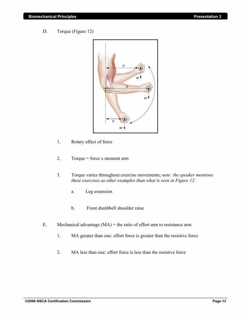
D. Torque (Figure 12)
1. Rotary effect of force 2. Torque = force x moment arm
3. Torque varies throughout exercise movements; note: the speaker mentions these exercises as other examples than what is seen in Figure 12:
a. Leg extension b. Front dumbbell shoulder raise
E. Mechanical advantage (MA) = the ratio of effort arm to resistance arm
1. MA greater than one: effort force is greater than the resistive force 2. MA less than one: effort force is less than the resistive force

Biomechanical Principles Presentation 3
©2006 NSCA Certification Commission Page 14
3. Examples
a. Elbow joint: the mechanical advantage is the greatest at 90° elbow flexion (Figure 13)
b. Knee joint: the patella optimizes the perpendicular distance from
the axis to the line of force to allow maximal torque capabilities (Figure 14)
II. Strength A. Contributory factors
1. Neural: muscle force is varied by altering the amount of motor unit activity
a. Motor unit recruitment: motor units are activated in a set sequence
Biomechanical Principles Presentation 3
©2006 NSCA Certification Commission Page 15
b. Motor unit firing rate: frequency of action potentials c. Methods to increase force production
2. Muscular: muscle force development depends on the mechanical properties
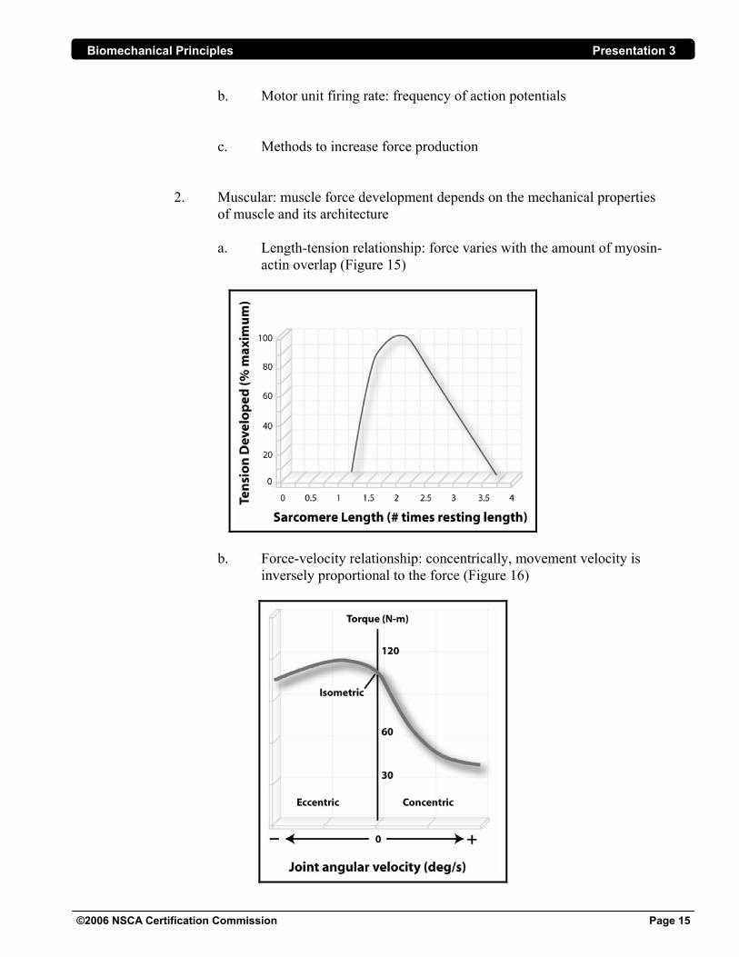
of muscle and its architecture a. Length-tension relationship: force varies with the amount of myosin-
actin overlap (Figure 15)
b. Force-velocity relationship: concentrically, movement velocity is
inversely proportional to the force (Figure 16)
Biomechanical Principles Presentation 3
©2006 NSCA Certification Commission Page 16
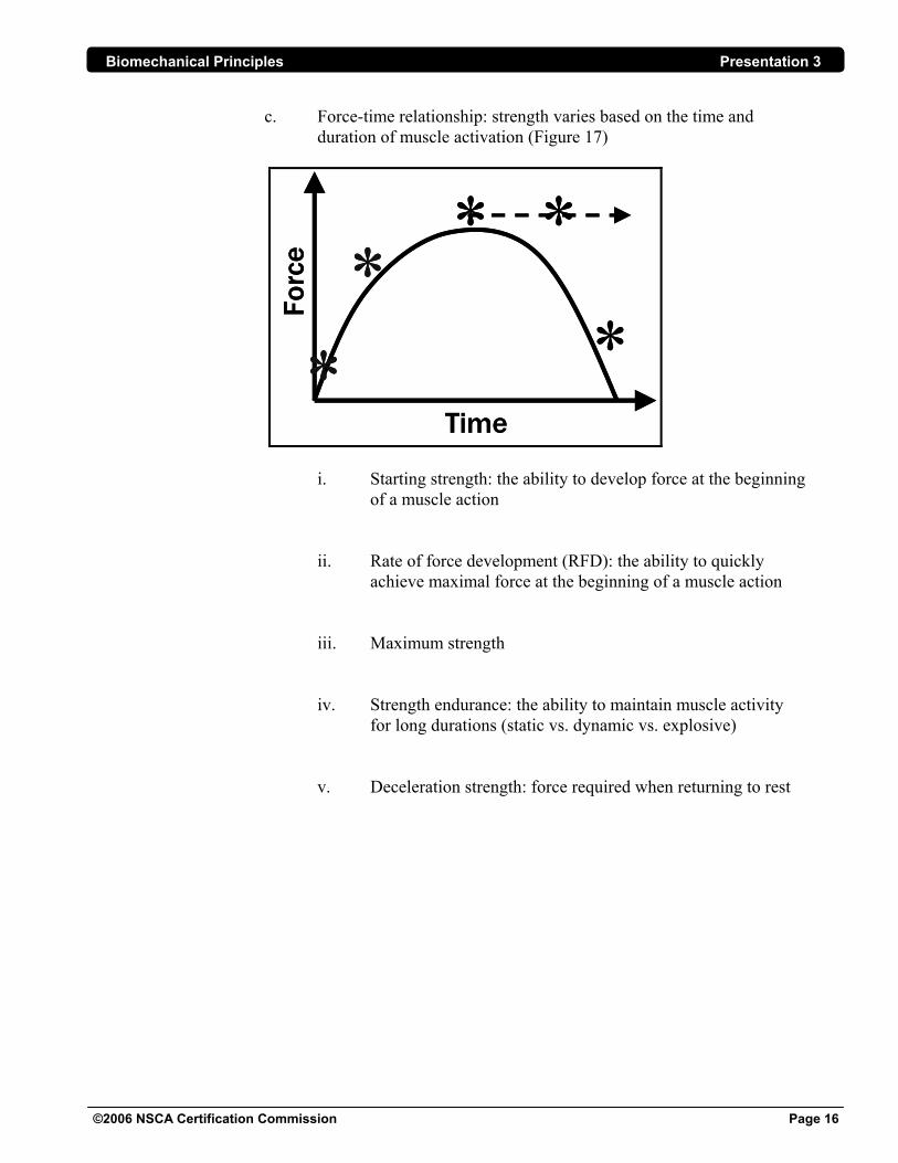
c. Force-time relationship: strength varies based on the time and duration of muscle activation (Figure 17)
**
* *
*Time
Forc
e
**
* *
*Time
Forc
e
i. Starting strength: the ability to develop force at the beginning of a muscle action ii. Rate of force development (RFD): the ability to quickly achieve maximal force at the beginning of a muscle action iii. Maximum strength iv. Strength endurance: the ability to maintain muscle activity for long durations (static vs. dynamic vs. explosive) v. Deceleration strength: force required when returning to rest
Biomechanical Principles Presentation 3
©2006 NSCA Certification Commission Page 17
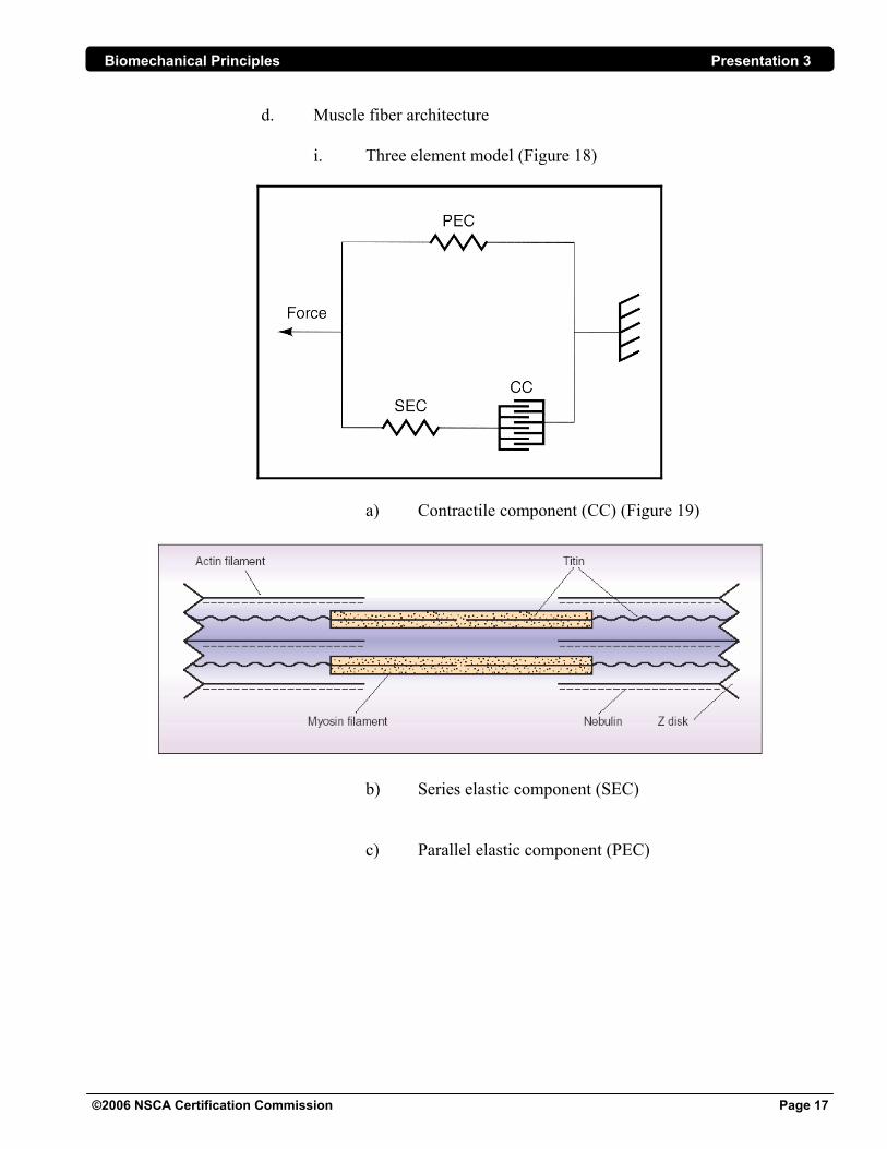
d. Muscle fiber architecture
i. Three element model (Figure 18)
a) Contractile component (CC) (Figure 19)
b) Series elastic component (SEC)
c) Parallel elastic component (PEC)
Biomechanical Principles Presentation 3
©2006 NSCA Certification Commission Page 18
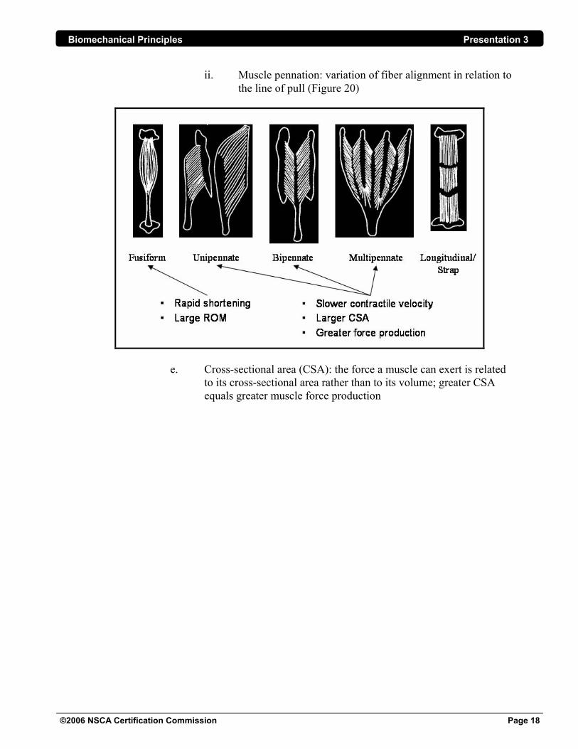
ii. Muscle pennation: variation of fiber alignment in relation to the line of pull (Figure 20)
e. Cross-sectional area (CSA): the force a muscle can exert is related
to its cross-sectional area rather than to its volume; greater CSA equals greater muscle force production
Biomechanical Principles Presentation 3
©2006 NSCA Certification Commission Page 19
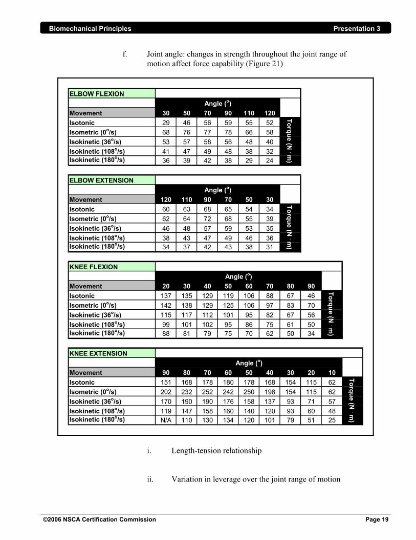
f. Joint angle: changes in strength throughout the joint range of motion affect force capability (Figure 21)
ELBOW FLEXION
Movement 30 50 70 90 110 120Isotonic 29 46 56 59 55 52Isometric (0o/s) 68 76 77 78 66 58Isokinetic (36o/s) 53 57 58 56 48 40Isokinetic (108o/s) 41 47 49 48 38 32Isokinetic (180o/s) 36 39 42 38 29 24
ELBOW EXTENSION
Movement 120 110 90 70 50 30Isotonic 60 63 68 65 54 34Isometric (0o/s) 62 64 72 68 55 39Isokinetic (36o/s) 46 48 57 59 53 35Isokinetic (108o/s) 38 43 47 49 46 36Isokinetic (180o/s) 34 37 42 43 38 31
KNEE FLEXION
Movement 20 30 40 50 60 70 80 90Isotonic 137 135 129 119 106 88 67 46Isometric (0o/s) 142 138 129 125 106 97 83 70Isokinetic (36o/s) 115 117 112 101 95 82 67 56Isokinetic (108o/s) 99 101 102 95 86 75 61 50Isokinetic (180o/s) 88 81 79 75 70 62 50 34
KNEE EXTENSION
Movement 90 80 70 60 50 40 30 20 10Isotonic 151 168 178 180 178 168 154 115 62Isometric (0o/s) 202 232 252 242 250 198 154 115 62Isokinetic (36o/s) 170 190 190 176 158 137 93 71 57Isokinetic (108o/s) 119 147 158 160 140 120 93 60 48Isokinetic (180o/s) N/A 110 130 134 120 101 79 51 25
Angle (o)
Torque (N . m
)Torque (N
. m)
Angle (o)
Torque (N . m
)
Angle (o)
Angle (o)
Torque (N . m
)
i. Length-tension relationship ii. Variation in leverage over the joint range of motion
Biomechanical Principles Presentation 3
©2006 NSCA Certification Commission Page 20
B. Types of forces 1. Gravity: resistance due to weight in a vertical direction; center of gravity
2. Ground reaction force 3. Tension 4. Compression 5. Shear
6. Axial loading
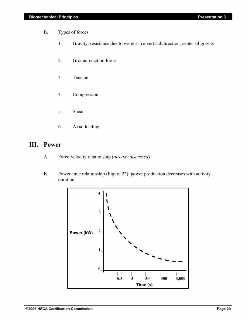
III. Power A. Force-velocity relationship (already discussed) B. Power-time relationship (Figure 22): power production decreases with activity
duration
4__
3__
2__
1__
0__ | | | | | 0.3 3 30 300 3,000
Time (s)
Power (kW)
Biomechanical Principles Presentation 3
©2006 NSCA Certification Commission Page 21
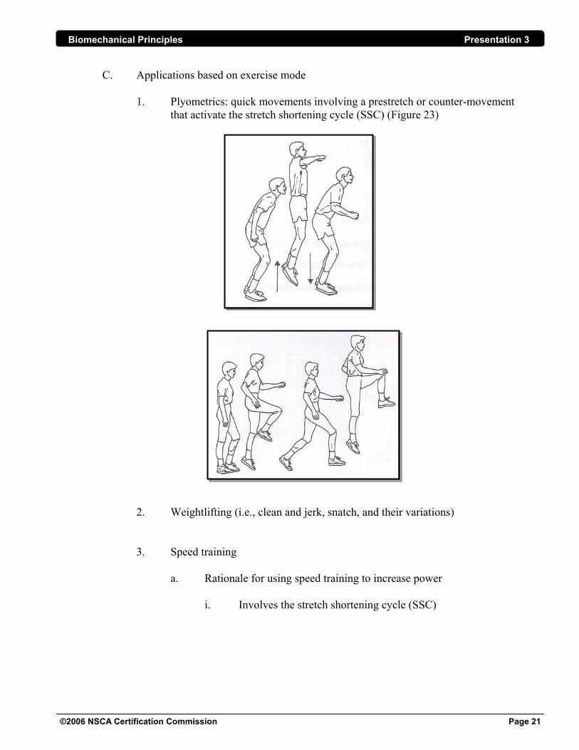
C. Applications based on exercise mode 1. Plyometrics: quick movements involving a prestretch or counter-movement that activate the stretch shortening cycle (SSC) (Figure 23)
2. Weightlifting (i.e., clean and jerk, snatch, and their variations) 3. Speed training
a. Rationale for using speed training to increase power i. Involves the stretch shortening cycle (SSC)
Biomechanical Principles Presentation 3
©2006 NSCA Certification Commission Page 22
ii. Requires speed-strength: the application of maximal force at high velocities (e.g., sprint training) b. Types of sprint training
i. Technique training ii. Sprint-assisted training iii. Resisted sprinting
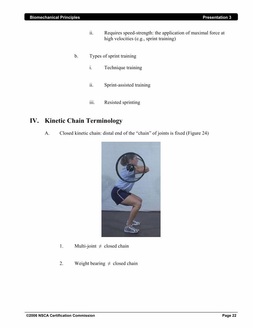
IV. Kinetic Chain Terminology A. Closed kinetic chain: distal end of the “chain” of joints is fixed (Figure 24)
1. Multi-joint ≠ closed chain 2. Weight bearing ≠ closed chain
Biomechanical Principles Presentation 3
©2006 NSCA Certification Commission Page 23
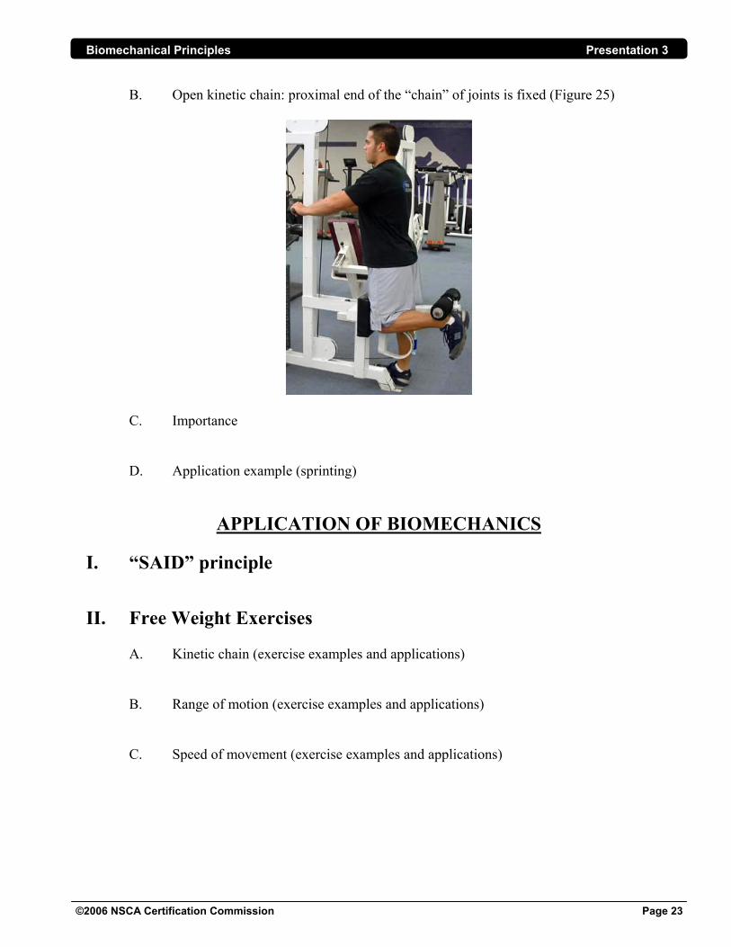
B. Open kinetic chain: proximal end of the “chain” of joints is fixed (Figure 25)
C. Importance
D. Application example (sprinting)
APPLICATION OF BIOMECHANICS I. “SAID” principle II. Free Weight Exercises
A. Kinetic chain (exercise examples and applications)
B. Range of motion (exercise examples and applications) C. Speed of movement (exercise examples and applications)
Biomechanical Principles Presentation 3
©2006 NSCA Certification Commission Page 24
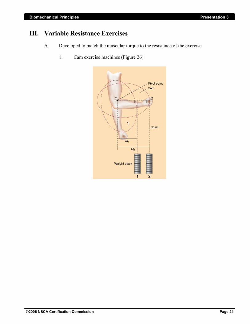
III. Variable Resistance Exercises
A. Developed to match the muscular torque to the resistance of the exercise 1. Cam exercise machines (Figure 26)
Biomechanical Principles Presentation 3
©2006 NSCA Certification Commission Page 25
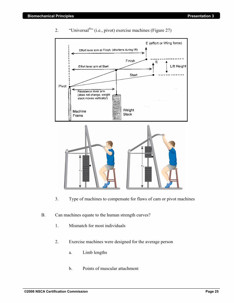
2. “Universal®” (i.e., pivot) exercise machines (Figure 27)
3. Type of machines to compensate for flaws of cam or pivot machines
B. Can machines equate to the human strength curves?
1. Mismatch for most individuals 2. Exercise machines were designed for the average person a. Limb lengths b. Points of muscular attachment
Biomechanical Principles Presentation 3
©2006 NSCA Certification Commission Page 26
GLOSSARY OF TERMS Acceleration—the rate at which speed changes Agonist—the muscle most directly involved in creating movement Anatomical position—the body standing erect with arms down and palms forward Angle of pennation—the angle between the overall muscle and its fibers (0o = no pennation) Angular displacement—the angle through which an object rotates Angular velocity—an object’s rotational speed (radians/sec or deg/sec) Antagonist—a muscle that can slow down or stop a movement Biomechanics—the mechanical analysis of biological systems Concentric muscle action—when a muscle shortens because its contractile force is greater than the
resistive force Eccentric muscle action—when a muscle lengthens because its contractile force is less than the resistive force Fluid resistance—the resisting force encountered by an object moving through a fluid or by a fluid moving past or around an object Friction—the resistive force encountered while attempting to move two surfaces in contact relative to each other Frontal plane—a vertical plane dividing the standing body into anterior and posterior halves Fulcrum—the pivot point of a lever Inertial force—the force required to accelerate an object (equals the mass of the object times its acceleration) Isometric muscle action—when muscle length does not change because the contractile force is equal to the resistive force Lever—an object that, when subjected to a force whose line of action does not pass through its pivot point, exerts force on any object impeding its tendency to rotate Lordosis—an arched back
Biomechanical Principles Presentation 3
©2006 NSCA Certification Commission Page 27
Mechanical advantage—the ratio of the movement arm through which an applied force acts to that through which a resistive force acts (e.g., a pliers with 9” handles with 1.5” jaws has a mechanical advantage of 9/1.5 = 6; a 10 pound squeeze on the handle generates a 6 x 10 pound = 60 pound squeeze at the jaws) Moment arm—the perpendicular distance from the line of action of a force on a lever to its fulcrum Muscle force—force generated by biochemical activity which tends to draw the opposite ends of a muscle towards each other Pennate muscle—a muscle whose fibers have a featherlike arrangement Power—the time rate of doing work (equals work/time or force x velocity) Resistive force—force which arts contrary to muscle force (i.e., gravity, inertia, friction) Rotational power—rotational work per unit time or torque times angular velocity Rotational work—the product of torque and angular displacement Sagittal plane—a vertical plane dividing the standing body into left and right halves Specificity—the concept that training is most effective when resistance exercises bear key similarities to the sports activity in which the improvement is sought; to train in a specific manner to achieve a specific outcome Strength—the maximal force that can be exerted under specified conditions Synergist—a muscle that assists indirectly in a movement Tendon—a fibrous attachment between muscle and bone that blends into and is continuous with both the muscle sheath and the connective tissue surrounding the bone Torque—the tendency of a force to rotate an object about a fulcrum (equals the magnitude of the force times the length of its moment arm); ability to create a turning effect Transverse plane—a horizontal plane dividing the standing body into superior and inferior halves Weight—the downward force on an object due to gravity (equal to the object’s mass times the local acceleration of gravity) Work—the product of the force exerted on an object and the distance the object moves in the direction in which the force is exerted
Biomechanical Principles Presentation 3
©2006 NSCA Certification Commission Page 28
PRESENTATION REFERENCES 1. Baechle, T.R. and R.W. Earle. (Eds.). Essentials of Strength Training and Conditioning (2nd
ed.). Champaign, IL: Human Kinetics. 2000. 2. Earle, R.W. and T.R. Baechle. (Eds.). NSCA’s Essentials of Personal Training. Champaign, IL: Human Kinetics. 2004. 3. Enoka, R.M. Neuromechanics of Human Movement (3rd ed.). Champaign, IL: Human Kinetics. 2002. 4. Harman, E. and P.N. Frykman. The effects of knee wraps on weightlifting performance and injury. NSCA Journal 12(5): 30-35. 1990. 5. Harman, E.A., Johnson, M. and P.N. Frykman. A movement-oriented approach to exercise prescription. NSCA Journal 14(1): 47-54. 1992. 6. Luttgens, K. and N. Hamilton. Kinesiology: Scientific Basis of Human Motion (10th ed.). Madison, WI: Brown & Benchmark. 2002. 7. Marieb, E.N. Human Anatomy and Physiology (2nd ed.). Redwood City, CA: Benjamin/ Cummings. 1992. 8. Özkaya, N. and M. Nordin. Fundamentals of Biomechanics: Equilibrium, Motion, and Deformation (2nd ed.). New York, NY: Springer Publishers. 1999. 9. Rasch, P.J. Kinesiology and Applied Anatomy (7th ed.). Philadelphia, PA: Lea & Febiger. 1993. 10. Tortora, G.J. Principles of Human Anatomy (10th ed.). Indianapolis, IN: Wiley Publishing, Inc. 2004.
Biomechanical Principles Presentation 3
©2006 NSCA Certification Commission Page 29
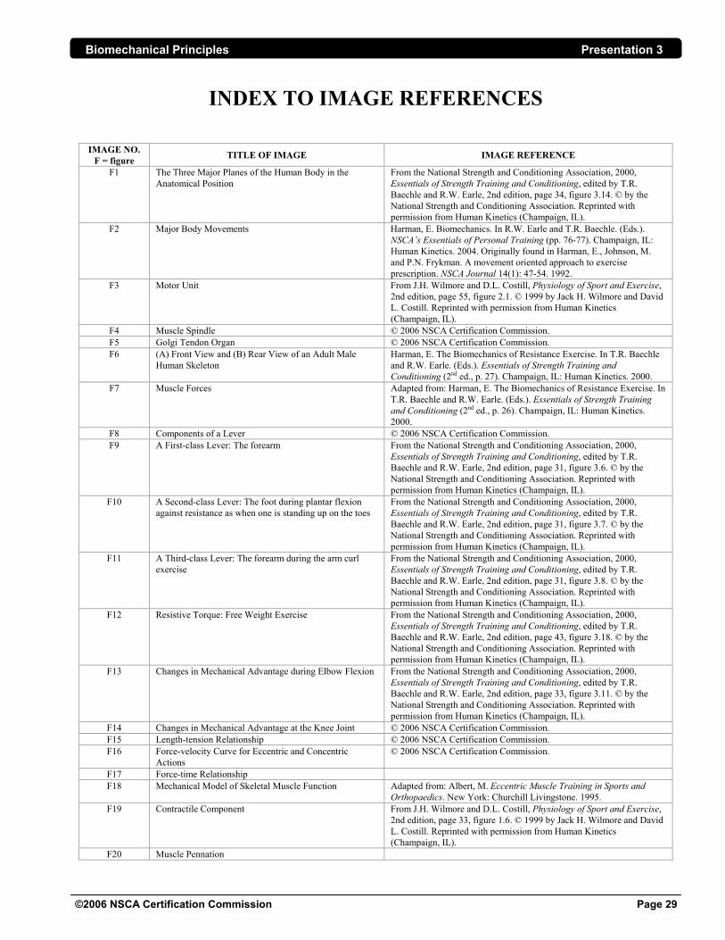
INDEX TO IMAGE REFERENCES IMAGE NO.
F = figure TITLE OF IMAGE IMAGE REFERENCE
F1 The Three Major Planes of the Human Body in the Anatomical Position
From the National Strength and Conditioning Association, 2000, Essentials of Strength Training and Conditioning, edited by T.R. Baechle and R.W. Earle, 2nd edition, page 34, figure 3.14. © by the National Strength and Conditioning Association. Reprinted with permission from Human Kinetics (Champaign, IL).
F2 Major Body Movements Harman, E. Biomechanics. In R.W. Earle and T.R. Baechle. (Eds.). NSCA’s Essentials of Personal Training (pp. 76-77). Champaign, IL: Human Kinetics. 2004. Originally found in Harman, E., Johnson, M. and P.N. Frykman. A movement oriented approach to exercise prescription. NSCA Journal 14(1): 47-54. 1992.
F3 Motor Unit From J.H. Wilmore and D.L. Costill, Physiology of Sport and Exercise, 2nd edition, page 55, figure 2.1. © 1999 by Jack H. Wilmore and David L. Costill. Reprinted with permission from Human Kinetics (Champaign, IL).
F4 Muscle Spindle © 2006 NSCA Certification Commission. F5 Golgi Tendon Organ © 2006 NSCA Certification Commission. F6 (A) Front View and (B) Rear View of an Adult Male
Human Skeleton Harman, E. The Biomechanics of Resistance Exercise. In T.R. Baechle and R.W. Earle. (Eds.). Essentials of Strength Training and Conditioning (2nd ed., p. 27). Champaign, IL: Human Kinetics. 2000.
F7 Muscle Forces Adapted from: Harman, E. The Biomechanics of Resistance Exercise. In T.R. Baechle and R.W. Earle. (Eds.). Essentials of Strength Training and Conditioning (2nd ed., p. 26). Champaign, IL: Human Kinetics. 2000.
F8 Components of a Lever © 2006 NSCA Certification Commission. F9 A First-class Lever: The forearm From the National Strength and Conditioning Association, 2000,
Essentials of Strength Training and Conditioning, edited by T.R. Baechle and R.W. Earle, 2nd edition, page 31, figure 3.6. © by the National Strength and Conditioning Association. Reprinted with permission from Human Kinetics (Champaign, IL).
F10 A Second-class Lever: The foot during plantar flexion against resistance as when one is standing up on the toes
From the National Strength and Conditioning Association, 2000, Essentials of Strength Training and Conditioning, edited by T.R. Baechle and R.W. Earle, 2nd edition, page 31, figure 3.7. © by the National Strength and Conditioning Association. Reprinted with permission from Human Kinetics (Champaign, IL).
F11 A Third-class Lever: The forearm during the arm curl exercise
From the National Strength and Conditioning Association, 2000, Essentials of Strength Training and Conditioning, edited by T.R. Baechle and R.W. Earle, 2nd edition, page 31, figure 3.8. © by the National Strength and Conditioning Association. Reprinted with permission from Human Kinetics (Champaign, IL).
F12 Resistive Torque: Free Weight Exercise From the National Strength and Conditioning Association, 2000, Essentials of Strength Training and Conditioning, edited by T.R. Baechle and R.W. Earle, 2nd edition, page 43, figure 3.18. © by the National Strength and Conditioning Association. Reprinted with permission from Human Kinetics (Champaign, IL).
F13 Changes in Mechanical Advantage during Elbow Flexion From the National Strength and Conditioning Association, 2000, Essentials of Strength Training and Conditioning, edited by T.R. Baechle and R.W. Earle, 2nd edition, page 33, figure 3.11. © by the National Strength and Conditioning Association. Reprinted with permission from Human Kinetics (Champaign, IL).
F14 Changes in Mechanical Advantage at the Knee Joint © 2006 NSCA Certification Commission. F15 Length-tension Relationship © 2006 NSCA Certification Commission. F16 Force-velocity Curve for Eccentric and Concentric
Actions © 2006 NSCA Certification Commission.
F17 Force-time Relationship F18 Mechanical Model of Skeletal Muscle Function Adapted from: Albert, M. Eccentric Muscle Training in Sports and
Orthopaedics. New York: Churchill Livingstone. 1995. F19 Contractile Component From J.H. Wilmore and D.L. Costill, Physiology of Sport and Exercise,
2nd edition, page 33, figure 1.6. © 1999 by Jack H. Wilmore and David L. Costill. Reprinted with permission from Human Kinetics (Champaign, IL).
F20 Muscle Pennation
Biomechanical Principles Presentation 3
©2006 NSCA Certification Commission Page 30
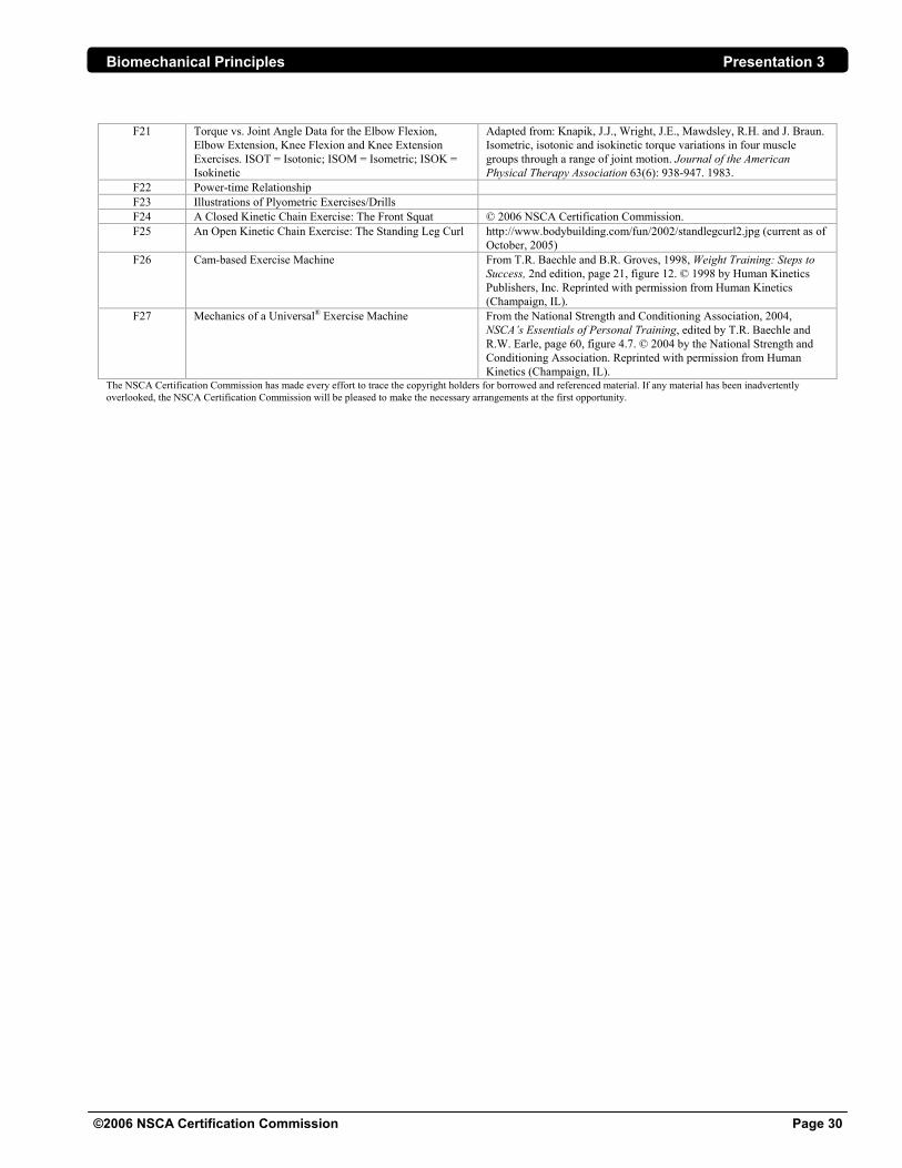
F21 Torque vs. Joint Angle Data for the Elbow Flexion,
Elbow Extension, Knee Flexion and Knee Extension Exercises. ISOT = Isotonic; ISOM = Isometric; ISOK = Isokinetic
Adapted from: Knapik, J.J., Wright, J.E., Mawdsley, R.H. and J. Braun. Isometric, isotonic and isokinetic torque variations in four muscle groups through a range of joint motion. Journal of the American Physical Therapy Association 63(6): 938-947. 1983.
F22 Power-time Relationship F23 Illustrations of Plyometric Exercises/Drills F24 A Closed Kinetic Chain Exercise: The Front Squat © 2006 NSCA Certification Commission. F25 An Open Kinetic Chain Exercise: The Standing Leg Curl http://www.bodybuilding.com/fun/2002/standlegcurl2.jpg (current as of
October, 2005) F26 Cam-based Exercise Machine From T.R. Baechle and B.R. Groves, 1998, Weight Training: Steps to
Success, 2nd edition, page 21, figure 12. © 1998 by Human Kinetics Publishers, Inc. Reprinted with permission from Human Kinetics (Champaign, IL).
F27 Mechanics of a Universal® Exercise Machine From the National Strength and Conditioning Association, 2004, NSCA’s Essentials of Personal Training, edited by T.R. Baechle and R.W. Earle, page 60, figure 4.7. © 2004 by the National Strength and Conditioning Association. Reprinted with permission from Human Kinetics (Champaign, IL).
The NSCA Certification Commission has made every effort to trace the copyright holders for borrowed and referenced material. If any material has been inadvertently overlooked, the NSCA Certification Commission will be pleased to make the necessary arrangements at the first opportunity.
Biomechanical Principles Presentation 3
MUSCULAR ANATOMY
A Supplement to “Biomechanical Principles” by
David H. Potach, MS, PT, CSCS,*D; NSCA-CPT,*D
SCAPULAR/DELTOID REGION
The movements of the shoulder girdle are accomplished by combinations of posterior and anterior
muscles that run between axial and appendicular skeletons. Muscles which insert on the
appendicular skeleton but originate from the axial skeleton are considered extrinsic muscles of the
limb; muscles that both originate from and insert upon the appendicular skeleton are intrinsic muscles
of the limb.
I. Superficial Extrinsic Muscles of the Back/Shoulder A. Trapezius
1. Trapezion = irregular four-sided figure
2. Origin: superior nuchal line; external occipital protuberance (of the occipital
bone); ligamentum nuchae
3. Insertion:
a. Part I (uppermost fibers): lateral 1/3 of clavicle
b. Part II (lower cervical/upper thoracic fibers): acromion (of scapula)
i. Thickest portion of trapezius
c. Part III (lower thoracic fibers): scapular spine
4. Actions:
a. Part I: elevation of scapula; upward rotation of scapula
b. Part II: retraction (adduction) of scapula
c. Part III: depression of scapula; upward rotation of scapula
d. Superior + inferior fibers (parts I & III): upward rotation of scapula
i. Upward rotation is similar to shrugging one's shoulders
ii. Upward rotation occurs when the acromion of the scapula
moves upward
©2006 NSCA Certification Commission Page 31
Biomechanical Principles Presentation 3
iii. The inferior angle of the scapula moves laterally and slightly up
during upward rotation
e. This large, flat, triangular muscle covers the posterior aspect of the
neck and superior half of the trunk. The superior fibers of the trapezius
elevate the scapula (i.e., pull it posteriorly toward the median plane),
and its inferior fibers depress the scapula and lower the shoulder. The
superior and inferior fibers act together in the superior rotation of the
scapula. The trapezius muscles brace the shoulders by pulling the
scapulae posteriorly, hence, weakness of these muscles results in
drooping of the shoulders.
B. Latissimus Dorsi
1. Latissimus dorsi = widest of the back
2. Origin: spinous processes of T-6 through S-2 (via thoracolumbar fascia);
posterior iliac crest; lower three ribs
3. Insertion: lesser tubercle on anterior aspect of humerus
a. Fibers converge, then turn 180 degrees to insert on lesser tubercle of
humerus
4. Actions:
a. Extension (of the humerus)
b. Adduction (of the humerus)
c. Medial rotation (of the humerus)
5. The latissimus dorsi covers the inferior half of the back (T-6 to the iliac crest).
This muscle is used when chopping wood, climbing, paddling a canoe and
swimming (particularly the crawl). When climbing, these muscles raise the
trunk; they raise the trunk to the arm, which occurs when performing chin-ups.
©2006 NSCA Certification Commission Page 32
Biomechanical Principles Presentation 3
The next muscle, although it belongs and is listed with the deep intrinsic muscles, is more
functionally suited to be included with the latissimus dorsi as its actions mimic those of the latissimus
dorsi.
6. Teres Major
a. Origin: inferior 1/3 of lateral border of scapula
b. Insertion: lesser tubercle (on anterior aspect of humerus)
i. Passes anterior to long head of triceps to crest of lesser tubercle
ii. Just medial to insertion of latissimus dorsi muscle
c. Actions:
i. Adduction of humerus
ii. Medial rotation of humerus
d. Teres major is an important stabilizer of the head of the humerus in the
glenoid cavity during abduction of the arm, i.e., it steadies the head of
the humerus in its socket
II. Deep Extrinsic Muscles of the Shoulder A. Levator Scapulae
1. Levator scapula = elevator of the scapula
2. Origin: transverse processes of upper cervical vertebrae (C1-C4)
3. Insertion: superior angle/upper vertebral border of scapula (superior to
scapular spine)
4. Actions:
a. Elevation of scapula
b. Downward rotation of scapula
i. Tilts glenoid cavity inferiorly by rotating the scapula
B. Rhomboids (minor and major)
1. Named for the shape of the muscle, i.e., shaped like a rhomboid, an oblique
parallelogram
2. Origin:
a. Minor: lower ligamentum nuchae & spinous processes of C-7 to T-1
b. Major: spinous processes of T-2 to T-5
©2006 NSCA Certification Commission Page 33
Biomechanical Principles Presentation 3
3. Insertion: vertebral border of scapula (spine to inferior angle)
4. Actions:
a. Retraction (adduction) of scapula
b. Downward rotation of scapula (depression of the glenoid cavity)
c. Fixation of scapula to body wall
5. Major is about two times wider than minor. The rhomboids are used when
forcibly lowering the raised upper limbs, e.g., when driving a stake with a
sledge hammer.
III. Intrinsic Muscles of the Shoulder A. Deltoid
1. Deltoid = triangular, i.e., it is shaped like the inverted Greek letter delta
2. Origin: scapular spine; posterior and lateral edge of acromion; lateral 1/3 of
clavicle
a. Approximates insertion of trapezius
3. Insertion: deltoid tuberosity (just above middle of humerus); fleshy portion (of
muscle) surrounds humeral head anteriorly, laterally and posteriorly
4. Actions:
a. Middle:
i. Especially strong, but has a short range of action. Action =
abduction (following initiation of movement by supraspinatus
muscle)
b. Anterior: flexion of the humerus at the shoulder joint
i. Internal (medial) rotation of humerus – minor movement for the
deltoid
c. Posterior: extension of the humerus at the shoulder joint
i. External (lateral) rotation of humerus – minor movement for the
deltoid
©2006 NSCA Certification Commission Page 34
Biomechanical Principles Presentation 3
5. To make these movements, the deltoid works with other muscles, e.g., the
anterior portion works with the pectoralis major and coracobrachialis when
flexing the humerus at the shoulder. The middle portion acts with the
supraspinatus when abducting the arm. Deltoids are used everyday when
swinging the arms during walking.
B. Deep intrinsic muscles
1. Supraspinatus
a. Supraspinatus = superior to the spine (of the scapula)
b. Origin: supraspinous fossa of scapula
c. Insertion: greater tubercle of humerus
i. Crosses over the top of the humeral head to insert on the
superior aspect of the greater tubercle
d. Actions:
i. Abduction of humerus (with deltoid)
ii. Stabilization of glenohumeral joint (with other rotator cuff
muscles)
e. The supraspinatus is the most commonly torn part of the rotator cuff
f. The supraspinatus acts strongly when a heavy weight is carried with the
upper arm adducted (e.g., when carrying a heavy suitcase)
2. Infraspinatus
a. Infraspinatus = inferior to spine of the scapula
b. Origin: infraspinous fossa of scapula
c. Insertion: greater tubercle of humerus
i. Passes transversely across the posterior aspect of humeral head
to insert into the upper part of the posterior aspect of the greater
tubercle
d. Actions:
i. External rotation of humerus
ii. Stabilization of glenohumeral joint (with other rotator cuff
muscles)
e. Infraspinatus occupies most of the infraspinous fossa of the scapula
©2006 NSCA Certification Commission Page 35
Biomechanical Principles Presentation 3
3. Teres Minor
a. Origin: middle 1/3 of lateral border of scapula
b. Insertion: greater tubercle of humerus
i. Passes posterior to origin of long head of triceps to insert into
the lower part of the posterior aspect of the greater tubercle
c. Actions:
i. External rotation of humerus
ii. Stabilization of glenohumeral joint (with other rotator cuff
muscles)
d. Teres minor is an elongated, tapering muscle (often inseparable from
infraspinatus), which lies along the inferior border of the infraspinatus.
4. Subscapularis
a. Subscapularis = underside of scapula
b. Origin: subscapular fossa, comprising anterior aspect of the scapula
c. Insertion: lesser tubercle (on anterior aspect of humerus)
d. Actions:
i. Internal rotation of humerus
ii. Stabilization of glenohumeral joint (with other rotator cuff
muscles)
PECTORAL REGION
The pectoral region contains four muscles, all of which are attached to the pectoral girdle and are
associated with movements of the pectoral girdle and upper limb.
I. Pectoral (Shoulder) Girdle A. Connects the upper limb to the axial skeleton (skull, vertebral column, ribs)
B. Formed by:
1. Clavicle
2. Scapula
©2006 NSCA Certification Commission Page 36
Biomechanical Principles Presentation 3
C. Although very mobile, the pectoral girdle is supported and stabilized by pectoral
muscles
II. Pectoral Muscles A. Pectoralis Major
1. Pectus = chest; major = greater
2. Origins:
a. Clavicular head: anterior surface of the medial 1/2 of clavicle
b. Sternocostal head:
i. Anterior surface of sternum
ii. Upper six costal cartilages
iii. Aponeurosis of external oblique muscle
3. Insertion: lateral lip of intertubercular groove of humerus
4. Actions:
a. Adduction of humerus
b. Internal rotation of humerus
c. When acting alone:
i. Clavicular head: flexion of humerus
ii. Sternoclavicular head: horizontal adduction
B. Pectoralis Minor
1. Pectus = chest; minor = lesser
2. Origin: ribs 3 to 5 near their costal cartilages
3. Insertion: coracoid process (of scapula)
a. Medial border and superior surface of coracoid process
4. Action: stabilization of scapula
a. Pectoralis minor draws the scapula inferiorly and anteriorly against the
body wall
b. Pectoralis minor also rotates the scapula, tilting the glenoid cavity
inferiorly (downward or inferior rotation)
©2006 NSCA Certification Commission Page 37
Biomechanical Principles Presentation 3
C. Serratus Anterior
1. Serratus = saw; anterior = front
2. Origin: external surfaces of lateral parts of ribs 1 to 8
3. Insertion: anterior surface of medial border of scapula
4. Actions:
a. Protraction of scapula; holds it against the body wall
i. Protraction is similar to continuing the bench press movement
past the perceived range of motion
b. Upward rotation of scapula
5. Because it is active during punching, serratus anterior has been called the
“boxer's muscle.” By fixing the scapula to the body, it acts as an anchor for
this bone to produce movements of the humerus.
ARM REGION
There are four muscles in the arm; three flexors in the anterior compartment and one extensor in the
posterior compartment.
I. Anterior Muscles of the Arm A. Biceps Brachii
1. Biceps = two heads (i.e., two origins for this muscle); brachii = arm
2. Origin:
a. Short head: coracoid process (of scapula)
b. Long head: supraglenoid tubercle (of scapula)
3. Insertion: radial tuberosity
4. Actions:
a. Supination of forearm
b. Flexion of forearm (only when it is supinated; palm facing up)
©2006 NSCA Certification Commission Page 38
Biomechanical Principles Presentation 3
5. Relationships:
a. Biceps brachii is an important flexor of the elbow, but is more
importantly the chief supinator of the forearm. There is no pronator
corresponding in strength or mechanical advantage to biceps brachii;
consequently, supination is a much stronger movement than pronation.
i. Screws are made to turn clockwise, enabling insertion of the
screw via supination by a right-handed person.
b. Biceps brachii has no attachment to the humerus
6. When the elbow is extended, the biceps is a simple flexor of the forearm; it is
also a powerful supinator when the forearm is flexed and when more power is
needed against resistance (e.g., turning a screw into hard wood). The biceps
barely operates at all during flexion of the prone forearm.
B. Brachialis
1. Brachialis = arm
2. Origin: distal half of anterior surface of humerus
3. Insertion: coronoid process and tuberosity (of ulna)
4. Action: flexion of forearm (in all positions)
5. Relationships:
a. Lies posterior to (deep to) biceps brachii. This is the main flexor of the
forearm. It flexes the forearm in all positions and in slow or quick
movements. When the forearm is being extended slowly, the brachialis
steadies the movement by slowly relaxing. It always contracts during
flexion of the elbow joint and is primarily responsible for maintaining
flexion. Because of this, it is the “workhorse” among the flexor
muscles of the elbow.
C. Brachioradialis
1. Brachi = arm; radialis = attachment on the radial side (thumb side) of the wrist
2. Origin: upper 2/3 of supracondylar ridge (of humerus)
3. Insertion: styloid process (of radius)
a. Becomes tendinous at mid-forearm
©2006 NSCA Certification Commission Page 39
Biomechanical Principles Presentation 3
4. Actions:
a. Flexion of forearm
i. Occurs when forearm is pronated, i.e., when the palm of the
hand faces the floor
II. Posterior Muscles of the Arm A. Triceps Brachii
1. Triceps = three heads (three origins for this muscle); brachii = arm
2. Origin:
a. Long head: infraglenoid tubercle (of scapula)
b. Lateral head: upper 1/2 of posterior surface of humerus. The long and
lateral heads of the triceps form a distinctive horseshoe shape in lean
individuals.
c. Medial head: lower 1/2 of posterior surface of humerus. The medial
head actually lies between the long and lateral heads of the triceps (i.e.,
between the two sides of the “horseshoe”).
3. Insertion: proximal end of olecranon process (of ulna)
4. Action: extension of the forearm
a. Triceps brachii is the chief extensor of the forearm
b. Long head steadies the head of abducted humerus
5. Because the long head of the triceps crosses the shoulder joint, it also aids in
both extension and adduction of the arm
©2006 NSCA Certification Commission Page 40
Biomechanical Principles Presentation 3
TRUNK REGION The abdominal wall serves four primary functions. It protects the abdominal organs (e.g., liver). It is
a repository for fat, especially for males. It aids in respiration and its muscles produce movements of
the vertebral column.
I. Anterior Muscles of the Trunk A. Rectus Abdominis
1. Rectus = straight or strap-like; abdomino = belly
2. Origin: pubic symphysis and pubic crest
3. Insertion: xiphoid process (of sternum) and costal cartilages 5 to 7
4. Action: flexion of trunk
a. Primary flexor of lumbar vertebrae
B. External Oblique
1. External = closer to surface; oblique = diagonal
2. Origin: outer surfaces of ribs 5 to 12 (lower 8)
a. Interdigitates with serratus anterior insertion
b. Interdigitates with latissimus dorsi origin
3. Insertions:
a. Linea alba
b. Pubic tubercle
c. Anterior 1/2 of iliac crest
4. Actions:
a. Flexion of trunk
i. When both act together
b. Flexion and rotation of trunk to opposite side
i. When acting separately
©2006 NSCA Certification Commission Page 41
Biomechanical Principles Presentation 3
5. Relationships:
a. Largest and most superficial of the three flat abdominal muscles
b. Its fibers run inferoanteriorly and medially
i. Similar to the direction one's fingers would be when inserting a
hand into a pocket
C. Internal Oblique
1. Internal = deep; oblique = diagonal
2. Origins:
a. Anterior 2/3 of iliac crest
b. Lateral 1/2 of inguinal ligament
3. Insertions:
a. Inferior borders of ribs 10 to 12
b. Linea alba
c. Pubis
4. Actions:
a. Flexion of trunk
i. When both act together
b. Flexion and rotation of trunk to same side
i. When acting separately and with a fixed pelvis (hip)
c. Flexion and rotation of trunk to opposite side
i. When acting separately and with a fixed thorax
5. Relationships:
a. Intermediate of the three flat abdominal muscles
i. External oblique = most superficial
ii. Transversus abdominis = innermost
b. Fibers run superoanteriorly and at right angles to those fibers of
external oblique
©2006 NSCA Certification Commission Page 42
Biomechanical Principles Presentation 3
D. Transversus Abdominis
1. Transverse = horizontal; abdomino = belly
2. Origins:
a. Internal surfaces of costal cartilages 7 to 12 (lower six)
b. Iliac crest
c. Lateral 1/3 of inguinal ligament
3. Insertions:
a. Linea alba (with internal oblique)
b. Pubic crest
4. Action: compression and support of abdominal organs
5. Relationships:
a. Innermost of the three flat abdominal muscles
b. Fibers run horizontally
II. Posterior Muscles of the Trunk – Muscles of the Back A. Erector Spinae
1. Erector = to erect; spinae = spinal column
2. Origin:
a. Broad tendon connected to the:
i. Iliac crest
ii. Sacrum
3. Insertions:
a. All of the ribs
b. Tips of transverse processes of all thoracic vertebrae
c. Spinous processes of upper lumbar vertebrae and all thoracic vertebrae
4. Actions:
a. Bilateral action
i. Extension of the head and vertebral column
b. Unilateral action
i. Lateral flexion of the head and vertebral column
©2006 NSCA Certification Commission Page 43
Biomechanical Principles Presentation 3
c. Erector spinae muscles are the chief extensors of the vertebral column
i. Extend vertebral column from flexed to straight position
ii. Extend vertebral column back, past straight
B. Quadratus Lumborum
1. Quad = four; lumb = lumbar region of back
2. Origins:
a. 12th rib (inferior border)
b. Transverse processes of lumbar vertebrae
3. Insertions:
a. Iliolumbar ligament
b. Iliac crest
4. Actions:
a. Extension of vertebral column
i. Primarily for stabilization
b. Lateral flexion of vertebral column
©2006 NSCA Certification Commission Page 44
Biomechanical Principles Presentation 3
HIP AND THIGH REGION Part I: Anterior Muscles
The primary purpose of the lower limb is to provide locomotion, bear the weight of the body, and
maintain equilibrium (balance). It consists of four primary components: the hip, the thigh, the leg
and the foot. Although there are major differences, the parts of the lower limb are similar to those of
the upper limb, i.e., the anatomy from the hip to the foot is similar to that from the shoulder to the
hand.
The hip and thigh area include the structures between the iliac crest and the knee. The hip is the
region between the iliac crest and the greater trochanter of the femur. The thigh includes the area
between the greater trochanter of the thigh and the knee, i.e., between the hip and knee.
I. Muscles of the Thigh A. Anterior thigh muscles
1. Iliopsoas (composed of psoas major and iliacus muscles)
a. Psoas Major
i. Psoa = muscle of loin; major = greater
ii. Origin: sides of T-12 to L-5 vertebrae and intervertebral discs
between them
iii. Insertion: lesser trochanter (of femur)
iv. Action: flexion of thigh at hip joint (major action)
a) Stabilization of hip joint
b) Flexion of the trunk
b. Iliacus
i. Iliacus = pertaining to the ilium (hip)
ii. Origins:
a) Iliac crest
b) Iliac fossa
c) Anterior sacroiliac ligaments
iii. Insertion: tendon of psoas major (to lesser trochanter)
a) Inserts on lateral side of psoas major
©2006 NSCA Certification Commission Page 45
Biomechanical Principles Presentation 3
iv. Action: flexion of thigh at hip joint (main action)
a) Stabilization of hip joint
b) Flexion of the trunk
c. Iliopsoas is the strongest flexor of the thigh at the hip joint
2. Tensor Fasciae Latae
a. Tensor = tightener; fascia = band; latus = wide
b. Origin: anterior superior iliac spine; part of iliac crest
c. Insertion: iliotibial tract (that attaches to lateral condyle of tibia)
d. Actions:
i. Tightens the fascia lata (a band of connective tissue that
surrounds the thigh muscles)
a) Enables thigh muscles to act with increased power
ii. Tightens iliotibial tract
a) Enables gluteus maximus muscle to keep knee joint in
the extended position
iii. Abduction of thigh at the hip joint
iv. Medial (inward) rotation of thigh at the hip joint
v. Flexion of thigh at the hip joint
3. Sartorius
a. Sartor = tailor
i. Sartorius is so named because it crosses the legs in the tailor's
squatting position
b. Origin: anterior superior iliac spine
c. Insertion: superior part of medial surface of tibia
i. The insertion of the sartorius, along with the gracilis and the
semitendinosus, is part of the pes anserinus (pes = foot;
anserinus = goose), a common fan-like insertion into the tibia.
©2006 NSCA Certification Commission Page 46
Biomechanical Principles Presentation 3
d. Actions:
i. At the hip
a) Flexion of thigh at the hip joint
b) Abduction of thigh at the hip joint
c) Lateral rotation of thigh at the hip joint
ii. At the knee
a) Flexion of leg at knee joint
iii. Sartorius only assists with these actions
e. Relationships:
i. Longest muscle in the body
ii. Most superficial muscle in the anterior thigh
4. Quadriceps Femoris
a. Quad = four; capital (“ceps”) = heads; femoris = thigh
b. Four parts (heads)
i. Rectus Femoris
a) Rectus = straight
b) Origin: anterior inferior iliac spine
c) Insertions:
i) Tibial tuberosity via patellar ligament
ii) Patellar base
d) Actions:
i) Extension of leg at knee joint
ii) Steadies hip joint
iii) Helps iliopsoas with flexion of thigh at hip
joint
ii. Vastus Lateralis
a) Vastus = large; lateral = toward outside (away from the
midline)
b) Origin: greater trochanter and lateral lip of linea aspera
of femur
©2006 NSCA Certification Commission Page 47
Biomechanical Principles Presentation 3
c) Insertions:
i) Tibial tuberosity via patellar ligament
ii) Patellar base
d) Action: extension of leg at knee joint
iii. Vastus Medialis
a) Vastus = large; medial = toward midline
b) Origin: intertrochanteric line and medial lip of linea
aspera of femur
c) Insertions:
i) Tibial tuberosity via patellar ligament
ii) Patellar base
d) Action: extension of leg at knee joint
iv. Vastus Intermedius
a) Vastus = large; intermedius = middle
b) Origin: anterior and lateral surfaces of femur
c) Insertions:
i) Tibial tuberosity via patellar ligament
ii) Patellar base
d) Action: extension of leg at knee joint
c. Relationships:
i. Tendons of the four parts of the muscle unite to form the
quadriceps tendon, which attaches to and surrounds the patella
and continues as the patellar tendon (or ligament) to attach to
the tibial tuberosity
ii. All parts of quadriceps femoris, acting through the patellar
ligament, extend the leg at the knee joint
iii. Used during climbing, running, jumping, and rising from a chair
©2006 NSCA Certification Commission Page 48
Biomechanical Principles Presentation 3
B. Medial thigh muscles
1. Adductor Longus
a. Longus = long
b. Origin: body of pubis, inferior to pubic crest
c. Insertion: middle 1/3 of linea aspera of femur
d. Action: adduction of thigh
e. Relationship: most anterior adductor muscle
2. Adductor Magnus
a. Magnus = large
b. There are two portions to this muscle
i. Adductor part
ii. Hamstring part
c. Origins:
i. Inferior ramus of pubis
ii. Ramus of ischium (adductor part)
iii. Ischial tuberosity
d. Insertions:
i. Adductor part
a) Gluteal tuberosity
b) Linea aspera
c) Supracondylar line
ii. Hamstring part
a) Adductor tubercle of femur
e. Actions:
i. Adduction of thigh
ii. Flexion of thigh at hip joint (adductor part)
iii. Extension of thigh at hip joint (hamstring part)
©2006 NSCA Certification Commission Page 49
Biomechanical Principles Presentation 3
3. Gracilis
a. Gracilis = slender
b. Origin: body and inferior ramus of pubis
c. Insertion: superior part of medial surface of tibia
i. The insertion of the gracilis, along with the sartorius and the
semitendinosus, is part of the pes anserinus (pes =foot;
anserinus = goose), a common fan-like insertion into the tibia.
d. Actions:
i. Adduction of thigh at hip joint
ii. Flexion of leg at knee joint
iii. Assists with medial rotation of leg at knee joint
e. Relationships:
i. Most superficial of the adductors
a) Therefore it is the most medial muscle of the thigh
ii. Weakest adductor muscle
a) Gracilis can be removed without noticeable loss of
function
b) Surgeons often transplant gracilis to replace a damaged
muscle
iii. Only adductor muscle to cross the knee joint
©2006 NSCA Certification Commission Page 50
Biomechanical Principles Presentation 3
GLUTEAL REGION
The gluteal region lies posterior to the pelvis. The region is bounded superiorly by the iliac crest and
inferiorly by the inferior (lower) border of the gluteus maximus muscle. The gluteal muscles consist
of the larger, more superficial gluteals (maximus, medius, and minimus) and a smaller, deeper group
which are primarily lateral rotators of the thigh (piriformis, obturator internus, gemelli, quadratus
femoris, and obturator externus).
I. Gluteal Muscles A. Gluteus Maximus
1. Gloutos = rump; maximus = greatest
2. Origins:
a. Iliac crest
b. Sacrum and coccyx
c. Sacrotuberous ligament
3. Insertions:
a. Iliotibial tract (to lateral condyle of tibia)
b. Gluteal tuberosity of femur
i. Very few fibers insert here (less than 1/4 the total number of
fibers)
4. Actions:
a. Extension of thigh at hip joint
i. Its main function is to bring the thigh from a flexed position
into line with the body (e.g., climbing stairs)
ii. Gluteus maximus is used very little during “normal” walking
b. Lateral rotation (assists)
c. Stabilization of knee joint
i. Due to its large insertion into the iliotibial tract, gluteus
maximus also helps keep the knee in extension, thereby
securing stability
©2006 NSCA Certification Commission Page 51
Biomechanical Principles Presentation 3
5. Relationships:
a. Gluteus maximus is one of the strongest muscles in the body
b. It is particularly well developed in humans (i.e., the upright position)
c. When standing (during hip extension), gluteus maximus covers the
ischial tuberosity; while seated (during hip flexion), the inferior border
of the muscle slides superiorly, leaving the ischial tuberosity a
subcutaneous structure
d. Gluteus maximus provides a very gross-type movement
i. One nerve fiber innervates a very large number of muscle fibers
(compared to the muscles of the eye)
e. If gluteus maximus is paralyzed, an individual will have difficulty
walking up stairs or an incline or standing from an upright position
i. Actions of the hamstring muscles will allow movement along
flat surfaces
B. Gluteus Medius
1. Gloutos = rump; medius = middle
2. Origin: external surface of ilium, between anterior and posterior gluteal lines
3. Insertion: lateral surface of greater trochanter of femur
4. Actions:
a. Abduction of thigh at hip joint
b. Medial rotation of femur at hip joint
c. Steadies pelvis
II. Lateral Rotators of the Thigh A. Piriformis
1. Piriform = pear-shaped
2. Origin:
a. Sacrum
b. Sacrotuberous ligament
c. SI joint capsule
©2006 NSCA Certification Commission Page 52
Biomechanical Principles Presentation 3
3. Insertion:
a. Trochanteric fossa of the femur
4. Actions:
a. External hip rotation (when thigh is extended)
b. Hip abduction (when thigh is flexed
c. SI and hip joint stabilization
B. Obturator Internus
C. Superior Gemellus
D. Inferior Gemellus
E. Quadratus Femoris
F. Obturator Externus
HIP AND THIGH REGION Part II: Posterior Muscles
The three large muscles in the posterior compartment of the thigh are collectively termed the
hamstring muscles. The hamstrings have a common origin at the ischial tuberosity. The hamstrings
span both the hip and knee joints and therefore act at both joints; however, they are not able to
perform both movements fully at the same time.
I. Posterior Thigh Muscles A. Semitendinosus
1. Semi = half; tendin = tendon (“half-tendon” for its long inferior tendon)
2. Origin: ischial tuberosity
3. Insertion: superomedial tibia
4. Actions:
a. Extension of thigh at hip joint, from a straight to an extended position
b. Flexion of leg at knee joint
c. Medial rotation of leg at knee joint
©2006 NSCA Certification Commission Page 53
Biomechanical Principles Presentation 3
B. Semimembranosus
1. Semi = half; membran = membrane (“half-membrane” for its superior
membranous portion)
2. Origin: ischial tuberosity
3. Insertion: posterior part of medial condyle of tibia
4. Actions: (same as semitendinosus)
a. Extension of thigh at hip joint, from a straight to an extended position
b. Flexion of leg at knee joint
c. Medial rotation of leg at knee joint
5. Relationships:
a. Lies deep to both semitendinosus and biceps femoris
C. Biceps Femoris
1. Origins:
a. Long head: ischial tuberosity
b. Short head: lateral lip of linea aspera and lateral supracondylar line
c. Short head of biceps femoris lies deep to long head of biceps femoris
and semitendinosus
2. Insertion: lateral side of fibular head
3. Actions:
a. Flexion of leg at knee joint
b. Lateral rotation of leg at knee joint
c. Extension of thigh at hip joint, from a straight to an extended position
(e.g., when starting to walk)
©2006 NSCA Certification Commission Page 54
Biomechanical Principles Presentation 3
LEG REGION
The leg is the inferior part of the lower limb, between the knee and ankle joints. While the entire lower
limb is commonly referred to as the leg, only the area between the knee and ankle joints is the anatomical
leg. The tibia and fibula are the bones of the leg, with the tibia being the primary weight bearing bone of
the leg.
I. Muscles of the Leg A. Anterior compartment
There are four muscles of the anterior compartment, all of which are primarily
concerned with dorsiflexion of the ankle joint and extension of the toes. Only one of
these four muscles will be discussed here.
1. Tibialis Anterior
a. Tibialis = tibia; anterior = front
b. Origin: superior 1/2 of lateral surface of tibia
c. Insertions:
i. Base of 1st metatarsal bone
ii. Medial cuneiform bone
d. Actions:
i. Dorsiflexion of foot at ankle joint
ii. Inversion of foot
e. Relationships:
i. Chief muscle to dorsiflex and invert the foot
ii. Used when walking to prevent stubbing of the toes as the
advancing limb swings forward
B. Lateral compartment (peronei) – evertors of the foot
1. The primary function of the evertors of the foot is to resist inversion of the
foot, especially in the plantar flexed position, when the ankle is least stable
2. Peroneus Brevis
©2006 NSCA Certification Commission Page 55
Biomechanical Principles Presentation 3
3. Peroneus Tertius
a. Actually part of the anterior compartment
4. Peroneus Longus
a. Perone = fibula; longus = long
b. Origin: head and superior 2/3 of lateral surface of fibula
c. Insertions:
i. Base of 1st metatarsal bone
ii. Medial cuneiform bone (located behind the first metatarsal)
a) These insertions are near those of tibialis anterior
d. Actions:
i. Eversion of foot
ii. Plantarflexion, weak movement
C. Posterior compartment
1. Gastrocnemius
a. Gaster = belly; kneme = leg
b. Origins:
i. Lateral head: lateral aspect of lateral condyle of femur
ii. Medial head: popliteal surface (area of the “back of the knee”)
of femur, superior to medial condyle of femur
c. Insertion: posterior calcaneus, via Achilles tendon
d. Actions:
i. Plantarflexion of foot at ankle joint
ii. Flexion of leg at knee joint
iii. Raises heel during walking
e. Relationships:
i. Most superficial posterior leg muscle
ii. In order for gastrocnemius to perform plantarflexion, the knee
must be in extension. Any muscle that crosses two joints
cannot simultaneously act maximally at both joints. Therefore,
to plantarflex, gastrocnemius must not be flexing the knee.
©2006 NSCA Certification Commission Page 56
Biomechanical Principles Presentation 3
©2006 NSCA Certification Commission Page 57
iii. The two heads form a single muscle less than halfway down the
calf, where it becomes a wide, flat tendon
2. Soleus
a. Sole = fish (so named due to its resemblance to sole, a flat fish)
b. Origins:
i. Posterior aspect of head of fibula
ii. Superior 1/4 posterior surface of fibula
iii. Soleal line of the tibia
iv. Medial border of tibia
c. Insertion: posterior calcaneus, via the Achilles’ tendon
d. Action: plantarflexion of foot at ankle joint (primary action)
e. Relationships:
i. Soleus is the largest muscle of the leg
ii. It is a very powerful muscle, but it has slower contractions than
gastrocnemius