Embed Size (px)
Citation preview

55
55
Biomechanical Evaluation of Crutch Design Variations
Adriana Segura, McNair Scholar, Penn State
Faculty Research AdviserStephen Piazza, PhD, Associate Professor
Departments of Kinesiology, Mechanical Engineering, Bioengineering,Orthopedics and Rehabilitation
Penn State
Abstract
The purpose of the study is to evaluate variations in the forces and moments applied toboth the axillary (armpit) pad and handgrip of standard axillary crutches. The modified axillarypad was tapered in the front rather than symmetric and the modified handgrip was angled toabout 17° rather than horizontal. The crutches used could be fitted with the standard or novelversions of both the axillary pad and handgrip, permitting four different crutch configurations.Using a six-axis load cell built into one of the crutches, forces and centers of pressure werecalculated at both the axillary pad and the handgrip for each configuration in ten subjects. Theonly significant difference found was in the location of the handgrip forces. The center ofpressure on the modified handgrip was more central that the location on the standard handgripsuggesting a more uniform force distribution.
Introduction
It is important for people with disabilities to be able to comfortably use crutches.Standing and walking allow for improved growth of bone, improved circulation of blood,reduced bladder infections and reduced pressure lesions (Shortell, Kucer, Neeley, & LeBlanc,2001). Crutch walking offers physiological and psychological advantages that a person cannotgain by sitting and using wheeled mobility. The use of crutches helps people with disabilities tobe able to move around freely. However, conventional crutches can present numerous problemsto the crutch user and therefore are sometimes a hindrance rather than a benefit.
Traditional axillary crutches transmit jarring forces to the wrists and shoulders and cancause injuries to the crutch user. Many crutch users suffer from a condition called crutch palsywhich occurs when the outer edge of the crutch saddle damages nerves in the axilla. This canimpair conduction in the damaged nerves and can lead to total or partial paralysis in some of themuscles of the arm and hand (Subramony, 1989). A patient can usually recover relatively quicklyfrom this condition by discontinuing the use of crutches. However, there are also more seriouscomplications resulting from prolonged axillary crutch use including formation of an aneurysmand axillary artery thrombosis (Poddar, Gitelis, Heydemann, & Piasecki, 1993). Also,conventional crutches are usually loud and crutch users sometimes feel uncomfortable with theloud noises as they are walking.
Crutches have been used for over 5,000 years and they have not changed much in thattime (LeBlanc, Carlson, & Nauenberg, 1993). There have been many attempts in the last centuryto modify the design of the standard axillary crutch including the development of Canadian

56
56
crutches which are a combination of axillary crutches and elbow crutches; spring-loadedcrutches; and rocker-bottom crutches. However, the designs have not generated much interestnor have they been successfully marketed to crutch users. It is important to develop a moreeffective, safe, and comfortable crutch for crutch users.
The benefits of developing a more effective crutch are not limited to a specific group ofpeople, but spread to many different groups. Crutches are used by many people includingamputees, paraplegics, people with broken bones, people with torn ligaments and many others.With a biomechanically favorable crutch, long-term crutch users as well as short-term crutchusers will be greatly helped. It will provide better mobility for them without the obstacles ofconventional axillary crutches. There will be less discomfort and harsh forces on the hands andaxilla.
Approximately twice as much energy is required to walk with crutches than to walkwithout assistance (Fisher & Patterson 1981). People with disabilities often do not want toexpend so much energy on a daily basis just for walking. With a less demanding and moreergonomically effective crutch, this energy gap can be lessened and more people will want to usecrutches instead of wheeled mobility because the effort of ambulation will not be as high.
The purpose of this study is to develop a refined design for a crutch that has a modifiedaxillary pad and handgrip and to determine if this new crutch distributes the axillary pad andhandgrip forces more evenly.
Literature Review
Literature was reviewed for the following topics: traditional crutches; complications ofcrutches; and modified crutch designs.
Traditional CrutchesCrutches and other walking aids have been used for over 5,000 years and in that time,
they have not changed much (LeBlanc et al., 1993). Currently, the two basic designs of crutchesthat are prescribed to most patients are axillary crutches and elbow crutches.
Axillary Crutches. Axillary crutches are a type of crutch that have a handgrip as well as apad that rests against the side of the body just under the armpit. This type of crutch is usedmostly by temporary crutch users (Shortell et al., 2001). Sometimes people avoid axillarycrutches because of potential problems that may arise from their use such as hand, arm, andaxilla problems (LeBlanc et al., 1993).
A study by Sankarankutty, Stallard, and Rose (1979) found that while subjects of theirstudy said that ambulation with axillary crutches was less tiring than ambulation with elbowcrutches, the percentage increase in heart rate from resting rate was about 20% higher forambulation with axillary crutches compared to ambulation with elbow crutches. The researchershypothesized that the increase in heart rate from the axillary crutches might have been due toartificial stimulation of the heart due to the contact of the top of the axillary crutch with thethoracic cage (Sankarankutty et al., 1979).
Instead of measuring energy expenditure by comparing heart rate, a study by Dounis,Rose, Wilson, and Steventon (1980) compared the amount of oxygen uptake for axillary crutchand elbow crutch ambulation. They found that oxygen uptake was less for ambulation with
57
57
axillary crutches than for ambulation with elbow crutches. They concluded that for their study,walking with axillary crutches required less energy than walking with elbow crutches.Additionally, the subjects of the study rated the use of three types of crutches according to aneffort scale provided by the researchers. All of the subjects were in complete agreement thatthere was less effort exerted when using axillary crutches than when using elbow crutches(Dounis, Rose, Wilson, & Steventon, 1980). However, other studies have not found significantdifferences in energy expenditure when subjects used axillary and elbow crutches for walking.Dounis, Steventon and Wilson (1980) found no difference in energy expenditure betweenaxillary and elbow crutches by comparing oxygen consumption. Similarly, Hall, J., Elvins,Burke, Ring, and Clarke (1991) found no differences between the heart rate of subjects whenusing axillary and elbow crutch designs for ambulation.
Elbow Crutches. Elbow crutches are also known as forearm crutches. Like axillarycrutches, they have a handgrip, but elbow crutches only extend to the elbow. There is no barunder or near the axilla. This type of crutch is used mostly by permanent crutch users (Shortell etal., 2001). Without the bar under the axilla, there are no jarring forces there, but there are stillforces at the hands and wrists. As mentioned in the previous section, there has been someevidence that elbow crutches require more energy expenditure than axillary crutches. In aperceived effort rating by subjects in a study using both types of crutches, all of the subjects saidthat ambulation with elbow crutches required more effort than ambulation with axillary crutches(Dounis, Rose, Wilson, & Steventon, 1980).
Complications of CrutchesCrutches have many physiological and psychological benefits to individuals who use
them by allowing them to walk instead of using wheeled mobility to get around. However, eventhough walking with crutches has many benefits, it also has many drawbacks that sometimeshinder individuals from using them.
Body Forces. There are harsh forces on the body due to crutch walking with axillarycrutches. Forces at the crutch tip are transferred directly to the hand and wrist and indirectly tothe axilla (Pariziale & Daniels, 1989). Wilson and Gilbert (1982) determined that the twoimportant forces acting on the body during crutch walking are the horizontal forces on the axillaand the total load on the hands. The study found that the whole body weight is supported by thehands along with additional inertial forces. However, the axilla only has horizontal forces actingon it. In Wilson and Gilbert’s (1982) study, it was determined that the peak body horizontalforces at the axilla occurred at the apex of swing-through. A force plate was used to measure theground reaction force at the crutch tip and a force transducer system was used to measure thehorizontal crutch reaction force on the axilla. According to this study by Wilson and Gilbert(1982), the crutch user’s hands support 1.1 to 3.4 times his/her body weight, and the axillasupport a horizontal load of about 3 to 11% of his/her body weight.
A similar study by Goh, Toh, and Bose (1986) found somewhat different results. Thestudy found that the peak force at the hand during crutch ambulation was 44.4% of body weightwhich was less than found in Wilson and Gilbert’s (1982) study. Also, Goh et al. (1986) testedthe differences in the axillary forces when the subjects used the crutches correctly andincorrectly. When the crutches were used correctly, the axillary load was about 5% of bodyweight, but when the crutches were used incorrectly, the load was about 34% of body weight.

58
58
When the subjects used the crutches correctly, the posterior upper strut of the crutch wassubjected to tension while the anterior strut was in compression during crutch stance phase.
While the forces on the body are greatly increased at the axilla and hands, the forces arealso increased on the supporting limb during ambulation with crutches. In the study, “Lower-limb vertical ground-reaction forces during crutch walking,” Stallard, Sankarankutty, and Rose(1978) measured the ground reaction forces on the supporting limb during crutch walking withboth axillary and elbow crutches. It monitored the forces when the subjects landed on one foot aswell as when they landed on two feet. For all single-foot landings with both types of crutches,the average increase was 24.5% and for all both-feet landings with both types of crutches, theaverage increase was 35.1% as compared to landing during normal walking. A similar study byStallard, Dounis, Major and Rose (1980) also found an increase on the supporting limb duringambulation. The study found increases in vertical ground reaction forces of about 16% ascompared to normal walking. In contrast to the findings of these two studies, a study by Li,Armstrong, and Cipriani (2001), found no increase in ground reaction force on the supportinglimb. However, the study did find that during partial weight bearing crutch gait, the stance phasedecreased significantly on the affected limb and increased significantly on the supporting limb.The center of gravity was shifted toward the supporting limb side of the body (Li et al., 2001).
A study by Shoup, Fletcher, and Merrill (1974) consisted of a literature search and adisplacement analysis of swing-through crutch gait in order to make recommendations for furthercrutch modifications. The researchers suggested three developments in crutch design from theresults of the study. They recommended that the vertical motion of the upper body be minimized,the shock absorption at the crutch tips be minimized, and the lateral motion of the crutch tipsshould be minimized.
Medical Conditions. Many cases of medical complications due to crutch walking havebeen documented. Crutch palsy is one of the least serious and is caused by axillary crutchwalking (Raikin & Froimson, 1997). This can cause patients to have lesions of the radial andulnar nerves which can cause denervation and conduction blocks along those nerves(Subramony, 1989). Crutch palsy can also lead to partial or total paralysis of muscles innervatedby the radial, median and ulnar nerves as seen in a case report by Poddar et al. (1993). In thereport, electromyography found radial nerve dysfunction with denervation of the radialinnervated muscles. The study by Raikin & Froimson (1997) suggests that patients can expectcomplete recovery once they discontinue the use of the crutches and have splinting as required.
Another condition that can be induced by crutch use is acne mechanica. Acne mechanicais when pressure, friction, or rubbing provoke acne lesions (Kang et al., 1999). Kang et al. (1999)presented a case where a long-time crutch user developed this condition.
A more serious condition is the formation of an aneurysm due to the rubbing of theaxillary pad of the crutch on the axilla of the user. There have been a number of documentedcases of aneurysms caused by axillary crutch use and three are presented in the case study byFeldman, Vujic, McKay, Callcott, and Uflacker (1995). In a case report presented by Thomasand Deshmukh (1973), the patient suffered from an aneurysm of the brachial artery which led tocomplete thrombosis. The patient had to undergo immediate surgery and a graft was used tobridge the gap in the artery.

59
59
Modified Crutch DesignsCanadian Crutches. There is a basic design of the axillary crutch, but there has also been
the development of a slightly modified version called the Canadian crutch. The Canadian crutchis basically a “Cuff” crutch that integrates aspects of axillary and elbow crutches. It has a handleas well as a “cuff” that wraps around the shoulder (Stallard, Sankarankutty, & Rose, “AComparison,” 1978). The cuff is designed to try to limit the amount of forces transmitted to theaxilla during crutch walking. Since the design of the Canadian crutch is so similar to thetraditional axillary crutch, it is sometimes put under the axillary crutch category (Hall J. et al.,1991).
In the study “A Comparison of Axillary, Elbow, and Canadian Crutches,” Stallard,Sankarankutty, and Rose (1978) compared the heart rates of participants when using axillary,elbow, and Canadian crutches for ambulation. The study found that the Canadian crutch eithergave the lowest heart rates at speeds comparable to those on the other types of crutches, or thehighest speeds at heart rates comparable to those on the other types of crutches. Overall, theCanadian crutches appeared to be less energy consuming than the axillary and elbow crutches. Arelated study by Sankarankutty, Stallard, and Rose (1979) found similar results. The study foundthat the increase of heart rate for ambulation with Canadian crutches was about 20% lower thanthe increase of heart rate for ambulation with elbow crutches and about 40% lower than theincrease of heart rate for ambulation with axillary crutches.
A study by Dounis, Steventon, and Wilson (1980) compared the energy use of subjectsusing elbow crutches and Canadian crutches by using a portable oxygen meter called the Oxylog.The results suggest that Canadian crutches are more efficient than elbow crutches becauseCanadian crutches allow a greater walking distance per unit of energy expenditure. While thisstudy only contained five subjects, a follow-up study with ten subjects obtained similar results(Dounis, Rose, Wilson, & Steventon, 1980). This study compared axillary, elbow, and Canadiancrutches. The study strongly suggests that Canadian crutches are preferred to both axillary andelbow crutches both objectively and subjectively.
Rocker-Bottom Crutches. The idea of the rocker-bottom crutch goes back for almost 90years. In 1918, Hall R. developed and built a modified crutch design which featured a metalrocker at the base of the crutch. He replicated the shoulder curve of the crutch as it rotates duringambulation, and applied the arc in the form of a metal rocker to the base of the crutch. WhileHall, R. (1918) described some of the advantages and disadvantages of the crutch design, noexperimental study was included in his paper. However, the rocker-bottom crutch did notdisappear after Hall’s preliminary design. In a study by LeBlanc et al. (1993), a quantitativecomparison of different axillary crutches was conducted. One of the crutches used in the studywas a rocker-bottom crutch. It was essentially a modified modern version of Hall’s rollingcrutch. Similar to what Hall, R. (1918) described in his paper, LeBlanc et al. (1993) found thatthe crutch provided a smooth gait and increased stride length. However the disadvantages of thecrutch were that it was awkward because of its size on stairs and aisles, it was heavy, and it washard to stabilize (Hall, R., 1918; LeBlanc et al., 1993).
Basford, Rhetta, and Schleusner (1990) wanted to determine differences between rockerbottom crutches and traditional axillary crutches in speed of ambulation, number of steps, heartrates and patient security. Even though the study found no significant differences between any ofthe above, it found that the subjects preferred the axillary crutches to the rocker-bottom crutches.A similar study by Nielson et al. (1990) found a subjective preference for the standard axillary

60
60
crutch for going up and down stairs, overall safety, and long-term use. This study also found nodifferences in walking performance including self-selected walking velocity, stride length,energy cost, gait efficiency, and relative exercise intensity.
Spring-loaded Crutches. The basis of spring-loaded crutches is that the extension post ofstandard crutches is replaced by a post with a spring mechanism in it (Pariziale & Daniels, 1989).In a study by Pariziale and Daniels, a basic design of a spring-loaded axillary crutch wascompared to a standard axillary crutch. According to the findings of the study, the spring-loadedcrutches reduced both the shock and maximum load at the hand and wrist when compared totraditional axillary crutches. There was a reduction of 20-25% in the stress put on the user’swrists. In a study by LeBlanc et al. (1993) that compared spring-loaded crutches to four othermodified crutch designs, advantages and disadvantages were listed. The advantages were that thecrutches had a lively feel, absorbed shock, and had energy return. The disadvantages found werethe moving parts, the lack of rigidity, and the difficulty in ground clearance during swing-through.
Another, more recent, attempt to design a new crutch was undertaken by Shortell et al.(2001). This new elbow crutch was made of carbon fiber composite material which incorporateda spring mechanism directly into the body of the crutch. Instead of an actual spring, theresearchers chose an S-curve design in which the two arcs of the S would deflect and act like aspring. Participants in the study were satisfied with the design, but felt that there was instabilitydue to the movement in the crutch handle (Shortell et al., 2001).
Handgrip Modifications. Complications of nerve impingement and callous formationsduring crutch use can be attributed to the angle of the handgrip and the contour of the woodenhandle (Yeakel & Margetis, 1969). The wrist naturally should be in slight ulnar deviation asopposed to radial deviation as it is during axillary crutch walking. A study by Yeakel andMargetis (1969) suggests that these problems can be eliminated with the use of poly (methylmethacrylate), a denture base repair resin. The material is putty-like so it can be molded to thehand of the specific crutch user. The study suggests that this allows the hand and wrist to be intheir best structural alignment and that the handgrip distributes the body weight over the entirepalm of the hand.
An article by Park, Malone, and Steglich (1952) argues for use of a tilted crutchhandpiece. The researchers explain that when 35 people grasped free rod, the angle found was ina range from 5 to 30 degrees to the horizontal and that 73% of those ranged from 20-25%. Wiley(1960) also suggests that similar modifications should be made to the handgrip in axillarycrutches by making it sloping to an angle of 15 degrees with the horizontal. He says that patientswho have used this angled handgrip feel that it is more comfortable than when the grip is at itstraditional horizontal position. Powers and Flatt (1962) suggest that further modifications shouldbe made to the crutch handle in addition to the sloping handgrip. The researchers suggest that thediameter of the handgrip should be made larger and should be tapered near the little finger toallow for the different degrees of flexion of the digits. The increased diameter allows for a betterpower grip of the handle by the crutch user (Powers & Flatt).
Other Crutch Designs. Other designs of crutches exist and some studies have comparedthem to traditional designs of crutches. In a study by Hinton and Cullen (1982), traditionalaxillary crutches were compared to Ortho crutches. The Ortho crutches were made of aluminum

61
61
with single uprights instead of double uprights like those found in traditional axillary crutches.The researchers suggested that for walking over a short distance, the Ortho crutch would be lesstiring for an inexperienced patient than axillary crutches.
A report done by Nova and Laura (1985) described various modifications in walkingaids. One of the aids described was the IMA crutch which featured a deformable underarmsupport and handgrip, full contact between crutch tip and the ground for any position of thecrutch, and a button that released the upper portion of the crutch. The researchers state thatunlike a normal axillary pad, the pad of the IMA crutch deflects when loaded. The telescopingaspect of the crutch allows the crutch to be reduced to a shorter length when the user is sitting(Nova & Laura). However, the study provides no scientific testing of the crutch design.
Wagstaff (1984) introduced a new design for a crutch called the Dublin crutch thatfeatured a single shaft with a protruding handgrip and slightly modified axillary pad. The studyfound that there was a slight significant decrease in energy expenditure when walking with theDublin crutch than when walking with a conventional axillary crutch.
Methods
This study evaluated whether or not there were differences in the forces and points ofapplication of the forces on the axillary pads and handgrips. Data about the forces and themoments of force on the crutch were collected in 29 Recreation Building, the BiomechanicsLaboratory.
A pair of Guardian Red Dot crutches fitted for individuals between 61 and 69 incheswere used in this study. Additional axillary pads and handgrips were designed and constructed inthe laboratory. The modified axillary pads were made of wood and were adapted from the shapeof the original pad. The front end of the pad was tapered to reduce the pressure on the front ofthe axilla. Both the standard and the modified axillary pads are shown in Figure 1. The modifiedhandgrips were made of wood and were angled to 17˚ which allowed for increased ulnardeviation. Both the crutch handles are shown in Figure 2. The crutches were adjustable to thetwo types of axillary pads and the two types of handgrips. This allowed for four configurationsof the crutches. The left crutch was also modified to accommodate a six-axis load cell made bySandia National Laboratories. Only three load cell readings were used in this study: Fx (force inx-direction), Fy (force in y direction) and Mz (moment about z-axis). Figure 3 shows how theload cell was incorporated into the crutch. Figure 4 shows the whole left crutch with the load cellbuilt into it. Figure 5 shows a sketch of the crutch with the forces and locations shown as well asthe coordinate system used.
Eight healthy subjects with no known musculoskeletal problems between the ages of 19-24 were used in the study. None of the subjects in the study needed any type of assistance forambulation. Each subject came into the Biomechanics Laboratory once and the session lastedless than one hour. All the subjects signed Informed Consent forms prior to their participation inthe study. Each subject was given instructions on how to walk with the crutches using a swing-through gait pattern. The subjects were explained that most of their body weight should be on thehands and not the axilla during swing-through.
For each of the four crutch configurations, each subject was given a five minute practicesession or as long as needed to feel comfortable with the crutches around the laboratory. Afterthe practice session, the subject was given a two minute rest period or as long as needed to feel

62
62
adequately rested. The subject then performed three good trials with each crutch configuration bywalking over the force plate with a swing-through crutch gait. A trial was considered good if theleft crutch tip struck the force plate and the subject adequately cleared the force plate.
The force plate used was a Kistler Company force plate. The Motion Analysis Systemused was the Eagle System made by Motion Analysis Corporation.
Figure 1: Standard (left) and modified (right) axillary pads
Figure 2: Standard (top) and modified (bottom) handgrips
Figure 3: Load cell incorporated into left crutch
Figure 4: Complete left crutch with load cell built into it
63
63
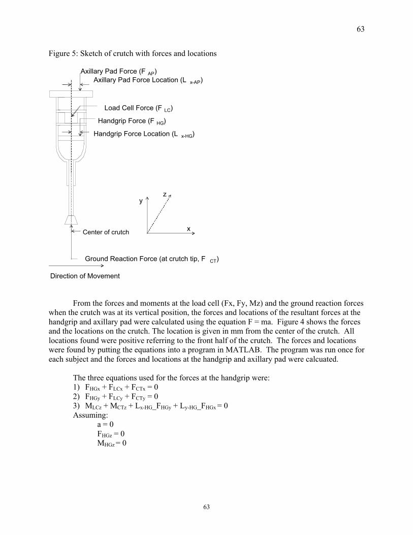
Figure 5: Sketch of crutch with forces and locations
Axillary Pad Force Location (L x-AP)
Direction of Movement
Handgrip Force Location (L x-HG)
Axillary Pad Force (F AP)
Handgrip Force (F HG)
Center of crutch x
yz
Ground Reaction Force (at crutch tip, F CT)
Load Cell Force (F LC)
From the forces and moments at the load cell (Fx, Fy, Mz) and the ground reaction forceswhen the crutch was at its vertical position, the forces and locations of the resultant forces at thehandgrip and axillary pad were calculated using the equation F = ma. Figure 4 shows the forcesand the locations on the crutch. The location is given in mm from the center of the crutch. Alllocations found were positive referring to the front half of the crutch. The forces and locationswere found by putting the equations into a program in MATLAB. The program was run once foreach subject and the forces and locations at the handgrip and axillary pad were calcuated.
The three equations used for the forces at the handgrip were:1) FHGx + FLCx + FCTx = 02) FHGy + FLCy + FCTy = 03) MLCz + MCTz + Lx-HG_FHGy + Ly-HG_FHGx = 0Assuming:
a = 0FHGz = 0MHGz = 0

64
64
The three equations used for the forces at the axillary pad were:1) FAPx + FLCx = 02) FAPy + FLCy = 03) MLCz + Lx-AP_FAPy + Ly-AP_FApx = 0Assuming:
a = 0FAPz = 0MAPz = 0
A two-way Analysis of Variance (ANOVA) with repeated measures was used to analyzethe effects of handgrip (standard and modified) and axillary pad (standard and modified). Ap-value of less than 0.05 was considered significant. The factors were the axillary pad and thehandgrip. The levels for each were standard and modified.
Results
The only significant difference found was in the locations of the force for the handgripdesigns. The forces on the handgrip and the forces and locations on the axillary pad were notsignificant.
Figure 6: Forces on the standard and modified handgripsHandgrip Forces
0
50
100
150
200
250
300
350
400
Standard Modified
Handgrip Configuration
Han
dg
rip
Fo
rce
(N)
No significant difference
There was no significant difference found in the forces on the standard and modified handgripdesigns (p =0.471).

65
65
Figure 7: Force Locations for the standard and modified handgripsHandgrip Force Locations
0
10
20
30
40
50
60
70
80
90
100
110
Standard Modified
Handgrip Configuration
Han
dg
rip
Fo
rce
Lo
cati
on
(m
m) p = 0.020
There was a signficant difference found in the force location on the standard and modifiedhandgrip designs (p = 0.020). The average standard handgrip force location was 52.9mm fromthe center of the crutch. The average modified handgrip force location was 27.9mm from thecenter of the crutch.
Figure 8: Forces on the standard and modified axillary pads
Axillary Pad Forces
0
10
20
30
40
50
60
70
80
90
100
110
Standard Modified
Axillary Pad Configuration
Axi
llary
Pad
Fo
rce
(N)
No significant difference
There was no significant difference found in the forces on the standard and modified axillary paddesigns (p= 0.434).

66
66
Figure 9: Force Locations for the standard and modified handgrips
Axillary Pad Force Locations
0
10
20
30
40
50
60
70
80
Standard Modified
Axillary Pad Configuration
Axi
llary
Pad
Fo
rce
Lo
cati
on
(m
m) No significant difference
There was no significant difference found in the force location on the standard and modifiedaxillary pad designs (p= 0.699).
The subjects were asked to rate the comfort level of the each of the crutch configurations.Five of the eight subjects preferred both the standard handgrip and axillary pad. The main reasoncited for preferring the standard handgrip over the modified handgrip was that the modifiedhandgrip was larger and harder to grip.
Discussion
The point of application of the resultant handgrip force is closer to the center of thehandgrip for the modified design than for the standard design. Since the force location is closerto the center of the crutch, this suggests that the body weight is distributed more evenly on thehand as predicted. For the standard design, the resultant force is applied much more to the frontof the hand. This suggests that the forces are not distributed evenly in this configuration. Forlong-term crutch users, using this modified design for the handgrip may help to lower the jarringforces on the hand by the crutch handle. The angled handgrip design can potentially providemore comfort to crutch users by distributing the forces along the hand instead of concentratingthe forces on only the front of the hand.
Five of the eight subjects preferred both the standard handgrip and axillary pad designs.However, since the main reason given for preferring the standard handgrip was that it wassmaller and easier to hold, the angled shape of the modified handgrip may not have been takenmuch into account. The study aimed to compare the shapes of the two handgrips, but thecomfort to the subjects may have relied more heavily on the size rather than the shape.
The sample size of eight people that was used for the study was very limited and was notable to be used to make generalizations for all crutch users. The subjects who participated in the

67
67
study are all from the State College, PA area and therefore are not representative of the wholepopulation. The demographics of State College may not be similar to the demographics of thewhole population. If more subjects had been used, then perhaps more significant difference mayhave been detected in the forces at the handgrip and forces and locations at the axillary pad.However, the differences found were so slight that hundreds of subjects may have been neededto detect any real significant difference.
Additionally, the subjects that were used were healthy college-age people. The data forthe subjects may not be representative of actual crutch users because the subjects in the study didnot have any problems with ambulation. The swing-through portion of the gait cycle of thesubjects will be considerably different from the swing-through portion of many crutch usersbecause many crutch users wear immobilizing leg casts. Those crutch users need extra room forthe straightened leg to clear the ground. Two of the participants were experienced crutch users sotheir ambulation with the crutches was likely more efficient than the rest of the participants whodid not have as much practice with crutches. Some of the data may not have been representativeof what would have been found by using experienced or long-term crutch users.
The leading cause of non-traumatic lower-leg amputation is diabetes mellitus (Mathur &Shiel, 2004). According to Mathur & Shiel (2004), recent information estimates that 13 millionpeople in the United States have diabetes. Many diabetics are overweight or obese and thereforecrutch dynamics for those individuals may be different than the crutch dynamics of healthysubjects. None of the subjects used in the study were obese and therefore they may not havetested these crutch biomechanics.
In future studies, the modified designs for both the standard and modified handgrips andaxillary pads should be made from the same material and should be the same size whencomparing the shapes alone. Also, using only experienced crutch users or giving inexperiencedcrutch users more practice time would help to make sure that all the subjects were walking asefficiently as possible. The energy expenditure of the subjects using each of the crutchconfigurations was not measured in this study. However, the extra energy required for crutchambulation is a large hindrance to many crutch users. It is one of the main reasons why manydisabled people choose wheeled mobility instead of using crutches. Comparing the energyexpenditure between the four combinations would be another good indication of the efficiency ofthe designs. Also, further modifications besides simply changing the handgrip and axillary padswould be good to explore. For example, adding a spring to the bottom of the crutch in hopes toabsorb some of the shock might also produce more significant results in the future. Also, testingdifferent handgrip and axillary pad shapes than those used in this study would also be helpful.
While this study only found significance in the location of the handgrip forces, thatinformation shows promise in reducing the jarring pressure at the hand. The information found inthis study provides groundwork for future studies in the area of crutch dynamics.

68
68
References
Basford, J.R., Rhetta, M.L., & Schleusner, M.P. (1990). Clinical Evaluation of the RockerBottom Crutch. Orthopedics, 13 (4), 457-450.
Dounis, E., Rose, G.K., Wilson, R.S.E., & Steventon, R.D. (1980). A Comparison of Efficiencyof Three Types of Crutches Using Oxygen Consumption. Rheumatology andRehabilitation, 19, 252-255.
Dounis, E., Steventon, R.D., & Wilson, R.S.E. (1980). The use of portable oxygen consumptionmeter (Oxylog) for assessing the efficiency of crutch walking. Journal of MedicalEngineering and Technology, 4(6), 296-298.
Feldman, D.R., Vujic, I., McKay, D., Callcott, F., & Uflacker, R. (1995). Crutch-InducedAxillary Artery Injury. Cardiovasc Intervent Radiol, 18, 295-299.
Fisher, S.V., & Patterson, R.P. (1981). Energy Cost of Ambulation with Crutches. Archives ofPhysical Medicine & Rehabilitation, 62, 250-256.
Goh, J.C.H., Toh, S.L., & Bose K. Biomechanical study on axillary crutches during single-legswing-through gait. Prosthetics and Orthotics International, 10, 89-95.
Hall, J., Elvins, D.M., Burke, S.J., Ring, E.F. J., & Clarke, A.K. (1991). Heart rate evaluation ofaxillary and elbow crutches. Journal of Medical Engineering and Technology, 15(6),232-238.
Hall, R. (1918). A Rolling Crutch. J.A.M.A., 70, 666-668
Hinton, C.A., Cullen, K.E. (1982). Energy Expenditure During Ambulation with Ortho Crutchesand Axillary Crutches. Physical Therapy, 62(6), 813-819.
Kang, Y.C., Choi, E.H., Hwang, S.m., Lee, W.S., Lee, S.H., & Ahn, S.K. (1999). AcneMechanica Due to an Orthopedic Crutch. Cutis, 64, 97-98.
LeBlanc, M.A., Carlson, L.E., & Nauenberg, T. (1993). A Quantitative Comparison of FourExperimental Axillary Crutches. Journal of Prosthetics and Orthotics, 5(1), 20-28.
Li, S., Armstrong, C.W., & Cipriani, D. (2001). Three-Point Gait Crutch Walking: Variability inGround Reaction Force During Weight Bearing. Archives of Physical Medicine &Rehabilitation, 82, 86-92.
Mathur, R., & Shiel, W.C. Jr. (June 2004). Diabetes – David Meets Goliath. Retrieved June 15,2004, from http://www.medicinenet.com/script/main/art.asp?articlekey=17281

69
69
Nielsen, D.H., Harris, J.M., Minton, Y.M., Motley, N.S., Rowley, J.L, & Wadsworth, C.T.(1990). Energy Cost, Exercise Intensity, and Gait Efficiency of Standard Versus Rocker-Bottom Axillary Crutch Walking. Physical Therapy, 70(8), 487-493.
Nova, L.C., & Laura, P.A.A. Design and Use of Improved Walking Aids. (1985). Journal ofBiomedical Engineering, 7, 329-333.
Pariziale, J., & Daniels J. (1989). The mechanical performance of ambulation using spring-loaded axillary crutches. American Journal of Physical Medicine and Rehabilitation,68(4),193-195.
Park, H.W., Malone, E.W., & Steglich, R. (1952). The Tilted Crutch Handpiece. Archives ofPhysical Medicine, 731-733.
Poddar, S.B., Gitelis, S., Heydemann, P.T., & Piasecki, P. (1993). Bilateral predominant radial nerve crutch palsy. Clinical Orthopaedics and Related Research. A Case Report, 297,
245-6.
Powers, W.R., & Flatt A.E. (1962). Modified Crutch Handle. Archives of Physical Medicine &Rehabilitation, 43, 570-573.
Raikin, S., & Froimson, M. (1997). Bilateral Brachial Plexus Compressive Neuropathy (CrutchPalsy). Journal of Orthopedic Trauma, 11(2), 136-137.
Sankarankutty, M., Stallard, J., & Rose, G.K. (1979). The Relative Efficiency of ‘SwingThrough’ Gait on Axillary, Elbow and Canadian Crutches Compared to Normal Walking.Journal of Biomedical Engineering, 1(1), 55-57.
Shortell, D., Kucer, J., Neeley, W.L., & LeBlanc, M. (2001). The design of a compliantcomposite crutch. Journal of Rehabilitation Research and Development, 38(1), 23-32.
Shoup, T.E., Fletcher, L.S., & Merrill, B.R. (1974). Biomechanics of Crutch Locomotion.Journal of Biomechanics, 7, 11-19.
Stallard, J., Dounis E., Major, R.E., & Rose, G.K. (1980). One Leg Swing Through Gait UsingTwo Crutches. Acta orthop. Scand., 51, 71-77.
Stallard, J., Sankarankutty, M., & Rose, G.K. (1978). A Comparison of Axillary, Elbow, andCanadian Crutches. Rheumatology and Rehabilitation, 17, 237-239.
Stallard, J., Sankarankutty, M., & Rose, G.K. (1978). Lower-limb vertical ground-reaction forcesduring crutch walking. Journal of Medical Engineering and Technology, 2(4), 201-202.
Subramony, S.H. (1989). Electrophysiological findings in crutch palsy. Electromyogr. clin.Neurophysiol., 29(5), 281-285.

70
70
Thomas, J.M., & Deshmukh, N. (1973). Aneurysm of the Brachial Artery with CompleteThrombosis Caused by a Crutch. The American Surgeon, 389-399.
Wagstaff, P.S. (1984). The Energetics of Walking using Axillary Crutches and the Prototype of aNew Design Termed the Dublin Crutch. Physiotherapy, 70(11), 422-424.
Wiley, B.C. (1960). Crutch with Sloping Handgrip. J.A.M.A., 172(7), 694-695.
Wilson, J.F., & Gilbert, J.A. (1982). Dynamic Body Forces on Axillary Crutch Walkers DuringSwing-Through Gait. American Journal of Physical Medicine, 61(2), 85-92.
Yeakel, M.H., & Margetis, P.M. (1969). A Customized Crutch Hand Grip. The AmericanJournal of Occupational Therapy, 23(5), 410-412.