Embed Size (px)
Citation preview

Essential Biomaterials Science: Professor David Williams
Biomaterials in Regenerative Medicine v Basic concepts in regenerative medicine and
the essential tissue engineering paradigm v Cells for tissue engineering v Biomolecules and nutrients
v Biomaterials and biofabrication v The clinical reality of tissue engineering v Tissue engineering and drug testing

Basic concepts in regenera.ve medicine and the essen.al
.ssue engineering paradigm
Tissue Engineering
Tissue engineering is the crea.on of new .ssue for the therapeu.c reconstruc.on of the human body, by the deliberate and controlled s.mula.on of selected target cells through a systema.c combina.on of molecular and mechanical signals
Williams D.F. To engineer is to create, Trends
in Biotechnology, 2006, 24, 4-‐8

Regenerative MedicineAlternative therapies to treat disease and injury by the regeneration of functional tissue / organ structures instead of replacement by medical devices or palliative care through pharmaceuticals
Tissue EngineeringThe creation of new tissue for the therapeutic reconstruction of the human body, by the deliberate and controlled stimulation of selected target cells through a systematic combination of molecular and mechanical signals
Gene Therapy
The technique for correcting defective g e n e s r e s p o n s i b l e f o r d i s e a s e development. In which copies of a gene is inserted into living cells to replace abnormal, disease-causing gene.
Cell TherapyThe process of introducing new cells into a tissue in order to treat a disease.
A
B C
May be combined

The Essence of Conventional Tissue Engineering
THE STARTING POINT The creation of new tissue for the therapeutic reconstruction of the human body, by the deliberate and controlled stimulation of
selected target cells through a systematic combination of molecular and mechanical signals
STEP ONE Obtain suitable cells, either stem cells or fully differentiated cells.
Manipulate cells to optimize cell number and phenotype
STEP TWO Prepare suitable biomaterial support
Arrange appropriate morphology and architecture of support Arrange appropriate surface characteristics
Seed cells into biomaterial support
STEP THREE Prepare suitable culture medium
Arrange appropriate growth factors and other molecular signaling agents Arrange appropriate bioreactor conditions
STEP FOUR
Incorporate tissue engineered construct into tissues of patient

Tissue Engineering Templates There will need to be control over the volume and, in many cases, shape, of the
region in which the cells are operating. This may simply be to confine the activity to the region of the lesion or target tissue or to give shape to the tissue that is being generated.
There may need to be careful control of the mechanical signaling to the cells, which is unlikely to happen in most situations where they are simply injected into tissue.
Cell-to-cell contact and signaling may be required in order to optimize their performance, again not readily achieved following free injection.
Under ex vivo conditions, the supply of nutrients, including oxygen, needs to be regulated,
Molecular signaling through growth factors, or alteration of cell phenotype through gene transfer may be required, and this may need spatiotemporal control,
In cases of complex tissue regeneration, more than one cell type is required for the expression of different types of ECM and this will definitely need spatiotemporal control; this becomes even more complex when we consider whole organ printing,

TARGET CELL(S)
REGENERATED TISSUE, FUNCTIONALLY INCORPORATED INTO HOST
Sourced (autologous, allogeneic,)
Selected
Expanded
Differentiated
Seeded into template
Cultured in ex vivo bioreactor
Test
Implant into host
Cell-free template implantedinto host
Resident cells recruited into template
Tissue generation takesplace in vivo
Ex V
ivo
Bior
eact
or P
arad
igm
Hum
an Body Bioreactor Paradigm

Cell Sources q Embryonic stem cells
q Induced pluripotent stem cells q Adult stem cells
q Hematopoietic stem cells q Mesenchymal stem cells
q Adipose-tissue derived stem cells q Amniotic fluid-derived stem cells
q Tooth derived stem cells q Peripheral blood stem cells q Autologous mature cells

Fully Differentiated Cells Tissue / Organ Stem / Progenitor Cells
Skeletal myoblasts
Adult chondrocytes
Fetal / neonatal chondrocytes
Valvular interstitial cells
Urothelial cells
Hepatocytes
Epithelial cells
Fibroblasts
Keratinocytes
Myocardium
Bone
Cartilage
Skeletal muscle
Heart valve
Blood vessel endothelium
Blood vessel SMCs
Bladder / urinary tract
Peripheral nerve
Brain
Liver
Skin
Trachea
Embryonic SCs
Cardiac PCs
Endothelial PCs
Peripheral blood SCs
Umbilical cord /Placenta derived SCs
Amniotic fluid SCs
Adipose SCs
Bone Marrow MSCs
Neural SCs / PCs IPS cells

Factors used in the forma.on of Induced Pluripotent Stem Cells in Humans
Cell Factors Fibroblast OKSM
OSLN Peripheral blood cells OKSM Cord blood endothelial cells OSLN Adipose-‐derived stem cells OKS Hepatocytes OKSM Kera.nocytes OKSM Neural stem cells O Amnio.c fluid derived stem cells OSN O Oct 4; K Klf4 S Sox2 M c-‐myc L Lin28 N Nnanog

Cell Sources ; Issues
Teratoma formation with embryonic stem cells Immunogenicity of allogeneic / xenogeneic cells Expansion and proliferation of mature adult cells
Regulatory issues Product consistency

Biomolecules and nutrients The culture medium used in in vitro / ex vivo tissue engineering is based on relatively standard
solutions with some specific additions depending on individual requirements. Culture media are either simple, complex or chemically defined. Simple, or balanced salt solutions are ionic buffers with energy and protein additions. Complex media are supplemented with amino acids, vitamins and some other nutrients. Chemically-defined media, or serum-free media, is comprised solely of
biochemically-defined low molecular weight constituents, entirely free of animal-derived substances.
Inorganic salts usually include NaCl (6.5-7.5 g/L), NaHCO3 (1.2-3.8 g/L), KCl (0.4 g/L), NaH2PO4
(0.1-0.8 g/L) and MgSO4 (<0.1 g/L).
Amino acids usually include L-Alanine (0.009 g/L), L-Arginine (0.1-0.2 g/L), L-Asparagine (0.01-0.05 g/L), L-Cystine, (0.02-0.06 g/L), L-Glutamic Acid (0.01-0.02 g/L), L-Glutamine (0.1-0.6 g/L), Glycine (0.01-0.03 g/L), L-Histidine (0.01-0.04 g/L), L-Isoleucine (0.003-0.1 g/L), L-Leucine (0.01-0.1 g/L),
L-Lysine (0.03-0.15 g/L), L-Methionine (0.005-0.03 g/L), L-Phenylalanine (0.005-0.07g/L), L-Prolene (0.01-0.04 g/L), L-Serine (0.01-0.04 g/L), L-Trytophan (0.005-0.02 g/L) and L-Tyrosine (0.002-0.05
g/L).
Vitamins frequently include Folic acid (0.001 g/L), Niacinamide (0.001 g/L), D-Pantothenic acid (0.001 g/L), Riboflavin (0.0001 g/L), Thiamin (0.001 g/L), Vitamin B-

Biomolecules and nutrients
Growth factor Tissues Angiopoietin 1 & 2 (Ang-1 and Ang-2) Blood vessels, heart Fibroblast growth factor 2 (FGF-2) Blood vessels, bone, skin, nerve, muscle
Bone morphogenetic protein 2 (BMP-2) Bone, cartilage Bone morphogenetic protein 7 (BMP-7) Cartilage, kidney
Epidermal growth factor (EGF) Skin, nerve Hepatocyte growth factor (HGF) Bone, liver, muscle
Insulin-like growth factor 1 (IGF-1) Bone, cartilage, lung, kidney, nerve, skin Nerve growth factor (NGF) Nerve, brain
Platelet derived growth factor (PGDF) Blood vessels, muscle, skin, cartilage Transforming growth factor α (TGFα) Brain, skin
Transforming growth factor β (TGFβ Bone, cartilage Vascular endothelial growth factor (VEGF) Blood vessels
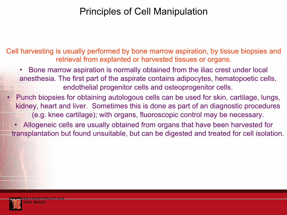
Principles of Cell Manipulation
Cell harvesting is usually performed by bone marrow aspiration, by tissue biopsies and retrieval from explanted or harvested tissues or organs.
• Bone marrow aspiration is normally obtained from the iliac crest under local anesthesia. The first part of the aspirate contains adipocytes, hematopoetic cells,
endothelial progenitor cells and osteoprogenitor cells. • Punch biopsies for obtaining autologous cells can be used for skin, cartilage, lungs,
kidney, heart and liver. Sometimes this is done as part of an diagnostic procedures (e.g. knee cartilage); with organs, fluoroscopic control may be necessary.
• Allogeneic cells are usually obtained from organs that have been harvested for transplantation but found unsuitable, but can be digested and treated for cell isolation.

Principles of Cell Manipulation
Cell selection is usually necessary because harvested cells will consist of several different types. There are several ways of selecting and separating cells.
• Density gradient centrifugation is often performed with Percoll medium, which consists of PVP-coated colloidal silica particles of 15-30 nm diameter (23% w/w in
water). Cells are loaded onto the gradient and centrifuged, when they are separated either on the basis of size or density.
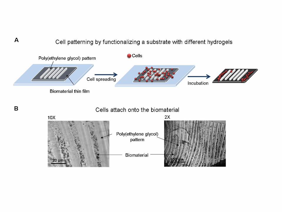
• Selective adhesion is based on the difference in adhesion properties of cells on surfaces such as fibronectin-coated plastics,
• Antibody techniques use the detection of antigens on cell surfaces by selective antibodies, which are linked to a fluorescent dye and separated by fluorescence
activated cell sorting (FACS).

Principles of Cell Manipulation Cell expansion is required in order to achieve the appropriate number of cells
for tissue engineering processes. • Some cells may be expanded in suspension culture, using a semi-solid
medium such as agar or collagen where they proliferate under the right media conditions,
• Anchorage dependent cells may be expanded in monolayer culture, where they adhere to the substrate, which could be conventional tissue culture
plastic, or a surface coated with laminin, fibronectin or collagen, where they proliferate.
• This process may be enhanced by using microcarriers to increase the surface area,
• The process may also be enhanced by using feeder cells, typically mouse embryonic fibroblasts, which have several supportive mechanisms for cell
proliferation. Cell differentiation very much depends on the cell system in question.
Mechanical signaling may be the most important process, • .

Tissue Engineering Templates
In order to achieve spatio-temporal control, we need biomaterials that can be formed into a suitable template which:
• May be pre-formed or injectable / in situ setting or gelling • Has the appropriate mechanical and biophysical properties • Can incorporate and release of necessary specific biomolecules • Degrades with the appropriate kinetics and lack of toxicity • Has the appropriate biocompatibility characteristics. But The biomaterial alone cannot perform all of the required functions, it needs
to be in a form that has the appropriate architecture and morphology, and needs to convey and deliver relevant biomolecules.
A Tissue Engineering Template comprises: BIOMATERIAL + ARCHITECTURE + BIOMOLECULES
We have no procedures that are able to assess these complex templates

Tissue Engineering Templates
Tissue engineering involves cells that are stimulated to generate new tissue by a combination of molecular and mechanical signals. Biomaterials are used to
facilitate the delivery of these signals.
Conventionally the material structures have been described as scaffolds, but they should be more than mechanical supports for the target cells.
The biomaterial component is better considered as a template, which incorporates
concepts of shape and volume, the chemistry and architecture necessary to support cell function, and the mechanical characteristics that provide both
mechanotransduction, elasticity and strength.
The template may be prefabricated for use in a bioreactor or for direct implantation, or may be injectable with appropriate cross-linking, gelation or self-assembly.
The template must be conducive to the control of nutrient, oxygen and
biomolecules supply to the cells and generating tissue.
The template should have appropriate degradation profiles, with the biocompatibility necessary to support the processes of tissue generation without
significant inflammation or immune responses.
The template should be conducive to incorporation of the generated tissue into the host, with vascularization and innervation.

Scaffolds, Matrices and Templates
Some Existing Scaffold Materials
• Synthetic biodegradable polymers • Synthetic non biodegradable polymers (? as hybrids) • Natural biopolymers (proteins, polysaccharides, silk) • Self assembled biological structures • Tissue derived structures (SIS) • Bioactive ceramics and glass ceramics • Composites, including nanocomposites • Multilayered structures • Temperature / pH responsive polymers (cell sheet engineering)

Specifications for Template Biomaterials Mandatory
• The material should be capable of recapitulating the architecture of the niche of the target cells,
• Moreover, since the cell niche is changeable over time, the material should be capable of adapting to the
constantly changing microenvironment, • The material should have elastic properties, particularly
stiffness, that favor mechanical signaling to the target cells in order to optimize differentiation, proliferation and
gene expression, • The material should have optimal surface or interfacial
energy characteristics to facilitate cell adhesion and function,

Specifications for Template Biomaterials Mandatory
• The material should be capable of orchestrating molecular signaling to the target cells, either by directing
endogenous molecules or delivering exogenous molecules,
• The material should be of a physical form that provides appropriate shape and size to the regenerated tissue,
• The material should be capable of forming into an architecture that optimizes cell, nutrient, gas and
biomolecule transport, either or both ex vivo or in vivo, and facilitates blood vessel and nerve development,
• The material should be intrinsically non-cytotoxic and non-immunogenic, and minimally pro-inflammatory,

Specifications for Template Biomaterials Optional
• The material should be degradable if that is desired, with
appropriate degradation kinetics and appropriate morphological and chemical degradation profiles,
• The material should be injectable if that is desired, with the appropriate rheological characteristics and
transformation mechanisms and kinetics, • Where necessary, the material should be compatible the processing techniques that simultaneously pattern both
the material and living cells,

Specifications for Template Biomaterials Optional
• Where multiple cell types are involved, the material
properties should be tunable in order to accommodate variable cellular requirements, with spatio-temporal
control as appropriate, • When used in a significantly stressed in vivo
environment, the material must have sufficient strength and toughness,
• In those situations where the biomaterial encapsulates cells, optimal diffusion characteristics concerning key
molecules is required.

Options for Template Biomaterials
Porous solids * Hybrid nano / micro-structured blends of synthetic
(PHB) / natural (silk-elastin) materials Hydrogels
*Engineered peptide hydrogels Decellularized ECM materials
NOTE; Standard biodegradable synthetic polymers
prepared by standard fabrication routes are unlikely to provide optimal tissue engineering templates and prior
FDA approval of materials used in medical devices is not an appropriate specification for a template

DISCARDED HUMAN KIDNEYS AS A SOURCE OF EXTRACELLULAR MARTIX SCAFFOLD FOR KIDNEY REGENERATION TECHNOLOGIES Dr. GIUSEPPE ORLANDO WAKE FOREST UNIVERSITY

A schematic of the NT3-chitosan tube, composed of an chitosan outer tube stuffed with NT3-coupled chitosan carriers.
Zhaoyang Yang et al. PNAS 2015;112:13354-13359
©2015 by National Academy of Sciences

Essential Biomaterials Science: Professor David Williams
Biomaterials in Drug and Gene Delivery Rationale for active molecule delivery u Non-oral delivery of conventional drugs u Drug release from monolithic devices u Micro- and nano-particulate systems
u Polymer therapeutics u Cancer; chemotherapy / immunotherapy
u Gene therapy and transfer u Vaccines
u Anti-microbial agents u Combination systems

Rationale for drug delivery systems GROUP A Conventional drugs given by conventional routes, such as medication given orally, or by
subcutaneous and intravenous injection or by topical application.
GROUP B Orally-delivered medication where tablets are modified to improve efficiency of delivery, such as pH sensitive coatings to control absorption in GI tract.
GROUP C Techniques to enhance non-oral delivery by increasing efficiency ot passage through
barriers, such as electrical charge or microneedles to assist transdermal delivery.
GROUP D Technologies to deliver drugs to specific tissue sites, such as infusion pumps.
GROUP E Devices that facilitate prolonged and sustained delivery such as depos placed in the eye and contraceptive devices.
GROUP F Systems that allow highly incompatible agents, such as toxic chemotherapeutic drugs, to ne
targeted to cells, for example by complexation with polymers.
GROUP G Systems with a high degree of cellular and sub-cellular targeting, for example with highly specific targeting of biomarkers on cells in immunotherapy.
GROUP H Combination products where drugs combined with devices improve their performance.
GROUP I Combined therapeutic and diagnostic systems.
GROUP J Delivery systems for ex vivo tissue engineering.

Vaccine delivery and nanoparticles
LIPOSOMES In clinical trials for delivery of vaccine against non-small cell lung cancer. Good
biodegradability and biocompatibility but quickly cleared and not good for water soluble proteins.
VIRUS LIKE PARTICLES One product marketed for vaccination against human papillomavirus, where the HPV
major caspid protein L1 self assembles as the virus-like nanoparticle. Some general safety concerns and only applicable to cancers caused by viruses.
PLGA
Good anti-tumor effects seen in mice. Good biocompatibility and biodegradation. Good manufacturing.
GOLD NANOPARTICLES May be coated with DNA, evaluated for vaccination against melanoma.
MAGNETITE
Stimulates tumor-specific T cell activity but non biodegradable. Has been in clinical trials for melanoma treatment
GELATIN Readily available, easily manufactured, with functional groups for immunoglobulin binding, active tumor targeting
and may be pegylated for better opsonization.
NANOEMULSIONS They can have long circulating times and good take-up by antigen presenting cells, possibly used for protection
against Hepatitis B, HIV and influenze by a mucosal route

Summary of viral vectors RETROVIRUSES
Viruses that can create double-stranded DNA copies of their RNA genomes, which can be integrated into the chromosomes of host cells. Human immunodeficiency virus (HIV) is a retrovirus. They usually only infect dividing cells, so that most applications are in ex vivo gene transfer. Direct administration to humans is usually ineffective. Some success has been achieved in the treatment of cancer, targeting
T cell receptors in melanoma to give complete tumor regression. The subset of lentiviruses can transduce non-dividing cells but there are safety issues
ADENOVIRUSES
Viruses with double-stranded DNA genomes that cause respiratory, intestinal, and eye infections in humans. Although they have a number of theoretical advantages, and can result in high levels of systemic gene transfer, results in humans are problematic, and they can result in severe levels of
toxicity at the levels required for clinical efficiency.
ADENO-ASSOCIATED VIRUSES Small, single-stranded DNA viruses that can insert their genetic material at a specific site on
chromosome 19. These are easily prepared and purified but are very limited because of the small insertion site.
HERPES SIMPLEX VIRUSES
Double-stranded DNA viruses that infect neurons. Herpes simplex virus type 1 is a common human pathogen that causes cold sores. They have the advantage of being able to infect a very wide range
of cells, both dividing and non-dividing. They do have significant immunogenicity and toxicity problems. It is possible to engineer these viruses such that they can specifically infect tumor cells –
giving oncolytic HSVs- which have been used in clinical trials for recurrent breast cancer, gliomas and liver metastases from colon cancer.

Mouth
Stomach
Esophagus
Small Intestine
Large Intestine
Rectum
Saliva, Minimum pH, 5.6 – 6.2 Maximum pH, 7.4 - 7.9
Resting pH, 4 – 5After ingestion, pH 1 - 2
Duodenum pH, 6Ileum pH, 8
pH range, 5.5 - 7

Drug reservoir
Drug reservoir
Impermeable backing Rate controlling membrane
Release liner
Release liner
Adhesive

v v
v v
v
v
v v
v v v
v v
v
v
v
v
Water
Diffusion Erosion Osmosis
Waterabsorption
Drugrelease
Drug release as surface erodes
Polymer degrades afterdrug release
Drug release

Polymeric drug
Polymer-proteinconjugate
Polymer-DNAComplex
Polymer-drugconjugate
Polymeric micelle
Polymer Protein
Polymer DNA
Drug Polymer Linker
Drug Hydrophilic block
Hydrophobic block

G1 S
G2M
Gap 1 or growth phase, high biosynthetic activity,Preparation for chromosome replication
S phase, with DNA synthesis
Gap 2, Preparation for mitosis
Mitosis, cell division
Alkylating agents, impair cell activity by covalent attachment to proteins and DNAe.g. Cisplatin, ifosfamide, cyclophosphamide
Antimetabolites, substitute formetabolites normally incorporatedinto DNA and RNAe.g. Methotrexate, 5-fluorouracil,gemcitabine
Anti-tumor antibiotics, intercalate DNA at specific sequences and cause strand breakage, e.g. doxorubicin, anthracycline
Taxoids, interfere with disassembly of microtubules, inhibiting cell function,e.g. docetaxel, paclitaxel
Vinca alkaloids, bind to tubuline.g. vincristine

Lipophilic carriers DOPE
DOTAP
DSPE
Polyca.onic carriers
Linear PEI Chitosan
Branched PEI
PAMAM PLL
Cyclodextrin
PLL-‐lipid
PEO-‐b-‐P(CL-‐g-‐SP) (Spermine graPed PEO-‐b-‐PCL)
PEI-‐PEG

Essential Biomaterials Science: Professor David Williams
Biomaterials in Imaging Systems Molecular imaging and biomaterial-based
contrast agents • Magnetic resonance imaging
• CT imaging • PET and SPECT imaging
• Ultrasound imaging • Multi-modal imaging
• Theranostics • Biosensors

Magnetic Resonance Imaging Targets: Anatomical, physiological and molecular information
Main imaging agents: Gadolinium, Iron oxides, Manganese oxide Spatial resolution: 10-100 µm
Advantages: No radiation, no depth limit, good soft tissue contrast, quantitative Disadvantages: Expensive, long acquisition time, limited sensitivity
Computed Tomography
Targets: Anatomical and physiological information Main imaging agents: Iodine
Spatial resolution: 50 µm Advantages: No depth limit, high resolution, short acquisition time, quantitative Disadvantages: Moderately expensive, some radiation, poor soft tissue contrast
Positive Emission Tomography
Targets: Physiological and molecular information Main imaging agents: 18F and 11C labeled compounds
Spatial resolution: 1 – 2 mm Advantages: No depth limit, quantitative, high sensitivity
Disadvantages: Expensive, long acquisition time, limited sensitivity
Ultrasound Targets: Physiological and anatomical information
Main imaging agents: Microbubbles Spatial resolution: 50 µm
Advantages: Low cost and ease of use, quantitative, high resolution Disadvantages: Limited imaging depth, confined to vasculature
Optical Imaging
Targets: Physiological and molecular information Main imaging agents: Fluorophores
Spatial resolution: 1-5 mm Advantages: High sensitivity, no radiation
Disadvantages: Limited imaging depth, whole body imaging not possible
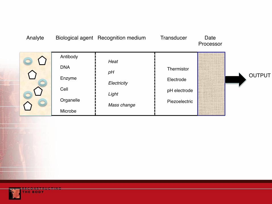
Analyte Biological agent Recognition medium Transducer Date Processor
OUTPUT
Antibody
DNA
Enzyme
Cell
Organelle
Microbe
Heat
pH
Electricity
Light
Mass change
Thermistor
Electrode
pH electrode
Piezoelectric