Embed Size (px)
Citation preview

Julia E. BabenseeWallace H. Coulter Department of Biomedical Engineering,
Georgia Institute of Technology and Emory University, Atlanta, GA
Biomaterial/GlycanCell Interactions

Dendritic Cell (DC) and Immunity
Medzhitov R., Nature Rev Immunology (2001)
DCs BridgeInnate and Adaptive Immunity
DCs Maintain ImmunologicalHomeostasis
http://www.dkfz.de/en/immungenetik/Heiko_group/Heiko-2.jpg
Key roles in:Maintaining self toleranceImmunity to pathogensCancerAutoimmunity

P.M. Kou
Distinct DC PhenotypesandImmunologicalOutcomes

Biomaterial Immunomodulatory Effect
M. M. Matzelle & J.E. Babensee, Biomaterials 25:295-304 (2004).
1
10
100
1000
10000
100000
1000000
10000000
2 3 4 8 12 18
Time (weeks)[A
nti
-OV
A I
gG
] (n
g/m
L)
PBS CFA 75/25 PLGA MP 75/25 PLGA SC
++
+ + ++
*
+
*
+
*
+*
+
*
+
*
+
*
+
*
+
*
+
*
+
*
+*
+* *
* * * *
an
ti-O
VA
Ig
G(n
g/m
l)time (weeks)
2 3 4 8 12 18
1
10
100
1000
10000
100000
1000000
10000000
2 3 4 8 12 18
Time (weeks)[A
nti
-OV
A I
gG
] (n
g/m
L)
PBS CFA 75/25 PLGA MP 75/25 PLGA SC
++
+ + ++
*
+
*
+
*
+*
+
*
+
*
+
*
+
*
+
*
+
*
+
*
+*
+* *
* * * *
an
ti-O
VA
Ig
G(n
g/m
l)time (weeks)
2 3 4 8 12 18
PBS CFA 75/25 PLGA MP 75/25 PLGA SCPBSPBS CFACFA 75/25 PLGA MP75/25 PLGA MP 75/25 PLGA SC
Biomaterial Immunomodulatory Effect
Combination Product
Activated APCsMφ, dendritic cells
Activated T cells
Adaptive Immunity
(Immune Response)
Innate Immunity
(Inflammation)
biomaterialcomponent
biologicalcomponent
PLGA Adjuvant Effect

Prime Mechanism By Which AdjuvantsEnhance Immune Responses
Maturation Of Dendritic Cells (DCs)

Dendritic Cells InfiltratePLGA Scaffolds
Plain PLGA PLGA with Adsorbed OVA
A. Paranjpe
Anti-DEC-205 labelingDay 7Original Magnification, 40X

Derivation of DCs forBiomaterial Treatment
1. Flow cytometry forexpression of surfacemarkers
2. Cytokine quantification3. Allostimulatory MLR4. Morphology5. Viability
Day 0 Day 5 Day 6
Human peripheralblood mononuclear cells
Biomaterial or controltreatment

DC Phenotype UponBiomaterial Contact
CD86
Agarose
Hyaluronic Acid
Alginate
Chitosan
PLGA
mDC
iDC
Supports DC maturation
Does not supportDC maturation
Supports DC Maturation
InhibitsDC maturation
Does not supportDC maturation
Fold increases over the immature DCs are shown to compare between different donors.★: p < 0.05, statistically higher than immature DC (=1); : p < 0.05, statistically lower than immature DC (=1); Bracket: p < 0.05, statistically different between two biomaterial treatments;‘┴’ indicates ‘or’.mean±SD, n=6 donors
Original magnification: 40×.
J. Park

Cytokine Profiles forBiomaterial-Treated DCs
(a) Pro-inflammatory cytokines(b) Chemokines(c) Anti-inflammatory cytokines
Proteins released into supernatanthave been measured using Bio-Pleximmunoassay (mean±SD, n=6 donors).
(a) (b)
(c)
J. Park

Red – Isotype Green – Foxp3
CD4+CD25+Quadrantonly
Untreated MNCs
MNCstreated with TGF-beta
iDC
mDC
PLGA
Agarose
CD4
CD25
J. Park
Autologous T cell Polarization by Biomaterial-Treated DCs

No
T ce
llC
o-cu
lture
with
T c
ells
Co-culture with Autologous CD3+ T cells
PLGAmDC iDC Chitosan Alginate HA Agarose
DCs collected
Inducer of Th1 (IFN-γ)with or without antigens
Inducer of Th1(IL-12p70) &Th2 (IL-10)withoutantigens
Inducer of Th1(IL-12p70) &Th2 (IL-10) withantigens
Inducer of IL-12p70without antigens
Inducer of IL-10with or withoutantigens
Inducer of IL-10with antigens
Inducer of IL-12p70with antigens
CD4+CD25+,Foxp3+Inducedwith antigentreatment
CD4+Inhibited with antigentreatment
Autologous T Cell Phenotype And Polarization By Biomaterial-Treated DCs
J. Park
Foxp3+

In Vivo Biomaterial Adjuvant EffectCorrelates with DC Response In Vitro
n = 6-7. Data are shown as replicates with mean represented as a line.
* p<0.05. ND = none detected
PB
S+
OV
AA
ga
ros
e O
VA
SC
PL
GA
OV
A S
C
CF
A+
OV
A
PB
S+
OV
AA
ga
ros
e O
VA
SC
PL
GA
OV
A S
C
CF
A+
OV
A
PB
S+
OV
AA
ga
ros
e O
VA
SC
PL
GA
OV
A S
C
CF
A+
OV
A
PB
S+
OV
AA
ga
ros
e O
VA
SC
PL
GA
OV
A S
C
CF
A+
OV
A
PB
S+
OV
AA
ga
ros
e O
VA
SC
PL
GA
OV
A S
C
CF
A+
OV
A
1
10
100
1000
10000
100000
2 Weeks 3 Weeks 4 Weeks 8 Weeks 12 Weeks
* *
* *
*
Treatment Group
An
ti-O
VA
Ig
G1
Co
ncen
trati
on
(n
g/m
l)
NDND
Norton, L., Park, J., Babensee, J.E., J. Control. Rel., in press

Babensee, J.E. and Paranjpe A., JBMR (2005). Bennewitz, N.L. and Babensee J.E., Biomaterials (2005).
Agarose PLGAChitosan
Hyaluronic acid
Biomaterial Adjuvant Effect
• Biomaterials induced differential levels of DCmaturation
• PLGA, but not agarose, enhanced humoralresponse to a model antigen (ovalbumin) in mice
• Control biomaterial systems are needed tocorrelate DC response to biomaterial properties
matureimmature

DC Recognition of Biomaterials
P. Kou
Role of CLRs in Immunity and Tolerance
S. J. van Vliet, J. J. Garcia-Vallejo, Y. van Kooyk, Immunology and Cell Biology (2008).

C-Type Lectin Receptors (CLRs)
T.B.H. Geijtenbeek and S.I. Gringhuis, Nature Rev Immunol (2009).

CLRs and Homeostatic and Pathologic Functions
J.J. Garcia-Vallejo, Y. van Kooyk, ImmunologicalReviews (2009).

CLRs, Recognition Sites and Signaling
• Classified as mannose or galactose binding• Recognize pathogen-specific glycans and endogenous
glycans (e.g. mediate cell interactions, tumor immuneevasion, autoimmunity)
• Differential expression on cells (e.g. DC subsets) allowfor specific targeting with then different types of immuneresponses induced
• Immuno-stimulatory and immuno-inhibitory• Some CLRs with signaling ability due to motifs• Collaboration (+ve and –ve) with other receptors eg.
TLRs

Hypothesis• Glycans and glycoproteins have functional
immuno-stimulatory or -inhibitory effects on DCs– The form, context, and/or density of glycan presentation
will affect DC phenotype– Glycan moieties from serum glycoproteins can
functionally modulate DC response– Glycan modification of biomaterials have the ability to
modulate the in vivo host response to a material orvaccine delivery vehicle

Differential carbohydrate profileson SAMs of Alkanethiols
X axis: carbohydrate ligand (lectin probe)
10% serum
Y ax
is: a
bsor
banc
e at
405
nm
0
0.5
1
1.5
2
2.5
3
3.5
4
Mannose (NPA) Sialyl (SNA-1) GlcNAc (UEA-II) mannose alpha(1,3)
and alpha(1,6)
mannose, D-
mannose (HHA)
CH3
OH
COOH
NH2
Mannose Family Galactose family
All SAM > CH3
NH2 > All SAM
OH > CH3 SAM
0
0.05
0.1
0.15
0.2
_-galactose(AIA) GalNAc (BPA) _-galactose(PNA)
CH3
OH
COOH
NH2
COOH >CH3 SAM
COOH >NH2 SAM
mean±SD, n=6 trials
S. Shankar, I.I. Chen, B. Keselowsky, A.J. Garcia, J.E. Babensee JBMR (2010).
N.D.
Avidin/alkaline phosphatase
Plant Lectin Probe
Biotin conjugate
CarbohydrateAu/Ti surface with alkanethiol
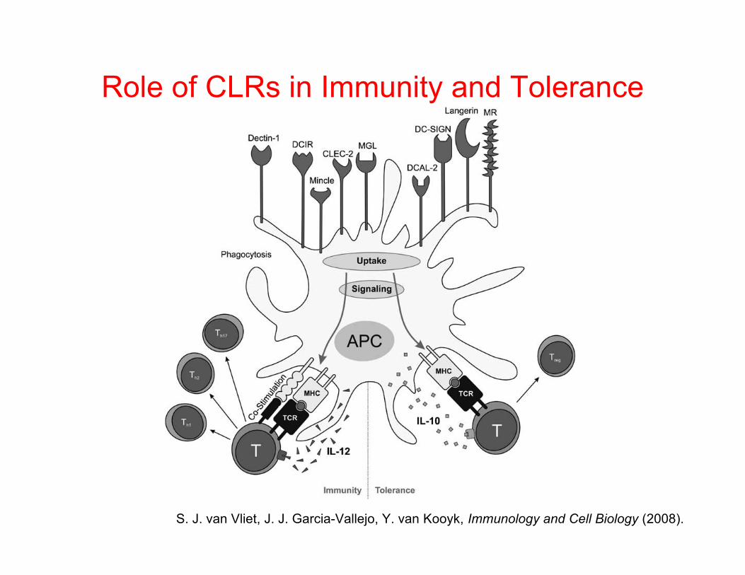
HTP Methodology
Day 0
FluorescentMicroplate
Reader
Day 5 Day 6
Incubate withanti-CD86-PEand anti-DC-SIGN-FITC
for 1 hr at 4oC
Fix in 0.03%FA for 30 – 45
min at RT
Cytotoxicitytest & Store forcytokineprofiling
FITC and PEfluorescencemeasured by
microplatereader
Zoom in the well
Day 6
Kou P. and Babensee JE. Acta Biomaterialia (2010)

Glycan Isolation and Biotinylation
HPLCforSepara-onofMan5‐9
HPLC&MALDI
FreeReducingGlycans(Man5‐9)
FreeReducingGlycans
Glycan‐AEAB‐LC‐Bio-n
2. 3.
4. 5.
1.
RibonucleaseBRibonucleaseB
6. 7.
N. Hotaling in collaboration with R. Cummings, Emory University

Glycan Purification and CharacterizationHPLC of Oligomannose 5-9 afterAEAB functionalization and afterseparation (before biotinylation)
Total amount of biotinylatedglycan-AEAB made
MALDI of biotinylatedglycans-AEAB
Man5-AEAB
Man6-AEAB
Man7-AEAB
Man8-AEAB
Man9-AEAB
Man5-AEAB-biotin
Man6-AEAB-biotin
Man7-AEAB-biotin
Man8-AEAB-biotin
Man9-AEAB-biotin

Enyme Link Lectin Assay on biotinylated glycans immobilized on TCPS by surface adsorbed SA. Asaturation assay was performed to determine what concentration of glycan was needed to saturate SAbinding sites for 4 different sizes of glycan A) a monosaccharaide, mannose, B) a trisaccharide, Lewisa,C) heptasaccaride, Oligomannose 5, and D) an undecasaccharide, Oligomannose 9. Brackets representstatistical difference from all indicated columns P ≤ 0.05.
Detection of Immobilized Glycans
N. Hotaling in collaboration with R. Cummings, Emory University

DC Response to 27 Biotinylated Sugars on SA Coated TCPS(n=7) TCPS iDC Normalized
TCPS
mD
C
SA iD
CSA
mD
C
a-G
lca-
Neu
5Ac
a-Fu
cb-
Gal
b-G
lcN
Ac
fuca
1-3G
lcN
Ac
Lact
ose
LN S'LN
Lex
DiL
exTr
iLex
SLex
Lec
SLec
Lea
Lea-
LC
SLea
Ley
Ley-
Lex
a-M
anTr
iMan
Man
5M
an6
Man
7M
an8
Man
9
0
2
4
6 **
#
#+ + + + + +
CD86
/DCS
IGN
Fold
Cha
nge
DC response to 27 biotinylated glycans (4000pmol/well).* represents statistical difference from iDC p≤ 0.05.# represents statistical difference from streptavidin iDC p≤ 0.05.+ represents no difference from TCPS mDC or streptavidin mDC.
DC Responses to Glycans
N. Hotaling in collaboration with R. Cummings, Emory University

n=6 independent determinations, mean ±S.D.* Different from iDCs on TCPS, p < 0.05; # Different from iDCs on SA coated TCPS, p<0.05.
DC Response to OVA and CationizedBSA Functionalized with Mannose or
Synthetic Oligomannose 5 (Syn-Man5)
N. Hotaling in collaboration with D. Ratner, University of Washington
Bio-Man5 fromR. Cummings
Syn-Man5 fromD. Ratner

Summary• DCs may use mechanisms analogous to
pathogen recognition to respond tobiomaterials (e.g. TLRs, CLRs)
• Glycans recognizable by DC CLRs aredetected associated with the adsorbed serumprotein layer, as directed by the underlyingsurface chemistry. This correlates with adifferential DC phenotypic response.
• Protein glycosylations are important forimmune cell (e.g. DC) interaction with abiomaterial
• Context of glycan presentation to DCs isimportant for an immunofunctional effect.
• Identify immunomodulatory glycans andoptimal mode of presentation for DC effects
• Modification of biomaterial surfaces andvaccine carriers can be used to direct hostimmune responses
T.B.H. Geijtenbeek and S.I. Gringhuis, Nature Rev Immunol (2009).

AcknowledgementsFunding1RO1EB004633-01A1, National Institutes of Health, NIBIB/NHLBIGeorgia Tech/Emory Center for the Engineering of Living Tissue(GTEC) grant, an NSF ERC, EEC-9731643CAREER Award, National Science FoundationArthritis Investigator Grant, The Arthritis FoundationJ&J – Georgia Institute of Technology Healthcare Innovation AwardConsortium for Functional Glycomics (NIH/NHLBI)Wallace H. Coulter GT/Emory-PKU BME Collaborative Seed Grant
Students/Postdocs
Melissa Stein Matzelle
Leah Moore
Mutsumi Yoshida
Saul Lee
Abhijit Paranjpe
Nancy Bennewitz
Sucharita Shankar
Peng Meng Kou
Jessica Mata
Jaehyung Park
Kate Lee
Todd Rogers
Christina Duden
Inn Inn Chen
Nathan Hotaling
Dr. Lori Norton
Collaborators
Judith Kapp, Christian Larsen, Bali Pulendran, R. Cummings,
D. Smith (EU)
Mary Marovich and Mike Eller (Walter Reed)
Polly Matzinger (NIAID, NIH)
John L. Brash and Rena Cornelius (McMaster University)
Andres Garcia, Ben Benjamin Keselowsky, Jeffrey Capadona,
Dr.Brent Carter, Johnafel Crowe (GT)
D. Ratner (University of Washington)