Embed Size (px)
Citation preview
Accepted Manuscript
Title: Biomarkers to identify and isolate senescent cells
Author: Mantas Matjusaitis Greg Chin Ethan AndersSarnoski Alexandra Stolzing
PII: S1568-1637(16)30079-4DOI: http://dx.doi.org/doi:10.1016/j.arr.2016.05.003Reference: ARR 670
To appear in: Ageing Research Reviews
Received date: 4-2-2016Revised date: 4-5-2016Accepted date: 11-5-2016
Please cite this article as: Matjusaitis, Mantas, Chin, Greg, Sarnoski, Ethan Anders,Stolzing,Alexandra,Biomarkers to identify and isolate senescent cells.AgeingResearchReviews http://dx.doi.org/10.1016/j.arr.2016.05.003
This is a PDF file of an unedited manuscript that has been accepted for publication.As a service to our customers we are providing this early version of the manuscript.The manuscript will undergo copyediting, typesetting, and review of the resulting proofbefore it is published in its final form. Please note that during the production processerrors may be discovered which could affect the content, and all legal disclaimers thatapply to the journal pertain.
Biomarkers to identify and isolate senescent cells
Mantas Matjusaitis1, Greg Chin, Ethan Anders Sarnoski2, Alexandra Stolzing3,4
1Scottish Centre for Regenerative Medicine, The University of Edinburgh, Edinburgh,
England
2Department of Molecular, Cellular, and Developmental Biology, Yale University, New
Haven, CT, USA
3Institute IZBI, University of Leipzig, Leipzig, Germany
4Loughborough University, Loughborough, England
Corresponding author: Alexandra Stolzing, PhD, Loughborough University, Wolfson School,
Epinal Way, LE11 3TU, Loughborough, UK. +44-1509-227577
Highlights
There is no single biomarker which can robustly identify senescent cells.
Widely used senescent cell biomarkers should be used in combination for accuracy.
Technologies like tangential flow filtration can be used to isolate senescent cells.
Other methods, like senolytic viruses, could be used to remove senescent cells.
Abstract
Aging is the main risk factor for many degenerative diseases and declining health. Senescent
cells are part of the underlying mechanism for time-dependent tissue dysfunction. These cells
can negatively affect neighbouring cells through an altered secretory phenotype: the
senescence-associated secretory phenotype (SASP). The SASP induces senescence in healthy
cells, promotes tumour formation and progression, and contributes to other age-related
diseases such as atherosclerosis, immune-senescence and neurodegeneration. Removal of
senescent cells was recently demonstrated to delay age-related degeneration and extend
lifespan.
To better understand cell aging and to reap the benefits of senescent cell removal, it is
necessary to have a reliable biomarker to identify these cells. Following an introduction to
cellular senescence, we discuss several classes of biomarkers in the context of their utility in
identifying and/or removing senescent cells from tissues. Although senescence can be
induced by a variety of stimuli, senescent cells share some characteristics that enable their
identification both in vitro and in vivo. Nevertheless, it may prove difficult to identify a single
biomarker capable of distinguishing senescence in all cell types. Therefore, this will not be a
comprehensive review of all senescence biomarkers but rather an outlook on technologies
and markers that are most suitable to identify and isolate senescent cells.
Keywords: Aging, senescence, biomarkers, cell biology
1. Introduction
1.1. Aging and cellular senescence
Our society is rapidly aging and the incidence of age-related diseases, such as
Alzheimer’s, diabetes and cancer is increasing (Christensen et al., 2009). If these trends
continue, aging will become a major economic and social burden (Harper, 2014; Kankeu et
al., 2013; Wimo et al., 2013). To avert this impending crisis, we must better understand why
we age. Aging is a heterogenic process at both the organismal and cellular level. The number
of contributing internal and external factors, such as epigenetic changes (Sinclair and
Oberdoerffer, 2009) and the environment, make it difficult to categorize and prioritize the
importance of each component. Such diversity has given rise to multiple theories regarding
the root cause of aging (Harman, 1956; Park and Yeo, 2013; Wei et al., 2001), which
sometimes contradict but more often complement one another. The variety of associated
causes implies that aging is likely to be multifactorial in nature (Riess and Krüger, 1999;
Sheikh et al., 2013).
One known aging factor is cellular senescence. Senescent cells accumulate with age in
organisms, albeit at different rates in the various organs (Erusalimsky and Kurz, 2005; Herbig
et al., 2006; Jeyapalan et al., 2007; Paradis et al., 2001). Originally, cellular senescence was
defined as a loss of replicative capacity (Hayflick, 1965) caused by a progressive shortening
of the tandem repeats protecting chromosome ends (telomeres). This eventually leads to
chromosomal damage and replicative arrest (Campisi, 1997). Interestingly, cellular
senescence can also be induced by stress (Toussaint et al., 2000) and oncogenes (Bartkova et
al., 2006), demonstrating that cellular senescence is not only caused by exhaustion of
replicative capacity as first thought. Such heterogeneity of cellular senescence, which we will
briefly discuss in the next paragraph, has led a field to sometimes unnecessary ambiguity,
leaving researchers to disagree what cellular senescence entails (Burton and Faragher, 2015).
For the purposes of this review, we define cellular senescence as a permanent (under
physiological conditions) cell cycle arrest that is a result of cellular stress or damage,
including but not limited to abnormal activation of oncogenes, telomere shortening and
macromolecule accumulation (De Cecco et al., 2011). With this description we deliberately
exclude developmental senescence, quiescent cells, and post-mitotic cells.
[Table 1]
1.2. Senescent cells in health and disease
Cellular senescence is thought to have developed as a safeguard to prevent damaged cells
from accumulating and either becoming cancerous or causing cancer (Figure 1). However,
accumulation of senescent cells in tissues is detrimental to the animal (Herbig et al., 2006;
Jeyapalan et al., 2007) as these non-functional cells directly and indirectly damage
surrounding cells (Salama et al., 2014). Examples of such damage include occupying niches
required by competent cells to function (Lynch, 2004), secreting transforming, inflammatory
and otherwise damaging components of the SASP (Campisi and d’Adda di Fagagna, 2007;
Coppé et al., 2010, 2008), promoting tumour formation (Leikam et al., 2015; Zacarias-Fluck
et al., 2015) and contributing to various age-related diseases such as atherosclerosis (Irvine et
al., 2014; Wang and Bennett, 2012).
While long-term accumulation of senescent cells is harmful to the organism, short-term
senescence events prevent cancer (Kuilman et al., 2008), guide development (Muñoz-Espín et
al., 2013) and improve tissue repair and wound healing (Demaria et al., 2014; Rodier and
Campisi, 2011). One proposed mechanism to negate the long-term detrimental effects of
senescent cells, while retaining their short-term beneficial functions, is to periodically purge
them from the body. Regular elimination of p16-positive senescent cells from functionally
wild-type mice slows time-dependent functional decline and extends median lifespan ~30%
(Baker et al., 2016, 2011). Moreover, recent studies have demonstrated clearance of
senescent cells from wild-type mice using small molecules which target the BCL-2 protein
family (Chang et al., 2016; Zhu et al., 2015), lending credence to this approach as a
therapeutic strategy. However, no method currently exists to accomplish this in humans, in
large part because senescent cells cannot yet be reliably identified in living tissue. In the
following sections, we will discuss biomarkers and their utility in identifying or eliminating
senescent cells in a living organism.
1.3. Characteristics of useful biomarkers
A biomarker is a biological signature of a condition which enables one to evaluate if the
biological system (organism, cell, etc.) possesses that condition or not. Many molecules, such
as proteins, nucleic acids, and lipids, can be used as a biomarker. They can be found within
the cell, in the adjacent extracellular area, or even systemically in the circulatory system.
Importantly, no single marker currently provides an accurate representation of cellular
senescence.
A useful biomarker must display several important features. First, it should be robustly
associated with the condition. Although it is likely that (i) context, such as cell type, will be
relevant and that (ii) it may not identify all cases of cellular senescence, it is crucial that the
presence of the marker strongly correlates with a specific condition. Second, it is essential to
know the threshold at which a marker becomes representative of the specific feature. Most
proteins are expressed at basal levels in many cells, and simple evaluation of the presence or
absence of the protein is not informative. For discrimination purposes, it is imperative to
identify a clear threshold value which defines the cellular status. Finally, to be practical, a
marker must be quantifiable using current technologies. Even a comprehensive understanding
of a marker is not practically helpful unless we are able to monitor its levels or purify cells
positive for it.
In this review, we will emphasize markers of cellular senescence that would be practical
to assay. Only single-cell markers of senescence will be discussed. Detailed reviews on
systemic aging markers have been published previously (Falandry et al., 2013; Pallis et al.,
2014).
1.4. The challenge and benefits of finding robust cellular senescence markers
While cellular senescence has been linked to a number of predictable phenotypic traits
and representative biomarkers (Table 1), senescent cells are still a heterogeneous population,
and this fact significantly complicates a search for a robust senescence biomarker. Senescent
cells arising from different stimuli exhibit measurably distinct proteomes (Aan et al., 2013;
Dierick et al., 2002; G. J. Aan, 2011). Moreover, gene expression profiles of senescent cells
are cell type-specific (Schnabl et al., 2003). Considering recent breakthroughs in our
understanding of a need for personalized medicine, it is likely that variation will be seen not
only between different senescent cell types but also between individuals. Importantly,
potential reversibility of senescent cells has to be taken into account when evaluating what is
and what is not cellular senescence. Although the main consensus in the field is to grant
senescence a permanent and irreversible status, a number of studies have shown that cell
cycle inhibition can be reversed in human senescent fibroblasts by inhibition of p53 and/or
p16, two key pathways used to established cellular senescence (Beauséjour et al., 2003; Ide et
al., 1983). More recently, it has been shown that inhibition of p38α/β MAPK lowers p16
levels and restores replicative capacity of aged mouse muscle stem cells (Cosgrove et al.,
2014). Unfortunately, these studies did not look into other senescence marker in their models.
Therefore, it is still not clear if these aged cells are only forced into the cell cycle from p16-
dependant growth arrest or if is it is a complete or partial reversal of cellular senescence.
Interestingly, cellular senescence can be prevented by activating autophagy, either with
rapamycin treatment or Atg7 overexpression, in aged satellite mouse and human muscle cells.
However, it is not clear from this data if cellular senescence can be reversed (García-Prat et
al., 2016). Even if senescence could not be reversed, it is clear that a large senescence
biomarkers can be affected by defined molecules and conditions, which is an important
consideration when looking for and validating such markers.
As cancer research has demonstrated, having robust, practical markers can enhance the
research field in many ways (Henry and Hayes, 2012; Pallis et al., 2014). A reliable readout
that identifies senescent cells would facilitate high-throughput screens as well as allow
quantification of aging in various animal models. Most importantly, therapeutic techniques
would benefit from robust markers to enable removal of senescent cells from tissues (such as
blood filtering, Figure 3), stem cell preparations from aged autologous donors (Melk et al.,
2009) and identification of replicative exhaustion in stem cell expansions. One example of
how such markers could be used is a strategy using selectively-lytic viruses, which are
already used in oncology (Elsedawy and Russell, 2013). These are genetically modified
viruses which replicate, lyse and kill cells in the presence (or absence) of specific gene
products and, therefore, allow selective targeting. This strategy has successfully reached
regulatory approvals in China and the USA for cancer treatment (Garber, 2006; Pol et al.,
2015). Theoretically, such a therapy could be applied to senescent cell removal as well.
Currently, due to the heterogeneity of cellular senescence and ambiguity of the term
(Table 1), there is no known universal biomarker which can selectively but broadly identify
senescent cells in different tissues and extracellular environments. In this review, we will
discuss a number of studies which have made significant strides towards the identification
and validation of cellular senescence biomarkers.
2. Surface markers & secretion profile
Surface and external factors are ideal biomarkers, as they can be detected without
intracellular delivery of a probe and without harming the cell. Multiple surface markers have
been associated with senescent cells, some more robust than others. Three major groups of
such molecules will be covered: secreted factors, plasma membrane (PM)-associated proteins
and PM lipid composition.
2.1. Secreted biomarkers
Secreted factors have excellent potential as biomarkers because detection methods can be
contact-free, allowing measurement without disturbing the cells. However, secreted markers
are intractable to cell isolation until technology is available which can detect the secretion of
single-cells during the selection process.
The senescence-associated secretory phenotype (SASP) has been observed in many
different cell types (Acosta et al. 2008; Campisi et al. 2011). One study has defined many of
the factors which are secreted by aged human fibroblasts and epithelial cells (Campisi et al.,
2008). This list of factors includes but is not limited to inflammatory mediators (such as IL-6
and MIP-3a), growth factors (such as HGF, GRO, and IGF-binding proteins), detached cell
surface molecules, and extracellular matrix components. Multiple studies have validated
SASP and its related factors as robust markers of senescence, which makes them very
promising candidates (Acosta et al., 2013; Sandeman et al., 2001).
Interestingly, another secretory profile of senescent cells recently has been identified. The
telomere-associated secretory phenotype (TASP) has been associated with replicative
senescence (Braig et al., 2014; Jiang et al., 2008). It is not clear if this phenotype should be
classified as separate from the SASP phenotype. However, it does provide some new and
unique markers to consider. These include cathelin-related antimicrobial peptide (CRAMP),
chitinase, stathmin, and EF-1α. Interestingly, one study has shown that CRAMP and chitinase
levels highly correlate not only with replicative senescence but also with human aging in
general (Lu et al., 2014).
These secretory profiles are useful tools for evaluating general tissue or cell culture
senescence. Some recent publications covered various ways of detecting such phenotypes.
One such method is the SASP-responsive alkaline phosphatase (SASP-RAP) assay, where
activity of secreted alkaline phosphatase is measured by a commercial chemiluminescence
kit. SASP-RAP reliably detected senescence in rat renal tubular epithelial cells and mouse
embryonic fibroblasts treated with etoposide, a DNA-damaging agent (Gu and Kitamura,
2012). ELISA protocols have also been developed for an array of SASP components,
including IL-6 (Rodier, 2013). Moreover, it also may be possible to detect SASP by
monitoring the known effects on surrounding cells. For example, senescent fibroblasts affect
epithelial cell proliferation which can, in turn, be quantified as an indirect marker (Parrinello
et al., 2005).
Nevertheless, it is important to understand that there are some major limitations inherent
to secretory markers. Firstly, the SASP may vary between cell types and between different
stages of senescence (Coppé et al., 2010; Maciel-Barón et al., 2016; Rodier et al., 2009).
Secondly, current technology does not permit the rapid, high-throughput analysis of single-
cell secretory phenotypes necessary to isolate a population on this basis. Although this
technological gap currently prevents the use of a secreted marker for selective cell removal,
secreted biomarkers are still applicable for general identification of senescent cells and
potentially for diagnostic tests aiming to quantify the burden of cellular senescence in an
individual.
2.2. Plasma membrane-associated proteins
Another set of surface structures which could be used for identifying and isolating
senescent cells are plasma membrane-associated proteins, such as receptors and
glycoproteins. As these structures are the main means of interaction between extracellular
and intracellular compartments, their composition and functionality is essential for cell
homeostasis.
Several independent research groups have shown that plasma membrane (PM)
glycoprotein composition is altered in aged cells (Poot et al., 1986; Wu et al., 2009).
Glycoproteins are important components and play a role in many functions, including
signalling in immune system and hormone responses (Ervasti, 2000; Li et al., 2010). There is
a wide range of possible glycoprotein variants, and specific glycoprotein compositions have
been linked to a senescence profile. For example, senescent human fibroblasts exhibit higher
levels of concanavalin A, fucose, and glucosamine (Blondal et al., 1985).
Expression changes in the concentration of PM-associated proteins could also be used
as a senescence biomarker. Althubiti and colleagues have identified 107 PM-associated
proteins upregulated in a human bladder cancer cell line induced to senesce by ectopic
expression of p16 and p21. Ten of these upregulation events were validated by western blots
and immunocytochemistry (Althubiti et al., 2014), while several additional candidates like
ICAM-1 had previously been reported to increase in senescent cells (Schnabl et al., 2003).
Several standard methods exist to measure expression changes in PM proteins, including
immunocytochemistry (Obradovic and Jurisic, 2012) and techniques utilizing other protein-
specific probes (Liu et al., 2005). Changes in the expression of PM proteins can also be
detected with transductionally targeted recombinant viruses, which are able to enter only
those cells with the appropriate ligand exposed on the PM. This has been accomplished in
cancer cells already and readers are referred to an excellent review by Everts and Curiel for
more information (Everts and Curiel, 2004). A similar strategy could be used to target
senescent cells. The specific targeting of senescent cells holds promise both as a senescent
assay and as a potential therapeutic. Transductionally targeted viruses could be engineered to
selectively kill senescent cells or to carry a fluorescence reporter gene for identification
(Perez et al., 2013) (Figure 4).
2.3. Plasma membrane lipid composition
The third type of marker which can be used to identify and isolate senescence cells is
linked to PM lipid composition (Sud et al., 2007). Although PM lipid-based senescence
biomarkers are less established and harder to detect compared to other surface signatures,
some promising candidates and techniques have emerged which hold great potential for
senescent cell identification and isolation.
Membrane lipid composition and membrane biophysical properties change in senescent
cells. Plasma membranes, due to altered lipid composition, become more rigid with age
(Fulop et al., 2012a; Momchilova et al., 2014). Moreover, senescent cells increase in size
compared to non-senescent analogues (Kim et al., 2015; Rodier and Campisi, 2011). This
modifies many essential functions within the cell, such as diffusion, cell size, membrane
fusion, chemical and electrical processes, cell elasticity and membrane stiffness (Pontes et al.,
2013). Such cellular mechanics have already been used as biomarkers to identify cancer cells,
malaria-infected cells and primary cells (Darling et al., 2008; Suresh et al., 2005).
A number of technologies have emerged to detect PM biomechanical and lipid
composition differences between cells. For example, tangential flow filtration (Cai et al.,
2015) discriminates between cells based on membrane stiffness. Such a method has been
successfully used to separate differentiated cells from pluripotent ones (Willoughby et al.,
2016). Moreover, a technology which selects cells based on cell mechanics has been used to
sort red blood cells based on shape and deformation (Beech et al., 2012), which make this
sub-group of senescence markers even more viable.
Another structure which could be used as a marker is a lipid raft (LR). LRs are small,
dynamic structures in the plasma membrane that are high in cholesterol and low in
unsaturated phospholipids (Brown, 2006). These structures recruit signalling components to
the plasma membrane (Kabouridis, 2006). It has been shown that T cells from young subjects
have a different LR distribution and functionality compared to T cells from older individuals,
although it is still not clear if these changes are linked to senescent or aged cells in general
(Fulop et al., 2012b; Larbi et al., 2006, 2004).
LR dispersal can be measured using CTxB fluorescent molecules that bind to GM1
gangliosides, which are LR markers (Holleran, 2003). Moreover, it is possible to use specific
fluorescent LR probes, such that quantifying the number of probes bound could be used as a
means of identifying senescent cells (Mikhalyov and Samsonov, 2011). For a full analysis of
the most current methods for studying membrane microdomains (including LRs and the
signalosome) in intact cells, readers are advised to refer to Lagerholm’s review (Lagerholm et
al., 2005).
In summary, membrane lipid composition and its effects on cell biophysical properties
represent a promising line of exploration for developing reliable, quantitative senescence
markers.
3. Intracellular senescence biomarkers
Many intrinsic changes have been observed in senescent cells. While currently known
intrinsic changes are more discrete and better defined compared to surface ones, they are
harder to measure in live tissue. Three major groups of potential biomarkers will be covered:
DNA-related markers, protective mechanism or damage signatures, and cell cycle genes.
3.1. DNA-related senescence biomarkers
It is still not clear if mutations significantly contribute to aging, but the cumulative
number of mutations and the rate at which they accumulate increases with age in model
organisms and senescent cells (Kennedy et al., 2012; Sedelnikova et al., 2004). Moreover,
accelerated-aging diseases, such as Werner and Cockayne Syndromes, are linked to
malfunctioning DNA repair mechanisms (Rossi et al., 2010). Such findings make it tempting
to speculate that DNA quality, metabolism, and maintenance are key mechanisms in the
biology of aging. That being said, most of the changes which will be discussed are not
exclusive to senescent cells and therefore use as a sole marker would yield a significant
proportion of false positives.
Various markers for DNA damage, such as γH2AX, 53BPI foci, Rad17, ATM and
MDC1, are commonly used as conditional markers of cellular senescence (Lawless et al.
2010; Wang et al. 2009; Sharma et al. 2012). γH2AX immunocytochemistry has been shown
to effectively mark senescent fibroblasts (von Zglinicki et al., 2005). Although these markers
are well established, they do not represent cellular senescence directly. γH2AX is a marker
for DNA double-strand breaks (DSBs), which is neither necessary for nor exclusive to
cellular senescence (Chapman et al., 2012). Similar limitations exist for other DNA damage
related biomarkers (Awasthi et al., 2015).
DNA synthesis markers can also serve as negative indicators for cellular senescence.
DNA synthesis rate can be analyzed in 5-bromo-2’deoxyuridine (BrdU) or 5-ethynyl-2’-
dexyuridine (EdU) incorporation assays (Gratzner, 1982; Salic and Mitchison, 2008). Only
cells that are undergoing DNA synthesis are able to incorporate transiently available BrdU or
EdU nucleosides. The incorporation rate of such synthetic nucleosides is low in senescent
cells, which suggests that DNA replication is near-absent (Voutetakis et al., 2015). Another
assay for cell proliferation is based on immunostaining for marker Ki-67 (Urruticoechea et
al., 2005). However, assays based on DNA synthesis would also identify other non-
replicative cells, such as quiescent cells and post-mitotic cells, which make such markers sub-
optimal if used alone.
Telomere length can also be used as a biomarker for replicative senescence (Mather et
al. 2011; Zietzer & Hillmeister 2014; Bekaert et al. 2005.). However, the shortening of
telomeres associated with replicative senescence has yet to be translated into a common
assay. There are several ways to measure telomere status in the cell. Measuring telomere
length by quantitative PCR is one way (Cawthon 2002). Moreover, protocols have been
designed which allow measurement of absolute telomere length (O’Callaghan and Fenech,
2011) or telomere length of a single-cell (Wang et al., 2013). Unfortunately, these PCR-based
approaches to telomere measurement require destruction of the cell. Fluorescence in situ
hybridization (FISH) can be used to visualize the length of telomeres in fixed but intact cells
(O’Sullivan et al., 2005; Ourliac-Garnier and Londoño-Vallejo, 2011), which retains
information such as subcellular localization. Moreover, FISH can be combined with flow
cytometry to provide information on telomere length in thousands of cells (Baerlocher et al.,
2006; Hultdin et al., 1998).
In addition to the direct measurement of telomere length, another potential aging
marker is telomere dysfunction-induced foci (TIF) (Brugat et al., 2010; Sahin et al., 2011).
TIF is a term used to describe telomeres which accumulated various DNA damage factors,
such as γH2AX, ATM and Mre11 (Badie et al., 2010; Takai et al., 2003). TIF has been
shown to increase in baboon fibroblasts that have undergone replicative senescence
(Jeyapalan et al., 2007) and also has an effect on the metabolic status of the cell (Sahin et al.,
2011). Therefore, it may be possible to identify senescent cells by immunostaining for these
factors at telomeres.
Epigenetic changes, including senescence-associated heterochromatic foci (SAHF),
can also be used as biomarkers for cellular senescence. Epigenetics largely determine cell-
state differences in a genetically identical population, such as within an organism (Sinclair
and Oberdoerffer, 2009). Although epigenetic markers vary depending on the environmental
context and type of cell (Kosar et al., 2011), epigenetic profiles have been defined for various
conditions, including cancer (Dumitrescu, 2012; Mäbert et al., 2014) and aged human cells
(Horvath, 2013). A specific heterochromatin profile has been linked to cellular senescence
(Kosar et al., 2011; Narita et al., 2003). SAHFs, domains of heterochromatin that contribute
to silencing of proliferative genes in senescent cells (Narita, 2007), can be detected by
multiple methods, including DAPI staining and immunocytochemistry against SAHF
components, such as HP1 (Aird and Zhang, 2013). Another novel assay, called chromatin in
vivo assay (CiA), has the potential to be used in high-throughput screening for
heterochromatin alterations (Jones, 2012). It may be possible to adapt it to report SAHFs.
In summary, DNA-related signatures are some of the most prominent cellular senescence
biomarkers. In addition to the fact that one or more of these markers can robustly be found in
most senescent cells, the broad availability of methods for their detection makes this
biomarker group extremely potent. Further discussion of how they can be used in
combination with other markers to increase specificity will be discussed later in the review.
3.2. Protective mechanism and damage markers
Aging is closely linked to loss of damage repair and/or stress response capabilities, which
results in the accumulation of various toxic by-products and other macromolecules (Chen et
al., 2007; Hipkiss, 2006). As a result, enzymatic by-products and activities of various
lysosomal and proteosomal enzymes, which are responsible for aggregate clearance, are often
altered in aged cells (García-Prat et al., 2016). The ability to detect such changes would help
to identify and possibly isolate senescence cells.
Perhaps the most frequently used biomarker for senescent cells is increased activity of β-
galactosidase (β-gal) (Bassaneze et al., 2008; Dimri et al., 1995), which reflects increased
lysosomal mass (Kurz et al., 2000). Several techniques exist for β-gal detection, including
fluorescence-based and cytochemical methods (Debacq-Chainiaux et al., 2009). However, β-
gal activity also increases in quiescence and in response to various forms of stress (Yang and
Hu, 2005). Thus, β-gal as a sole marker may often yield false-positives and is best used in
combination with other markers. A similar marker, α-fucosidase, has recently been reported
(Hildebrand et al., 2013). While this marker boasts similar ease of detection, it remains
uncertain if it is equally, more, or less specific for senescence than β-gal.
Proteasomes, protein complexes responsible for degrading unneeded or damaged
proteins, may also act as useful biomarkers for senescence. For example, 26 S proteasome
activity is reduced in human primary senescent fibroblasts (Chondrogianni et al., 2003;
Reinheckel et al., 1998; Torres and Perez, 2008), and 26 S proteasome activity has recently
been measured using fluorogenic peptide substrates (Georgila et al., 2014). While this assay
has not yet been applied to senescent cells, it may serve as a secondary marker in conjunction
with other indicators of cellular senescence.
Reactive oxygen species (ROS) can also be used for senescent cell detection. ROS are
involved in oxidative stress, signalling and differentiation processes. ROS increase with age
and correlate with many age-related cellular changes (Liochev, 2013). Furthermore, elements
associated with protection against oxidative stress (Cox2, SOD, Mn and other antioxidants)
often show reduced functionality in aged organisms (Espinoza et al., 2008; Paul et al., 2007).
However, oxidative stress, like many other markers mentioned in this review, is only an
indirect marker of senescence. It is not necessary for cells to have high ROS activity to
become senescent. Furthermore, a low level of ROS, is not only useful but essential for
normal cellular functions (Liochev, 2013). Conversely, high ROS levels are not always
associated with senescence as they are sometimes linked to differentiation (Paul et al., 2014).
This may make interpretation of results problematic if clear thresholds (and their deviation
depending on environment) are not determined. Various assays, including commercially
available kits, exist to measure oxidative stress markers and levels of intracellular ROS
(Starkov, 2010; Fan and Li, 2014). Most of these techniques rely on fluorescence detection
with either flow cytometry/fluorescence microscopy or chemiluminescence.
In summary, markers of cellular damage and protection mechanisms, such as ROS and β-
Gal, are well-known signatures of cellular senescence. While not exclusive to cellular
senescence, these relatively easy-to-assay markers are frequently employed as companion
diagnostics.
3.3. Cell cycle genes
Arrest of cell proliferation is a hallmark of a senescent cell, which means that genes
responsible for such regulation can potentially be used as markers to identify which cells are
senescent.
During growth arrest, various cell cycle inhibitors such as p53 and p21 are activated
(Campisi, 2005). p21waf is a cyclin-dependant kinase (CDK) inhibitor which acts by
mediating pRB dephosphorylation. Its overexpression promotes cellular senescence as shown
by β-gal activity, telomere length and DNA damage monitoring (Huang et al., 2004).
Moreover, if p21waf is down-regulated in senescent cells, replicative capacity is restored
(Schnabl et al., 2003).
Another molecular alteration that is specific to senescent cells is the phosphorylation (and
activation) of p53 at serine 15. This phosphorylation has been observed in normal human
fibroblasts that have undergone replicative senescence (Webley et al., 2000), oncogene-
induced senescence (Ferbeyre et al., 2000), and other forms of senescence (Ghosh et al.,
2008). p53 and the related tumor suppressor p16INK4a are relatively robust markers of
senescence as their expression is required to induce the characteristic permanent cell cycle
arrest (Campisi and d’Adda di Fagagna, 2007; Krishnamurthy et al., 2004; Vandenberk et al.,
2011). However, these genes are not absolute markers of cellular senescence. For example,
p53 is also responsible for apoptotic programmed cell death (Amaral et al., 2010). An assay
relying solely on p53 levels, therefore, would not be able to discriminate senescent cells from
those undergoing apoptosis. Furthermore, mutations may shift p53 activity patterns (Muller
and Vousden, 2013). In such cases, p53 mutations, although tumorigenic, may not be linked
to cellular senescence. Moreover, a reversible growth arrest state, known as quiescence, also
depends on the aforementioned cell cycle inhibitors (Li and Bhatia, 2011). Although such
context specificity would make these biomarkers unreliable if used alone, their use in a
combinatorial strategy could be very potent.
4. Summary of most promising senescence biomarkers and combinatory approach
4.1. Synopsis of the most practical markers for identification and isolation of
senescent cells.
Some of the cellular senescence biomarkers are more practically viable either because of
their relative robustness to accurately select senescent cells or existing technologies for their
detection. In the following paragraph, we offer our opinion of the most promising senescence
biomarkers.
Firstly, markers for cell cycle genes (p16, p21, p53, Rb) and DNA synthesis (BrdU
incorporation) provide a robust way to distinguish dividing cells from non-dividing cells.
Considering that cell cycle arrest is a hallmark of cellular senescence, such markers carry
great potential. That being said, if used alone, these assays would also identify non-senescent,
non-proliferative cells, such as post-mitotic neurons or quiescent stem cells. Other senescence
markers should be used in combination to alleviate such off-target effects.
Secondly, due to the non-proliferative and dysfunctional nature of senescent cells, an
accumulation of various intracellular junk and a stressed cellular repair mechanism are often
observed. This makes a number of pathways a potential source for biomarkers, the most
prominent example being β-gal activity in lysosomes. With a number of assays now available
to detect such changes, this category of biomarkers may offer several strong candidates for
diagnostic assays.
Thirdly, considering recent developments in the ability to quantify cell biomechanical
properties (Beech et al., 2012; Darling et al., 2008; Suresh, 2007) and plasma membrane
elasticity alterations observed in senescent cells, new technologies could arise that would
enable senescent cell identification and isolation. Similar approaches, based on tangential
flow filtration, for example, have been used successfully to separate pluripotent cells from
heterogeneous populations (Willoughby et al., 2016).
Lastly, senescent cell surface components, such as ICAM-1 and NOTCH3, are very
attractive biomarkers due to possibility of screening live cells without damaging them.
Moreover, a number of techniques are available to achieve such aims, including
immunocytochemistry, transductionally targeted viruses, FACS or magnetic cell sorting.
Recent studies have also identified a large number of novel surface markers, which
potentially may join the list of commonly used signatures for senescence (Althubiti et al.,
2014).
4.2. Using combinations of markers for a more robust and specific assay
To achieve the highest possible specificity of an assay, a combination of the most
prominent markers should be used. To date, it is common best practice in the cellular
senescence research field to use a panel of markers to validate that cells are senescent (Acosta
et al., 2013; Guo et al., 2009). p53, p16, β-gal and telomere length remain the most
commonly used markers and their combination should accurately pinpoint senescent cells.
Although essential, a four-piece requirement for identification and isolation is practically
challenging and cost- and time-ineffective. We propose that a synthetic reporter (or lytic)
virus could potentially be used as a single system with combinatorial potential.
Similar strategies have been successfully used in the past for oncolytic viruses where
input from multiple sources (genes, receptors etc.) was required for the function of the virus
(Barker et al., 2003; Larson et al., 2015; Lee et al., 2010; Singh et al., 2012). For example,
one study constructed an adenovirus in which gene E1a was under the control of the hTERT
(human telomerase reverse transcriptase) promoter while the viral gene E1b was under the
control of the HRE (hypoxia response element) promoter (Wang et al., 2008). Such a system
allowed viral replication only in hypoxic cells with active telomerase, which are very
common signatures for cancer cells. Although this particular example utilized two different
promoters, much more elaborate bio-sensor circuits could be built to allow even greater
specificity. Theoretically, most transcriptional signatures can be targeted with these viruses
by genetically modifying viral promoters. Moreover, viral coat engineering allows specifying
to which surface markers a viral particle can bind and consequently infect a target cell
(Verheije and Rottier, 2012).
5. Outlook
In this review, we have covered a number of biological signatures, outlining their
advantages and disadvantages as cellular senescence biomarkers. Some of them are widely
accepted to mark an aspect of aging (telomeres, ROS etc.), while other molecules, such as
lipid rafts, still require further validation as a practical marker.
One of the major limitations of all these markers is the fact that the change is only
relative. For example, ROS is always present in the cells and small amounts are essential for
intrinsic signalling. Although an increase in ROS levels is considered to be a marker of
oxidative stress and possibly cellular senescence, at what point does the difference become a
biomarker? Furthermore, can we robustly classify a measurement that can be considered a
definitive indicator of senescence in all circumstances? These questions remain to be
answered. Another important aspect is use of biomarker combinations. None of the markers
can accurately and robustly detect senescent cells in all given instances. This is why it may be
useful to use a combination of markers. Such combinations of complementing markers, such
as the ones we have suggested, would offer a diagnostic with better accuracy and robustness.
Although senescent cell markers are widely used in basic research, such signatures
may also be useful in the clinic as well. Removal of senescent cells has been shown to have a
positive effect on murine health. With increasing knowledge in immuno- and viro-therapies,
new opportunities may arise to use senescence biomarkers in senescent cell removal
procedures for therapeutic benefit.
Acknowledgments
The work presented in this paper was made possible by funding from the German Federal
Ministry of Education and Research (BMBF1315883). We thank Claudia Garcia-Diaz for
helpful comments and advice on the figures.
References
Aan, G.J., Hairi, H.A., Makpol, S., Rahman, M.A., Karsani, S.A., 2013. Differences in
protein changes between stress-induced premature senescence and replicative
senescence states. Electrophoresis 34, 2209–17. doi:10.1002/elps.201300086
Acosta, J.C., Banito, A., Wuestefeld, T., Georgilis, A., Janich, P., Morton, J.P., Athineos, D.,
Kang, T.-W., Lasitschka, F., Andrulis, M., Pascual, G., Morris, K.J., Khan, S., Jin, H.,
Dharmalingam, G., Snijders, A.P., Carroll, T., Capper, D., Pritchard, C., Inman, G.J.,
Longerich, T., Sansom, O.J., Benitah, S.A., Zender, L., Gil, J., 2013. A complex
secretory program orchestrated by the inflammasome controls paracrine senescence.
Nat. Cell Biol. 15, 978–90. doi:10.1038/ncb2784
Acosta, J.C., O’Loghlen, A., Banito, A., Guijarro, M. V, Augert, A., Raguz, S., Fumagalli,
M., Da Costa, M., Brown, C., Popov, N., Takatsu, Y., Melamed, J., d’Adda di Fagagna,
F., Bernard, D., Hernando, E., Gil, J., 2008. Chemokine signaling via the CXCR2
receptor reinforces senescence. Cell 133, 1006–18. doi:10.1016/j.cell.2008.03.038
Aird, K.M., Zhang, R., 2013. Detection of senescence-associated heterochromatin foci
(SAHF). Methods Mol. Biol. 965, 185–96. doi:10.1007/978-1-62703-239-1_12
Althubiti, M., Lezina, L., Carrera, S., Jukes-Jones, R., Giblett, S.M., Antonov, A., Barlev, N.,
Saldanha, G.S., Pritchard, C.A., Cain, K., Macip, S., 2014. Characterization of novel
markers of senescence and their prognostic potential in cancer. Cell Death Dis. 5, e1528.
doi:10.1038/cddis.2014.489
Amaral, J.D., Xavier, J.M., Steer, C.J., Rodrigues, C.M., 2010. The role of p53 in apoptosis.
Discov. Med. 9, 145–52.
Awasthi, P., Foiani, M., Kumar, A., 2015. ATM and ATR signaling at a glance. J. Cell Sci.
128, 4255–62. doi:10.1242/jcs.169730
Badie, S., Escandell, J.M., Bouwman, P., Carlos, A.R., Thanasoula, M., Gallardo, M.M.,
Suram, A., Jaco, I., Benitez, J., Herbig, U., Blasco, M.A., Jonkers, J., Tarsounas, M.,
2010. BRCA2 acts as a RAD51 loader to facilitate telomere replication and capping.
Nat. Struct. Mol. Biol. 17, 1461–9. doi:10.1038/nsmb.1943
Baerlocher, G.M., Vulto, I., de Jong, G., Lansdorp, P.M., 2006. Flow cytometry and FISH to
measure the average length of telomeres (flow FISH). Nat. Protoc. 1, 2365–76.
doi:10.1038/nprot.2006.263
Baker, D.J., Childs, B.G., Durik, M., Wijers, M.E., Sieben, C.J., Zhong, J., Saltness, R.A.,
Jeganathan, K.B., Casaclang Verzosa, G., Pezeshki, A., Khazaie, K., Miller, J.D., Van
Deursen, J.M., 2016. Naturally occurring p16 Ink4a -positive cells shorten healthy
lifespan. Nature 530, 184–189. doi:10.1038/nature16932
Barker, S.D., Dmitriev, I.P., Nettelbeck, D.M., Liu, B., Rivera, A.A., Alvarez, R.D., Curiel,
D.T., Hemminki, A., 2003. Combined transcriptional and transductional targeting
improves the specificity and efficacy of adenoviral gene delivery to ovarian carcinoma.
Gene Ther. 10, 1198–204. doi:10.1038/sj.gt.3301974
Bartkova, J., Rezaei, N., Liontos, M., Karakaidos, P., Kletsas, D., Issaeva, N., Vassiliou, L.-
V.F., Kolettas, E., Niforou, K., Zoumpourlis, V.C., Takaoka, M., Nakagawa, H., Tort,
F., Fugger, K., Johansson, F., Sehested, M., Andersen, C.L., Dyrskjot, L., Ørntoft, T.,
Lukas, J., Kittas, C., Helleday, T., Halazonetis, T.D., Bartek, J., Gorgoulis, V.G., 2006.
Oncogene-induced senescence is part of the tumorigenesis barrier imposed by DNA
damage checkpoints. Nature 444, 633–7. doi:10.1038/nature05268
Bassaneze, V., Miyakawa, A.A., Krieger, J.E., 2008. A quantitative chemiluminescent
method for studying replicative and stress-induced premature senescence in cell
cultures. Anal. Biochem. 372, 198–203. doi:10.1016/j.ab.2007.08.016
Beauséjour, C.M., Krtolica, A., Galimi, F., Narita, M., Lowe, S.W., Yaswen, P., Campisi, J.,
2003. Reversal of human cellular senescence: Roles of the p53 and p16 pathways.
EMBO J. 22, 4212–4222. doi:10.1093/emboj/cdg417
Beech, J.P., Holm, S.H., Adolfsson, K., Tegenfeldt, J.O., 2012. Sorting cells by size, shape
and deformability. Lab Chip 12, 1048–51. doi:10.1039/c2lc21083e
Bekaert, S., De Meyer, T., Van Oostveldt, P., 2005. Telomere attrition as ageing biomarker.
Anticancer Res. 25, 3011–21.
Biran, A., Krizhanovsky, V., 2015. Senescent cells talk frankly with their neighbors. Cell
Cycle 14, 2181–2. doi:10.1080/15384101.2015.1056608
Blondal, J.A., Dick, J.E., Wright, J.A., 1985. Membrane glycoprotein changes during the
senescence of normal human diploid fibroblasts in culture. Mech. Ageing Dev. 30, 273–
83.
Borton, M., Docherty, J.R., 1989. The effects of ageing on neuronal uptake of noradrenaline
in the rat. Naunyn. Schmiedebergs. Arch. Pharmacol. 340, 139–43.
Braig, M., Pällmann, N., Preukschas, M., Steinemann, D., Hofmann, W., Gompf, A.,
Streichert, T., Braunschweig, T., Copland, M., Rudolph, K.L., Bokemeyer, C.,
Koschmieder, S., Schuppert, A., Balabanov, S., Brümmendorf, T.H., 2014. A “telomere-
associated secretory phenotype” cooperates with BCR-ABL to drive malignant
proliferation of leukemic cells. Leukemia. doi:10.1038/leu.2014.95
Brown, D.A., 2006. Lipid rafts, detergent-resistant membranes, and raft targeting signals.
Physiology (Bethesda). 21, 430–9. doi:10.1152/physiol.00032.2006
Brugat, T., Nguyen-Khac, F., Grelier, A., Merle-Béral, H., Delic, J., 2010. Telomere
dysfunction-induced foci arise with the onset of telomeric deletions and complex
chromosomal aberrations in resistant chronic lymphocytic leukemia cells. Blood 116,
239–49. doi:10.1182/blood-2009-12-257618
Burton, D.G.A., Faragher, R.G.A., 2015. Cellular senescence: from growth arrest to
immunogenic conversion. Age (Omaha). 37, 1–19. doi:10.1007/s11357-015-9764-2
Cai, L., Yang, Y., Jiao, N., Zhang, R., 2015. Evaluation of Tangential Flow Filtration for the
Concentration and Separation of Bacteria and Viruses in Contrasting Marine
Environments. PLoS One 10, e0136741. doi:10.1371/journal.pone.0136741
Campisi, J., 2005. Senescent cells, tumor suppression, and organismal aging: good citizens,
bad neighbors. Cell 120, 513–22. doi:10.1016/j.cell.2005.02.003
Campisi, J., 1997. The biology of replicative senescence. Eur. J. Cancer 33, 703–9.
doi:10.1016/S0959-8049(96)00058-5
Campisi, J., Andersen, J.K., Kapahi, P., Melov, S., 2011. Cellular senescence: a link between
cancer and age-related degenerative disease? Semin. Cancer Biol. 21, 354–9.
doi:10.1016/j.semcancer.2011.09.001
Campisi, J., d’Adda di Fagagna, F., 2007. Cellular senescence: when bad things happen to
good cells. Nat. Rev. Mol. Cell Biol. 8, 729–40. doi:10.1038/nrm2233
Campisi, J., Nelson, P., Patil, C., Coppé, J.-P., Rodier, F., Sun, Y., Goldstein, J.,
Munoz, D., Desprez, P.-Y., 2008. Senescence-Associated Secretory Phenotypes Reveal
Cell-Nonautonomous Functions of Oncogenic RAS and the p53 Tumor Suppressor.
Public Libr. Sci.
Cawthon, R.M., 2009. Telomere length measurement by a novel monochrome multiplex
quantitative PCR method. Nucleic Acids Res. 37, e21. doi:10.1093/nar/gkn1027
Cawthon, R.M., 2002. Telomere measurement by quantitative PCR. Nucleic Acids Res. 30,
e47.
Chang, J., Wang, Y., Shao, L., Laberge, R.-M., Demaria, M., Campisi, J., Janakiraman, K.,
Sharpless, N.E., Ding, S., Feng, W., Luo, Y., Wang, X., Aykin-Burns, N., Krager, K.,
Ponnappan, U., Hauer-Jensen, M., Meng, A., Zhou, D., 2016. Clearance of senescent
cells by ABT263 rejuvenates aged hematopoietic stem cells in mice. Nat. Med. 22, 78–
83. doi:10.1038/nm.4010
Chapman, J.R., Taylor, M.R.G., Boulton, S.J., 2012. Playing the end game: DNA double-
strand break repair pathway choice. Mol. Cell 47, 497–510.
doi:10.1016/j.molcel.2012.07.029
Chen, J.-H., Hales, C.N., Ozanne, S.E., 2007. DNA damage, cellular senescence and
organismal ageing: causal or correlative? Nucleic Acids Res. 35, 7417–28.
doi:10.1093/nar/gkm681
Chondrogianni, N., Gonos, E.S., 2004. Proteasome inhibition induces a senescence-like
phenotype in primary human fibroblasts cultures. Biogerontology 5, 55–61.
Chondrogianni, N., Stratford, F.L.L., Trougakos, I.P., Friguet, B., Rivett, A.J., Gonos, E.S.,
2003. Central role of the proteasome in senescence and survival of human fibroblasts:
induction of a senescence-like phenotype upon its inhibition and resistance to stress
upon its activation. J. Biol. Chem. 278, 28026–37. doi:10.1074/jbc.M301048200
Christensen, K., Doblhammer, G., Rau, R., Vaupel, J.W., 2009. Ageing populations: the
challenges ahead. Lancet 374, 1196–208. doi:10.1016/S0140-6736(09)61460-4
Coppé, J.-P., Desprez, P.-Y., Krtolica, A., Campisi, J., 2010. The senescence-associated
secretory phenotype: the dark side of tumor suppression. Annu. Rev. Pathol. 5, 99–118.
doi:10.1146/annurev-pathol-121808-102144
Coppé, J.-P., Patil, C.K., Rodier, F., Sun, Y., Muñoz, D.P., Goldstein, J., Nelson, P.S.,
Desprez, P.-Y., Campisi, J., 2008. Senescence-associated secretory phenotypes reveal
cell-nonautonomous functions of oncogenic RAS and the p53 tumor suppressor. PLoS
Biol. 6, 2853–68. doi:10.1371/journal.pbio.0060301
Cosgrove, B.D., Gilbert, P.M., Porpiglia, E., Mourkioti, F., Lee, S.P., Corbel, S.Y.,
Llewellyn, M.E., Delp, S.L., Blau, H.M., 2014. Rejuvenation of the muscle stem cell
population restores strength to injured aged muscles. Nat. Med. 20, 255–264.
doi:10.1038/nm.3464
Darling, E.M., Topel, M., Zauscher, S., Vail, T.P., Guilak, F., 2008. Viscoelastic properties
of human mesenchymally-derived stem cells and primary osteoblasts, chondrocytes, and
adipocytes. J. Biomech. 41, 454–64. doi:10.1016/j.jbiomech.2007.06.019
De Cecco, M., Jeyapalan, J., Zhao, X., Tamamori-Adachi, M., Sedivy, J.M., 2011. Nuclear
protein accumulation in cellular senescence and organismal aging revealed with a novel
single-cell resolution fluorescence microscopy assay. Aging (Albany. NY). 3, 955–67.
Debacq-Chainiaux, F., Erusalimsky, J.D., Campisi, J., Toussaint, O., 2009. Protocols to
detect senescence-associated beta-galactosidase (SA-betagal) activity, a biomarker of
senescent cells in culture and in vivo. Nat. Protoc. 4, 1798–806.
doi:10.1038/nprot.2009.191
Demaria, M., Ohtani, N., Youssef, S.A., Rodier, F., Toussaint, W., Mitchell, J.R., Laberge,
R.-M., Vijg, J., Van Steeg, H., Dollé, M.E.T., Hoeijmakers, J.H.J., de Bruin, A., Hara,
E., Campisi, J., 2014. An essential role for senescent cells in optimal wound healing
through secretion of PDGF-AA. Dev. Cell 31, 722–33.
doi:10.1016/j.devcel.2014.11.012
Dierick, J.-F., Eliaers, F., Remacle, J., Raes, M., Fey, S.J., Larsen, P.M., Toussaint, O., 2002.
Stress-induced premature senescence and replicative senescence are different
phenotypes, proteomic evidence. Biochem. Pharmacol. 64, 1011–7.
Dimri, G.P., Lee, X., Basile, G., Acosta, M., Scott, G., Roskelley, C., Medrano, E.E.,
Linskens, M., Rubelj, I., Pereira-Smith, O., 1995. A biomarker that identifies senescent
human cells in culture and in aging skin in vivo. Proc. Natl. Acad. Sci. U. S. A. 92,
9363–7.
Dumitrescu, R.G., 2012. Epigenetic markers of early tumor development. Methods Mol. Biol.
863, 3–14. doi:10.1007/978-1-61779-612-8_1
Elsedawy, N.B., Russell, S.J., 2013. Oncolytic vaccines. Expert Rev. Vaccines 12, 1155–72.
doi:10.1586/14760584.2013.836912
Erusalimsky, J.D., Kurz, D.J., 2005. Cellular senescence in vivo: its relevance in ageing and
cardiovascular disease. Exp. Gerontol. 40, 634–42. doi:10.1016/j.exger.2005.04.010
Ervasti, J.M., 2000. Structure and Function of the Dystrophin-Glycoprotein Complex.
Espinoza, S.E., Guo, H., Fedarko, N., DeZern, A., Fried, L.P., Xue, Q.-L., Leng, S., Beamer,
B., Walston, J.D., 2008. Glutathione peroxidase enzyme activity in aging. J. Gerontol.
A. Biol. Sci. Med. Sci. 63, 505–9.
Everts, M., Curiel, D.T., 2004. Transductional targeting of adenoviral cancer gene therapy.
Curr. Gene Ther. 4, 337–46.
Falandry, C., Gilson, E., Rudolph, K.L., 2013. Are aging biomarkers clinically relevant in
oncogeriatrics? Crit. Rev. Oncol. Hematol. 85, 257–65.
doi:10.1016/j.critrevonc.2012.08.004
Fan, L.M., Li, J.-M.,. Evaluation of methods of detecting cell reactive oxygen species
production for drug screening and cell cycle studies. J. Pharmacol. Toxicol. Methods 70,
40–7. doi:10.1016/j.vascn.2014.03.173
Ferbeyre, G., de Stanchina, E., Querido, E., Baptiste, N., Prives, C., Lowe, S.W., 2000. PML
is induced by oncogenic ras and promotes premature senescence. Genes Dev. 14, 2015–
27.
Fulop, T., Le Page, A., Garneau, H., Azimi, N., Baehl, S., Dupuis, G., Pawelec, G., Larbi, A.,
2012a. Aging, immunosenescence and membrane rafts: the lipid connection. Longev.
Heal. 1, 6. doi:10.1186/2046-2395-1-6
Fulop, T., Le Page, A., Garneau, H., Azimi, N., Baehl, S., Dupuis, G., Pawelec, G., Larbi, A.,
2012b. Aging, immunosenescence and membrane rafts: the lipid connection. Longev.
Heal. 1, 6. doi:10.1186/2046-2395-1-6
Fumagalli, M., Rossiello, F., Clerici, M., Barozzi, S., Cittaro, D., Kaplunov, J.M., Bucci, G.,
Dobreva, M., Matti, V., Beausejour, C.M., Herbig, U., Longhese, M.P., d’Adda di
Fagagna, F., 2012. Telomeric DNA damage is irreparable and causes persistent DNA-
damage-response activation. Nat. Cell Biol. 14, 355–65. doi:10.1038/ncb2466
Fischer, B.M., Wong, J.K., Degan, S., Kummarapurugu, A.B., Zheng, S., Haridass, P.,
Voynow, J.A., 2013. Increased expression of senescence markers in cystic fibrosis
airways. Am. J. Physiol. Lung Cell. Mol. Physiol. 304, L394–400.
doi:10.1152/ajplung.00091.2012
G. J. Aan, H.A.H., 2011. Differential Protein Expression in Senescent Human Skin
Fibroblasts and Stress Induced Premature Senescence (SIPS) Fibroblasts. Sains
Malaysiana 40, 1247 – 1253.
Garber, K., 2006. China approves world’s first oncolytic virus therapy for cancer treatment. J.
Natl. Cancer Inst. 98, 298–300. doi:10.1093/jnci/djj111
García-Prat, L., Martínez-Vicente, M., Perdiguero, E., Ortet, L., Rodríguez-Ubreva, J.,
Rebollo, E., Ruiz-Bonilla, V., Gutarra, S., Ballestar, E., Serrano, A.L., Sandri, M.,
Muñoz-Cánoves, P., 2016. Autophagy maintains stemness by preventing senescence.
Nature 529, 37–42. doi:10.1038/nature16187
Georgila, K., Voutetakis, K., Delitsikou, V., Chondrogianni, N., Gonos, E.S., 2014.
Optimization of in vitro measurement of proteasome activity in mammalian cells using
fluorogenic substrates. Free Radic. Biol. Med. 75 Suppl 1, S31.
doi:10.1016/j.freeradbiomed.2014.10.762
Ghosh, A.K., Kanda, T., Steele, R., Ray, R.B., 2008. Knockdown of MBP-1 in human
foreskin fibroblasts induces p53-p21 dependent senescence. PLoS One 3, e3384.
doi:10.1371/journal.pone.0003384
Golde, T.E., Miller, V.M., 2009. Proteinopathy-induced neuronal senescence: a hypothesis
for brain failure in Alzheimer’s and other neurodegenerative diseases. Alzheimers. Res.
Ther. 1, 5. doi:10.1186/alzrt5
Gratzner, H.G., 1982. Monoclonal antibody to 5-bromo- and 5-iododeoxyuridine: A new
reagent for detection of DNA replication. Science 218, 474–5.
Gu, L., Kitamura, M., 2012. Sensitive detection and monitoring of senescence-associated
secretory phenotype by SASP-RAP assay. PLoS One 7, e52305.
doi:10.1371/journal.pone.0052305
Guo, X., Keyes, W.M., Papazoglu, C., Zuber, J., Li, W., Lowe, S.W., Vogel, H., Mills, A.A.,
2009. TAp63 induces senescence and suppresses tumorigenesis in vivo. Nat. Cell Biol.
11, 1451–7. doi:10.1038/ncb1988
Harman, D., 1956. Aging: a theory based on free radical and radiation chemistry. J. Gerontol.
11, 298–300.
Harper, S., 2014. Economic and social implications of aging societies. Science 346, 587–91.
doi:10.1126/science.1254405
Hayflick, L., 1965. The limited in vitro lifetime of human diploid cell strains. Exp. Cell Res.
37, 614–36.
Henry, N.L., Hayes, D.F., 2012. Cancer biomarkers. Mol. Oncol. 6, 140–6.
doi:10.1016/j.molonc.2012.01.010
Herbig, U., Ferreira, M., Condel, L., Carey, D., Sedivy, J.M., 2006. Cellular senescence in
aging primates. Science 311, 1257. doi:10.1126/science.1122446
Hildebrand, D.G., Lehle, S., Borst, A., Haferkamp, S., Essmann, F., Schulze-Osthoff, K.,
2013. α-Fucosidase as a novel convenient biomarker for cellular senescence. Cell Cycle
12, 1922–7. doi:10.4161/cc.24944
Hipkiss, A.R., 2006. Accumulation of altered proteins and ageing: causes and effects. Exp.
Gerontol. 41, 464–73. doi:10.1016/j.exger.2006.03.004
Holleran, B.J., 2003. Differential recruitment of alpha2beta1 and alpha4beta1 integrins to
lipid rafts in Jurkat T lymphocytes exposed to collagen type IV and fibronectin. J.
Leukoc. Biol. 73, 243–252. doi:10.1189/jlb.0902439
Horvath, S., 2013. DNA methylation age of human tissues and cell types. Genome Biol. 14,
R115. doi:10.1186/gb-2013-14-10-r115
Huang, Y., Corbley, M.J., Tang, Z., Yang, L., Peng, Y., Zhang, Z.Y., Tong, T.J., 2004.
Down-regulation of p21WAF1 promotes apoptosis in senescent human fibroblasts:
involvement of retinoblastoma protein phosphorylation and delay of cellular aging. J.
Cell. Physiol. 201, 483–91. doi:10.1002/jcp.20125
Hultdin, M., Gronlund, E., Norrback, K.-F., Eriksson-Lindstrom, E., Roos, G., Just, T., 1998.
Telomere analysis by fluorescence in situ hybridization and flow cytometry. Nucleic
Acids Res. 26, 3651–3656. doi:10.1093/nar/26.16.3651
Ide, T., Tsuji, Y., Ishibashi, S., Mitsui, Y., 1983. Reinitiation of host DNA synthesis in
senescent human diploid cells by infection with Simian virus 40. Exp. Cell Res. 143,
343–9.
Irvine, K.M., Skoien, R., Bokil, N.J., Melino, M., Thomas, G.P., Loo, D., Gabrielli, B., Hill,
M.M., Sweet, M.J., Clouston, A.D., Powell, E.E., 2014. Senescent human hepatocytes
express a unique secretory phenotype and promote macrophage migration. World J.
Gastroenterol. 20, 17851–62. doi:10.3748/wjg.v20.i47.17851
Jeyapalan, J.C., Ferreira, M., Sedivy, J.M., Herbig, U., 2007. Accumulation of senescent cells
in mitotic tissue of aging primates. Mech. Ageing Dev. 128, 36–44.
doi:10.1016/j.mad.2006.11.008
Jiang, H., Schiffer, E., Song, Z., Wang, J., Zürbig, P., Thedieck, K., Moes, S., Bantel, H.,
Saal, N., Jantos, J., Brecht, M., Jenö, P., Hall, M.N., Hager, K., Manns, M.P., Hecker,
H., Ganser, A., Döhner, K., Bartke, A., Meissner, C., Mischak, H., Ju, Z., Rudolph,
K.L., 2008. Proteins induced by telomere dysfunction and DNA damage represent
biomarkers of human aging and disease. Proc. Natl. Acad. Sci. U. S. A. 105, 11299–304.
doi:10.1073/pnas.0801457105
Jones, B., 2012. Epigenetics: detecting the dynamics and memory of heterochromatin. Nat.
Rev. Genet. 13, 517. doi:10.1038/nrg3283
Kabouridis, P.S., 2006. Lipid rafts in T cell receptor signalling . Mol. Membr. Biol. 23, 49–
57. doi:10.1080/09687860500453673
Kankeu, H.T., Saksena, P., Xu, K., Evans, D.B., 2013. The financial burden from non-
communicable diseases in low- and middle-income countries: a literature review. Health
Res. Policy Syst. 11, 31. doi:10.1186/1478-4505-11-31
Kennedy, S.R., Loeb, L.A., Herr, A.J., 2012. Somatic mutations in aging, cancer and
neurodegeneration. Mech. Ageing Dev. 133, 118–26. doi:10.1016/j.mad.2011.10.009
Kim, M.S., Jo, S., Park, J.T., Shin, H.Y., Kim, S.S., Gurel, O., Park, S.C., 2015. Method to
purify and analyze heterogeneous senescent cell populations using a microfluidic filter
with uniform fluidic profile. Anal. Chem. 87, 9584–8.
doi:10.1021/acs.analchem.5b00445
Kosar, M., Bartkova, J., Hubackova, S., Hodny, Z., Lukas, J., Bartek, J., 2011. Senescence-
associated heterochromatin foci are dispensable for cellular senescence, occur in a cell
type- and insult-dependent manner and follow expression of p16(ink4a). Cell Cycle 10,
457–68.
Krishnamurthy, J., Torrice, C., Ramsey, M.R., Kovalev, G.I., Al-Regaiey, K., Su, L.,
Sharpless, N.E., 2004. Ink4a/Arf expression is a biomarker of aging. J. Clin. Invest. 114,
1299–307. doi:10.1172/JCI22475
Kuilman, T., Michaloglou, C., Vredeveld, L.C.W., Douma, S., van Doorn, R., Desmet, C.J.,
Aarden, L.A., Mooi, W.J., Peeper, D.S., 2008. Oncogene-induced senescence relayed by
an interleukin-dependent inflammatory network. Cell 133, 1019–31.
doi:10.1016/j.cell.2008.03.039
Kurz, D.J., Decary, S., Hong, Y., Erusalimsky, J.D., 2000. Senescence-associated (beta)-
galactosidase reflects an increase in lysosomal mass during replicative ageing of human
endothelial cells. J. Cell Sci. 113 ( Pt 2, 3613–22.
Lagerholm, B.C., Weinreb, G.E., Jacobson, K., Thompson, N.L., 2005. Detecting
microdomains in intact cell membranes. Annu. Rev. Phys. Chem. 56, 309–36.
doi:10.1146/annurev.physchem.56.092503.141211
Larbi, A., Douziech, N., Dupuis, G., Khalil, A., Pelletier, H., 2004. Age-associated alterations
in the recruitment of signal- transduction proteins to lipid rafts in human T lymphocytes
Abstract : Aging is associated with a decline in T cell activation and proliferation , but
the underlying ings suggest that lipid rafts ac. J. Leukoc. Biol. 75, 373–381.
doi:10.1189/jlb.0703319.1
Larbi, A., Dupuis, G., Khalil, A., Douziech, N., Fortin, C., Fülöp, T., 2006. Differential role
of lipid rafts in the functions of CD4+ and CD8+ human T lymphocytes with aging.
Cell. Signal. 18, 1017–30. doi:10.1016/j.cellsig.2005.08.016
Larson, C., Oronsky, B., Scicinski, J., Fanger, G.R., Stirn, M., Oronsky, A., Reid, T.R., 2015.
Going viral: a review of replication-selective oncolytic adenoviruses. Oncotarget 6,
19976–89.
Lawless, C., Wang, C., Jurk, D., Merz, A., Zglinicki, T. von, Passos, J.F., 2010. Quantitative
assessment of markers for cell senescence. Exp. Gerontol. 45, 772–8.
doi:10.1016/j.exger.2010.01.018
Lee, B.Y., Han, J.A., Im, J.S., Morrone, A., Johung, K., Goodwin, E.C., Kleijer, W.J.,
DiMaio, D., Hwang, E.S., 2006. Senescence-associated beta-galactosidase is lysosomal
beta-galactosidase. Aging Cell 5, 187–95. doi:10.1111/j.1474-9726.2006.00199.x
Lee, C.Y.F., Bu, L.X.X., DeBenedetti, A., Williams, B.J., Rennie, P.S., Jia, W.W.G., 2010.
Transcriptional and translational dual-regulated oncolytic herpes simplex virus type 1
for targeting prostate tumors. Mol. Ther. 18, 929–35. doi:10.1038/mt.2010.26
Leikam, C., Hufnagel, A.L., Otto, C., Murphy, D.J., Mühling, B., Kneitz, S., Nanda, I.,
Schmid, M., Wagner, T.U., Haferkamp, S., Bröcker, E.-B., Schartl, M., Meierjohann, S.,
2015. In vitro evidence for senescent multinucleated melanocytes as a source for tumor-
initiating cells. Cell Death Dis. 6, e1711. doi:10.1038/cddis.2015.71
Li, L., Bhatia, R., 2011. Stem cell quiescence. Clin. Cancer Res. 17, 4936–41.
doi:10.1158/1078-0432.CCR-10-1499
Li, Y., Yuan, H., Yang, K., Xu, W., Tang, W., Li, X., 2010. The structure and functions of P-
glycoprotein. Curr. Med. Chem. 17, 786–800.
Liang, M., 2012. Clinical development of oncolytic viruses in China. Curr. Pharm.
Biotechnol. 13, 1852–7.
Liggett, S.B., Tepe, N.M., Lorenz, J.N., Canning, A.M., Jantz, T.D., Mitarai, S., Yatani, A.,
Dorn, G.W., 2000. Early and delayed consequences of beta(2)-adrenergic receptor
overexpression in mouse hearts: critical role for expression level. Circulation 101, 1707–
14.
Liochev, S.I., 2013. Reactive oxygen species and the free radical theory of aging. Free Radic.
Biol. Med. 60, 1–4. doi:10.1016/j.freeradbiomed.2013.02.011
Liu, H., Ding, Y., Voskuhl, R.R., 2005. Method to detect functional estrogen receptor
expression using estrogen receptor probing compound. J. Immunoassay Immunochem.
26, 295–301. doi:10.1080/15321810500220894
Lu, Y.-Y., Yang, X., Chen, W.-Q., Ju, Z.-Y., Shou, Z.-F., Jin, J., Zhang, X.-H., Chen, J.-H.,
Jiang, H., 2014. Proteins induced by telomere dysfunction are associated with human
IgA nephropathy. J. Zhejiang Univ. Sci. B 15, 566–74. doi:10.1631/jzus.B1300115
Lynch, M.D., 2004. The role of cellular senescence may be to prevent proliferation of
neighboring cells within stem cell niches. Ann. N. Y. Acad. Sci. 1019, 191–4.
doi:10.1196/annals.1297.030
Mäbert, K., Cojoc, M., Peitzsch, C., Kurth, I., Souchelnytskyi, S., Dubrovska, A., 2014.
Cancer biomarker discovery: Current status and future perspectives. Int. J. Radiat. Biol.
90, 659–77. doi:10.3109/09553002.2014.892229
Maciel-Barón, L.A., Morales-Rosales, S.L., Aquino-Cruz, A.A., Triana-Martínez, F., Galván-
Arzate, S., Luna-López, A., González-Puertos, V.Y., López-Díazguerrero, N.E., Torres,
C., Königsberg, M., 2016. Senescence associated secretory phenotype profile from
primary lung mice fibroblasts depends on the senescence induction stimuli. Age (Dordr).
38, 26. doi:10.1007/s11357-016-9886-1
Madamanchi, A., 2007. Beta-adrenergic receptor signaling in cardiac function and heart
failure. Mcgill J. Med. 10, 99–104.
Mather, K.A., Jorm, A.F., Parslow, R.A., Christensen, H., 2011. Is telomere length a
biomarker of aging? A review. J. Gerontol. A. Biol. Sci. Med. Sci. 66, 202–13.
doi:10.1093/gerona/glq180
Baker, D.J., Childs, B.G., Durik, M., Wijers, M.E., Sieben, C.J., Zhong, J., Saltness, R.A.,
Jeganathan, K.B., Casaclang Verzosa, G., Pezeshki, A., Khazaie, K., Miller, J.D., Van
Deursen, J.M., 2016. Naturally occurring p16 Ink4a -positive cells shorten healthy
lifespan. Nature 530, 184–189. doi:10.1038/nature16932
Baker, D.J., Wijshake, T., Tchkonia, T., LeBrasseur, N.K., Childs, B.G., van de Sluis, B.,
Kirkland, J.L., van Deursen, J.M., 2011. Clearance of p16Ink4a-positive senescent cells
delays ageing-associated disorders. Nature 479, 232–6. doi:10.1038/nature10600
Bassaneze, V., Miyakawa, A.A., Krieger, J.E., 2008. A quantitative chemiluminescent
method for studying replicative and stress-induced premature senescence in cell
cultures. Anal. Biochem. 372, 198–203. doi:10.1016/j.ab.2007.08.016
Beauséjour, C.M., Krtolica, A., Galimi, F., Narita, M., Lowe, S.W., Yaswen, P., Campisi, J.,
2003. Reversal of human cellular senescence: Roles of the p53 and p16 pathways.
EMBO J. 22, 4212–4222. doi:10.1093/emboj/cdg417
Chang, J., Wang, Y., Shao, L., Laberge, R.-M., Demaria, M., Campisi, J., Janakiraman, K.,
Sharpless, N.E., Ding, S., Feng, W., Luo, Y., Wang, X., Aykin-Burns, N., Krager, K.,
Ponnappan, U., Hauer-Jensen, M., Meng, A., Zhou, D., 2016. Clearance of senescent
cells by ABT263 rejuvenates aged hematopoietic stem cells in mice. Nat. Med. 22, 78–
83. doi:10.1038/nm.4010
Christensen, K., Doblhammer, G., Rau, R., Vaupel, J.W., 2009. Ageing populations: the
challenges ahead. Lancet 374, 1196–208. doi:10.1016/S0140-6736(09)61460-4
Coppé, J.-P., Desprez, P.-Y., Krtolica, A., Campisi, J., 2010. The senescence-associated
secretory phenotype: the dark side of tumor suppression. Annu. Rev. Pathol. 5, 99–118.
doi:10.1146/annurev-pathol-121808-102144
De Cecco, M., Jeyapalan, J., Zhao, X., Tamamori-Adachi, M., Sedivy, J.M., 2011. Nuclear
protein accumulation in cellular senescence and organismal aging revealed with a novel
single-cell resolution fluorescence microscopy assay. Aging (Albany. NY). 3, 955–67.
Dimri, G.P., Lee, X., Basile, G., Acosta, M., Scott, G., Roskelley, C., Medrano, E.E.,
Linskens, M., Rubelj, I., Pereira-Smith, O., 1995. A biomarker that identifies senescent
human cells in culture and in aging skin in vivo. Proc. Natl. Acad. Sci. U. S. A. 92,
9363–7.
Fan, L.M., Li, J.-M., 2014. Evaluation of methods of detecting cell reactive oxygen species
production for drug screening and cell cycle studies. J. Pharmacol. Toxicol. Methods 70,
40–7. doi:10.1016/j.vascn.2014.03.173
Ferbeyre, G., de Stanchina, E., Querido, E., Baptiste, N., Prives, C., Lowe, S.W., 2000. PML
is induced by oncogenic ras and promotes premature senescence. Genes Dev. 14, 2015–
27.
Garber, K., 2006. China approves world’s first oncolytic virus therapy for cancer treatment. J.
Natl. Cancer Inst. 98, 298–300. doi:10.1093/jnci/djj111
Ghosh, A.K., Kanda, T., Steele, R., Ray, R.B., 2008. Knockdown of MBP-1 in human
foreskin fibroblasts induces p53-p21 dependent senescence. PLoS One 3, e3384.
doi:10.1371/journal.pone.0003384
Harman, D., 1956. Aging: a theory based on free radical and radiation chemistry. J. Gerontol.
11, 298–300.
Harper, S., 2014. Economic and social implications of aging societies. Science 346, 587–91.
doi:10.1126/science.1254405
Hayflick, L., 1965. The limited in vitro lifetime of human diploid cell strains. Exp. Cell Res.
37, 614–36.
Hildebrand, D.G., Lehle, S., Borst, A., Haferkamp, S., Essmann, F., Schulze-Osthoff, K.,
2013. α-Fucosidase as a novel convenient biomarker for cellular senescence. Cell Cycle
12, 1922–7. doi:10.4161/cc.24944
Ide, T., Tsuji, Y., Ishibashi, S., Mitsui, Y., 1983. Reinitiation of host DNA synthesis in
senescent human diploid cells by infection with Simian virus 40. Exp. Cell Res. 143,
343–9.
Kankeu, H.T., Saksena, P., Xu, K., Evans, D.B., 2013. The financial burden from non-
communicable diseases in low- and middle-income countries: a literature review. Health
Res. Policy Syst. 11, 31. doi:10.1186/1478-4505-11-31
Kurz, D.J., Decary, S., Hong, Y., Erusalimsky, J.D., 2000. Senescence-associated (beta)-
galactosidase reflects an increase in lysosomal mass during replicative ageing of human
endothelial cells. J. Cell Sci. 113 ( Pt 2, 3613–22.
Lynch, M.D., 2004. The role of cellular senescence may be to prevent proliferation of
neighboring cells within stem cell niches. Ann. N. Y. Acad. Sci. 1019, 191–4.
doi:10.1196/annals.1297.030
Maciel-Barón, L.A., Morales-Rosales, S.L., Aquino-Cruz, A.A., Triana-Martínez, F., Galván-
Arzate, S., Luna-López, A., González-Puertos, V.Y., López-Díazguerrero, N.E., Torres,
C., Königsberg, M., 2016. Senescence associated secretory phenotype profile from
primary lung mice fibroblasts depends on the senescence induction stimuli. Age (Dordr).
38, 26. doi:10.1007/s11357-016-9886-1
Melk, A., Schmidt, B.M.W., Braun, H., Vongwiwatana, A., Urmson, J., Zhu, L.-F., Rayner,
D., Halloran, P.F., 2009. Effects of donor age and cell senescence on kidney allograft
survival. Am. J. Transplant 9, 114–23. doi:10.1111/j.1600-6143.2008.02500.x
Narita, M., 2007. Cellular senescence and chromatin organisation. Br. J. Cancer 96, 686–91.
doi:10.1038/sj.bjc.6603636
Park, D.C., Yeo, S.G., 2013. Aging. Korean J. Audiol. 17, 39–44.
doi:10.7874/kja.2013.17.2.39
Pol, J., Kroemer, G., Galluzzi, L., 2015. First oncolytic virus approved for melanoma
immunotherapy. Oncoimmunology 5, e1115641. doi:10.1080/2162402X.2015.1115641
Rodier, F., Coppé, J.-P., Patil, C.K., Hoeijmakers, W.A.M., Muñoz, D.P., Raza, S.R., Freund,
A., Campeau, E., Davalos, A.R., Campisi, J., 2009. Persistent DNA damage signalling
triggers senescence-associated inflammatory cytokine secretion. Nat. Cell Biol. 11, 973–
9. doi:10.1038/ncb1909
Sinclair, D.A., Oberdoerffer, P., 2009. The ageing epigenome: damaged beyond repair?
Ageing Res. Rev. 8, 189–98. doi:10.1016/j.arr.2009.04.004
Wei, Y.H., Ma, Y.S., Lee, H.C., Lee, C.F., Lu, C.Y., 2001. Mitochondrial theory of aging
matures--roles of mtDNA mutation and oxidative stress in human aging. Zhonghua Yi
Xue Za Zhi (Taipei). 64, 259–70.
Willoughby, N.A., Bock, H., Hoeve, M.A., Pells, S., Williams, C., McPhee, G., Freile, P.,
Choudhury, D., De Sousa, P.A., 2016. A scalable label-free approach to separate human
pluripotent cells from differentiated derivatives. Biomicrofluidics 10, 014107.
doi:10.1063/1.4939946
Wimo, A., Jönsson, L., Bond, J., Prince, M., Winblad, B., 2013. The worldwide economic
impact of dementia 2010. Alzheimers. Dement. 9, 1–11.e3.
doi:10.1016/j.jalz.2012.11.006
Yang, N.-C., Hu, M.-L., 2005. The limitations and validities of senescence associated-beta-
galactosidase activity as an aging marker for human foreskin fibroblast Hs68 cells. Exp.
Gerontol. 40, 813–9. doi:10.1016/j.exger.2005.07.011
Zhu, Y., Tchkonia, T., Fuhrmann-Stroissnigg, H., Dai, H.M., Ling, Y.Y., Stout, M.B.,
Pirtskhalava, T., Giorgadze, N., Johnson, K.O., Giles, C.B., Wren, J.D., Niedernhofer,
L.J., Robbins, P.D., Kirkland, J.L., 2015. Identification of a Novel Senolytic Agent,
Navitoclax, Targeting the Bcl-2 Family of Anti-Apoptotic Factors. Aging Cell.
doi:10.1111/acel.12445
Mikhalyov, I., Samsonov, A., 2011. Lipid raft detecting in membranes of live erythrocytes.
Biochim. Biophys. Acta 1808, 1930–9. doi:10.1016/j.bbamem.2011.04.002
Momchilova, A., Petkova, D., Staneva, G., Markovska, T., Pankov, R., Skrobanska, R.,
Nikolova-Karakashian, M., Koumanov, K., 2014. Resveratrol alters the lipid
composition, metabolism and peroxide level in senescent rat hepatocytes. Chem. Biol.
Interact. 207, 74–80. doi:10.1016/j.cbi.2013.10.016
Muller, P.A.J., Vousden, K.H., 2013. p53 mutations in cancer. Nat. Cell Biol. 15, 2–8.
doi:10.1038/ncb2641
Muñoz-Espín, D., Cañamero, M., Maraver, A., Gómez-López, G., Contreras, J., Murillo-
Cuesta, S., Rodríguez-Baeza, A., Varela-Nieto, I., Ruberte, J., Collado, M., Serrano, M.,
2013. Programmed cell senescence during mammalian embryonic development. Cell
155, 1104–18. doi:10.1016/j.cell.2013.10.019
Narita, M., Nũnez, S., Heard, E., Narita, M., Lin, A.W., Hearn, S.A., Spector, D.L., Hannon,
G.J., Lowe, S.W., 2003. Rb-mediated heterochromatin formation and silencing of E2F
target genes during cellular senescence. Cell 113, 703–16.
Narita, M., 2007. Cellular senescence and chromatin organisation. Br. J. Cancer 96, 686–91.
doi:10.1038/sj.bjc.6603636
Nishimatsu, H., Suzuki, E., Saito, Y., Niimi, A., Nomiya, A., Fukuhara, H., Kume, H.,
Homma, Y., 2015. Senescent Cells Impair Erectile Function through Induction of
Endothelial Dysfunction and Nerve Injury in Mice. PLoS One 10, e0124129.
doi:10.1371/journal.pone.0124129
O’Callaghan, N.J., Fenech, M., 2011. A quantitative PCR method for measuring absolute
telomere length. Biol. Proced. Online 13, 3. doi:10.1186/1480-9222-13-3
O’Sullivan, J.N., Finley, J.C., Risques, R.-A., Shen, W.-T., Gollahon, K.A., Rabinovitch,
P.S., 2005. Quantitative fluorescence in situ hybridization (QFISH) of telomere lengths
in tissue and cells. Curr. Protoc. Cytom. Chapter 12, Unit 12.6.
doi:10.1002/0471142956.cy1206s33
Obradovic, J., Jurisic, V., 2012. Evaluation of current methods to detect the mutations of
epidermal growth factor receptor in non-small cell lung cancer patients. Multidiscip.
Respir. Med. 7, 52. doi:10.1186/2049-6958-7-52
Ourliac-Garnier, I., Londoño-Vallejo, A., 2011. Telomere length analysis by quantitative
fluorescent in situ hybridization (Q-FISH). Methods Mol. Biol. 735, 21–31.
doi:10.1007/978-1-61779-092-8_3
Oishi, T., Imai, H., Go, Y., Imamura, M., Hirai, H., Takada, M., 2014. Sporadic premature
aging in a Japanese monkey: a primate model for progeria. PLoS One 9, e111867.
doi:10.1371/journal.pone.0111867
Pallis, A.G., Hatse, S., Brouwers, B., Pawelec, G., Falandry, C., Wedding, U., Lago, L.D.,
Repetto, L., Ring, A., Wildiers, H., 2014. Evaluating the physiological reserves of older
patients with cancer: the value of potential biomarkers of aging? J. Geriatr. Oncol. 5,
204–18. doi:10.1016/j.jgo.2013.09.001
Paradis, V., Youssef, N., Dargère, D., Bâ, N., Bonvoust, F., Deschatrette, J., Bedossa, P.,
2001. Replicative senescence in normal liver, chronic hepatitis C, and hepatocellular
carcinomas. Hum. Pathol. 32, 327–32. doi:10.1053/hupa.2001.22747
Park, D.C., Yeo, S.G., 2013. Aging. Korean J. Audiol. 17, 39–44.
doi:10.7874/kja.2013.17.2.39
Parrinello, S., Coppe, J.-P., Krtolica, A., Campisi, J., 2005. Stromal-epithelial interactions in
aging and cancer: senescent fibroblasts alter epithelial cell differentiation. J. Cell Sci.
118, 485–96. doi:10.1242/jcs.01635
Paul, A., Belton, A., Nag, S., Martin, I., Grotewiel, M.S., Duttaroy, A., 2007. Reduced
mitochondrial SOD displays mortality characteristics reminiscent of natural aging.
Mech. Ageing Dev. 128, 706–16. doi:10.1016/j.mad.2007.10.013
Paul, M.K., Bisht, B., Darmawan, D.O., Chiou, R., Ha, V.L., Wallace, W.D., Chon, A.T.,
Hegab, A.E., Grogan, T., Elashoff, D.A., Alva-Ornelas, J.A., Gomperts, B.N., 2014.
Dynamic changes in intracellular ROS levels regulate airway basal stem cell
homeostasis through Nrf2-dependent Notch signaling. Cell Stem Cell 15, 199–214.
doi:10.1016/j.stem.2014.05.009
Perez, J.T., García-Sastre, A., Manicassamy, B., 2013. Insertion of a GFP reporter gene in
influenza virus. Curr. Protoc. Microbiol. Chapter 15, Unit 15G.4.
doi:10.1002/9780471729259.mc15g04s29
Pol, J., Kroemer, G., Galluzzi, L., 2015. First oncolytic virus approved for melanoma
immunotherapy. Oncoimmunology 5, e1115641. doi:10.1080/2162402X.2015.1115641
Pontes, B., Ayala, Y., Fonseca, A.C.C., Romão, L.F., Amaral, R.F., Salgado, L.T., Lima,
F.R., Farina, M., Viana, N.B., Moura-Neto, V., Nussenzveig, H.M., 2013. Membrane
elastic properties and cell function. PLoS One 8, e67708.
doi:10.1371/journal.pone.0067708
Poot, M., Verkerk, A., Jongkind, J.F., 1986. Accumulation of a high molecular weight
glycoprotein during in vitro ageing and contact inhibition of growth. Mech. Ageing Dev.
34, 219–32.
Reinheckel, T., Sitte, N., Ullrich, O., Kuckelkorn, U., Davies, K.J., Grune, T., 1998.
Comparative resistance of the 20S and 26S proteasome to oxidative stress. Biochem. J.
335 ( Pt 3, 637–42.
Riess, O., Krüger, R., 1999. Parkinson’s disease--a multifactorial neurodegenerative disorder.
J. Neural Transm. Suppl. 56, 113–25.
Rodier, F., 2013. Detection of the senescence-associated secretory phenotype (SASP).
Methods Mol. Biol. 965, 165–73. doi:10.1007/978-1-62703-239-1_10
Rodier, F., Campisi, J., 2011. Four faces of cellular senescence. J. Cell Biol. 192, 547–56.
doi:10.1083/jcb.201009094
Rodier, F., Coppé, J.-P., Patil, C.K., Hoeijmakers, W.A.M., Muñoz, D.P., Raza, S.R., Freund,
A., Campeau, E., Davalos, A.R., Campisi, J., 2009. Persistent DNA damage signalling
triggers senescence-associated inflammatory cytokine secretion. Nat. Cell Biol. 11, 973–
9. doi:10.1038/ncb1909
Rossi, M.L., Ghosh, A.K., Bohr, V.A., 2010. Roles of Werner syndrome protein in protection
of genome integrity. DNA Repair (Amst). 9, 331–44. doi:10.1016/j.dnarep.2009.12.011
Sahin, E., Colla, S., Liesa, M., Moslehi, J., Müller, F.L., Guo, M., Cooper, M., Kotton, D.,
Fabian, A.J., Walkey, C., Maser, R.S., Tonon, G., Foerster, F., Xiong, R., Wang, Y.A.,
Shukla, S.A., Jaskelioff, M., Martin, E.S., Heffernan, T.P., Protopopov, A., Ivanova, E.,
Mahoney, J.E., Kost-Alimova, M., Perry, S.R., Bronson, R., Liao, R., Mulligan, R.,
Shirihai, O.S., Chin, L., DePinho, R.A., 2011. Telomere dysfunction induces metabolic
and mitochondrial compromise. Nature 470, 359–65. doi:10.1038/nature09787
Salama, R., Sadaie, M., Hoare, M., Narita, M., 2014. Cellular senescence and its effector
programs. Genes Dev. 28, 99–114. doi:10.1101/gad.235184.113
Salic, A., Mitchison, T.J., 2008. A chemical method for fast and sensitive detection of DNA
synthesis in vivo. Proc. Natl. Acad. Sci. U. S. A. 105, 2415–20.
doi:10.1073/pnas.0712168105
Sandeman, S.R., Faragher, R.G., Allen, M.C., Liu, C., Lloyd, A.W., 2001. Does MMP-2
expression and secretion change with increasing serial passage of keratocytes in culture?
Mech. Ageing Dev. 122, 157–67.
Schnabl, B., Purbeck, C.A., Choi, Y.H., Hagedorn, C.H., Brenner, D., 2003. Replicative
senescence of activated human hepatic stellate cells is accompanied by a pronounced
inflammatory but less fibrogenic phenotype. Hepatology 37, 653–64.
doi:10.1053/jhep.2003.50097
Sedelnikova, O.A., Horikawa, I., Zimonjic, D.B., Popescu, N.C., Bonner, W.M., Barrett, J.C.,
2004. Senescing human cells and ageing mice accumulate DNA lesions with
unrepairable double-strand breaks. Nat. Cell Biol. 6, 168–70. doi:10.1038/ncb1095
Sharma, A., Singh, K., Almasan, A., 2012. Histone H2AX phosphorylation: a marker for
DNA damage. Methods Mol. Biol. 920, 613–26. doi:10.1007/978-1-61779-998-3_40
Sharpless, N.E., Sherr, C.J., 2015. Forging a signature of in vivo senescence. Nat. Rev.
Cancer 15, 397–408. doi:10.1038/nrc3960
Shay, J.W., Pereira-Smith, O.M., Wright, W.E., 1991. A role for both RB and p53 in the
regulation of human cellular senescence. Exp. Cell Res. 196, 33–9.
Sheikh, S., Haque, E., Mir, S.S., 2013. Neurodegenerative Diseases: Multifactorial
Conformational Diseases and Their Therapeutic Interventions. J. Neurodegener. Dis.
2013, 1–8. doi:10.1155/2013/563481
Sinclair, D.A., Oberdoerffer, P., 2009. The ageing epigenome: damaged beyond repair?
Ageing Res. Rev. 8, 189–98. doi:10.1016/j.arr.2009.04.004
Singh, P.K., Doley, J., Kumar, G.R., Sahoo, A.P., Tiwari, A.K., 2012. Oncolytic viruses &
their specific targeting to tumour cells. Indian J. Med. Res. 136, 571–84.
Starkov, A.A., 2010. Measurement of mitochondrial ROS production. Methods Mol. Biol.
648, 245–55. doi:10.1007/978-1-60761-756-3_16
Sud, M., Fahy, E., Cotter, D., Brown, A., Dennis, E.A., Glass, C.K., Merrill, A.H., Murphy,
R.C., Raetz, C.R.H., Russell, D.W., Subramaniam, S., 2007. LMSD: LIPID MAPS
structure database. Nucleic Acids Res. 35, D527–32. doi:10.1093/nar/gkl838
Suresh, S., 2007. Biomechanics and biophysics of cancer cells. Acta Biomater. 3, 413–38.
doi:10.1016/j.actbio.2007.04.002
Suresh, S., Spatz, J., Mills, J.P., Micoulet, A., Dao, M., Lim, C.T., Beil, M., Seufferlein, T.,
2005. Connections between single-cell biomechanics and human disease states:
gastrointestinal cancer and malaria. Acta Biomater. 1, 15–30.
doi:10.1016/j.actbio.2004.09.001
Takai, H., Smogorzewska, A., de Lange, T., 2003. DNA Damage Foci at Dysfunctional
Telomeres. Curr. Biol. 13, 1549–1556. doi:10.1016/S0960-9822(03)00542-6
Tominaga, K., 2015. The emerging role of senescent cells in tissue homeostasis and
pathophysiology. Pathobiol. Aging Age Relat. Dis. 5, 27743.
Torres, C.A., Perez, V.I., 2008. Proteasome modulates mitochondrial function during cellular
senescence. Free Radic. Biol. Med. 44, 403–14.
doi:10.1016/j.freeradbiomed.2007.10.002
Toussaint, O., Medrano, E.E., von Zglinicki, T., 2000. Cellular and molecular mechanisms of
stress-induced premature senescence (SIPS) of human diploid fibroblasts and
melanocytes. Exp. Gerontol. 35, 927–45.
Urruticoechea, A., Smith, I.E., Dowsett, M., 2005. Proliferation marker Ki-67 in early breast
cancer. J. Clin. Oncol. 23, 7212–20. doi:10.1200/JCO.2005.07.501
Vacchelli, E., Eggermont, A., Sautès-Fridman, C., Galon, J., Zitvogel, L., Kroemer, G.,
Galluzzi, L., 2013. Trial watch: Oncolytic viruses for cancer therapy. Oncoimmunology
2, e24612. doi:10.4161/onci.24612
Vandenberk, B., Brouwers, B., Hatse, S., Wildiers, H., 2011. p16INK4a: A central player in
cellular senescence and a promising aging biomarker in elderly cancer patients. J.
Geriatr. Oncol. 2, 259–269. doi:10.1016/j.jgo.2011.08.004
Verheije, M.H., Rottier, P.J.M., 2012. Retargeting of viruses to generate oncolytic agents.
Adv. Virol. 2012, 798526. doi:10.1155/2012/798526
von Zglinicki, T., Saretzki, G., Ladhoff, J., d’Adda di Fagagna, F., Jackson, S.P., 2005.
Human cell senescence as a DNA damage response. Mech. Ageing Dev. 126, 111–7.
doi:10.1016/j.mad.2004.09.034
Voutetakis, K., Chatziioannou, A., Gonos, E.S., Trougakos, I.P., 2015. Comparative Meta-
Analysis of Transcriptomics Data during Cellular Senescence and In Vivo Tissue
Ageing. Oxid. Med. Cell. Longev. 2015, 1–17. doi:10.1155/2015/732914
Wang, C., Jurk, D., Maddick, M., Nelson, G., Martin-Ruiz, C., von Zglinicki, T., 2009. DNA
damage response and cellular senescence in tissues of aging mice. Aging Cell 8, 311–23.
doi:10.1111/j.1474-9726.2009.00481.x
Wang, F., Pan, X., Kalmbach, K., Seth-Smith, M.L., Ye, X., Antumes, D.M.F., Yin, Y., Liu,
L., Keefe, D.L., Weissman, S.M., 2013. Robust measurement of telomere length in
single cells. Proc. Natl. Acad. Sci. U. S. A. 110, E1906–12.
doi:10.1073/pnas.1306639110
Wang, J.C., Bennett, M., 2012. Aging and Atherosclerosis: Mechanisms, Functional
Consequences, and Potential Therapeutics for Cellular Senescence. Circ. Res. 111, 245–
259. doi:10.1161/CIRCRESAHA.111.261388
Wang, X., Su, C., Cao, H., Li, K., Chen, J., Jiang, L., Zhang, Q., Wu, X., Jia, X., Liu, Y.,
Wang, W., Liu, X., Wu, M., Qian, Q., 2008. A novel triple-regulated oncolytic
adenovirus carrying p53 gene exerts potent antitumor efficacy on common human solid
cancers. Mol. Cancer Ther. 7, 1598–603. doi:10.1158/1535-7163.MCT-07-2429
Webley, K., Bond, J.A., Jones, C.J., Blaydes, J.P., Craig, A., Hupp, T., Wynford-Thomas, D.,
2000. Posttranslational modifications of p53 in replicative senescence overlapping but
distinct from those induced by DNA damage. Mol. Cell. Biol.
Wei, Y.H., Ma, Y.S., Lee, H.C., Lee, C.F., Lu, C.Y., 2001. Mitochondrial theory of aging
matures--roles of mtDNA mutation and oxidative stress in human aging. Zhonghua Yi
Xue Za Zhi (Taipei). 64, 259–70.
Willoughby, N.A., Bock, H., Hoeve, M.A., Pells, S., Williams, C., McPhee, G., Freile, P.,
Choudhury, D., De Sousa, P.A., 2016. A scalable label-free approach to separate human
pluripotent cells from differentiated derivatives. Biomicrofluidics 10, 014107.
doi:10.1063/1.4939946
Wimo, A., Jönsson, L., Bond, J., Prince, M., Winblad, B., 2013. The worldwide economic
impact of dementia 2010. Alzheimers. Dement. 9, 1–11.e3.
doi:10.1016/j.jalz.2012.11.006
Wu, B., Ueno, M., Onodera, M., Kusaka, T., Huang, C., Hosomi, N., Kanenishi, K.,
Sakamoto, H., 2009. Age-related changes in P-glycoprotein expression in senescence-
accelerated mouse. Curr. Aging Sci. 2, 187–92.
Yang, N.-C., Hu, M.-L., 2005. The limitations and validities of senescence associated-beta-
galactosidase activity as an aging marker for human foreskin fibroblast Hs68 cells. Exp.
Gerontol. 40, 813–9. doi:10.1016/j.exger.2005.07.011
Zacarias-Fluck, M.F., Morancho, B., Vicario, R., Luque Garcia, A., Escorihuela, M.,
Villanueva, J., Rubio, I.T., Arribas, J., 2015. Effect of cellular senescence on the growth
of HER2-positive breast cancers. J. Natl. Cancer Inst. 107, djv020–.
doi:10.1093/jnci/djv020
Zietzer, A., Hillmeister, P., 2014. Leucocyte telomere length as marker for cardiovascular
ageing. Acta Physiol. (Oxf). 211, 251–6. doi:10.1111/apha.12284
Zhu, Y., Tchkonia, T., Fuhrmann-Stroissnigg, H., Dai, H.M., Ling, Y.Y., Stout, M.B.,
Pirtskhalava, T., Giorgadze, N., Johnson, K.O., Giles, C.B., Wren, J.D., Niedernhofer,
L.J., Robbins, P.D., Kirkland, J.L., 2015. Identification of a Novel Senolytic Agent,
Navitoclax, Targeting the Bcl-2 Family of Anti-Apoptotic Factors. Aging Cell.
doi:10.1111/acel.12445
Zhu, Y., Tchkonia, T., Pirtskhalava, T., Gower, A.C., Ding, H., Giorgadze, N., Palmer, A.K.,
Ikeno, Y., Hubbard, G.B., Lenburg, M., O’Hara, S.P., LaRusso, N.F., Miller, J.D., Roos,
C.M., Verzosa, G.C., LeBrasseur, N.K., Wren, J.D., Farr, J.N., Khosla, S., Stout, M.B.,
McGowan, S.J., Fuhrmann-Stroissnigg, H., Gurkar, A.U., Zhao, J., Colangelo, D.,
Dorronsoro, A., Ling, Y.Y., Barghouthy, A.S., Navarro, D.C., Sano, T., Robbins, P.D.,
Niedernhofer, L.J., Kirkland, J.L., 2015. The Achilles’ heel of senescent cells: from
transcriptome to senolytic drugs. Aging Cell 14, 644–58. doi:10.1111/acel.12344
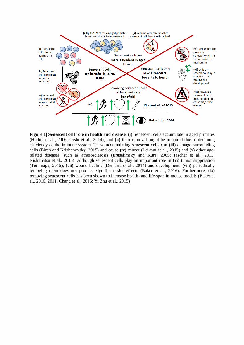
Figure 1| Senescent cell role in health and disease. (i) Senescent cells accumulate in aged primates
(Herbig et al., 2006; Oishi et al., 2014), and (ii) their removal might be impaired due to declining
efficiency of the immune system. These accumulating senescent cells can (iii) damage surrounding
cells (Biran and Krizhanovsky, 2015) and cause (iv) cancer (Leikam et al., 2015) and (v) other age-
related diseases, such as atherosclerosis (Erusalimsky and Kurz, 2005; Fischer et al., 2013;
Nishimatsu et al., 2015). Although senescent cells play an important role in (vi) tumor suppression
(Tominaga, 2015), (vii) wound healing (Demaria et al., 2014) and development, (viii) periodically
removing them does not produce significant side-effects (Baker et al., 2016). Furthermore, (ix)
removing senescent cells has been shown to increase health- and life-span in mouse models (Baker et
al., 2016, 2011; Chang et al., 2016; Yi Zhu et al., 2015)
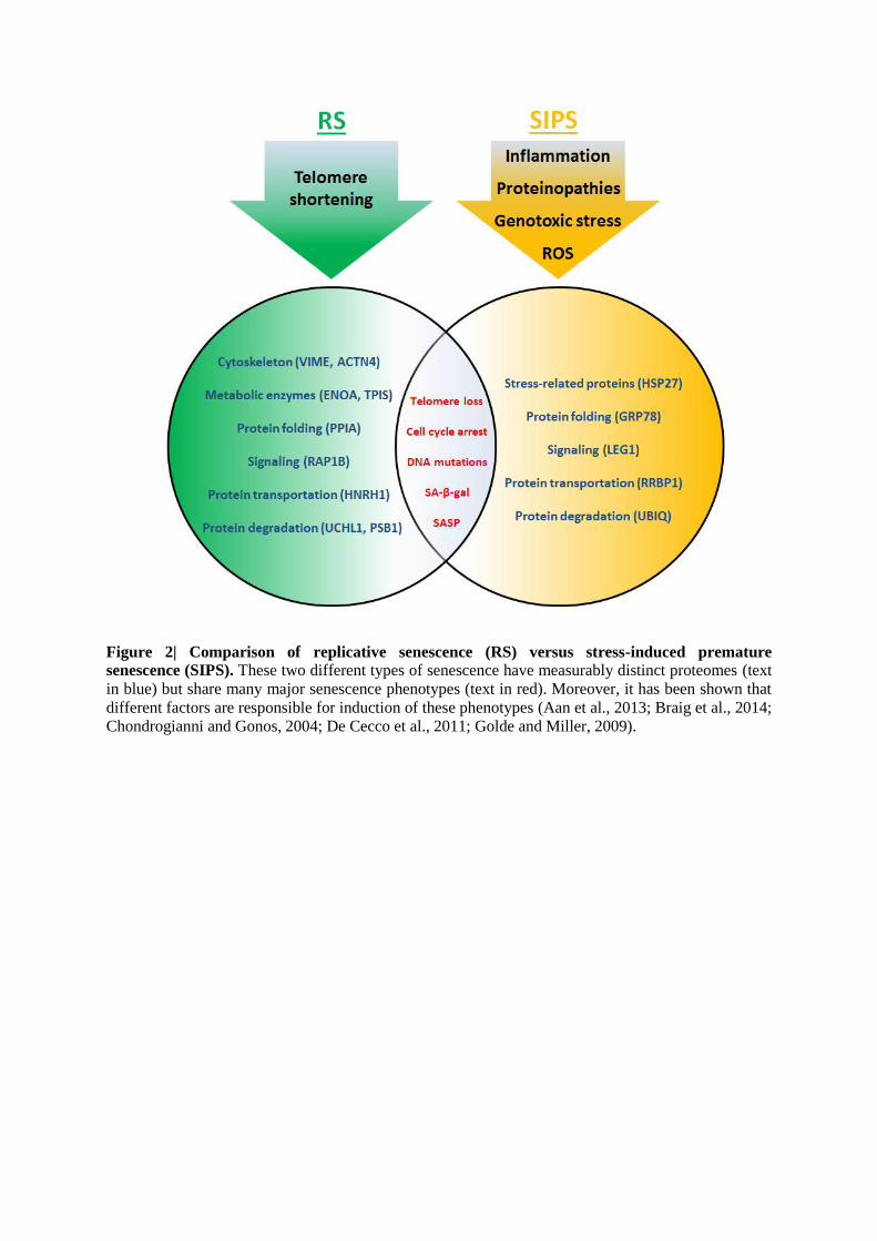
Figure 2| Comparison of replicative senescence (RS) versus stress-induced premature
senescence (SIPS). These two different types of senescence have measurably distinct proteomes (text
in blue) but share many major senescence phenotypes (text in red). Moreover, it has been shown that
different factors are responsible for induction of these phenotypes (Aan et al., 2013; Braig et al., 2014;
Chondrogianni and Gonos, 2004; De Cecco et al., 2011; Golde and Miller, 2009).
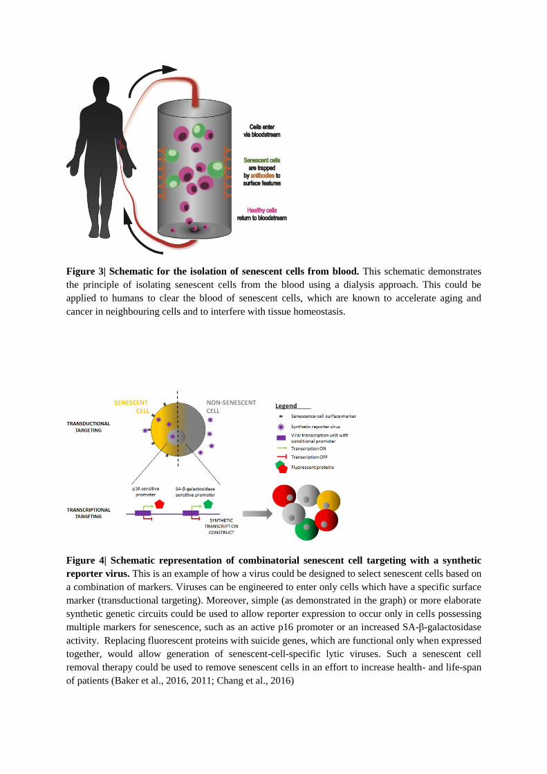
Figure 3| Schematic for the isolation of senescent cells from blood. This schematic demonstrates
the principle of isolating senescent cells from the blood using a dialysis approach. This could be
applied to humans to clear the blood of senescent cells, which are known to accelerate aging and
cancer in neighbouring cells and to interfere with tissue homeostasis.
Figure 4| Schematic representation of combinatorial senescent cell targeting with a synthetic
reporter virus. This is an example of how a virus could be designed to select senescent cells based on
a combination of markers. Viruses can be engineered to enter only cells which have a specific surface
marker (transductional targeting). Moreover, simple (as demonstrated in the graph) or more elaborate
synthetic genetic circuits could be used to allow reporter expression to occur only in cells possessing
multiple markers for senescence, such as an active p16 promoter or an increased SA-β-galactosidase
activity. Replacing fluorescent proteins with suicide genes, which are functional only when expressed
together, would allow generation of senescent-cell-specific lytic viruses. Such a senescent cell
removal therapy could be used to remove senescent cells in an effort to increase health- and life-span
of patients (Baker et al., 2016, 2011; Chang et al., 2016)
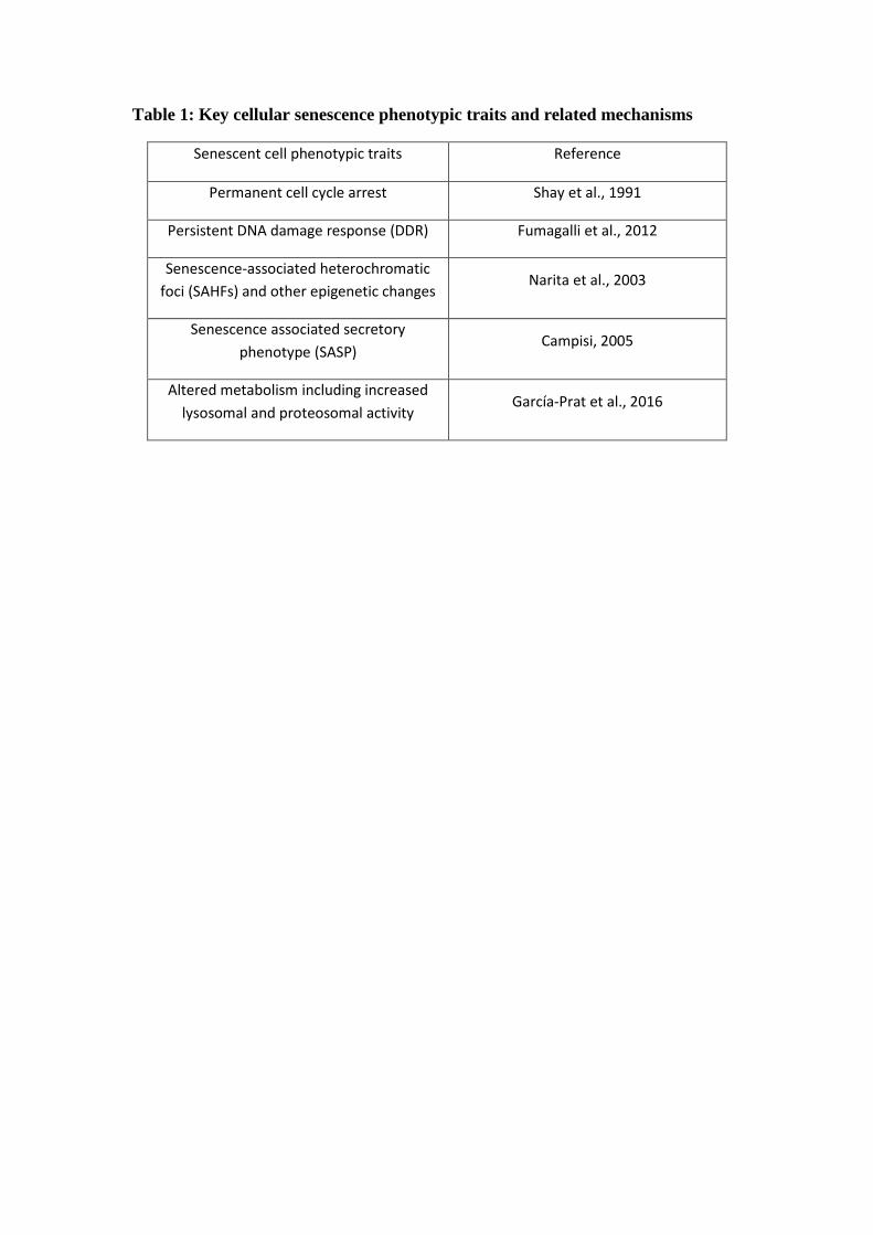
Table 1: Key cellular senescence phenotypic traits and related mechanisms
Senescent cell phenotypic traits Reference
Permanent cell cycle arrest Shay et al., 1991
Persistent DNA damage response (DDR) Fumagalli et al., 2012
Senescence-associated heterochromatic
foci (SAHFs) and other epigenetic changes Narita et al., 2003
Senescence associated secretory
phenotype (SASP) Campisi, 2005
Altered metabolism including increased
lysosomal and proteosomal activity García-Prat et al., 2016
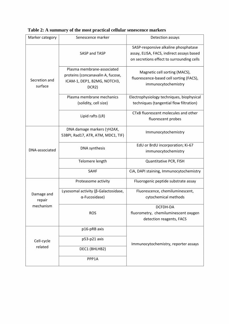
Table 2: A summary of the most practical cellular senescence markers
Marker category Senescence marker Detection assays
Secretion and
surface
SASP and TASP
SASP-responsive alkaline phosphatase
assay, ELISA, FACS, indirect assays based
on secretions effect to surrounding cells
Plasma membrane-associated
proteins (concanavalin A, fucose,
ICAM-1, DEP1, B2MG, NOTCH3,
DCR2)
Magnetic cell sorting (MACS),
fluorescence-based cell sorting (FACS),
immunocytochemistry
Plasma membrane mechanics
(solidity, cell size)
Electrophysiology techniques, biophysical
techniques (tangential flow filtration)
Lipid rafts (LR) CTxB fluorescent molecules and other
fluorescent probes
DNA-associated
DNA damage markers (γH2AX,
53BPI, Rad17, ATR, ATM, MDC1, TIF) Immunocytochemistry
DNA synthesis EdU or BrdU incorporation; Ki-67
immunocytochemistry
Telomere length Quantitative PCR, FISH
SAHF CiA, DAPI staining, Immunocytochemistry
Damage and
repair
mechanism
Proteasome activity Fluorogenic peptide substrate assay
Lysosomal activity (β-Galactosidase,
α-Fucosidase)
Fluorescence, chemiluminescent,
cytochemical methods
ROS
DCFDH-DA
fluorometry, chemiluminescent oxygen
detection reagents, FACS
Cell-cycle
related
p16-pRB axis
Immunocytochemistry, reporter assays p53-p21 axis
DEC1 (BHLHB2)
PPP1A
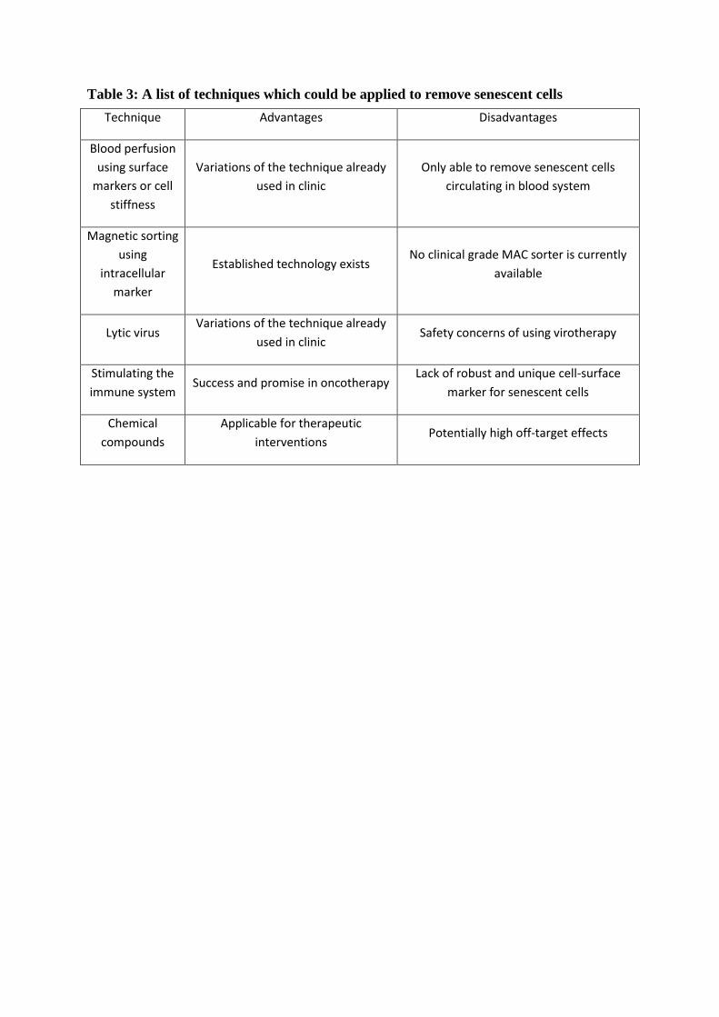
Table 3: A list of techniques which could be applied to remove senescent cells
Technique Advantages Disadvantages
Blood perfusion
using surface
markers or cell
stiffness
Variations of the technique already
used in clinic
Only able to remove senescent cells
circulating in blood system
Magnetic sorting
using
intracellular
marker
Established technology exists No clinical grade MAC sorter is currently
available
Lytic virus Variations of the technique already
used in clinic Safety concerns of using virotherapy
Stimulating the
immune system Success and promise in oncotherapy
Lack of robust and unique cell-surface
marker for senescent cells
Chemical
compounds
Applicable for therapeutic
interventions Potentially high off-target effects