Embed Size (px)
Citation preview

Biomarkers of inflammation
Maureen L. McGary, DNP, NP-C
Assistant Professor
Georgetown University

Presenter Disclosure InformationPennsylvania Council of Nurse Practitioners
Biomarkers of Inflammation
I will not discuss off label use or investigational use in my presentation.
I don’t have financial relationships to disclose related to the topic I will present.

Rheumatologic inflammatory and disease-specific biomarkers: An update for primary care.
Learning Goals:
Upon completion of this presentation the participant will be able to:
Discuss the appropriate biomarker testing for specified major rheumatologic disorders.
Describe role of presented cytokines in inflammation for specified major rheumatologic disorders.
Apply the results of biomarker testing to improve the referral process for patients to rheumatology specialists.
Apply the results of biomarker testing to improve the initial treatment for patients while awaiting evaluation by a rheumatology specialist.

Epidemiology of Rheumatic Disorders in the United States
• For the period 2010-2012, an estimated 52.5 million (22.7%) adults reported doctor diagnosed arthritis or other form of RA, gout, lupus, or fibromyalgia (CDC, 2013a)
• Total direct costs attributable to arthritis and other rheumatologic causes were $80.8 billion ($115 billion in 2013 dollars; CDC, 2007).
• The most common cause of disability in the US is rheumatologic conditions, including arthritis (CDC,2014a).

Epidemiology of Rheumatic Disorders in the United States
• Approximately 294,000 American children under the age of 18 have arthritis or other rheumatologic disorders. (Sacks, Helmick, Luo, Ilowite, & Bowyer, 2006).
• Arthritis and rheumatologic disorders affect women more often than men, those of African decent and whites more than those of Hispanic or Asian decent (CDC, 2013a).
• 67 million adults in the U.S. will have
arthritis per CDC estimates by 2030 (CDC, 2014a).

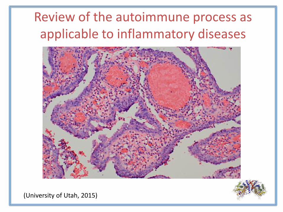
Review of the autoimmune process as applicable to inflammatory diseases
Autoimmune diseases occur when something triggers the immune system to fail to recognize self. The regulatory T cells no longer function to maintain the immune system’s recognition of self, an attack on self begins, and this begins the hallmark inflammatory process.
There are a number of causes of this error. These include stress, viral and bacterial infections, sunlight, solvents, among others. Research on exactly which trigger is the culprit for certain diseases continues.

Review of the autoimmune process as applicable to inflammatory diseases
(Nature Reviews, 2015)

Inflammation
PMN
(University of Utah, 2015)

Review of the autoimmune process as applicable to inflammatory diseases
(University of Utah, 2015)
Review of the autoimmune process as applicable to inflammatory diseases
(University of Utah, 2015)

Review of the autoimmune process as applicable to inflammatory diseases

THE ROLE OF COMPLEMENTS

Biomarkers of Inflammation
…measurable substances, processes, or structuresthat are found in the body or in body productsused to predict or influence the occurrence of adisease process or the outcomes of the disease.
Types and roles

Acute Phase Reactants
• C-Reactive Proteins (CRP)
• Erythrocyte Sedimentation Rate (ESR)
• Serum Complement Proteins
• Platelets

C-Reactive Protein
Information from the American Association
for Clinical Chemistry(AACC) on hs-CRP used for cardiac risk screening
http://labtestsonline.org/understanding/analytes/hscrp/tab/test/
and CRP used for generalized inflammation such as infection or autoimmune diseases
http://labtestsonline.org/understanding/analytes/crp/tab/test/
Be sure to order the appropriate test based on which diagnosis you are exploring.

Two of significance in humans:
C Reactive Protein is expressed during acute phase response to tissue injury or inflammation
Serum amyloid P component may have a role in atherosclerosis and amyloidosis
Pentraxins
Acute phase proteins (APP)

Erythrocyte Sedimentation Rate
The distance that erythrocytes fall (in mm) during one hour in a venous sample.
Normal values: 0-15 mm/hrNewer test allow for 30 minute turnaround

Platelets
Process of increased platelet count
Significance of Increased platelet count
Low to low normal platelet count

Serum Complement Proteins
C3
C4
CH50

Antinuclear Antibodies (ANA)

Anti - Carbamylated Protein (Anti -CarP) Antibodies
Recently identified antibodies that may be present years before RA onset
Associated with erosive RA
Test done by Rheumatology

Rheumatoid Arthritis: anti-CCP (ACPA)
As sensitive as, and more specific than, IgM rheumatoid factors (RF) in early and fully established disease
May predict the eventual development into RA when found in undifferentiated arthritis
A marker of erosive disease in RA
May be detected in healthy individuals years before onset of clinical RA
Normal range: 0-5 U/mL

IgM Rheumatoid Factor (RF) Is found in 60-80% of persons with established RA but only 50-60% of
persons with early RA
Is present in many other disorders
While it does not correlate with disease activity, higher levels do suggest the likelihood of more severe disease and extra-articular manifestations of RA.
Test is for IgM class but around 15% have IgG RF
Normal range < 20, but check normals for your lab.
High lipid levels may affect this test, elderly may have higher results

The Identification of and Role of Biomarkers in Specific Disease States
Inflammatory Arthropathies
Rheumatoid Arthritis
Juvenile Idiopathic Arthritis
Psoriatic Arthritis
Ankylosing Spondylitis

Rheumatoid Arthritis:
Physical Assessment
(ACR, 2015)

Looking for RA: Lab assessment
Complete blood count (CBC)
Comprehensive metabolic panel (CMP)
Rheumatoid Factor (RF)
Antibodies to citrullinated peptides including anti-CCP (also called ACPA)
Erythrocyte Sedimentation Rate (ESR)
C-reactive protein (CRP)(ACR, 2015)

http://www.rheumatology.org/Portals/0/Files/ra_class_slides.pdf

http://www.rheumatology.org/Portals/0/Files/ra_class_slides.pdf
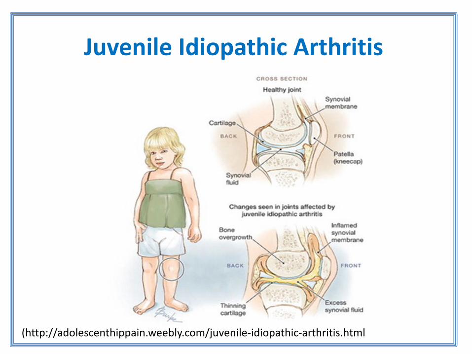
Juvenile Idiopathic Arthritis
(http://adolescenthippain.weebly.com/juvenile-idiopathic-arthritis.html

Juvenile Idiopathic Arthritis
Primarily diagnosed based on clinical presentation
Markers of inflammation ESR CRP
CBC: anemia of chronic disease RF & anti-CCP are not diagnostic but high
titers of anti-CCP may indicate more erosive disease
(Kim & Dong, 2010)

Juvenile Idiopathic Arthritis

Psoriatic Arthritis
http://www.healthcentral.com/rheumatoid-arthritis/cf/slideshows/7-facts-about-rheumatoid-arthritis-diagnosis

Psoriatic Arthritis

SPONDYLOARTHRITISAnkylosing Spondylitis
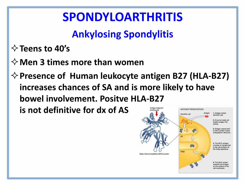
SPONDYLOARTHRITISAnkylosing Spondylitis
Teens to 40’s
Men 3 times more than women
Presence of Human leukocyte antigen B27 (HLA-B27) increases chances of SA and is more likely to have bowel involvement. Positve HLA-B27 is not definitive for dx of AS

Vasculitides
Polymyalgia Rheumatica
Giant Cell Arteritis
Behçet's Disease

Polymyalgia Rheumatica

Polymyalgia Rheumatica
ESR: elevated > 30 but may be over 100 (90-94% have elevated ESR)
Negative RF and ANA the prevalence of positive assays for antinuclear antibody and rheumatoid factor rise with age.
Normocytic anemia may be present
Many will have CRP > 5 (99%)

Polymyalgia Rheumatica
http://bestpractice.bmj.com/best-practice/monograph/153/diagnosis/criteria.html

Polymyalgia Rheumatica

Giant Cell Arteritis
(Temporal Arteritis)

Giant Cell Arteritis (Temporal Arteritis)
Occurs in about 10 % of patients with PMR
Is associated with PMR
Elevated CRP & ESR
Normocytic anemia
Thrombocytosis

El-Dairi et a., 2015

Behçet's Disease/Syndrome

Behçet's Disease/Syndrome
No specific tests
Vessel biopsy showing vasculitis
Pathergy Test: however only small percent will be positive but a positive test is informative

Behçet's Disease/Syndrome
For diagnosis: must have one required criteria and 2 minor criteriaRequired CriteriaRecurrent oral ulcerations: minor aphthous, major aphtous or herpetiformulceration observed by physician or patient, which recurred at least 3 times in one 12-month periodMinor Criteria• Recurrent genital ulceration: aphthous ulceration or scarring observed by
physician or patient• Eye lesion: anterior uveitis, posterior uveitis, or cells in vitreous on slit lamp
examination or retinal vasculitis observed by ophthalmologist• Skin lesions: erythema nodosum observed by physician or patient,
pseudofolliculitis or papulopustular lesions, or acneform nodules observed by physician in post-adolescent patients not on corticosteroid treatment
• Positive pathergy test (Behcetine test) read by physician 24-48 hours.
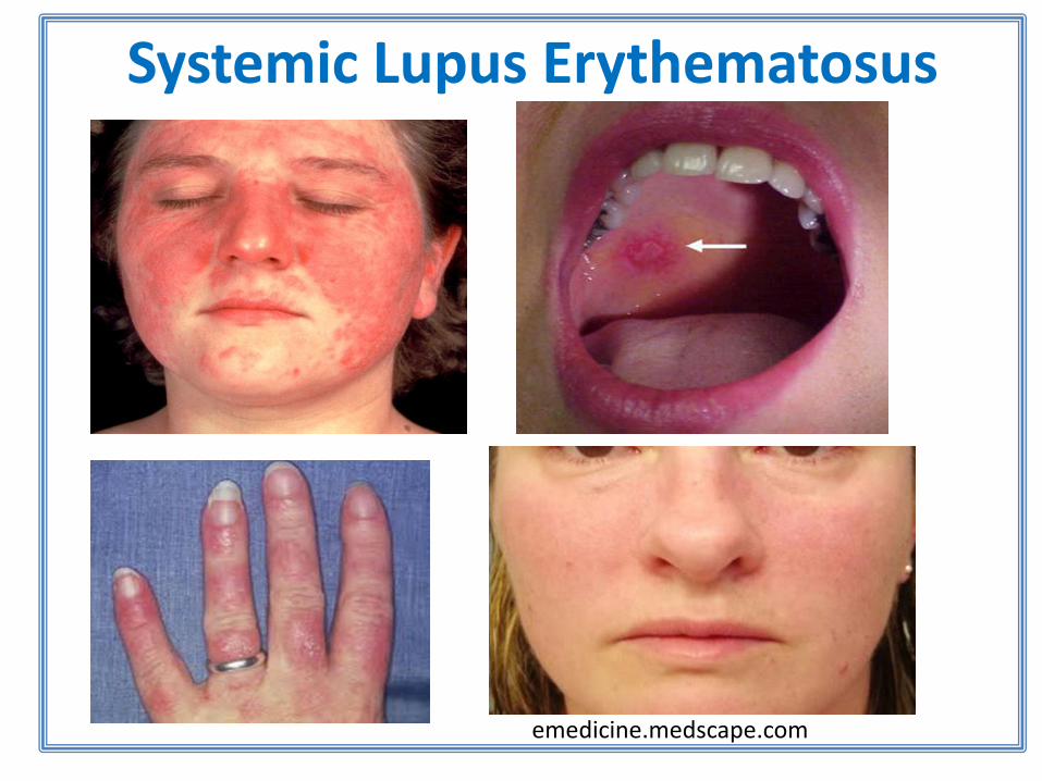
Systemic Lupus Erythematosus
emedicine.medscape.com

For primary care screening in the presence of symptoms:
Positive ANA
Positive Anti dsDNA
Positive Anti-phospholipid antibody
Low Complements C3, C4, CH50
U/A: check for proteinuria
Systemic Lupus Erythematosus

(Lam, Ghetu, & Bieniek, 2016)

Sjögren's Syndrome
www.reviewofoptometry.com
drnadiakidermandentist.wordpress.com

American-European Consensus Criteria for Sjögren’s SyndromeIn order to make a diagnosis of Sjögren’s syndrome, the following criteria must be met:
I. Ocular Symptoms (at least one)Symptoms of dry eyes for at least 3 monthsA foreign body sensation in the eyesUse of artificial tears 3 or more times per day
II. Oral Symptoms (at least one)Symptoms of dry mouth for at least 3 monthsRecurrent or persistently swollen salivary glandsNeed for liquids to swallow dry foods
Sjögren's Syndrome

III. Ocular Signs (at least one)Abnormal Schirmer’s test, (without anesthesia; ≤5 mm/5 minutes)Positive vital dye staining of the eye surface
IV. HistopathologyLip biopsy showing focal lymphocytic sialoadenitis (focus score ≥1 per 4 mm2)
V. Oral Signs (at least one)Unstimulated whole salivary flow (≤1.5 mL in 15 minutes)Abnormal parotid sialographyAbnormal salivary scintigraphy
VI. For a primary Sjögren’s syndrome diagnosis:Any 4 of the 6 criteria, must include either item IV (Histopathology) or VI (Autoantibodies)Any 3 of the 4 objective criteria (III, IV, V, VI)
Sjögren's Syndrome

VI. Autoantibodies (at least one)
Anti-SSA (Ro) or
Anti-SSB (La)
or both
Sjögren's Syndrome

Sjögren's Syndrome
Tran.V.K. (n.d.).
Anti-SSA (Ro)
Primarily associated with Sjögren’s
Detected in 76% of patients with primary Sjögren’s
Detected in only 10-15% of patients with secondary Sjögren’s
Detected in 50% of patients with subacute cutaneous lupus
Associated with other conditions, for example: neonatal lupus
syndrome and congenital heart block
Anti-SSB (La)
Detected in 40-60% of patients with Sjögren’s
Rarely detected without SSA
Associated with ANA-negative SLE and Scleroderma
Utilization of laboratory values in improving the
referral process

Current Research
Research into triggers of RA such as the possible role of oral bacteria, most likely Porphyromonas gingivalis. (Bingham, 2015)
Elevated plasma levels of Citrullinated Proteins and hydroxyproline may be a useful biomarkers for early osteoarthritis. When used in an algorithm that includes anti-CCP, early OA can be distinguished. (Ahmend et al.. 2015).
Osteoarthritis was thought to be related to increase weight bearing load in obese patients. Newer research indicates that there is a connection between negative effects on joint tissues from ø
http://www.hopkinsarthritis.org/arthritis-info/rheumatoid-arthritis/ra-pathophysiology-2/

ReferencesAbeles, A.M. & Abeles, M. (2016). The clinical utility of a positive antinuclear
antibody test result. The American Journal of Medicine. 126(4), 342-348. Retrieved from http://dx.doi.org/10.1016/j.amjmed.2012.09.014
Aggarwal, R., Liao, K., Nair, R., Ringold, S.,& Karen H. Costenbader, K. H.(2010). Anti-Citrullinated Peptide Antibody (ACPA) Assays and their Role in the Diagnosis of Rheumatoid Arthritis. Arthritis and Rheumatism (Arthritis Care and Research). doi: 10.1002/art.24827
Bhagat, M., Sehra, S. T., Shahane, A., Kwan, M. (2014). Utility of immunologic testing in suspected rheumatologic disease. Current Allergy and Asthma Reports, 14 (1), 405-414.
Breda, L., Nozzi, M., De Sanctis, S., Chiarelli, F. (2010). Laboratory tests in the diagnosis and follow-up of pediatric rheumatic diseases: An update./seminars in Arthritis an Rheumatism., 40(1). doi: 10.1016/j.semarthrit.2008.12.001
Fitch-Rogalsky, C., Steber, W., Mahler, M., Lupton, T., Martin, L., Barr, S.G., Mosher, D.P., Wick, J., Fritzler, M.J. (2014). Clinical and Serological Features of Patients referred through a rheumatology triage system because of positive antinuclear antibodies. PLoS One, 9 (4). doi: 10.1371/journal.pone.0093812

ReferencesLam, N.V., Ghetu, M., & Bieniek, M.L. (2016). Systemic Lupus Erythematosus:
Primary care approach to diagnosis and management. American Family Physician. 94(4), 284- 294.
Habets, K.L., Huizinga, T.W., Toes, R.E. (2013). Platelets and autoimmunity. European Journal of Clinical Investigation. 43(7). doi: 10.1111/eci.12101
Healthtap. (2015) Graphics. Retrieved from https://www.healthtap.com/topics/signs-of-autoimmune-diseases-with-negative-blood-tests
Meroni, P.L., Chan, E.K., Tincani, A., de la Torre, I.G., & Andrade, L.E. (2013). Antinuclear antibody test: When to order? [Letter to the editor]. The American Journal of Medicine. 126(10, e17). Retrieved from http://dx.doi.org/10.1016/j.amjmed.2013.04.022
Mills, K. (2004). Nature Reviews Immunology 4, 841-855 (November 2004)
doi:10.1038/nri1485

ReferencesNardi N., Brito-Zerón P., Ramos-Casals M., et al. (2006). Circulating auto-
antibodies against nuclear and non-nuclear antigens in primary Sjögren's syndrome: Prevalence and clinical significance in 335 patients. Clinical Rheumatology, 25, 341-346
Soto, M.E., Hern ´andez-Becerril, N., Perez-Chiney, A.C., Hern ´andez- Rizo, A., Telich-Tarriba, J.E., Ju ´arez-Orozco, L.E., Melendez, G., Bojalil, R. (2013). Predictive value of antinuclear antibodies in autoimmune diseases classified by clinical criteria: Analytical study in a specialized health institute, 1 year follow-up. Results in Immunology. Retrieved from: http://www.sciencedirect.com/science/article/pii/S221128391300017
Thijssen, E., van Caam, A., van der Kraan, P. M. (2015). Obesity and osteoarthritis, more than just wear and tear: Pivotal roles for inflamed adipose tissue and dyslipidaemia in obesity-induced osteoarthritis. Rheumatology 54 (4): 588-600. doi: 10.1093/rheumatology/keu464

References
Tran.V.K. (n.d.). Rheumatology panel for primary care [PowerPoint]. Retrieved August 31, 2016 from http://slideplayer.com/slide/8986308/
Willemze, A., Toes, R.E.M., Huizinga, T.W.J., & Trouw, L.A. (2012). New biomarkers in rheumatoid arthritis. The Netherlands Journal of Medicine,7 0 (9), 393-398.