Embed Size (px)
Citation preview
Biology, Prognosis, and Therapyof Waldenström Macroglobulinemia
Jorge J. Castillo, Irene M. Ghobrial and Steven P. Treon
Abstract
WaldenströmMacroglobulinemia (WM) is a rare B-cell lymphoma characterizedby the uncontrolled accumulation of malignant lymphoplasmacytic cells, mainlyin the bone marrow, and monoclonal IgM production. Despite its rarity, ourunderstanding of the biology of this disease has improved significantly in recentyears with the identification of recurrent mutations in the MYD88 and CXCR4genes. Based on the diversity of clinical features observed in WM patients,therapy should be highly personalized having into account several factors suchas age, co-morbidities, IgM levels, and presence of hyperviscosity, coagulop-athy, cryoglobulinemia, or cold agglutinin disease. In this chapter, we review therecent advances in the biology of WM and the current therapeutic options foruntreated and relapsed WM patients. Finally, we discuss the role of prognosticfactors and current evidence supporting an improvement in the survival of WMpatients in the last decade.
Keywords
Waldenström Macroglobulinemia � MYD88 � CXCR4 � Biology � Therapy �Survival
J.J. Castillo (&) � I.M. Ghobrial � S.P. TreonDivision of Hematologic Malignancies, Dana-Farber Cancer Institute, Harvard MedicalSchool, 450 Brookline Ave, M221, 02215 Boston, MA, USAe-mail: [email protected]
© Springer International Publishing Switzerland 2015A.M. Evens and K.A. Blum (eds.), Non-Hodgkin Lymphoma,Cancer Treatment and Research 165, DOI 10.1007/978-3-319-13150-4_7
177
Contents
1 Introduction .......................................................................................................................... 1782 Clinical Features of WM..................................................................................................... 1783 Diagnosis of WM ................................................................................................................ 1794 Biology of WM ................................................................................................................... 1795 Criteria for Initiation of Therapy......................................................................................... 1806 Frontline Therapy for WM.................................................................................................. 180
6.1 Proteasome Inhibitor-Based Therapy ......................................................................... 1836.2 Alkylator-based Therapy ............................................................................................ 1846.3 Nucleoside Analog-Based Therapy ............................................................................ 1846.4 Other Options.............................................................................................................. 185
7 Salvage Therapy for Relapsed/Refractory WM.................................................................. 1867.1 Bortezomib.................................................................................................................. 1867.2 Nucleoside Analogs and Alkylating Agents .............................................................. 1867.3 Ofatumumab................................................................................................................ 1867.4 Everolimus .................................................................................................................. 1877.5 Ibrutinib....................................................................................................................... 187
8 Maintenance Therapy........................................................................................................... 1879 High-Dose Therapy and Stem Cell Transplantation ........................................................... 18810 Prognostic Factors for Survival in WM............................................................................ 18811 Trends in Survival in WM ................................................................................................ 19012 Conclusion ......................................................................................................................... 191References .................................................................................................................................. 192
1 Introduction
Waldenström Macroglobulinemia (WM) is a rare B-cell lymphoproliferative dis-order characterized by the uncontrolled accumulation of malignant immunoglobulinM (IgM)-secreting lymphoplasmacytic cells. WM belongs to the lymphoplasma-cytic lymphoma (LPL) category as defined by the 2008 World Health Organizationclassification [1]. However, over 95 % of the cases of LPL are WM with theremainder comprised by IgA, IgG, and non-secreting LPL. WM occurs in adultswith a median age in the 60s with a slight male predominance. A familial predis-position has been described in approximately 25 % of the patients with WM, withfamilial patterns varying from presence of various B-cell malignancies, and familiesin which multiple cases of WM or IgM MGUS have been observed [2].
2 Clinical Features of WM
WM presents predominantly with bone marrow involvement and only a minority ofpatients (15–30 %) present with extramedullary disease such as lymphadenopathyor hepatosplenomegaly. The most common clinical features associate with anemia(i.e., fatigue, tiredness, and/or shortness of breath) due to overcrowding of the bonemarrow space by LPL or iron deficiency [3]. However, given the physicochemical
178 J.J. Castillo et al.
characteristics of IgM, patients can experience signs and symptoms associated withother mechanisms such as hyperviscosity, cryoglobulinemia, peripheral neuropathy,coagulopathy, cold agglutinins, and tissue deposition in the skin (Schnitzler syn-drome), gastrointestinal tract, central nervous system (Bing-Neel syndrome), orkidneys. Patients can rarely present with amyloid deposition, which can causeedema, hepatomegaly, macroglossia, cardiac, liver and kidney dysfunction, andaxonal peripheral neuropathy.
3 Diagnosis of WM
The diagnostic criteria for WM are shown in Table 1. A bone marrow aspirationand biopsy is a key component of the diagnostic work-up. The immunopheno-typical profile of WM cells shows expression of surface IgM, CD19, CD20, CD22,CD38, and CD79. Up to 20 % of cases may express CD5, CD10, or CD23. Anincreased number of mast cells in the bone marrow may help differentiate WM fromother indolent B-cell lymphomas. A great variety of cytogenetic abnormalities havebeen described in WM; however, chromosome 6q deletions have been observed inhalf of the patients [4]. More recently, a recurrent mutation in the MYD88 gene(MYD88 L265P) has been identified in over 90 % of cases with WM [5]. Theoccurrence of this mutation in WM has since been validated in several independentcohorts [6–9]. In contrast, the MYD88 L265P gene mutation was not detected inpatients with IgM myeloma and was detected in less than 10 % of patients withmarginal zone lymphoma. The high specificity and sensitivity of the MYD88L265P gene mutation has obvious diagnostic implications in patients in whom adiagnosis of WM is suspected but uncertain.
4 Biology of WM
The MYD88 L265P gene mutation has shown to support growth and survival ofWM cells in several studies. A knockdown model of MYD88 showed decreasedsurvival of MYD88 L265P expressing WM cells, whereas survival was moreenhanced by knock-in of mutant versus wild-type MYD88 [10]. MYD88 acts as anadaptor molecule in toll-like receptor (TLR) and interleukin-1 receptor (IL-1R)signaling [11]. Following stimulation of TLR or IL-1R, MYD88 is recruited to theactivated receptor complex as a homodimer which complexes with IL-1R-associated
Table 1 Diagnostic criteriafor Waldenströmmacroglobulinemia
IgM monoclonal gammopathy of any concentration
Bone marrow infiltration by small lymphocytes showingplasmacytic differentiation
Intertrabecular pattern of bone marrow infiltration
Surface IgM+, CD19+, CD20+, CD22+, CD25+. CD27+,FMC7+, CD5±, CD10−, CD23−, CD103−
Biology, Prognosis and Therapy … 179
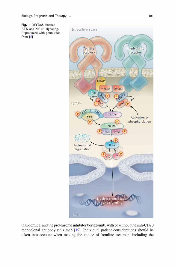
kinase 4 (IRAK4) and subsequently activates IRAK1 and IRAK2 [12]. IRAK1activation then leads to NF-κB activation via IκBα phosphorylation [13]. Recently, astudy has shown that MYD88 L265P also activates the Bruton’s Tyrosine Kinase(BTK) pathway [10]. In this preclinical study, the activation of BTK by MYD88could be abrogated by the use of BTK kinase inhibitors. A diagram of the activationof BTK and NF-κB via MYD88 is shown in Fig. 1.
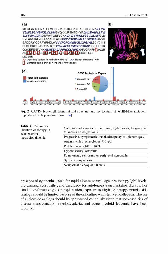
A recent study from our group first ever reported the occurrence of recurrentsomatic CXCR4 gene mutations in approximately 30 % of WM patients [14]. Thesomatic mutations occur in the C-terminal domain and are similar to those observedin patients with WHIM (Warts, Hypogammaglobulinemia, Infections, and Myelo-kathexis) syndrome. These mutations regulate signaling of CXCR4 by its ligandSDF-1a [15]. In WM patients, two classes of CXCR4 mutations occur: non-sense(CXCR4WHIM/NS) and frameshift (CXCR4WHIM/FS) mutations [14, 16]. Non-senseand frameshift mutations are almost equally divided among WM patients, and over30 different types of CXCR4 mutations have been identified. Preclinical studieswith the most common CXCR4 S338X mutation in WM have shown sustainedsignaling of AKT, ERK, and BTK following SDF-1a binding in comparison withwild-type CXCR4, as well increased cell growth and survival of WM cells [17].Figure 2 shows (a) the protein sequence for the full-length transcript, (b) the crystalstructure of CXCR4, and (c) the location of the WHIM-like mutations.
5 Criteria for Initiation of Therapy
Given the indolent and incurable nature of WM, a large proportion of patientswould not need immediate therapy upon diagnosis and will be placed on watchfulwaiting. Current guidelines do not recommend initiation of therapy based on IgMlevels alone since they might not correlate with clinical manifestations of the dis-ease [18]. Initiation of therapy is, however, reasonable in patients demonstratingrising IgM levels along with signs or symptoms associated with disease progres-sion. Criteria for initiation of therapy are shown in Table 2. In patients in whom animmediate control of the disease is warranted, such as symptomatic hyperviscosity,coagulopathy, cryoglobulinemia, or cold agglutinin disease, a rapid reduction of theIgM paraprotein should be achieved with plasmapheresis. Treatment directed atWM should follow as soon as possible since plasmapheresis does not constitutedefinitive therapy and IgM levels will rise and return to baseline levels within4 weeks.
6 Frontline Therapy for WM
There are several options for the frontline therapy of patients withWM. These optionsinclude alkylating agents (i.e., chlorambucil, cyclophosphamide, and bendamustine),nucleoside analogs (i.e., fludarabine and cladribine), the immunomodulator
180 J.J. Castillo et al.
thalidomide, and the proteasome inhibitor bortezomib, with or without the anti-CD20monoclonal antibody rituximab [19]. Individual patient considerations should betaken into account when making the choice of frontline treatment including the
Fig. 1 MYD88-directedBTK and NF-κB signaling.Reproduced with permissionfrom [5]
Biology, Prognosis and Therapy … 181
presence of cytopenias, need for rapid disease control, age, pre-therapy IgM levels,pre-existing neuropathy, and candidacy for autologous transplantation therapy. Forcandidates for autologous transplantation, exposure to alkylator therapy or nucleosideanalogs should be limited because of the difficulties with stem cell collection. The useof nucleoside analogs should be approached cautiously given that increased risk ofdisease transformation, myelodysplasia, and acute myeloid leukemia have beenreported.
Table 2 Criteria forinitiation of therapy inWaldenströmmacroglobulinemia
Constitutional symptoms (i.e., fever, night sweats, fatigue dueto anemia or weight loss)
Progressive, symptomatic lymphadenopathy or splenomegaly
Anemia with a hemoglobin ≤10 g/dl
Platelet count <100 × 109/L
Hyperviscosity syndrome
Symptomatic sensorimotor peripheral neuropathy
Systemic amyloidosis
Symptomatic cryoglobulinemia
Fig. 2 CXCR4 full-length transcript and structure, and the location of WHIM-like mutations.Reproduced with permission from [14]
182 J.J. Castillo et al.
6.1 Proteasome Inhibitor-Based Therapy
The WM Clinical Trial Group presented data on a study that included 23 patientsusing bortezomib, dexamethasone, and rituximab (BDR) for the primary therapy ofsymptomatic WM [20]. Bortezomib was administered at a dose of 1.3 mg/m2 IValong with dexamethasone 40 mg on days 1, 4, 8, and 11 and rituximab 375 mg/m2
IV on day 11 every 21 days for four induction cycles, followed by four maintenancecycles administered every 3 months. The overall response rate (ORR) was 96 % withthree complete responses (CR), two near complete responses (nCR), and three verygood partial responses (VGPR) for a VGPR or better of 35 %. The median time toresponse was 1.4 months, which makes BDR an excellent choice if a rapid control ofdisease is required or in young patients to decrease the risk of secondary myeloidmalignancies. With a median follow-up of 23 months, 18 out of 23 patients (78 %)remained free of progression. The most common toxicity was peripheral neuropathy;39 and 30 % of patients, respectively, experienced grade 2 and grade 3 peripheralneuropathy. Grade 2 and grade 3 neutropenia were also reported in 26 % and 26 % ofpatients, respectively. It is important to note that appropriate Herpes zoster pro-phylaxis should be instituted in all patients receiving proteasome inhibitor therapy.
Another study evaluated weekly bortezomib in combination with rituximab in 26untreated patients with WM [21]. In this study, bortezomib was administered at adose of 1.6 mg/m2 IV weekly on days 1, 8, and 15 every 28 days for six cycles withrituximab 375 mg/m2 weekly times four during cycles 1 and 4 only. The ORR was88 % with a VGPR or better of 8 %. The median time to best response was3.7 months. The 1-year PFS rate was 75 %. There was 54 % of peripheral neu-ropathy grade 2 or lower but no grade 3 or higher neuropathy was observed. Grade3 or higher neutropenia was seen in 12 % of patients. More recently, a study by theEuropean Myeloma Network evaluated the efficacy of weekly bortezomib, low-dose dexamethasone, and rituximab in 59 untreated WM patients [22]. Bortezomibwas administered as a single agent at 1.3 mg/m2 IV on days 1, 4, 8, and 11 (cycle 1)followed 3 weeks later by weekly bortezomib at 1.6 mg/m2 IV and dexamethasone40 mg IV on days 1, 8, 15, and 22 and rituximab 375 mg/m2 IV every 35 days(cycles 2–5). The ORR was 85 % with VGPR or better of 10 %. After a 32-monthfollow-up, the median PFS was 42 months. Grade 3 or higher peripheral neuropathyoccurred in 7 % of patients. The subcutaneous (SQ) administration of bortezomibhas shown to be effective and induces less neuropathy than the IV route in patientswith myeloma [23]; however, the safety and efficacy of bortezomib SQ has not beenformally studied in patients with WM.
A recent study evaluated the combination of carfilzomib, dexamethasone, andrituximab (CaRD) in 31 patients with WM [24]. Carfilzomib was administered at adose of 20 mg/m2 IV during cycle 1 and at 36 mg/m2 IV during cycles 2–6 withdexamethasone 20 mg IV on days 1, 2, 8, and 9 along with rituximab on days 2 and9 every three weeks during induction therapy. Maintenance therapy consisted oncarfilzomib 36 mg/m2 IV and dexamethasone 20 mg IV on days 1 and 2 andrituximab 375 mg/m2 on day 2 every 2 months for eight cycles. The ORR was 87 %
Biology, Prognosis and Therapy … 183
with one CR and ten VGPR for a VGPR or better of 35 %. The 15-month PFS ratewas 65 %. No grade 3 neuropathy was observed. Grade 3 hyperglycemia andhyperlipasemia were observed in 23 and 16 % of patients, respectively. Of note, therate of IgG hypogammaglobulinemia increased from 48 % at baseline to 90 %following therapy.
6.2 Alkylator-based Therapy
Cyclophosphamide-based therapy includes regimens in which cyclophosphamide iscombined with rituximab and prednisone (CP-R), rituximab, prednisone and vin-cristine (CVP-R), and rituximab, prednisone, vincristine and doxorubicin (CHOP-R). These regimens would induce an ORR of 70–80 % with CR rate around 10 %[25]. It is unclear if the addition of vincristine or doxorubicin translates into clinicalbenefits in patients with WM. A retrospective study compared the activity andtoxicity of CP-R, CVP-R, and CHOP-R in patients with WM [26]. In that study,CP-R was associated with similar response rates than CVP-R and CHOP-R.However, the rate of treatment complications such as febrile neutropenia, hospi-talizations, and neuropathy was lower. Based on these data, CP-R might bepreferred over CVP-R or CHOP-R in patients with WM.
The combination of bendamustine and rituximab (BR) has been compared withCHOP-R in a randomized controlled study by the Study Group for Lymphomas in acohort that included 42 patients with WM [27]. Bendamustine was administered ata dose of 90 mg/m2 IV on days 1 and 2 along with rituximab 375 mg/m2 IV on day1 at 3-week intervals for 6 cycles. The ORR for BR was 96 % and 94 % for CHOP-R. With a median follow-up of 26 months, the PFS rate for BR was 90 % comparedwith 59 % for CHOP-R. BR was associated with lower rates of grade 3 or higherneutropenia, infectious complications, and alopecia, but with higher rates of skinrash. BR might provide an excellent therapeutic option for patients with lym-phadenopathy or patients with pre-existing neuropathy.
6.3 Nucleoside Analog-Based Therapy
In the large randomized controlled study to date, which enrolled 414 patients (339WM, 37 MZL, and 38 LPL) from 101 centers in five countries, fludarabine wascompared with chlorambucil in untreated WM patients [28]. In this study, the ORRfor fludarabine was 48 % versus 39 % for chlorambucil. The median PFS was alsolonger for fludarabine (36 months) in comparison with chlorambucil (27 months).There was a significant improvement in median OS, which was not reached forfludarabine versus 70 months for chlorambucil. Finally, the rate of second malig-nancieswas higher in the chlorambucil arm than in thefludarabine arm (21%vs. 4%).
The long-term outcomes of the combination of fludarabine and rituximab (FR)were evaluated in 43 patients with WM, from which 27 patients were untreated
184 J.J. Castillo et al.
[29]. Therapy consisted of 8 infusions of rituximab at 375 mg/m2 IV per weekadministered at weeks 1–4, 17, 18, 30, and 31, along with 6 cycles of fludarabine25 mg/m2 IV daily given for 5 days at weeks 5, 9, 13, 19, 23, and 27. The ORR inuntreated patients was 96 %. The median time to response was 3.9 months, and themedian time to best response was 19 months. The median time to progression inuntreated patients was 78 months. Grade 3 or higher adverse events includedmyelosuppression and infections. Other observed complications were aggressivetransformation and secondary malignancies, although most of these events wereseen in the previously treated group of patients.
It is unclear at this time whether the combination of an alkylating agent such ascyclophosphamide, a nucleoside analog such as fludarabine, and rituximab (FCR)provides any additional benefits in patients with WM. A study evaluated FCR in 43patients with WM of which 28 were previously untreated [30]. The FCR regimenconsisted of rituximab 375 mg/m2 IV on day 1 and fludarabine 25 mg/m2 andcyclophosphamide 250 mg/m2 IV on days 2 through 4. Cycles were repeated everyfour weeks for a maximum of six cycles. The ORR was 79 % with no differencebetween untreated and previously treated patients. Grade 3 or higher neutropeniawas seen in 88 % of the patients. After a median follow-up of 39 months, 11patients (25 %) have died, including five from progressive disease, three frompneumonia, and one from acute myeloid leukemia.
6.4 Other Options
The combination of thalidomide and rituximab (TR) has been associated with anORR of 70 % and a median PFS of 3 years [31]. TR could be useful in myelo-suppressed patients in whom an immediate disease control is not required. Themain adverse event associated with thalidomide is neuropathy, which can be seen inup to 40 % of patients treated with TR.
The use of rituximab as single agent can be considered in selected patients withlow IgM levels or concurrent autoimmune processes such as cold agglutinin dis-ease, autoimmune thrombocytopenia, or IgM-related neuropathy. The ORR to 4weekly infusion of rituximab is 20–30 % [32] and to 4 weekly infusions followedby 4 weekly infusions 12 weeks later of 40–50 % [33]. The use of single-agentrituximab in patients with high IgM levels has been associated with IgM flares withan occurrence rate of 40–50 % [34]. These flares might lead to symptomatichyperviscosity as well as worsening IgM-related neuropathy or cryoglobulinemia.Hence, rituximab monotherapy should be avoided in patients with IgM lev-els >4,000 mg/dl.
Biology, Prognosis and Therapy … 185
7 Salvage Therapy for Relapsed/Refractory WM
For those patients with relapsed/refractory disease, any of the frontline therapiesmentioned above are appropriate options. Therapies in the relapsed setting shouldalso be personalized to the individual patient considering age, performance status,pre-existing neuropathy, IgM levels, and/or need for immediate control of thedisease. One must also have in mind to avoid the use of stem cell toxic therapy inpatients in whom high-dose chemotherapy with autologous stem cell rescue is beingconsidered.
7.1 Bortezomib
Bortezomib-based therapy has shown to be effective in relapsed or refractory WMpatients in several studies, either alone or in combination with rituximab [35–38].The ORR ranged between 30 and 80 % depending on the study with the mostcommon adverse events being peripheral neuropathy, neutropenia, and thrombo-cytopenia. Once-weekly administration of bortezomib has been associated withlower rates of neuropathy when compared with the twice-weekly regimen. In onestudy, bortezomib seemed to be less effective on inducing lymph node responses[35]. As mentioned previously, patients undergoing therapy with proteasomeinhibitors should receive prophylaxis against herpes zoster.
7.2 Nucleoside Analogs and Alkylating Agents
Fludarabine has single-agent activity in relapsed WM with an approximate 40 %ORR [39]. The combination FR showed high activity in relapsed/refractory WMpatients with ORR of 93 % [29]. The most common adverse events were neutro-penia and infections. There were also concerns about aggressive transformation andthe development of secondary myeloid malignancies.
The combination BR showed an ORR of 83 % with a median PFS of 13 months[40]. The most common adverse events were prolonged myelosuppression, spe-cifically in patients who had received prior nucleoside analog therapy.
7.3 Ofatumumab
The fully human anti-CD20 monoclonal antibody ofatumumab is also useful in therelapsed/refractory setting. Ofatumumab has shown to be effective and well toler-ated in WM patients who develop rituximab intolerance [40]. A study published inabstract form showed an ORR of 57 % in patients with relapsed/refractory WM[41]. The occurrence rate of IgM flare to ofatumumab in this study was 5 %. Themost common adverse events were infections.
186 J.J. Castillo et al.
7.4 Everolimus
Everolimus is an oral mammalian target of rapamycin inhibitor and has induced anORR of 50 % in patients with relapsed/refractory WM [42]. The median time toresponse in patients who achieved a PR was 2 months with a median PFS of21 months. The most common adverse events were anemia, leukopenia, throm-bocytopenia, and stomatitis. Everolimus has also been studied in the frontlinesetting [43]. In this multicenter prospective study, 33 WM patients who were notpreviously treated received everolimus at a dose of 10 mg PO once daily. The bestORR was 72 % with a median time to best response of 6 months. Interestingly, thebone marrow burden of disease remained unchanged. This bone marrow–IgMdiscordance complicated response assessment. The most common adverse eventswere anemia, oral ulcerations, and pneumonitis.
7.5 Ibrutinib
The oral Bruton tyrosine kinase inhibitor ibrutinib has been evaluated in 63 patientswith relapsed/refractory WM [44]. Ibrutinib was administered at a dose of 420 mgPO daily. The ORR was 81 % with a median time to response of 4 weeks. The mostcommon adverse events were thrombocytopenia and neutropenia. Of note, WMpatients with CXCR4 gene mutations experienced lower response rates to ibrutinibthan patients with wild-type CXCR4.
8 Maintenance Therapy
A retrospective study examined the outcome of 248 WM rituximab-naïve patientswho were treated with rituximab-containing regimen and then either observed orreceived maintenance rituximab [45]. In this study, further improved responses afterinduction therapy were seen in 10 % (16 out of 162) of observed patients and in42 % (36 out of 86) of patients who received maintenance rituximab. Both PFS (56vs. 29 months) and OS (>120 vs. 116 months) were longer in patients who receivedmaintenance rituximab. Improved PFS was evident despite previous treatmentstatus or type of induction therapy. Among patients receiving maintenance ritux-imab, an increased number of infectious events, predominantly grades 1 and 2sinusitis and bronchitis, were observed. A prospective study examining the role ofmaintenance rituximab in patients with WM has been initiated by the German STiLgroup [46]. After undergoing induction therapy with BR, 100 out 162 patientsresponded to BR (ORR 86 %). From these patients, 43 were then randomized toobservation and 47 to maintenance rituximab. Enrollment for this study is complete,and final results are awaited.
Biology, Prognosis and Therapy … 187
9 High-Dose Therapy and Stem Cell Transplantation
The use of stem cell transplantation (SCT) therapy has been explored in patientswith WM. Several small series have been reported for autologous and allogeneictransplantation, with variable outcomes. Kyriakou and colleagues reported thelargest experience using European Bone Marrow Transplant (EBMT) data for WMpatients receiving autologous SCT [47]. Among 158 relapsed/refractory WMpatients receiving an autologous SCT, the 5-year PFS and OS rates were 49 and69 %, respectively. Non-relapse mortality at 1 year was 4 %. Chemorefractorydisease and number of prior lines of therapy at time of autologous SCT were themost important prognostic factors for PFS and OS. When used as consolidation atfirst response, autologous SCT provided a PFS rate of 44 % at 5 years. Long-termoutcomes of WM patients undergoing allogeneic SCT from EBMT have beenreported [48]. A total of 86 patients received allograft by either myeloablative orreduced-intensity conditioning (RIC). The median age was 49 years, and 47 patientshad 3 or more previous lines of therapy. Eight patients failed prior autologous SCT.Fifty-nine patients (69 %) had chemotherapy-sensitive disease at the time of allo-geneic SCT. Non-relapse mortality rate at 3 years was 33 % for patients receivingmyeloablative transplant and 23 % for those who received RIC. The ORR was76 %. The relapse rates at 3 years were 11 % for myeloablative and 25 % for RICrecipients. Five-year PFS and OS rates for WM patients who received a my-eloablative allogeneic SCT were 56 and 62 %, respectively, and for patients whoreceived RIC were 49 and 64 %, respectively. The occurrence of chronic graft-versus-host disease was associated with improved PFS and suggested the existenceof relevant graft-versus-WM effect in this study.
10 Prognostic Factors for Survival in WM
In 2003, the Consensus Panel Recommendation from the Second InternationalWorkshop on WM emitted a statement on prognostic markers for survival inpatients with WM [18]. In this paper, prognostic factors such as age, hemoglobinlevel, white blood cell count, platelet count, weight loss, cryoglobulinemia, serumalbumin level, IgM level, and beta-2-microglobulin level were identified based onthree separate multivariate analyses. However, these studies reported on the sur-vival of WM patients treated with chemotherapy alone 5–10 years before the actualreport and, likely, are no longer representative of current outcomes. Since then,multiple studies have aimed at developing prognostic scores that can guide prac-titioners and WM patients.
Arguably, the most widely used prognostic score is the International PrognosticScoring System for WM (IPSSWM) [49]. In this study, data on 597 previouslyuntreated WM patients from seven institutions were analyzed. After univariate andmultivariate evaluation,five adverse prognostic factorswere identified: age >65 years,
188 J.J. Castillo et al.
hemoglobin ≤11.5 g/dl, platelet count ≤100 × 109/L, beta-2-microglobulin >3 mg/dl,and monoclonal IgM concentration >7 g/dl. The hazard ratio (HR) for OS of most ofthe factors ranged between 1.5 and 1.9, with the exception of age (HR 2.8). Patientswere stratified in three risk groups: low (0 or 1 factor except for age), intermediate (ageor 2 factors), and high risk (≥3 factors). The distribution of the groups was as follows:low (27 %), intermediate (38 %), and high risk (35 %). The median OS for the low,intermediate, and high-risk groups were 143, 99, and 44 months, respectively. One ofthe caveats of this study is that 63%of patients were treatedwith alkylating agents and32 % with fludarabine alone; only 4 % of patients received rituximab.
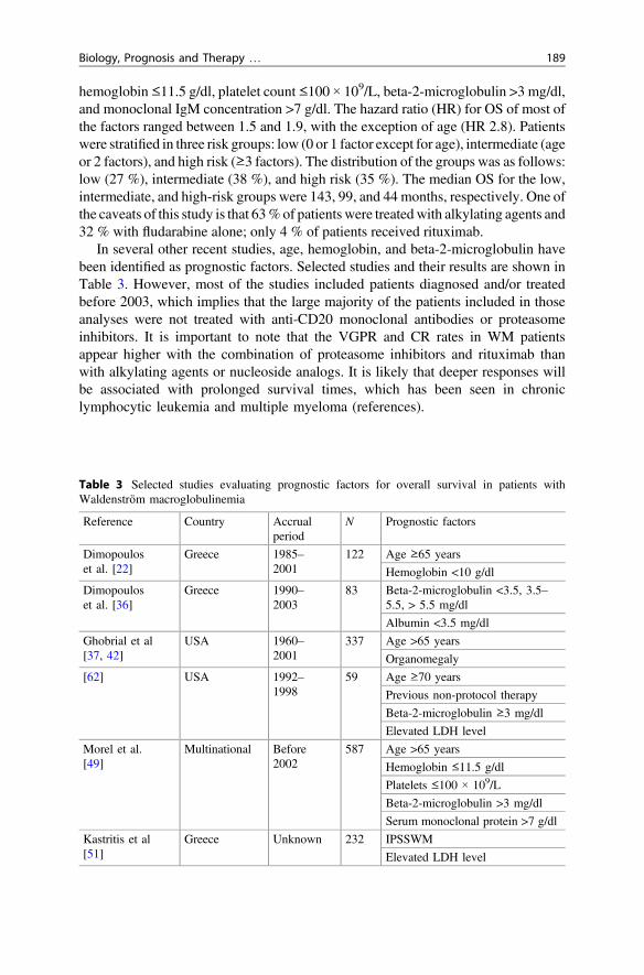
In several other recent studies, age, hemoglobin, and beta-2-microglobulin havebeen identified as prognostic factors. Selected studies and their results are shown inTable 3. However, most of the studies included patients diagnosed and/or treatedbefore 2003, which implies that the large majority of the patients included in thoseanalyses were not treated with anti-CD20 monoclonal antibodies or proteasomeinhibitors. It is important to note that the VGPR and CR rates in WM patientsappear higher with the combination of proteasome inhibitors and rituximab thanwith alkylating agents or nucleoside analogs. It is likely that deeper responses willbe associated with prolonged survival times, which has been seen in chroniclymphocytic leukemia and multiple myeloma (references).
Table 3 Selected studies evaluating prognostic factors for overall survival in patients withWaldenström macroglobulinemia
Reference Country Accrualperiod
N Prognostic factors
Dimopouloset al. [22]
Greece 1985–2001
122 Age ≥65 years
Hemoglobin <10 g/dl
Dimopouloset al. [36]
Greece 1990–2003
83 Beta-2-microglobulin <3.5, 3.5–5.5, > 5.5 mg/dl
Albumin <3.5 mg/dl
Ghobrial et al[37, 42]
USA 1960–2001
337 Age >65 years
Organomegaly
[62] USA 1992–1998
59 Age ≥70 years
Previous non-protocol therapy
Beta-2-microglobulin ≥3 mg/dl
Elevated LDH level
Morel et al.[49]
Multinational Before2002
587 Age >65 years
Hemoglobin ≤11.5 g/dl
Platelets ≤100 × 109/L
Beta-2-microglobulin >3 mg/dl
Serum monoclonal protein >7 g/dl
Kastritis et al[51]
Greece Unknown 232 IPSSWM
Elevated LDH level
Biology, Prognosis and Therapy … 189
A recent study evaluated differences in PFS in WM patients who obtained a CRor VGPR with rituximab-containing therapy [50]. In this study, 159 rituximab-naïve WM patients were treated with rituximab-containing regimens that includedrituximab alone or in combination with cyclophosphamide, fludarabine, immuno-modulatory agents, or bortezomib. Patients who achieved a CR or VGPR had amedian PFS over 75 months compared with 43 months and 31 months in patientswho achieved a PR or a minor response to therapy, respectively. Neither age, IgMlevel, hematocrit, platelet count, beta-2-microglobulin, IPSSWM, or treatmentgroup were predictors of the attainment of CR/VGPR.
11 Trends in Survival in WM
Previous epidemiologic studies suggested that the survival of WM patients could beprolonged and sometimes measured in decades. It is unclear, however, if the sur-vival of patients with WM has improved with the advent of novel therapies.
A Greek study included 345 patients with WM, of who 130 initiated therapybefore the year 2000 and 215 who started after 2000, and showed no evidence ofoverall survival improvement in the latter group. A survival benefit was expectedbased on the availability of the chimeric anti-CD20 antibody rituximab in Greeceafter 2000 [51]. However, the group treated after the year 2000 was older (70 vs.65 years) and presented with higher-risk disease when compared with those treatedbefore 2000. Also, the median follow-up for both groups was rather short,approximately 9 and 3 years for the groups before and after 2000, respectively. It ispossible that given the small sample size and short follow-up, the study might havebeen underpowered to detect the expected benefit.
A Swedish study included 1,555 patients with WM diagnosed between 1980 and2005 [52]. In this study, in which 1,187 patients were diagnosed before 2000 and368 after 2000, a continuous RS benefit was identified with improvements seen in1990s as well as the 2000s. Older patients and men had worse outcomes. Theauthors evaluated lead-time bias (i.e., longer survival in patients diagnosed earlierin the course of the disease) as one of the factors associated with the improvementseen in the outcomes of patients with WM and did not find differences in theproportion of asymptomatic patients diagnosed before or after 2000.
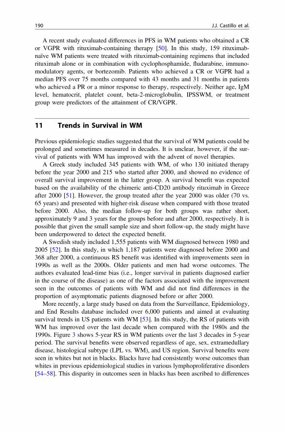
More recently, a large study based on data from the Surveillance, Epidemiology,and End Results database included over 6,000 patients and aimed at evaluatingsurvival trends in US patients with WM [53]. In this study, the RS of patients withWM has improved over the last decade when compared with the 1980s and the1990s. Figure 3 shows 5-year RS in WM patients over the last 3 decades in 5-yearperiod. The survival benefits were observed regardless of age, sex, extramedullarydisease, histological subtype (LPL vs. WM), and US region. Survival benefits wereseen in whites but not in blacks. Blacks have had consistently worse outcomes thanwhites in previous epidemiological studies in various lymphoproliferative disorders[54–58]. This disparity in outcomes seen in blacks has been ascribed to differences
190 J.J. Castillo et al.
in socioeconomical status, health insurance coverage, and patients’ or providers’attitudes toward therapy. Purely biological factors may also play a role, consideringthat blacks have an increased incidence of IgG and IgA MGUS but a lower inci-dence of IgM MGUS. It is unclear, however, if this difference is responsible for theworse outcomes seen in black patients with WM.
12 Conclusion
There are multiple options for the treatment of patients with WM in the frontline aswell as the relapsed setting. However, the treatment regimens would have to bethoughtfully selected depending on the patient’s characteristics such as age, co-morbidities, IgM levels, and genetic mutations. Age, beta-2-microglobulin level,and hematological parameters seemed to be the best prognostic factors associatedwith OS in patients with WM. However, studies evaluating these factors in largecohorts of patients treated with novel agents are lacking. Finally, the survival ofpatients with WM appears to be improving in the last decade, probably associatedwith the advent of novel agents and better supportive therapy. Novel agents such asthe phosphatidylinositol 3-kinase inhibitor idelalisib (CAL-101, GS-1101), the bcl-2 inhibitor GDC-199 (ABT-199), the oral proteasome inhibitor ixazomib(MLN9708), and the glycoengineered anti-CD20 monoclonal antibody obin-utuzumab (GA-101), to cite a few, will shortly enter clinical trials for WM. A recentphase II study studied idelalisib in 125 patients with relapsed/refractory indolent
Fig. 3 5-year relative survival estimates in patients with Waldenström Macroglobulinemia fromthe SEER database (1980–2010), according to 5-year period of diagnosis. Reproduced withpermission from [51]
Biology, Prognosis and Therapy … 191
lymphoma, of which 10 patients had LPL/WM [59]. From these, 2 patientsachieved a PR and 6 a minor response for an ORR of 80 %. In a smaller phase Istudy on 64 patients with relapsed/refractory indolent lymphoma [60], the ORR inLPL/WM patients was 55 % (5/9 patients). In a phase I study on 44 patients withrelapsed/refractory indolent lymphoma, GDC-199 induced a response in 3 out of 4WM patients (ORR 75 %), including one CR [61]. Promising agents with greaterefficacy, novel mechanisms of action, and better safety profiles are likely the futureof WM therapy.
References
1. Swerdlow SH, Berger F, Pileri SA et al (2008) Lymphoplasmacytic lymphoma. In: SwerdlowSH, Campo E, Harris NL et al (eds) WHO classification of tumours of haematopoietic andlymphoid tissues. Lyon, France, IARC, pp 194–195
2. Treon SP, Hunter ZR, Aggarwal A et al (2006) Characterization of familial waldenstrom’smacroglobulinemia. Ann Oncol 17:488–494
3. Ciccarelli BT, Patterson CJ, Hunter ZR et al (2011) Hepcidin is produced bylymphoplasmacytic cells and is associated with anemia in waldenstrom’smacroglobulinemia. Clin Lymphoma Myeloma Leuk 11:160–163
4. Schop RF, Kuehl WM, Van Wier SA, Ahmann GJ, Price-Troska T, Bailey RJ, Jalal SM, Qi Y,Kyle RA, Greipp PR, Fonseca R (2002) Waldenström macroglobulinemia neoplastic cells lackimmunoglobulin heavy chain locus translocations but have frequent 6q deletions. Blood 100(8):2996–3001.
5. Treon SP, Xu L, Yang G et al (2012) MYD88 L265P somatic mutation in Waldenstrom’smacroglobulinemia. N Engl J Med 367:826–833
6. Jimenez C, Sebastian E, Chillon MC et al (2013) MYD88 L265P is a marker highlycharacteristic of, but not restricted to waldenstrom’s macroglobulinemia. Leukemia 27:1722–1728
7. Mori N, Ohwashi M, Yoshinaga K et al (2013) L265P mutation of the MYD88 gene isfrequent in waldenstrom’s macroglobulinemia and its absence in myeloma. PLoS one 8:80088
8. Poulain S, Roumier C, Decambron A et al (2013) MYD88 L265P mutation in Waldenstrommacroglobulinemia. Blood 121:4504–4511
9. Varettoni M, Arcaini L, Zibellini S et al (2013) Prevalence and clinical significance of theMYD88 (L265P) somatic mutation in Waldenstrom’s macroglobulinemia and relatedlymphoid neoplasms. Blood 121:2522–2528
10. Yang G, Zhou Y, Liu X et al (2013) A mutation in MYD88 (L265P) supports the survival oflymphoplasmacytic cells by activation of Bruton tyrosine kinase in Waldenstrommacroglobulinemia. Blood 122:1222–1232
11. Watters TM, Kenny EF, O’Neill LA (2007) Structure, function and regulation of the Toll/IL-1receptor adaptor proteins. Immunol Cell Biol 85:411–419
12. Lin SC, Lo YC, Wu H (2010) Helical assembly in the MyD88-IRAK4-IRAK2 complex inTLR/IL-1R signalling. Nature 465:885–890
13. Kawagoe T, Sato S, Matsushita K et al (2008) Sequential control of Toll-like receptor-dependent responses by IRAK1 and IRAK2. Nat Immunol 9:684–691
14. Hunter ZR, Xu L, Yang G et al (2014) The genomic landscape of Waldenstrommacroglobulinemia is characterized by highly recurring MYD88 and WHIM-like CXCR4mutations, and small somatic deletions associated with B-cell lymphomagenesis. Blood123:1637–1646
15. Busillo JM, Benovic JL (2007) Regulation of CXCR4 signaling. Biochim Biophys Acta1768:952–963
192 J.J. Castillo et al.
16. Treon SP, Cao Y, Xu L et al (2014) Somatic mutations in MYD88 and CXCR4 aredeterminants of clinical presentation and overall survival in Waldenstrom macroglobulinemia.Blood 123:2791–2796
17. Roccaro AM, Sacco A, Jimenez C, Maiso P, Moschetta M, Mishima Y, Aljawai Y, Sahin I,Kuhne M, Cardarelli P, Cohen L, San Miguel JF, Garcia-Sanz R, Ghobrial IM (2014)C1013G/CXCR4 acts as a driver mutation of tumor progression and modulator of drugresistance in lymphoplasmacytic lymphoma. Blood 123(26):4120–4131
18. Kyle RA, Treon SP, Alexanian R et al (2003) Prognostic markers and criteria to initiatetherapy in Waldenstrom’s macroglobulinemia: consensus panel recommendations from thesecond international workshop on Waldenstrom’s macroglobulinemia. Semin Oncol 30:116–120
19. NCCN Clinical Practices Guidelines in Oncology (NCCN Guidelines). Waldenstrom’smacroglobulinemia/lymphoplasmacytic lymphoma. Available at http://www.nccn.org/professionals/physician_gls/pdf/waldenstroms.pdf . Accessed 1 June 2014
20. Treon SP, Ioakimidis L, Soumerai JD et al (2009) Primary therapy of Waldenstrommacroglobulinemia with bortezomib, dexamethasone, and rituximab: WMCTG clinical trial05-180. J Clin Oncol 27:3830–3835
21. Ghobrial IM, Xie W, Padmanabhan S et al (2010) Phase II trial of weekly bortezomib incombination with rituximab in untreated patients with Waldenstrom macroglobulinemia. Am JHematol 85:670–674
22. Dimopoulos MA, Garcia-Sanz R, Gavriatopoulou M et al (2013) Primary therapy ofWaldenstrom macroglobulinemia (WM) with weekly bortezomib, low-dose dexamethasone,and rituximab (BDR): long-term results of a phase 2 study of the European Myeloma Network(EMN). Blood 122:3276–3282
23. Moreau P, Pylypenko H, Grosicki S et al (2011) Subcutaneous versus intravenousadministration of bortezomib in patients with relapsed multiple myeloma: a randomised,phase 3, non-inferiority study. Lancet Oncol 12:431–440
24. Treon SP, Tripsas CK, Meid K, Kanan S, Sheehy P, Chuma S, Xu L, Cao Y, Yang G, Liu X,Patterson CJ, Warren D, Hunter ZR, Turnbull B, Ghobrial IM, Castillo JJ (2014) Carfilzomib,rituximab, and dexamethasone (CaRD) treatment offers a neuropathy-sparing approach fortreating Waldenström's macroglobulinemia. Blood 124(4):503–510
25. Treon SP (2009) How I treat Waldenstrom macroglobulinemia. Blood 114:2375–238526. Ioakimidis L, Patterson CJ, Hunter ZR et al (2009) Comparative outcomes following CP-R,
CVP-R, and CHOP-R in waldenstrom’s macroglobulinemia. Clin Lymphoma Myeloma 9:62–66
27. Rummel MJ, Niederle N, Maschmeyer G et al (2013) Bendamustine plus rituximab versusCHOP plus rituximab as first-line treatment for patients with indolent and mantle-celllymphomas: an open-label, multicentre, randomised, phase 3 non-inferiority trial. Lancet381:1203–1210
28. Leblond V, Johnson S, Chevret S et al (2013) Results of a randomized trial of chlorambucilversus fludarabine for patients with untreated Waldenstrom macroglobulinemia, marginal zonelymphoma, or lymphoplasmacytic lymphoma. J Clin Oncol 31:301–307
29. Treon SP, Branagan AR, Ioakimidis L et al (2009) Long-term outcomes to fludarabine andrituximab in Waldenstrom macroglobulinemia. Blood 113:3673–3678
30. Tedeschi A, Benevolo G, Varettoni M et al (2012) Fludarabine plus cyclophosphamide andrituximab in Waldenstrom macroglobulinemia: an effective but myelosuppressive regimen tobe offered to patients with advanced disease. Cancer 118:434–443
31. Treon SP, Soumerai JD, Branagan AR et al (2008) Thalidomide and rituximab inWaldenstrom macroglobulinemia. Blood 112:4452–4457
32. Gertz MA, Rue M, Blood E et al (2004) Multicenter phase 2 trial of rituximab forWaldenstrom macroglobulinemia (WM): an Eastern Cooperative Oncology Group Study(E3A98). Leuk Lymphoma 45:2047–2055
Biology, Prognosis and Therapy … 193
33. Treon SP, Emmanouilides C, Kimby E et al (2005) Extended rituximab therapy inwaldenstrom’s macroglobulinemia. Ann Oncol 16:132–138
34. Treon SP, Branagan AR, Hunter Z et al (2004) Paradoxical increases in serum IgM andviscosity levels following rituximab in waldenstrom’s macroglobulinemia. Ann Oncol15:1481–1483
35. Chen C, Kouroukis CT, White D et al (2009) Bortezomib in relapsed or refractorywaldenstrom’s macroglobulinemia. Clin Lymphoma Myeloma 9:74–76
36. Dimopoulos MA, Anagnostopoulos A, Kyrtsonis MC et al (2005) Treatment of relapsed orrefractory waldenstrom’s macroglobulinemia with bortezomib. Haematologica 90:1655–1658
37. Ghobrial IM, Hong F, Padmanabhan S et al (2010) Phase II trial of weekly bortezomib incombination with rituximab in relapsed or relapsed and refractory Waldenstrommacroglobulinemia. J Clin Oncol 28:1422–1428
38. Treon SP, Hunter ZR, Matous J et al (2007) Multicenter clinical trial of bortezomib inrelapsed/refractory waldenstrom’s macroglobulinemia: results of WMCTG Trial 03-248. ClinCancer Res 13:3320–3325
39. Zinzani PL, Gherlinzoni F, Bendandi M et al (1995) Fludarabine treatment in resistantwaldenstrom’s macroglobulinemia. Eur J Haematol 54:120–123
40. Treon SP, Hanzis C, Tripsas C et al (2011) Bendamustine therapy in patients with relapsed orrefractory waldenstrom’s macroglobulinemia. Clin Lymphoma Myeloma Leuk 11:133–135
41. Furman RR, Eradat H, DiRienzo CG et al (2011) A phase II trial of ofatumumab in subjectswith Waldenstrom’s macroglobulinemia. ASH Annu Meet Abstr 118:3701
42. Ghobrial IM, Gertz M, Laplant B et al (2010) Phase II trial of the oral mammalian target ofrapamycin inhibitor everolimus in relapsed or refractory Waldenstrom macroglobulinemia.J Clin Oncol 28:1408–1414
43. Treon SP, Tripsas CK, Ioakimidis L et al (2011) Prospective, multicenter study of the MTORinhibitor everolimus (RAD001) as primary therapy in Waldenstrom’s macroglobulinemia.ASH Annu Meet Abstr 118:2951
44. Treon SP, Tripsas CK, Yang G et al (2013) A prospective multicenter study of the Bruton’styrosine kinase inhibitor ibrutinib in patients with relapsed or refractory Waldenstrom’smacroglobulinemia. Blood 122:251
45. Treon SP, Hanzis C, Manning RJ et al (2011) Maintenance rituximab is associated withimproved clinical outcome in rituximab naive patients with Waldenstrom macroglobulinaemiawho respond to a rituximab-containing regimen. Br J Haematol 154:357–362
46. Rummel MJ, Lerchenmuller C, Greil R et al (2012) Bendamustin-rituximab inductionfollowed by observation or rituximab maintenance for newly diagnosed patients withwaldenstrom’s macroglobulinemia: results from a prospective, randomized, multicenter study(StiL NHL 7-2008 -MAINTAIN-; ClinicalTrials.gov Identifier: NCT00877214). ASH AnnMeet Abstr 120:2739
47. Kyriakou C, Canals C, Sibon D et al (2010) High-dose therapy and autologous stem-celltransplantation in Waldenstrom macroglobulinemia: the lymphoma working party of theeuropean group for blood and marrow transplantation. J Clin Oncol 28:2227–2232
48. Kyriakou C, Canals C, Cornelissen JJ et al (2010) Allogeneic stem-cell transplantation inpatients with Waldenstrom macroglobulinemia: report from the lymphoma working party ofthe european group for blood and marrow transplantation. J Clin Oncol 28:4926–4934
49. Morel P, Duhamel A, Gobbi P et al (2009) International prognostic scoring system forWaldenstrom macroglobulinemia. Blood 113:4163–4170
50. Treon SP, Yang G, Hanzis C et al (2011) Attainment of complete/very good partial responsefollowing rituximab-based therapy is an important determinant to progression-free survival,and is impacted by polymorphisms in FCGR3A in Waldenstrom macroglobulinaemia. Br JHaematol 154:223–228
51. Kastritis E, Kyrtsonis MC, Hatjiharissi E et al (2011) No significant improvement in theoutcome of patients with waldenstrom’s macroglobulinemia treated over the last 25 years. AmJ Hematol 86:479–483
194 J.J. Castillo et al.
52. Kristinsson SY, Eloranta S, Dickman PW et al (2013) Patterns of survival inlymphoplasmacytic lymphoma/Waldenstrom macroglobulinemia: a population-based studyof 1,555 patients diagnosed in Sweden from 1980 to 2005. Am J Hematol 88:60–65
53. Castillo JJ, Olszewski AJ, Cronin AM et al (2014) Survival trends in Waldenstrommacroglobulinemia: an analysis of the surveillance, epidemiology and end results database.Blood 123:3999–4000
54. Shenoy PJ, Malik N, Nooka A et al (2011) Racial differences in the presentation and outcomesof diffuse large B-cell lymphoma in the United States. Cancer 117:2530–2540
55. Shenoy P, Maggioncalda A, Malik N, Flowers CR (2011) Incidence patterns and outcomes forhodgkin lymphoma patients in the United States. Adv Hematol 2011:725219
56. Nabhan C, Aschebrook-Kilfoy B, Chiu BC et al. (2014) The impact of race, ethnicity, age, andsex on clinical outcome in chronic lymphocytic leukemia: a comprehensive SEER analysis inthe modern era. Leuk Lymphoma 2014
57. Nabhan C, Aschebrook-Kilfoy B, Chiu BC et al. (2014) The impact of race, age, and sex infollicular lymphoma: a comprehensive SEER analysis across consecutive treatment eras. Am JHematol, 89(6): 633–638
58. Castillo JJ, Winer ES, Olszewski AJ (2013) Population-based prognostic factors for survival inpatients with burkitt lymphoma: an analysis from the surveillance, epidemiology, and endresults database. Cancer 119:3672–3679
59. Gopal AK, Kahl BS, de Vos S et al (2014) PI3Kdelta inhibition by idelalisib in patients withrelapsed indolent lymphoma. N Engl J Med 370:1008–1018
60. Flinn IW, Kahl BS, Furman RR et al (2014) Idelalisib, a selective inhibitor ofphosphatidylinositol 3-kinase-δ, as therapy for previously treated indolent non-Hodgkinlymphoma. Blood 123:3406–3413
61. Davids SM, Seymour JF, Gerecitano JF et al (2014) Phase I study of ABT-199 (GDC-199) inpatients with relapsed/refractory non-Hodgkin lymphoma (NHL): responses observed indiffuse large B-cell lymphoma (DLBCL) and follicular lymphoma (FL) at higher cohort doses.J Clin Oncol 32:8522
62. Dhodapkar MV, Hoering A, Gertz MA, Rivkin S, Szymonifka J, Crowley J, Barlogie B (2009)Long-term survival in Waldenstrom macroglobulinemia: 10-year follow-up of SouthwestOncology Group-directed intergroup trial S9003. Blood 113(4):793–796
Biology, Prognosis and Therapy … 195