Embed Size (px)
Citation preview
The Biology of CancerFirst Edition
Chapter 7:Tumor Suppressor Genes
Copyright © Garland Science 2007
Robert A. Weinberg
Outlines:
-Cell fusion: cancer phenotype is recessive-TSG: Knudson’s two-hit theory
-Retinoblastoma tumor -Familial vs. sporadic forms
-Inactivation of TSGs-LOH-mutations-promoter hypermethylation
-TSGs and proteins function in diverse ways-pRB (Chap. 8)-p53 (Chap. 9)-NF1 protein acts as a negative regulator of Ras signaling -APC facilitates egress of cells from colonic crypts -Von Hippel-Lindau disease: pVHL modulates the hypoxic response
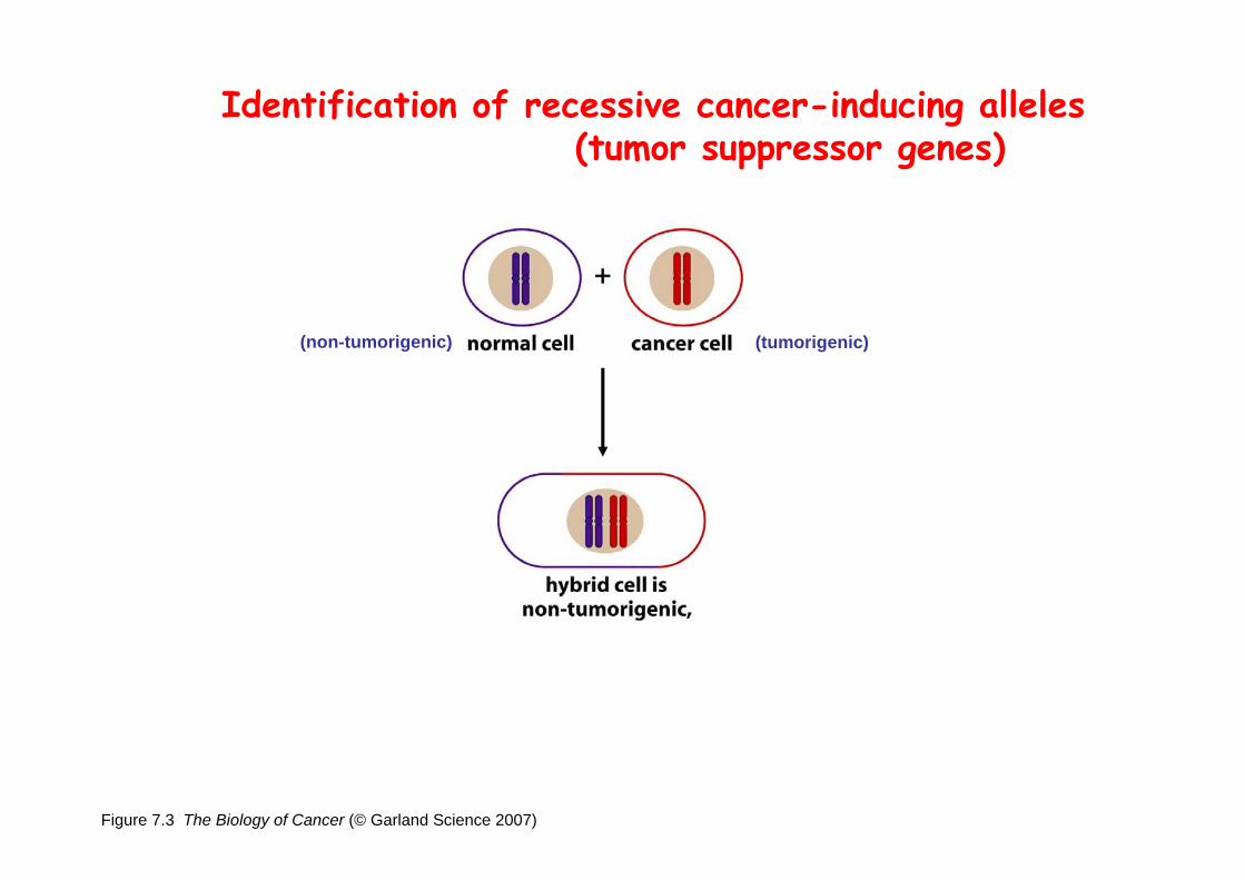
Figure 7.3 The Biology of Cancer (© Garland Science 2007)
Identification of recessive cancer-inducing alleles(tumor suppressor genes)
(non-tumorigenic) (tumorigenic)

Figure 7.3 The Biology of Cancer (© Garland Science 2007)
Identification of recessive cancer-inducing alleles(tumor suppressor genes)
(non-tumorigenic) (tumorigenic)
(from virus-induced tumors)
If the cancer cell had originally arisen without the involvement of a tumor virus, then its malignant phenotype was recessive when this cell was fused with a normal cell.
(from non-virus-induced human tumors, or from chemically induced rodent tumord)
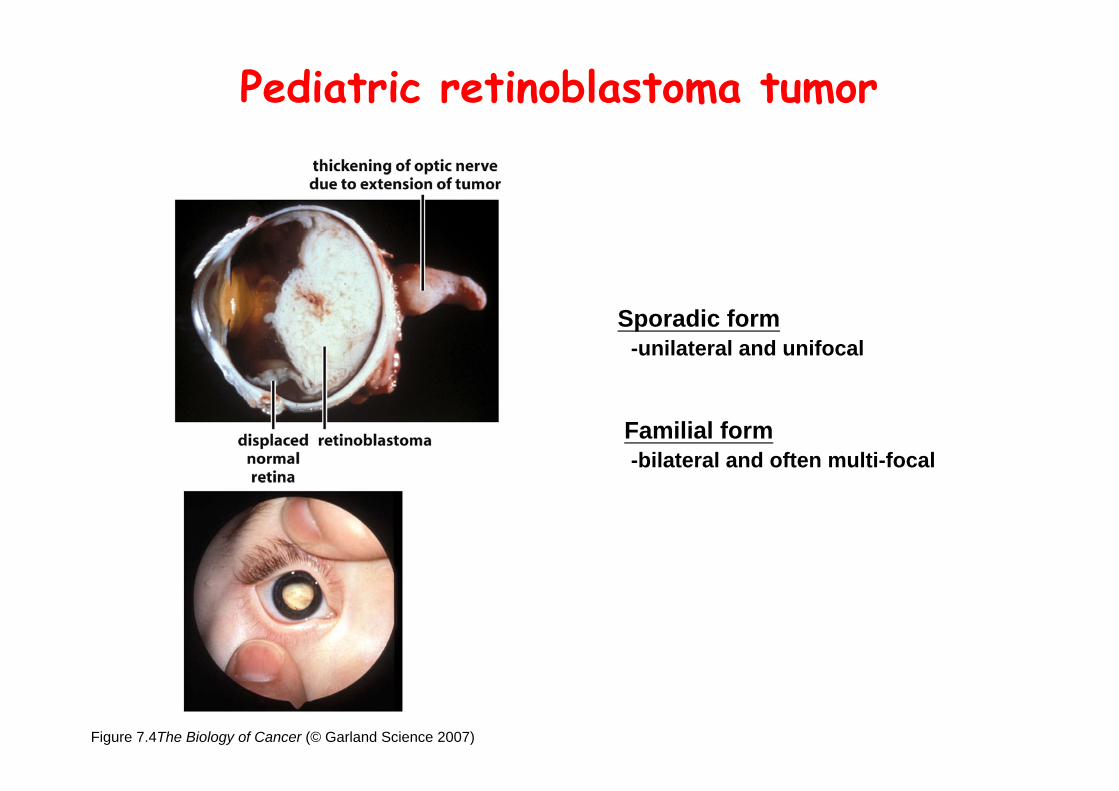
Figure 7.4The Biology of Cancer (© Garland Science 2007)
Pediatric retinoblastoma tumor
Sporadic form-unilateral and unifocal
Familial form-bilateral and often multi-focal
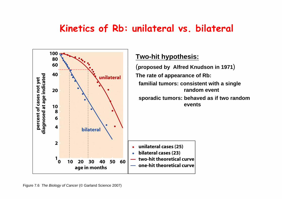
Figure 7.6 The Biology of Cancer (© Garland Science 2007)
Kinetics of Rb: unilateral vs. bilateral
Two-hit hypothesis:(proposed by Alfred Knudson in 1971)The rate of appearance of Rb:
familial tumors: consistent with a single random event
sporadic tumors: behaved as if two random events
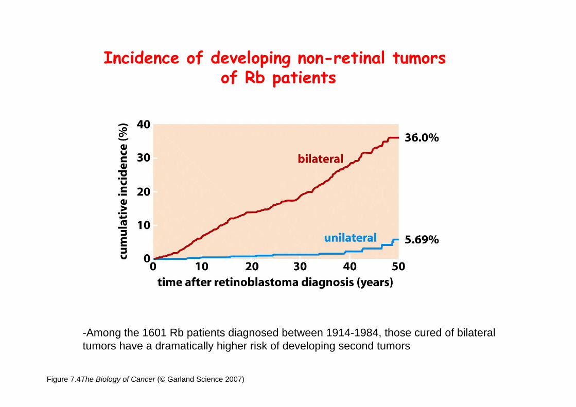
Figure 7.4The Biology of Cancer (© Garland Science 2007)
Incidence of developing non-retinal tumors of Rb patients
-Among the 1601 Rb patients diagnosed between 1914-1984, those cured of bilateral tumors have a dramatically higher risk of developing second tumors
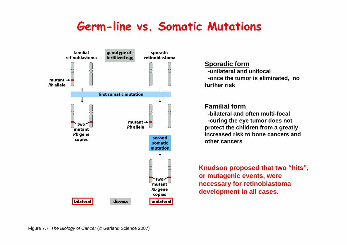
Figure 7.7 The Biology of Cancer (© Garland Science 2007)
Sporadic form-unilateral and unifocal-once the tumor is eliminated, no
further risk
Familial form-bilateral and often multi-focal-curing the eye tumor does not
protect the children from a greatly increased risk to bone cancers and other cancers
Knudson proposed that two “hits”, or mutagenic events, were necessary for retinoblastoma development in all cases.
Germ-line vs. Somatic Mutations
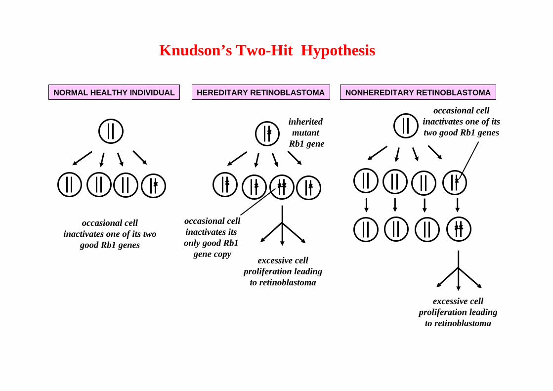
NORMAL HEALTHY INDIVIDUAL NONHEREDITARY RETINOBLASTOMAHEREDITARY RETINOBLASTOMA
inherited mutant
Rb1 gene
occasional cellinactivates one of its two
good Rb1 genes
occasional cellinactivates its only good Rb1
gene copyexcessive cell
proliferation leadingto retinoblastoma
occasional cell inactivates one of its two good Rb1 genes
excessive cellproliferation leading
to retinoblastoma
Knudson’s Two-Hit Hypothesis
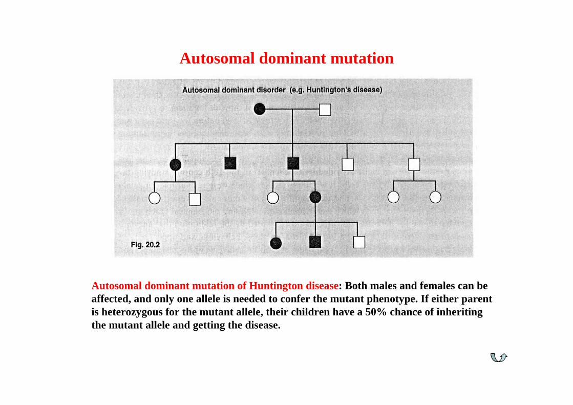
Autosomal dominant mutation of Huntington disease: Both males and females can be affected, and only one allele is needed to confer the mutant phenotype. If either parent is heterozygous for the mutant allele, their children have a 50% chance of inheriting the mutant allele and getting the disease.
Autosomal dominant mutation

Autosomal recessive mutation of cystic fibrosis: Both males and females can be affected but must carry two mutant alleles of the gene to show mutant phenotype. Both parents must be heterozygous carriers for their children to be at risk of being affected.
Autosomal recessive mutation
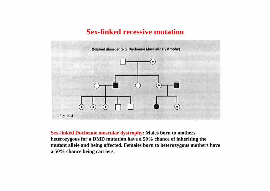
Sex-linked Duchenne muscular dystrophy: Males born to mothers heterozygous for a DMD mutation have a 50% chance of inheriting the mutant allele and being affected. Females born to heterozygous mothers have a 50% chance being carriers.
Sex-linked recessive mutation

(Affected individual: green-filled circles or squares)
Pedigree of a kindred afflicted with Rb
Somatic dominant transmission pattern of familial Rb
Mutant Rb genes are both dominant and recessive.-An individual who inherits a mutant, defective allele of Rb is almost certain to develop retinoblastoma at some point in childhood. -However, a cell carrying a mutant and a wild-type Rb gene copy would behave normally. The mutant Rb allele acts dominantly at the organismic level and recessively at cellular level.

Figure 7.10 The Biology of Cancer (© Garland Science 2007)
Chromosomal localization of Rb locus, 13q14
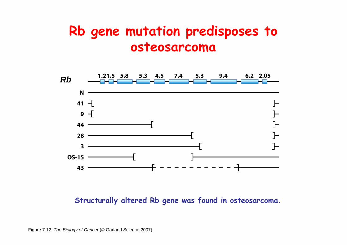
Figure 7.12 The Biology of Cancer (© Garland Science 2007)
Rb gene mutation predisposes to osteosarcoma
Structurally altered Rb gene was found in osteosarcoma.
Rb
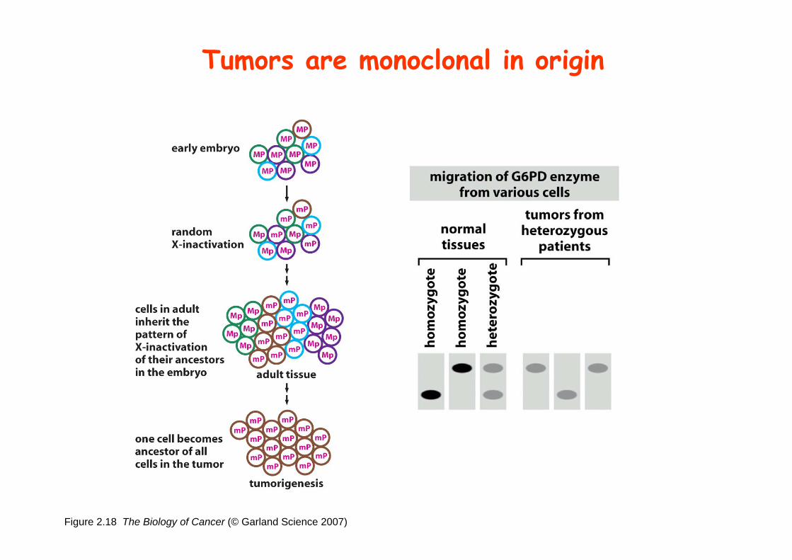
Figure 2.18 The Biology of Cancer (© Garland Science 2007)
Tumors are monoclonal in origin

Loss of heterozygosity: esterase D locus serves as a surrogate marker for RB
Figure 7.11 The Biology of Cancer (© Garland Science 2007)
Zymographic analysis of esterase D isoforms
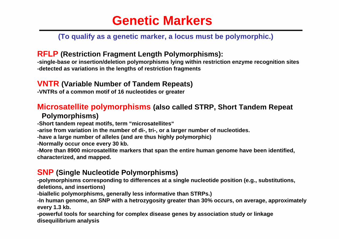
(To qualify as a genetic marker, a locus must be polymorphic.)
Genetic Markers
RFLP (Restriction Fragment Length Polymorphisms):-single-base or insertion/deletion polymorphisms lying within restriction enzyme recognition sites-detected as variations in the lengths of restriction fragments
VNTR (Variable Number of Tandem Repeats)-VNTRs of a common motif of 16 nucleotides or greater
Microsatellite polymorphisms (also called STRP, Short Tandem Repeat Polymorphisms)
-Short tandem repeat motifs, term “microsatellites“-arise from variation in the number of di-, tri-, or a larger number of nucleotides.-have a large number of alleles (and are thus highly polymorphic)-Normally occur once every 30 kb.-More than 8900 microsatellite markers that span the entire human genome have been identified, characterized, and mapped.
SNP (Single Nucleotide Polymorphisms)-polymorphisms corresponding to differences at a single nucleotide position (e.g., substitutions, deletions, and insertions)-biallelic polymorphisms, generally less informative than STRPs.)-In human genome, an SNP with a hetrozygosity greater than 30% occurs, on average, approximately every 1.3 kb.-powerful tools for searching for complex disease genes by association study or linkage disequilibrium analysis
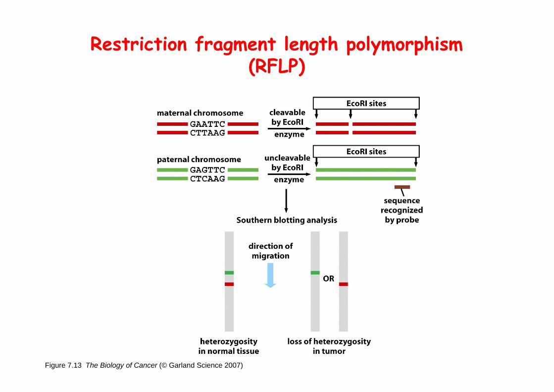
Figure 7.13 The Biology of Cancer (© Garland Science 2007)
Restriction fragment length polymorphism (RFLP)

Loss of Heterozygosity:Identifying Lost Tumor Suppressor Genes

Figure 7.15 The Biology of Cancer (© Garland Science 2007)
Single nucleatide polymorphism (SNP)
Allele-specific primer extension reaction
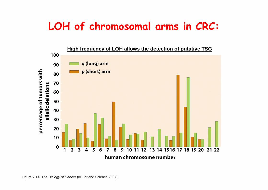
Figure 7.14 The Biology of Cancer (© Garland Science 2007)
LOH of chromosomal arms in CRC:
High frequency of LOH allows the detection of putative TSG

Linkage analysis: association of phenotype to genotype
the usage of polymorphic markers
hetero- homo-
12

Disease Gene Localization
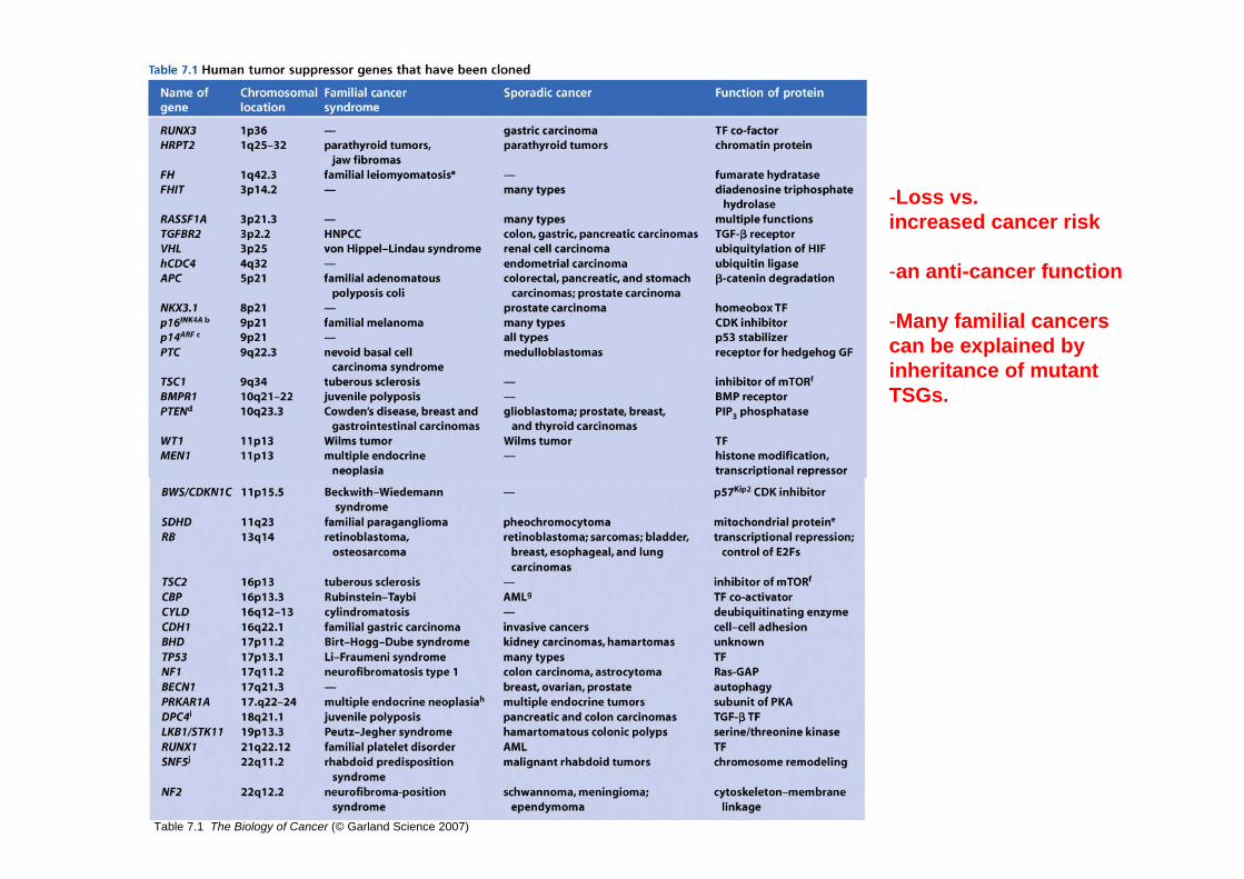
Table 7.1 The Biology of Cancer (© Garland Science 2007)
-Loss vs. increased cancer risk
-an anti-cancer function
-Many familial cancers can be explained by inheritance of mutant TSGs.
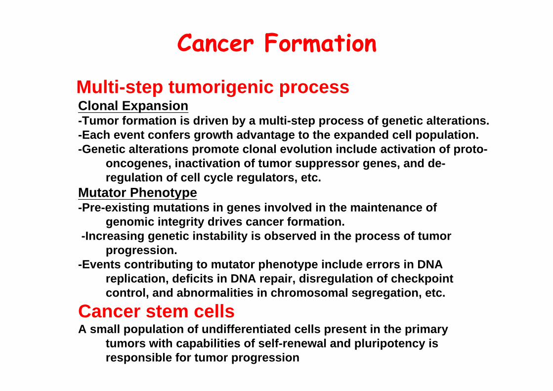
Cancer Formation
Multi-step tumorigenic processClonal Expansion-Tumor formation is driven by a multi-step process of genetic alterations.-Each event confers growth advantage to the expanded cell population.-Genetic alterations promote clonal evolution include activation of proto-
oncogenes, inactivation of tumor suppressor genes, and de-regulation of cell cycle regulators, etc.
Mutator Phenotype-Pre-existing mutations in genes involved in the maintenance of
genomic integrity drives cancer formation.-Increasing genetic instability is observed in the process of tumor
progression.-Events contributing to mutator phenotype include errors in DNA
replication, deficits in DNA repair, disregulation of checkpoint control, and abnormalities in chromosomal segregation, etc.
Cancer stem cellsA small population of undifferentiated cells present in the primary
tumors with capabilities of self-renewal and pluripotency is responsible for tumor progression
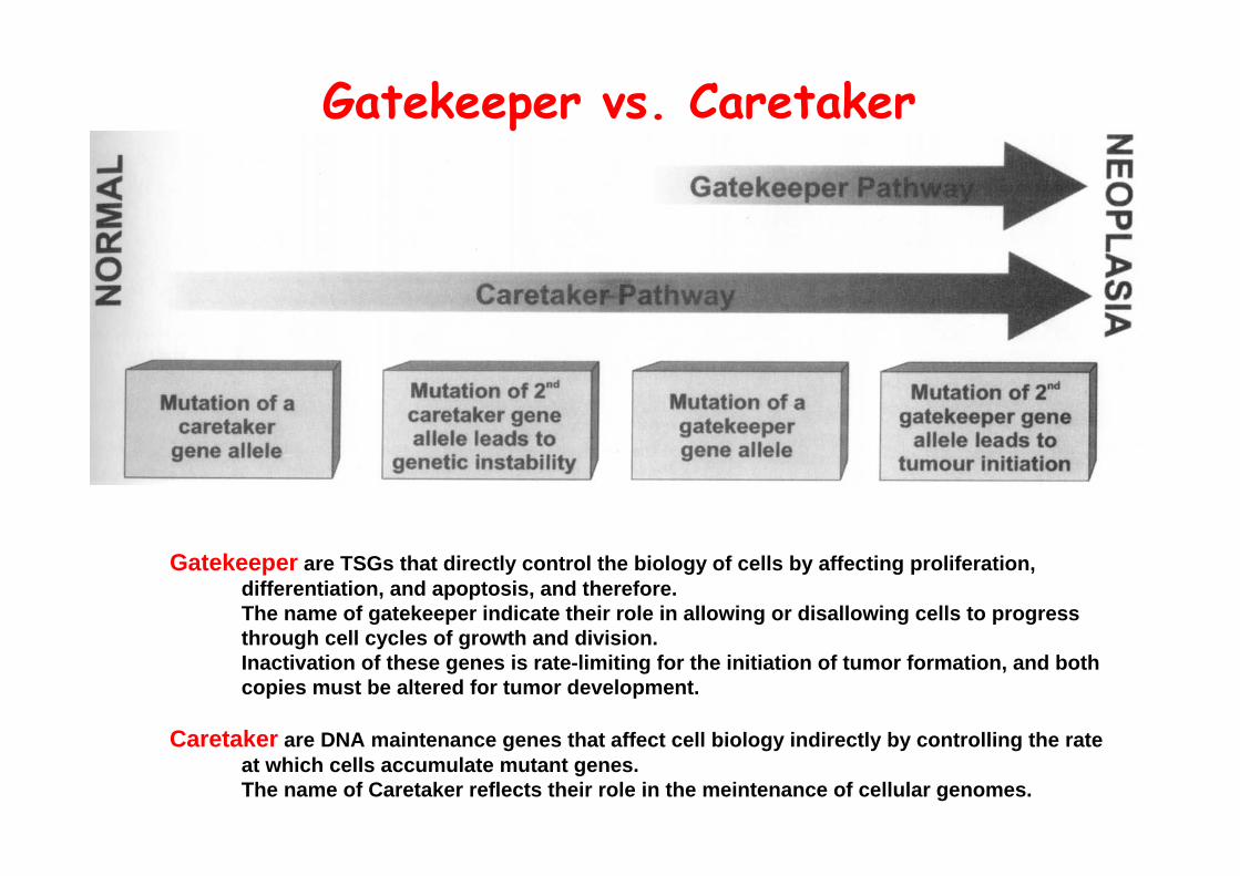
Gatekeeper vs. Caretaker
Gatekeeper are TSGs that directly control the biology of cells by affecting proliferation, differentiation, and apoptosis, and therefore.The name of gatekeeper indicate their role in allowing or disallowing cells to progress through cell cycles of growth and division. Inactivation of these genes is rate-limiting for the initiation of tumor formation, and both copies must be altered for tumor development.
Caretaker are DNA maintenance genes that affect cell biology indirectly by controlling the rate at which cells accumulate mutant genes.The name of Caretaker reflects their role in the meintenance of cellular genomes.

Inactivation of TSG:Genetic and epigenetic alterations

Epigenetic modification
-non-genetic changes-does not alter the nucleotide sequence of the DNA-Inactivation of TSG can be achieved by promoter methylation and/or promoter deacetylation-More than half of the TSGs are found to be silenced in sporadic cancers by promoter methylation.-The promoters of a variety of genes that inhibit tumor formation are also found in methylatedstatus.

Figure 7.16 The Biology of Cancer (© Garland Science 2007)
Maintenance of DNA methylation
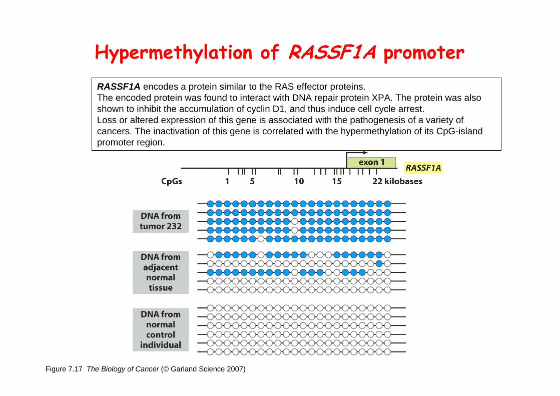
Figure 7.17 The Biology of Cancer (© Garland Science 2007)
Hypermethylation of RASSF1A promoterRASSF1A encodes a protein similar to the RAS effector proteins. The encoded protein was found to interact with DNA repair protein XPA. The protein was also shown to inhibit the accumulation of cyclin D1, and thus induce cell cycle arrest. Loss or altered expression of this gene is associated with the pathogenesis of a variety of cancers. The inactivation of this gene is correlated with the hypermethylation of its CpG-island promoter region.
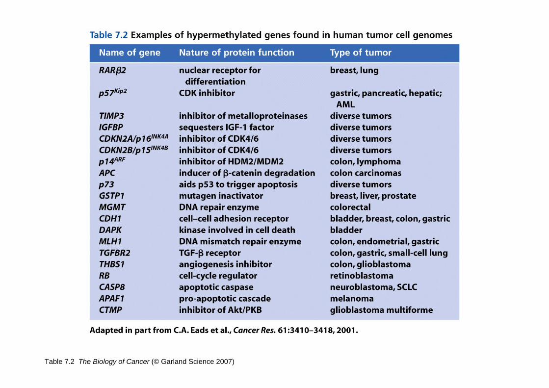
Table 7.2 The Biology of Cancer (© Garland Science 2007)

Figure 7.18 The Biology of Cancer (© Garland Science 2007)
ISH: a measurement of DNA methylation(In situ hybridization using a methylation-specific PCR reaction and in situ hybridization for p16INK4A promoter)
low-grade cervical ca. high-grade
normal breast normal breast
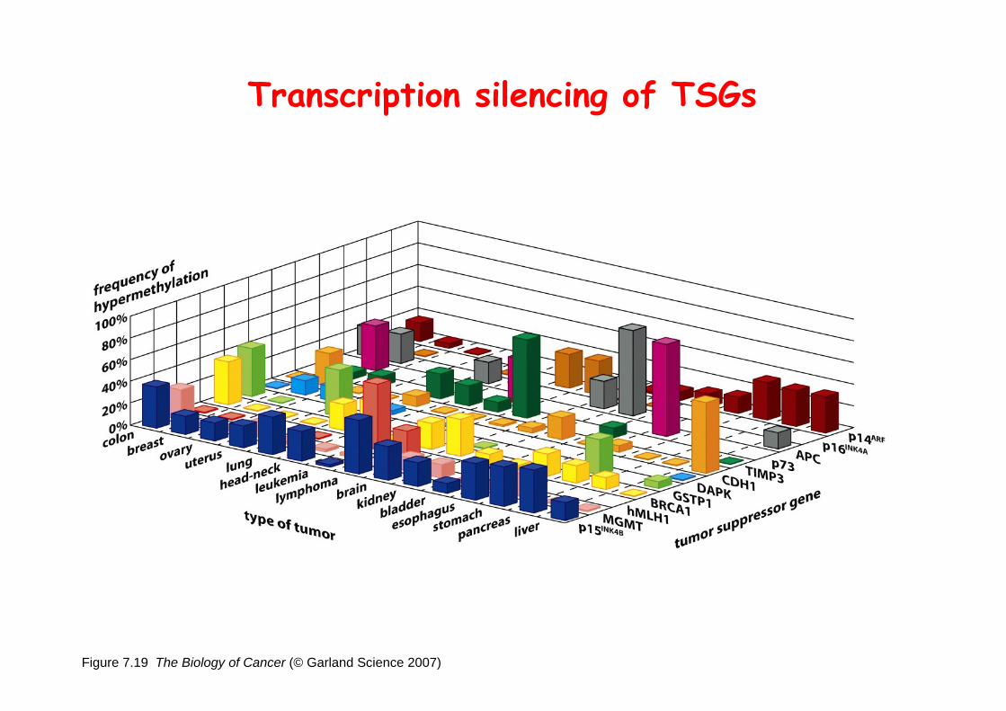
Figure 7.19 The Biology of Cancer (© Garland Science 2007)
Transcription silencing of TSGs
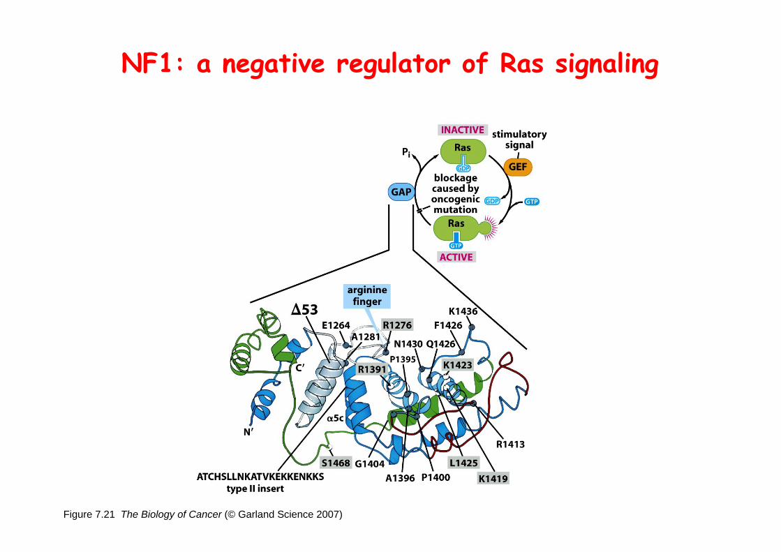
Figure 7.21 The Biology of Cancer (© Garland Science 2007)
NF1: a negative regulator of Ras signaling

Figure 7.23 The Biology of Cancer (© Garland Science 2007)
the wall of a normal colon
the wall of colon from a FAP patient
Adenomatous growths, polyps in colon-Pedunculated polys are shown as stalk-like growths.Sessil polys appearing as flat thickenings of colonic
wall are not shown.
polyps
Familial adenomatous polyposis (FAP)
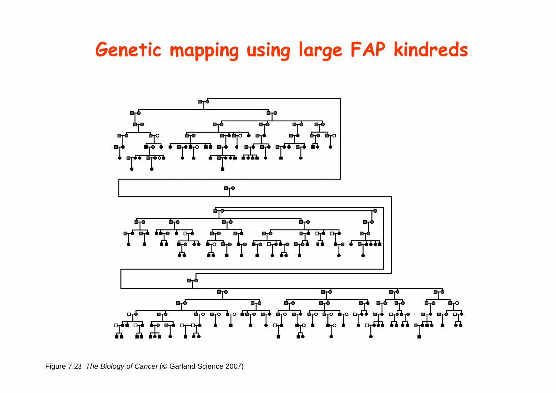
Figure 7.23 The Biology of Cancer (© Garland Science 2007)
Genetic mapping using large FAP kindreds
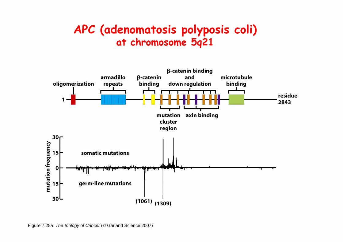
Figure 7.25a The Biology of Cancer (© Garland Science 2007)
APC (adenomatosis polyposis coli)at chromosome 5q21
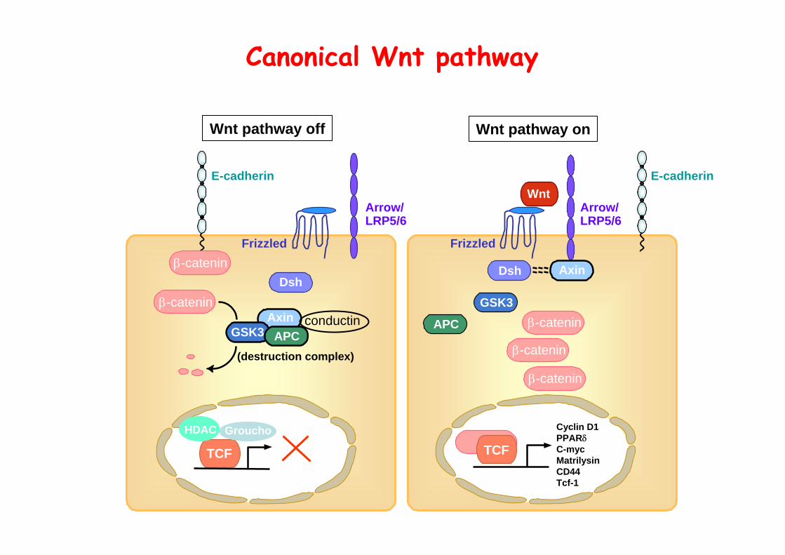
Wnt pathway off Wnt pathway on
Canonical Wnt pathway
Axin
Wnt
Dsh
Arrow/LRP5/6
Frizzled
E-cadherin
TCF
APC
GSK3β-catenin
β-catenin
β-catenin
Cyclin D1PPARδC-mycMatrilysinCD44Tcf-1
β-catenin
Axin
Dsh
Arrow/LRP5/6
Frizzled
E-cadherin
TCF
APCGSK3
β-catenin
GrouchoHDAC
conductin
(destruction complex)
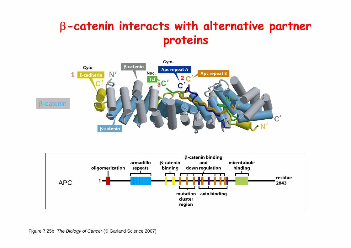
Figure 7.25b The Biology of Cancer (© Garland Science 2007)
β-catenin interacts with alternative partner proteins
APC
β-catenin
1
32
Cyto-Cyto-
Nuc.

TCF4-/-
Figure 7.24a The Biology of Cancer (© Garland Science 2007)
Role of β-catenin/TCF pathway
IHC of β-catenin in Min mouse
DAPI stain shows nuclei.
IHC of Ki67 in TCF-mice
TCF4+/-
enterocyteslumen of intestine
mesenchymal core
bottom of crypts
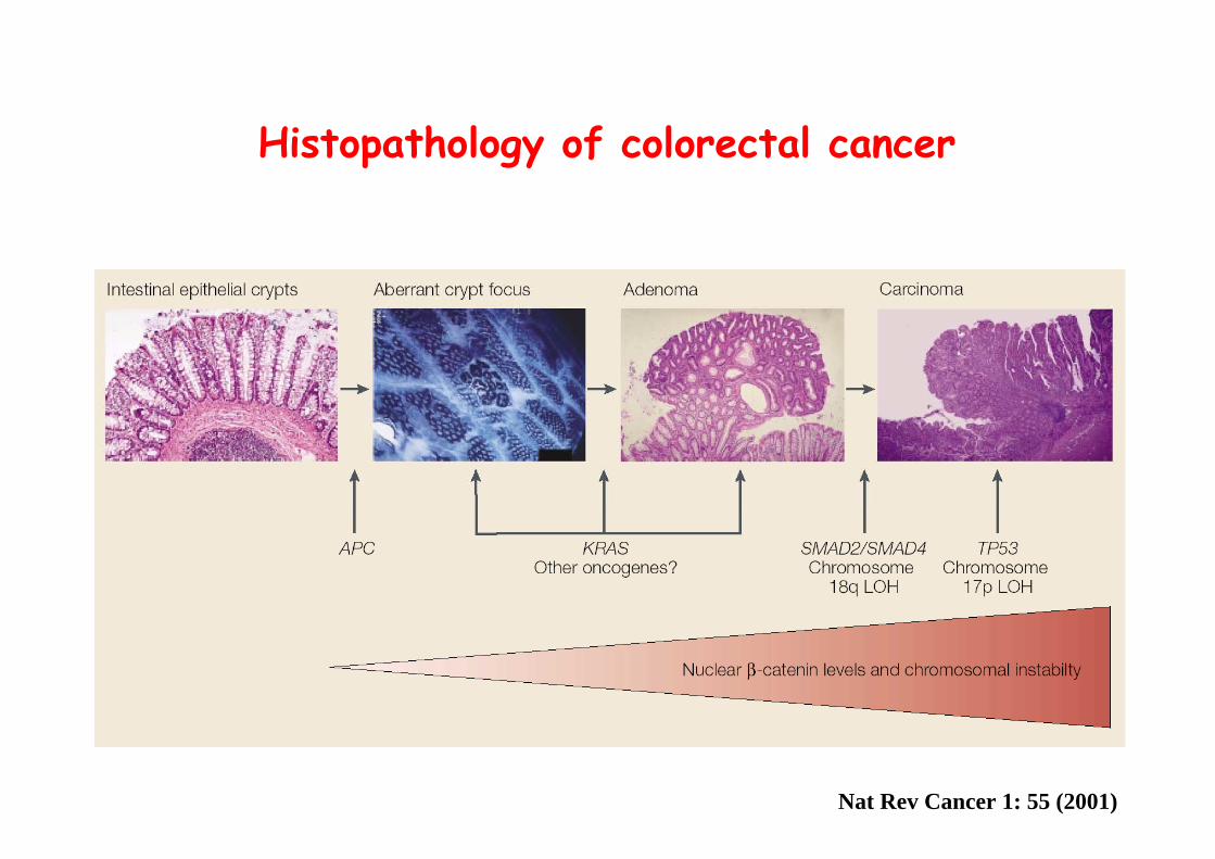
Histopathology of colorectal cancer
Nat Rev Cancer 1: 55 (2001)
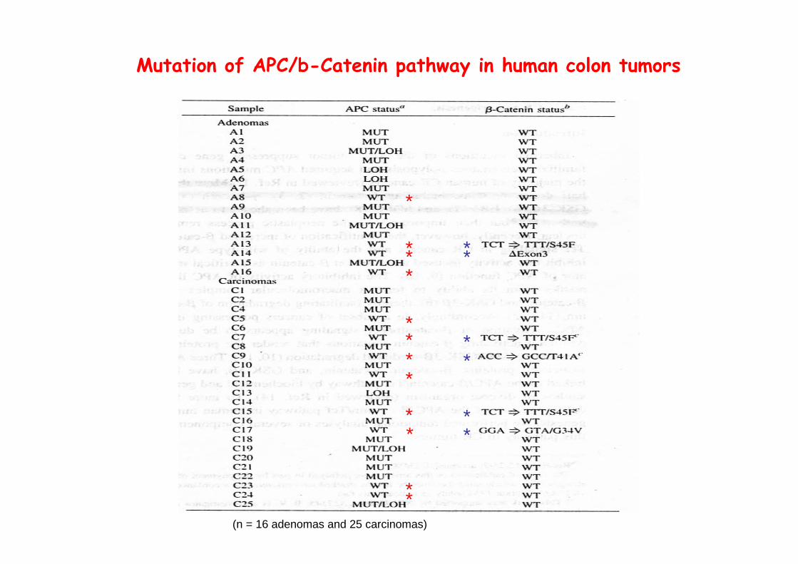
Mutation of APC/b-Catenin pathway in human colon tumors
*
***
**
*
**
**
*
**
*
** *
(n = 16 adenomas and 25 carcinomas)
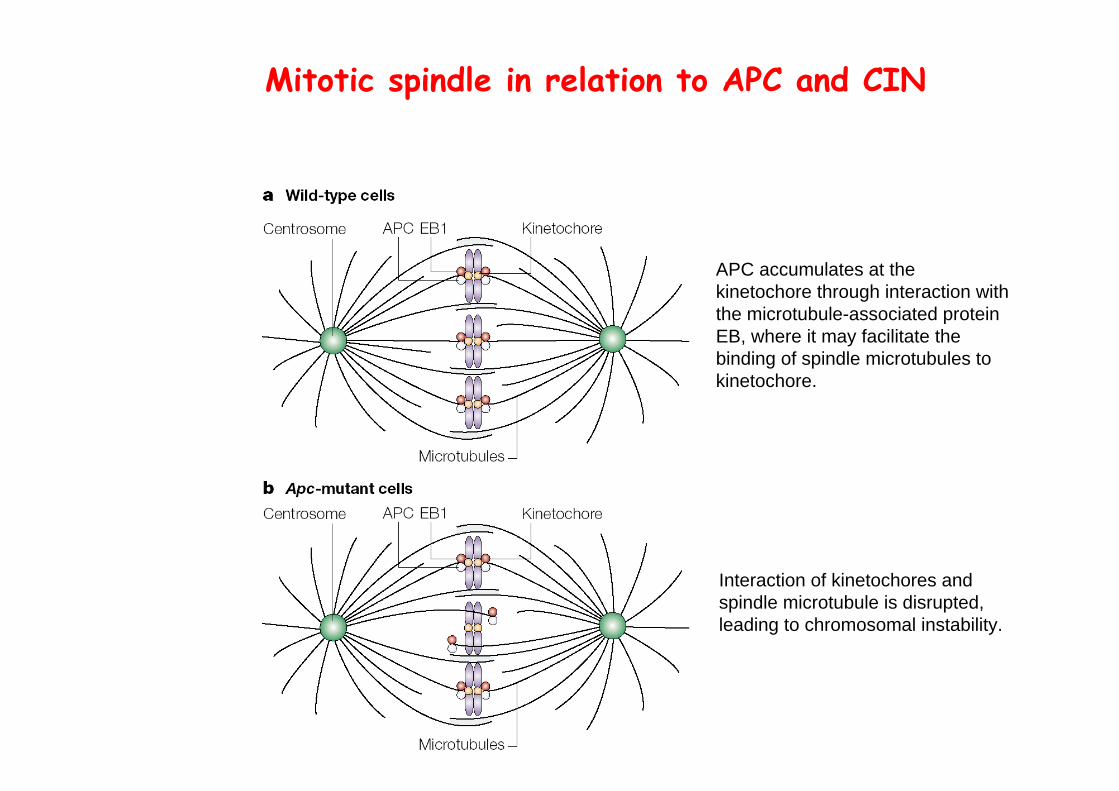
Mitotic spindle in relation to APC and CIN
Interaction of kinetochores and spindle microtubule is disrupted, leading to chromosomal instability.
APC accumulates at the kinetochore through interaction with the microtubule-associated protein EB, where it may facilitate the binding of spindle microtubules to kinetochore.
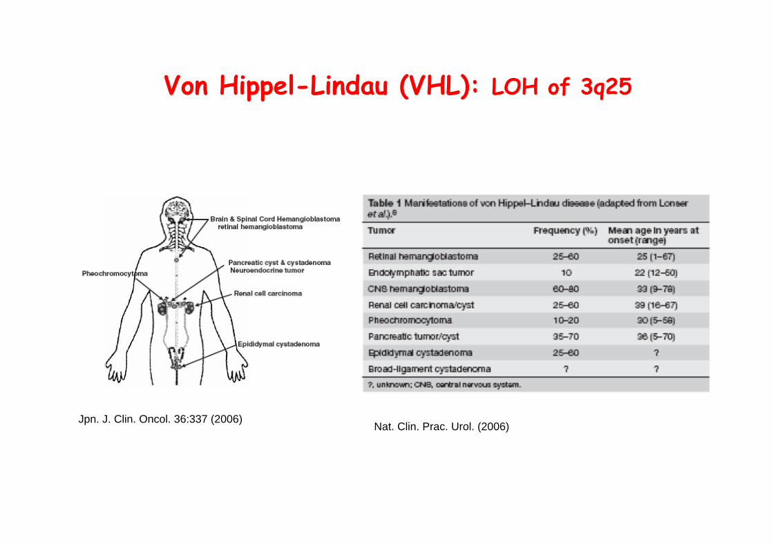
Von Hippel-Lindau (VHL): LOH of 3q25
Jpn. J. Clin. Oncol. 36:337 (2006)Nat. Clin. Prac. Urol. (2006)
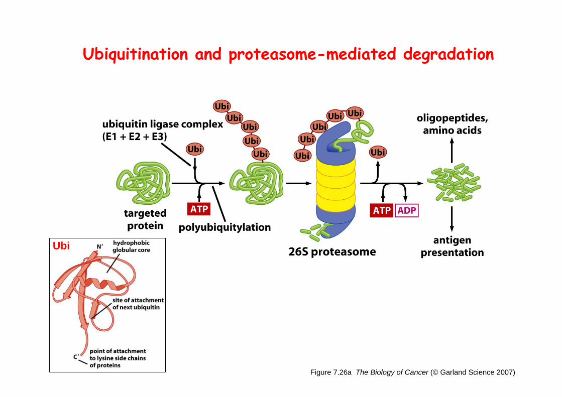
Figure 7.26a The Biology of Cancer (© Garland Science 2007)
Ubiquitination and proteasome-mediated degradation
Ubi
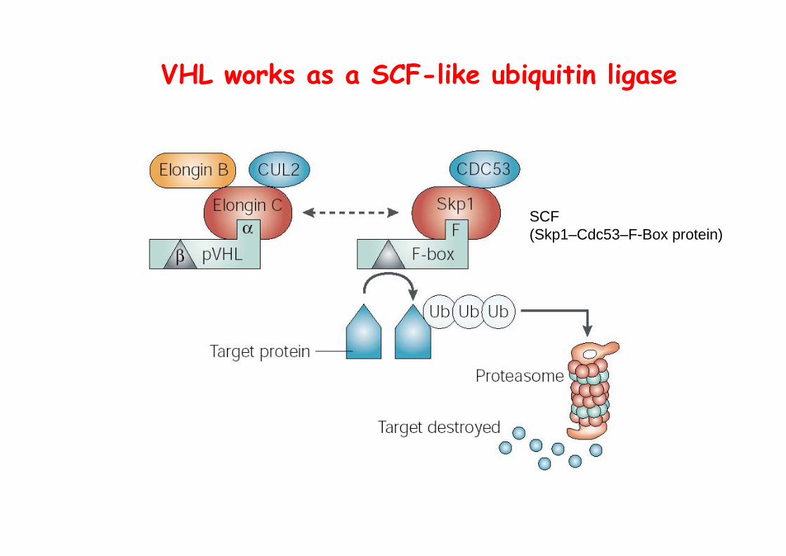
VHL works as a SCF-like ubiquitin ligase
SCF (Skp1–Cdc53–F-Box protein)
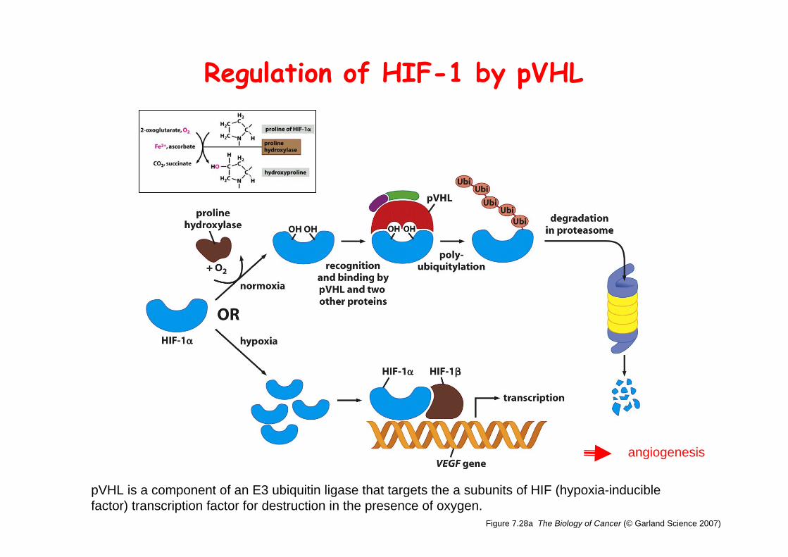
Figure 7.28a The Biology of Cancer (© Garland Science 2007)
Regulation of HIF-1 by pVHL
pVHL is a component of an E3 ubiquitin ligase that targets the a subunits of HIF (hypoxia-inducible factor) transcription factor for destruction in the presence of oxygen.
angiogenesis