Embed Size (px)
Citation preview
Sylv
ia S
. Ma
der
Copyright © The McGraw Hill Companies Inc. Permission required for reproduction or display
PowerPoint® Lecture Slides are prepared by Dr. Isaac Barjis, Biology Instructor
BIOLOGY 10th Edition
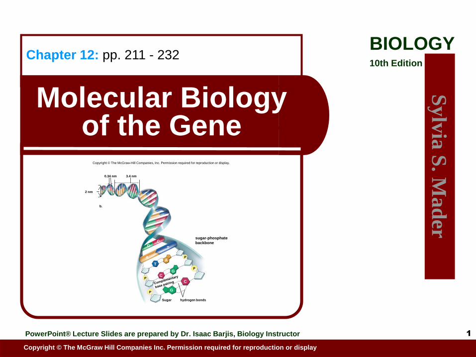
Molecular Biology of the Gene
Chapter 12: pp. 211 - 232
1
Copyright © The McGraw-Hill Companies, Inc. Permission required for reproduction or display.
b.
3.4 nm
2 nm
0.34 nm
G C
A
T
T A
P
P
P
P
C G
G
C
Sugar hydrogen bonds
sugar-phosphate
backbone

2
Outline
Genetic Material
Transformation
DNA Structure
Watson and Crick
DNA Replication
Prokaryotic versus Eukaryotic
Replication Errors
Transcription
Translation
Structure of Eukaryotic Chromosome
3
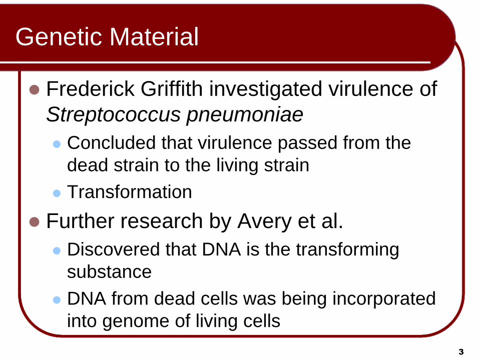
Genetic Material
Frederick Griffith investigated virulence of
Streptococcus pneumoniae
Concluded that virulence passed from the
dead strain to the living strain
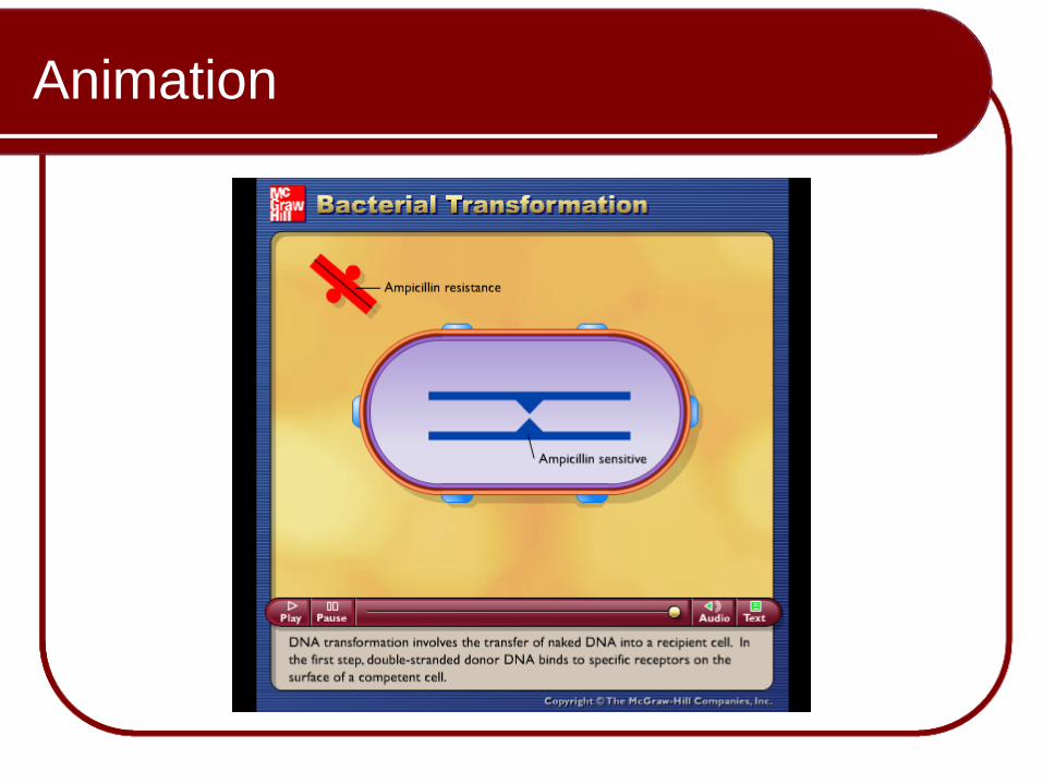
Transformation
Further research by Avery et al.
Discovered that DNA is the transforming
substance
DNA from dead cells was being incorporated
into genome of living cells

4
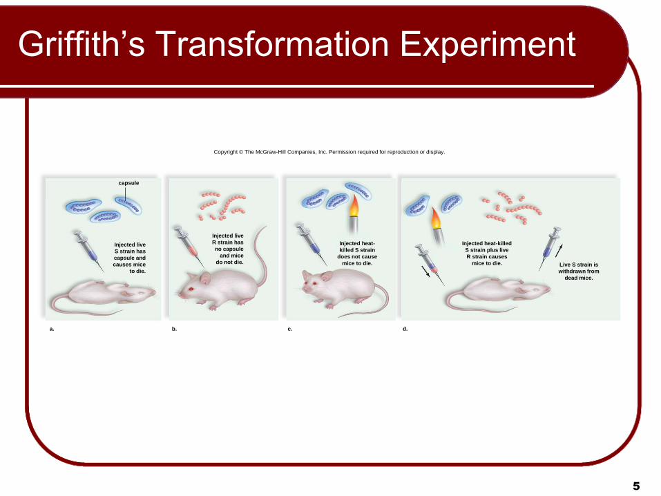
Griffith’s Transformation Experiment
Mice were injected with two strains of
pneumococcus: an encapsulated (S) strain
and a non-encapsulated (R) strain.
The S strain is virulent (the mice died); it has a
mucous capsule and forms “shiny” colonies.
The R strain is not virulent (the mice lived); it
has no capsule and forms “dull” colonies.
5
Griffith’s Transformation Experiment
Copyright © The McGraw-Hill Companies, Inc. Permission required for reproduction or display.
capsule
Injected live
S strain has
capsule and
causes mice
to die.
a. b.
Injected live
R strain has
no capsule
and mice
do not die.
c.
Injected heat-
killed S strain
does not cause
mice to die.
d.
Injected heat-killed
S strain plus live
R strain causes
mice to die. Live S strain is
withdrawn from
dead mice.
Animation
Please note that due to differing
operating systems, some animations
will not appear until the presentation is
viewed in Presentation Mode (Slide
Show view). You may see blank slides
in the “Normal” or “Slide Sorter” views.
All animations will appear after viewing
in Presentation Mode and playing each
animation. Most animations will require
the latest version of the Flash Player,
which is available at
http://get.adobe.com/flashplayer.

7
Transformation of Organisms Today
Result the so-called genetically modified
organisms (GMOs)
Invaluable tool in modern biotechnology today
Commercial products that are currently much used
Green fluorescent protein (GFP) used as a marker
A jellyfish gene codes for GFP
The jellyfish gene is isolated and then transferred to a
bacterium, or the embryo of a plant, pig, or mouse.
When this gene is transferred to another organism, the
organism glows in the dark

8
Transformation of Organisms
A normal canola plant (left) and a transgenic canola
plant expressing GFP (right) under a fluorescent light.
Copyright © The McGraw-Hill Companies, Inc. Permission required for reproduction or display.
(Bacteria): © Martin Shields/Photo Researchers, Inc.; (Jellyfish): © R. Jackman/OSF/Animals Animals/Earth Scenes; (Pigs): Courtesy Norrie Russell, The Roslin Institute;
(Mouse): © Eye of Science/Photo Researchers, Inc.; (Plant): © Dr. Neal Stewart

9
Structure of DNA
DNA contains:
Two Nucleotides with purine bases
Adenine (A)
Guanine (G)
Two Nucleotides with pyrimidine bases
Thymine (T)
Cytosine (C)

Animation
Please note that due to differing
operating systems, some animations
will not appear until the presentation is
viewed in Presentation Mode (Slide
Show view). You may see blank slides
in the “Normal” or “Slide Sorter” views.
All animations will appear after viewing
in Presentation Mode and playing each
animation. Most animations will require
the latest version of the Flash Player,
which is available at
http://get.adobe.com/flashplayer.
11
Chargaff’s Rules
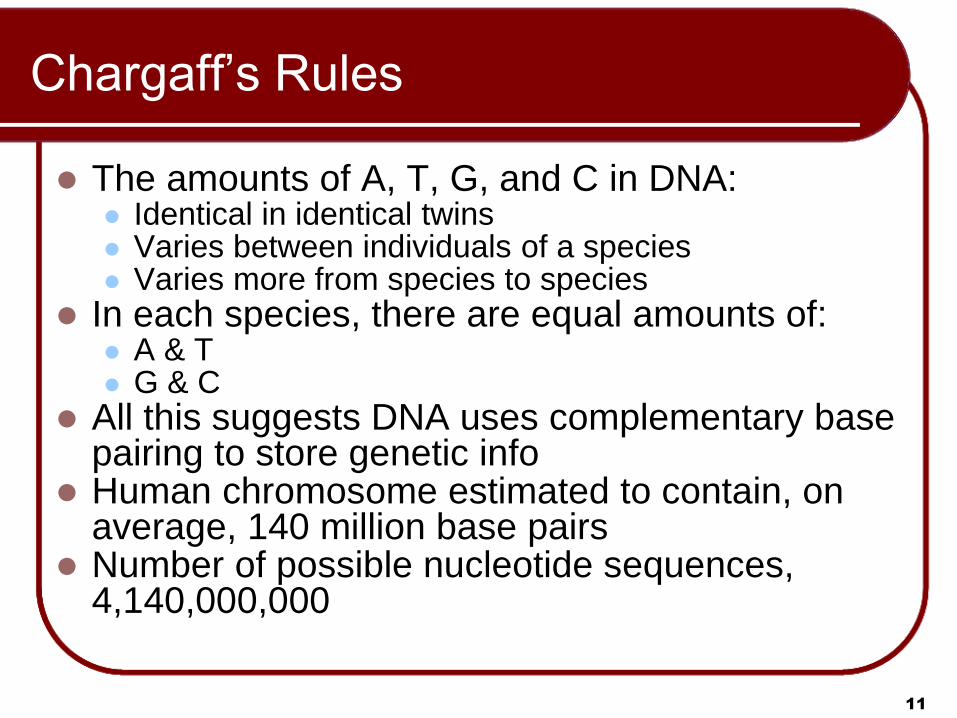
The amounts of A, T, G, and C in DNA: Identical in identical twins Varies between individuals of a species Varies more from species to species
In each species, there are equal amounts of: A & T G & C
All this suggests DNA uses complementary base pairing to store genetic info
Human chromosome estimated to contain, on average, 140 million base pairs
Number of possible nucleotide sequences, 4,140,000,000

12
Nucleotide Composition of DNA
Copyright © The McGraw-Hill Companies, Inc. Permission required for reproduction or display.
O
N
N
CH
CH
C
C
NH 2
cytosine (C)
3 C C
2
C 1
O HO P O
O
H
H H
H H
OH
CH 3
O
HN
N
C
CH
C
C
O HO P O
O
H
H H
H H
OH
HN
N
N
C CH
O
C
C C
N
H 2 N
C 2
C 2
C 1
C 1
O HO P O
O
guanine (G)
phosphate
H
H H
H H
OH
N
N
N
HC CH
NH 2
C
C C
N
4
3 C
2
C 1
5 O
O
O
O
O
O
H
H H
H H
OH
c. Chargaf f ’ s data
DN A Composition in V arious Species (%)
Species
Homo sapiens (human)
Drosophila melanogaster (fruit fly)
Zea mays (corn)
Neurospora crassa (fungus)
Escherichia coli (bacterium)
Bacillus subtilis (bacterium)
31.0
27.3
25.6
23.0
24.6
28.4
31.5
27.6
25.3
23.3
24.3
29.0
19.1
22.5
24.5
27.1
25.5
21.0
18.4
22.5
24.6
26.6
25.6
21.6
A T G C
a. Purine nucleotides b. Pyrimidine nucleotides
nitrogen-containing base
sugar = deoxyribose
thymine (T)
adenine (A)
HO P O CH 2
5 CH 2
5 CH 2
5 CH 2
C
4 C
4 C
4 C
C
3 C
3 C
Animation
Please note that due to differing
operating systems, some animations
will not appear until the presentation is
viewed in Presentation Mode (Slide
Show view). You may see blank slides
in the “Normal” or “Slide Sorter” views.
All animations will appear after viewing
in Presentation Mode and playing each
animation. Most animations will require
the latest version of the Flash Player,
which is available at
http://get.adobe.com/flashplayer.

Animation
14
Please note that due to differing
operating systems, some animations
will not appear until the presentation is
viewed in Presentation Mode (Slide
Show view). You may see blank slides
in the “Normal” or “Slide Sorter” views.
All animations will appear after viewing
in Presentation Mode and playing each
animation. Most animations will require
the latest version of the Flash Player,
which is available at
http://get.adobe.com/flashplayer.

15
Watson and Crick Model
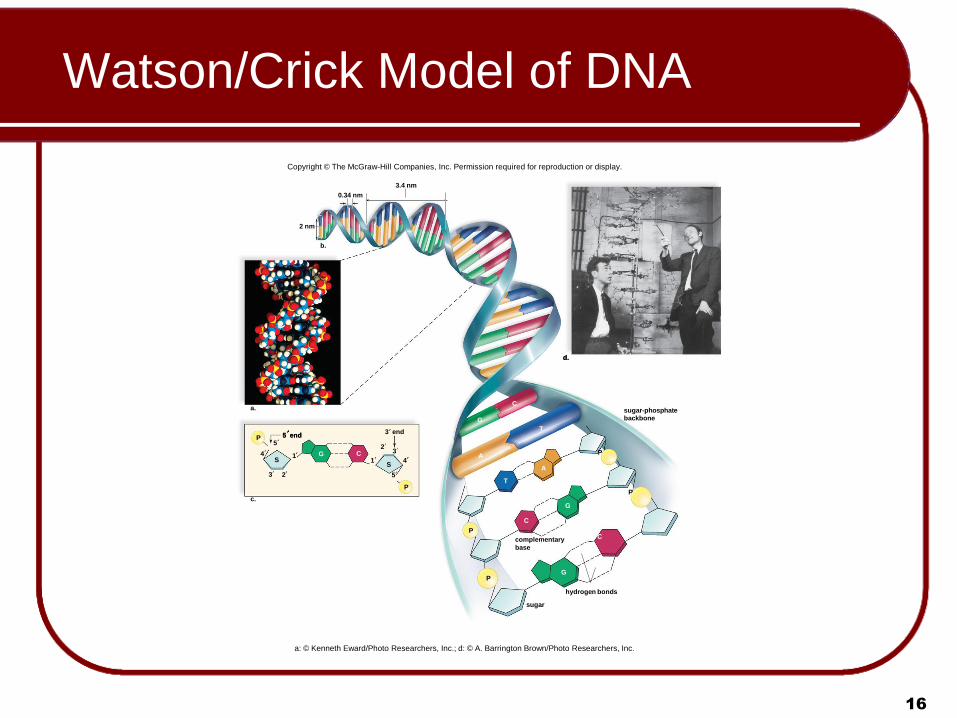
Watson and Crick, 1953
Constructed a model of DNA
Double-helix model is similar to a twisted
ladder
Sugar-phosphate backbones make up the sides
Hydrogen-bonded bases make up the rungs
Received a Nobel Prize in 1962
16
Watson/Crick Model of DNA
Copyright © The McGraw-Hill Companies, Inc. Permission required for reproduction or display.
a: © Kenneth Eward/Photo Researchers, Inc.; d: © A. Barrington Brown/Photo Researchers, Inc.
P
P
P
P
c.
b.
complementary
base
sugar-phosphate backbone
3.4 nm
2 nm
0.34 nm
P
P
S S
4
5 end 3 end
1 1
2 3
2 3
5
4
5
C G
G
C
C
G
T
T
A
A
C
G
a.
d. d.
5 end
sugar
hydrogen bonds

17
X-Ray Diffraction of DNA
Rosalind Franklin studied the structure of DNA
using X-rays.
She found that if a concentrated, viscous solution
of DNA is made, it can be separated into fibers.
Under the right conditions, the fibers can produce
X-ray diffraction pattern
She produced X-ray diffraction photographs.
This provided evidence that DNA had the following
features:
DNA is a helix.
Some portion of the helix is repeated.

18
X-Ray Diffraction of DNA
Copyright © The McGraw-Hill Companies, Inc. Permission required for reproduction or display.
© Photo Researchers, Inc.; c: © Science Source/Photo Researchers, Inc.
X-ray beam
b. c.
Rosalind Franklin
diffraction pattern
Crystalline
DNA
diffracted
X-rays
a.

19
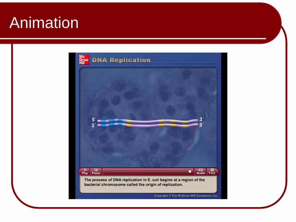
Replication of DNA
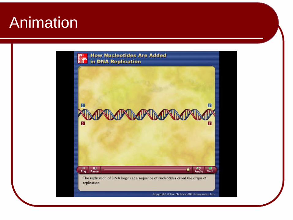
DNA replication is the process of copying a
DNA molecule.
Replication is semiconservative, with
each strand of the original double helix
(parental molecule) serving as a template
(mold or model) for a new strand in a
daughter molecule.

Animation
Please note that due to differing
operating systems, some animations
will not appear until the presentation is
viewed in Presentation Mode (Slide
Show view). You may see blank slides
in the “Normal” or “Slide Sorter” views.
All animations will appear after viewing
in Presentation Mode and playing each
animation. Most animations will require
the latest version of the Flash Player,
which is available at
http://get.adobe.com/flashplayer.

Animation
21
Please note that due to differing
operating systems, some animations
will not appear until the presentation is
viewed in Presentation Mode (Slide
Show view). You may see blank slides
in the “Normal” or “Slide Sorter” views.
All animations will appear after viewing
in Presentation Mode and playing each
animation. Most animations will require
the latest version of the Flash Player,
which is available at
http://get.adobe.com/flashplayer.
Animation
Please note that due to differing
operating systems, some animations
will not appear until the presentation is
viewed in Presentation Mode (Slide
Show view). You may see blank slides
in the “Normal” or “Slide Sorter” views.
All animations will appear after viewing
in Presentation Mode and playing each
animation. Most animations will require
the latest version of the Flash Player,
which is available at
http://get.adobe.com/flashplayer.
23
Replication: Eukaryotic
DNA replication begins at numerous points along linear chromosome
DNA unwinds and unzips into two strands
Each old strand of DNA serves as a template for a new strand
Complementary base-pairing forms new strand on each old strand
Requires enzyme DNA polymerase

Animation
Please note that due to differing
operating systems, some animations
will not appear until the presentation is
viewed in Presentation Mode (Slide
Show view). You may see blank slides
in the “Normal” or “Slide Sorter” views.
All animations will appear after viewing
in Presentation Mode and playing each
animation. Most animations will require
the latest version of the Flash Player,
which is available at
http://get.adobe.com/flashplayer.
26
Replication: Eukaryotic
Replication bubbles spread bi-directionally until they meet
The complementary nucleotides join to form new strands. Each daughter DNA molecule contains an old strand and a new strand.
Replication is semiconservative:
One original strand is conserved in each daughter molecule i.e. each daughter double helix has one parental strand and one new strand.

Animation
Please note that due to differing
operating systems, some animations
will not appear until the presentation is
viewed in Presentation Mode (Slide
Show view). You may see blank slides
in the “Normal” or “Slide Sorter” views.
All animations will appear after viewing
in Presentation Mode and playing each
animation. Most animations will require
the latest version of the Flash Player,
which is available at
http://get.adobe.com/flashplayer.

28
Semiconservative Replication of DNA
Copyright © The McGraw-Hill Companies, Inc. Permission required for reproduction or display.
region of parental
DNA double helix
G
G
G
T
A
A
C
C
3' 5'
A T
C G
A T
A
G
G C
C G
A
region of
replication:
new nucleotides
are pairing
with those of
parental strands
region of
completed
replication
daughter DNA double helix
old
strand
new
strand daughter DNA double helix
old
strand
new
strand
C C
A
A
T T
G
G
T A
T A
C
G
A
T
A
T
A
C G A
T A T
A
T A
C G
C G
A
G
T
A
C
G
C
G
A

Animation
29
Please note that due to differing
operating systems, some animations
will not appear until the presentation is
viewed in Presentation Mode (Slide
Show view). You may see blank slides
in the “Normal” or “Slide Sorter” views.
All animations will appear after viewing
in Presentation Mode and playing each
animation. Most animations will require
the latest version of the Flash Player,
which is available at
http://get.adobe.com/flashplayer.

Animation
Please note that due to differing
operating systems, some animations
will not appear until the presentation is
viewed in Presentation Mode (Slide
Show view). You may see blank slides
in the “Normal” or “Slide Sorter” views.
All animations will appear after viewing
in Presentation Mode and playing each
animation. Most animations will require
the latest version of the Flash Player,
which is available at
http://get.adobe.com/flashplayer.

31
Science Focus: Aspects of DNA
Replication
G C
A T
G C
G C
P
P
P
P
P
P
P
P
P
P
P
P is attached here OH
CH2
C
C C
C
H H
H
H H
OH
OH O
base is attached here
5 end
3 end 5 end
template strand
Direction of replication new strand
Deoxyribose molecule
RNA primer
3
3
5
3
5
5 parental DN A helix
helicase at replication fork
leading new strand
template strand
template strand
lagging strand
DN A polymerase
DNA polymerase DNA ligase
Okazaki fragment
Replication fork introduces complications
5
7
6
4
3
2
1
DNA polymerase
attaches a new
nucleotide to the 3
carbon of the
previous nucleotide.
Copyright © The McGraw-Hill Companies, Inc. Permission required for reproduction or display.
5
4
3 2
1
3 end
3
32
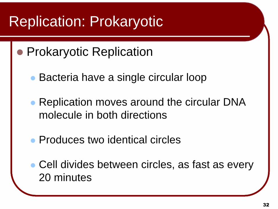
Replication: Prokaryotic
Prokaryotic Replication
Bacteria have a single circular loop
Replication moves around the circular DNA
molecule in both directions
Produces two identical circles
Cell divides between circles, as fast as every
20 minutes
33
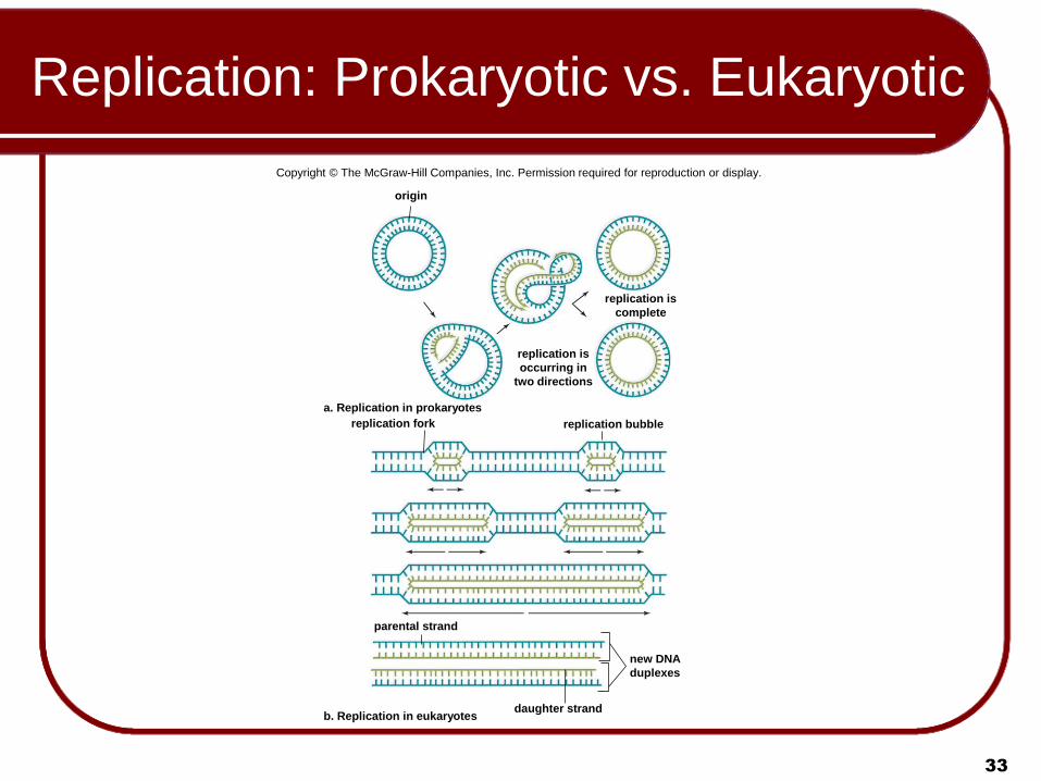
Replication: Prokaryotic vs. Eukaryotic
Copyright © The McGraw-Hill Companies, Inc. Permission required for reproduction or display.
origin
replication is
occurring in
two directions
replication is
complete
replication fork replication bubble
a. Replication in prokaryotes
parental strand
daughter strand
new DNA
duplexes
b. Replication in eukaryotes

34
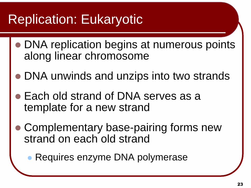
Replication Errors
Genetic variations are the raw material for evolutionary change
Mutation:
A permanent (but unplanned) change in base-pair sequence
Some due to errors in DNA replication
Others are due to to DNA damage
DNA repair enzymes are usually available to reverse most errors

35
Function of Genes
Genes Specify Enzymes Beadle and Tatum:
Experiments on fungus Neurospora crassa Proposed that each gene specifies the synthesis of
one enzyme One-gene-one-enzyme hypothesis
Genes Specify a Polypeptide A gene is a segment of DNA that specifies the
sequence of amino acids in a polypeptide Suggests that genetic mutations cause
changes in the primary structure of a protein
36
Protein Synthesis: From DNA to RNA to
Protein
The mechanism of gene expression
DNA in genes specify information, but information is not structure and function
Genetic info is expressed into structure & function through protein synthesis
The expression of genetic info into structure & function:
DNA in gene controls the sequence of nucleotides in an RNA molecule
RNA controls the primary structure of a protein

Animation
Please note that due to differing
operating systems, some animations
will not appear until the presentation is
viewed in Presentation Mode (Slide
Show view). You may see blank slides
in the “Normal” or “Slide Sorter” views.
All animations will appear after viewing
in Presentation Mode and playing each
animation. Most animations will require
the latest version of the Flash Player,
which is available at
http://get.adobe.com/flashplayer.
38
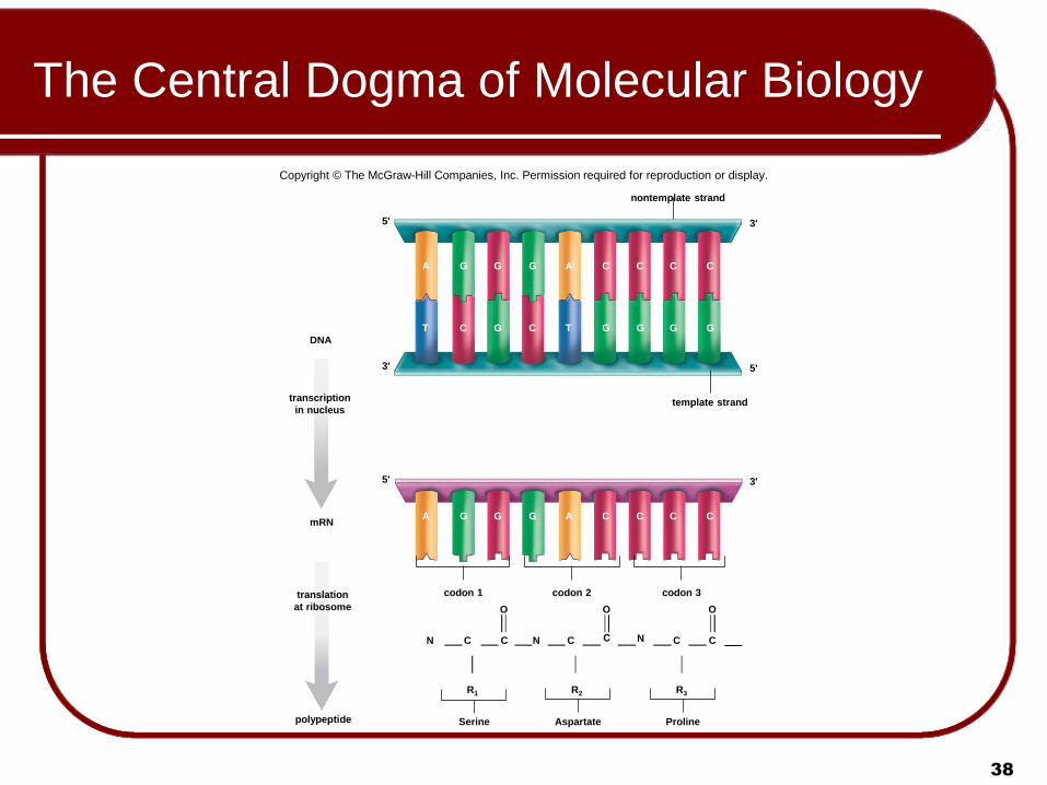
The Central Dogma of Molecular Biology
Copyright © The McGraw-Hill Companies, Inc. Permission required for reproduction or display.
nontemplate strand
3' 5'
A G G G A C C C C
T C G C T G G G G
5' 3'
template strand transcription
in nucleus
3' 5'
mRN
DNA
A G G G A C C C C
codon 1 codon 2 codon 3
polypeptide
translation
at ribosome
N N N C C C C C
R1 R2 R3
Serine Aspartate Proline
O O O
39
Types of RNA
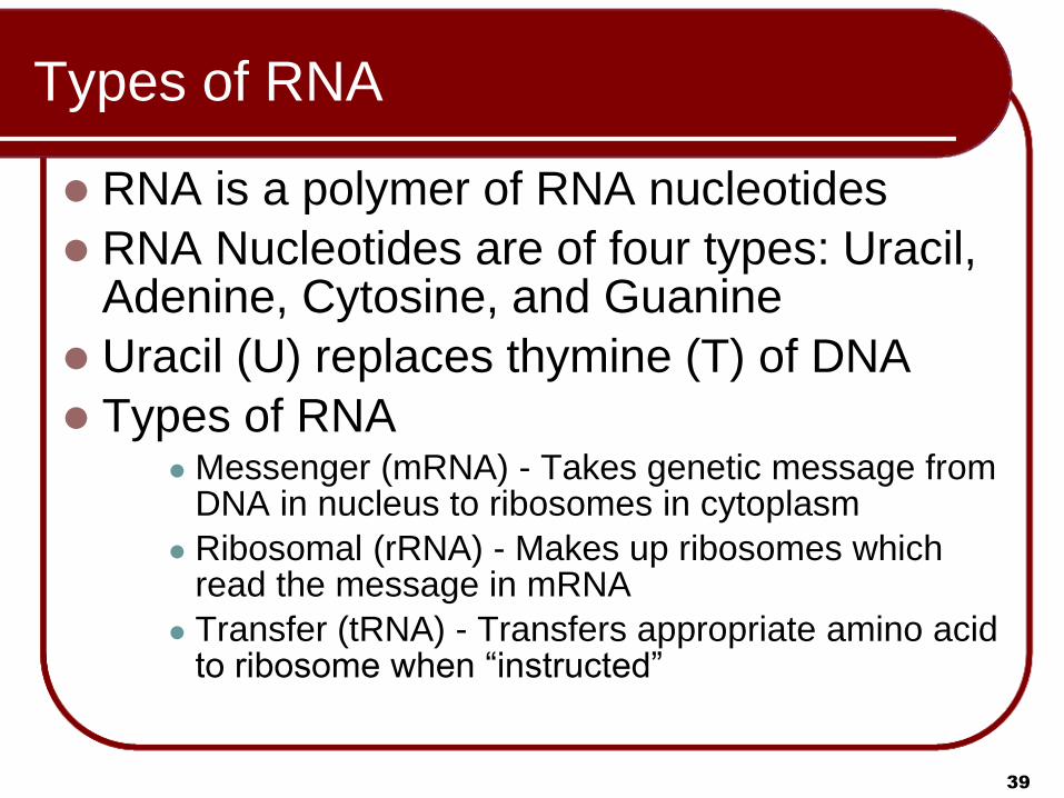
RNA is a polymer of RNA nucleotides
RNA Nucleotides are of four types: Uracil, Adenine, Cytosine, and Guanine
Uracil (U) replaces thymine (T) of DNA
Types of RNA Messenger (mRNA) - Takes genetic message from
DNA in nucleus to ribosomes in cytoplasm
Ribosomal (rRNA) - Makes up ribosomes which read the message in mRNA
Transfer (tRNA) - Transfers appropriate amino acid to ribosome when “instructed”
40
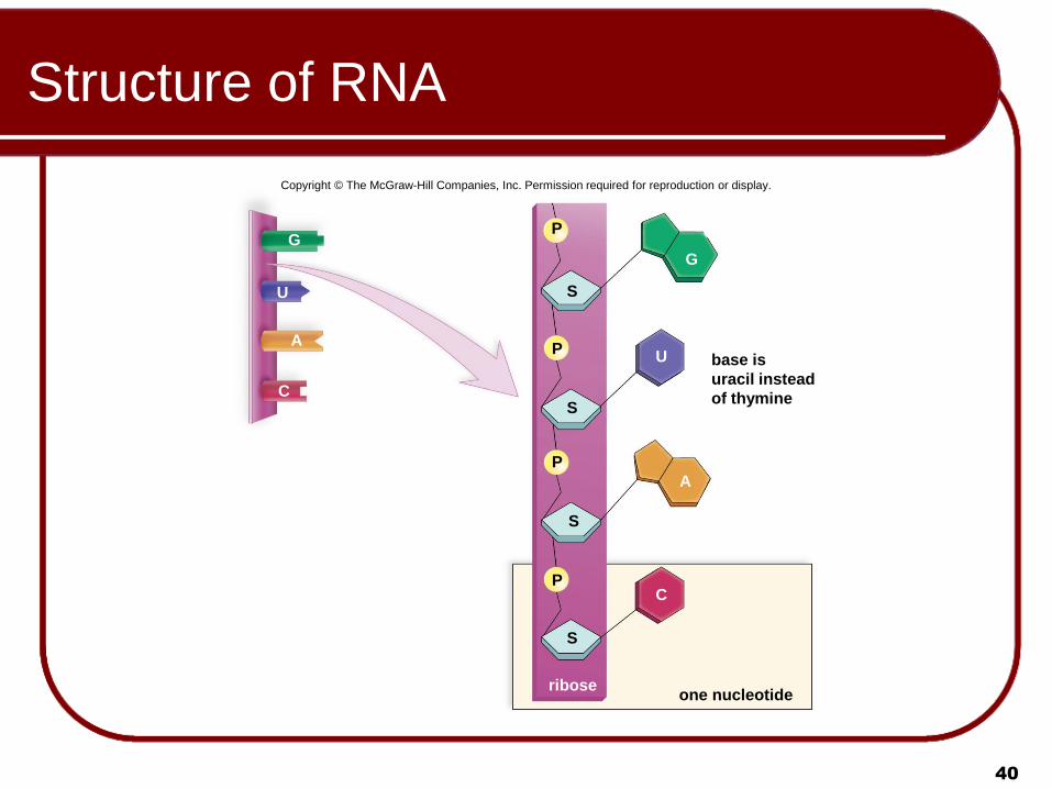
Structure of RNA
G
U
A
C
S
S
S
S
P
P
P
P
ribose
G
U base is
uracil instead
of thymine
A
C
one nucleotide
Copyright © The McGraw-Hill Companies, Inc. Permission required for reproduction or display.

41
RNA vs. DNA structure

42
The Genetic Code
Properties of the genetic code: Universal
With few exceptions, all organisms use the code the same way
Encode the same 20 amino acids with the same 64 triplets
Degenerate (redundant) There are 64 codons available for 20 amino acids Most amino acids encoded by two or more codons
Unambiguous (codons are exclusive) None of the codons code for two or more amino acids Each codon specifies only one of the 20 amino acids
Contains start and stop signals Punctuation codons Like the capital letter we use to signify the beginning of a
sentence, and the period to signify the end
43
The Genetic Code
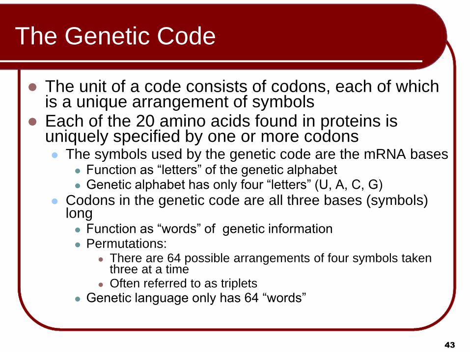
The unit of a code consists of codons, each of which is a unique arrangement of symbols
Each of the 20 amino acids found in proteins is uniquely specified by one or more codons The symbols used by the genetic code are the mRNA bases
Function as “letters” of the genetic alphabet Genetic alphabet has only four “letters” (U, A, C, G)
Codons in the genetic code are all three bases (symbols) long Function as “words” of genetic information Permutations:
There are 64 possible arrangements of four symbols taken three at a time
Often referred to as triplets
Genetic language only has 64 “words”
44
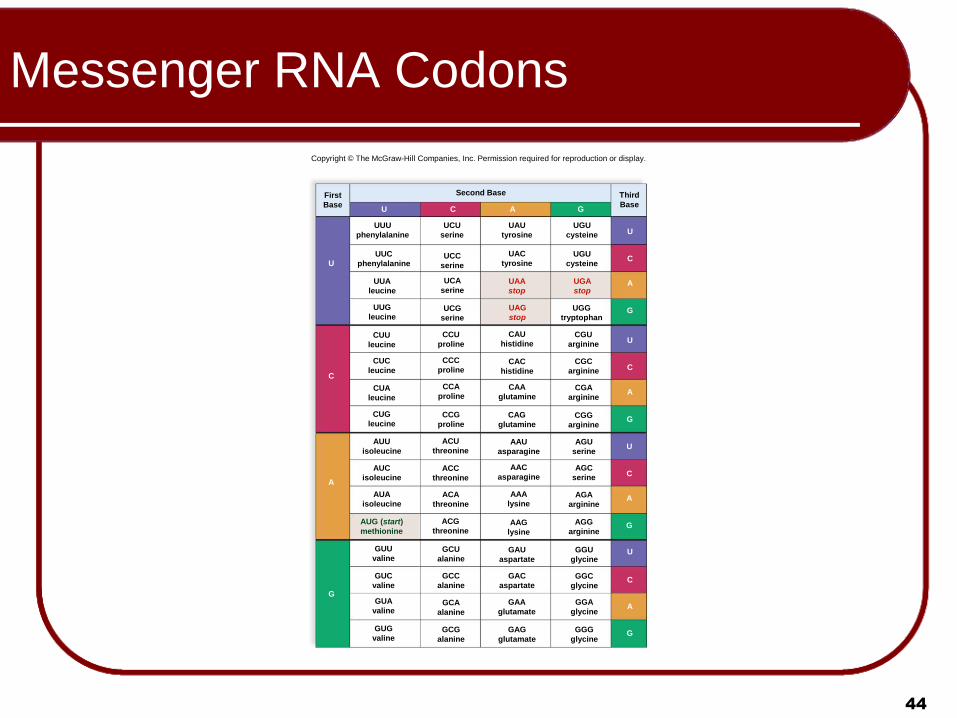
Messenger RNA Codons
Copyright © The McGraw-Hill Companies, Inc. Permission required for reproduction or display.
Second Base Third
Base
First
Base U C G A
U
C
A
G
UUU
phenylalanine
UCU
serine
UAU
tyrosine
UGU
cysteine
UUC
phenylalanine UCC
serine
UAC
tyrosine
UGU
cysteine
UCA
serine UUA
leucine
UCG
serine
UUG
leucine UGG
tryptophan
UGA
stop
UAA
stop
UAG
stop
U
C
A
G
CUU
leucine
CUC
leucine
CUA
leucine
CUG
leucine
CCU
proline
CCC
proline
CCA
proline
CCG
proline
CAC
histidine
CAA
glutamine
CAG
glutamine
CAU
histidine
CGA
arginine
CGG
arginine
CGU
arginine
CGC
arginine
U
C
A
G
AUG (start)
methionine
AUU
isoleucine
AUC
isoleucine
AUA
isoleucine
ACU
threonine
ACC
threonine
ACA
threonine
ACG
threonine
AAU
asparagine
AAC
asparagine
AAA
lysine
AAG
lysine
AGU
serine
AGC
serine
AGA
arginine
AGG
arginine
U
C
A
G
GUU
valine
GUC
valine
GUA
valine
GUG
valine
GCU
alanine
GCC
alanine
GCA
alanine
GCG
alanine
GAU
aspartate
GAC
aspartate
GAA
glutamate
GAG
glutamate
GGU
glycine
GGC
glycine
GGA
glycine
GGG
glycine
U
C
A
G

45
Steps in Gene Expression: Transcription
Transcription
Gene unzips and exposes unpaired bases
Serves as template for mRNA formation
Loose RNA nucleotides bind to exposed DNA
bases using the C=G & A=U rule
When entire gene is transcribed into mRNA,
result is a pre-mRNA transcript of the gene
The base sequence in the pre-mRNA is
complementary to the base sequence in DNA

Animation
Please note that due to differing
operating systems, some animations
will not appear until the presentation is
viewed in Presentation Mode (Slide
Show view). You may see blank slides
in the “Normal” or “Slide Sorter” views.
All animations will appear after viewing
in Presentation Mode and playing each
animation. Most animations will require
the latest version of the Flash Player,
which is available at
http://get.adobe.com/flashplayer.
47
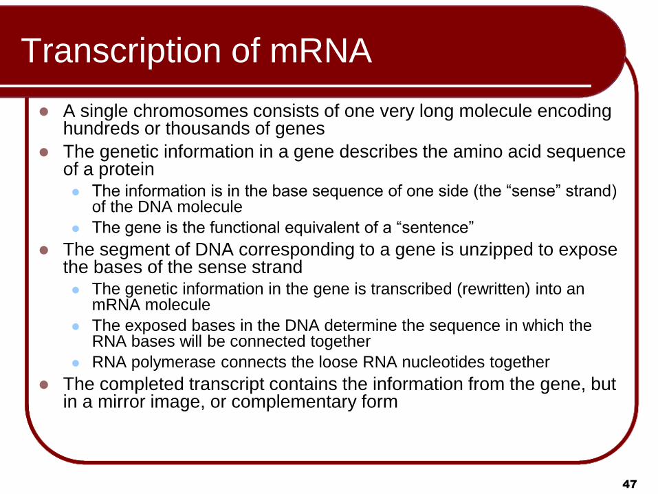
Transcription of mRNA
A single chromosomes consists of one very long molecule encoding hundreds or thousands of genes
The genetic information in a gene describes the amino acid sequence of a protein The information is in the base sequence of one side (the “sense” strand)
of the DNA molecule
The gene is the functional equivalent of a “sentence”
The segment of DNA corresponding to a gene is unzipped to expose the bases of the sense strand The genetic information in the gene is transcribed (rewritten) into an
mRNA molecule
The exposed bases in the DNA determine the sequence in which the RNA bases will be connected together
RNA polymerase connects the loose RNA nucleotides together
The completed transcript contains the information from the gene, but in a mirror image, or complementary form

Animation
Please note that due to differing
operating systems, some animations
will not appear until the presentation is
viewed in Presentation Mode (Slide
Show view). You may see blank slides
in the “Normal” or “Slide Sorter” views.
All animations will appear after viewing
in Presentation Mode and playing each
animation. Most animations will require
the latest version of the Flash Player,
which is available at
http://get.adobe.com/flashplayer.
49
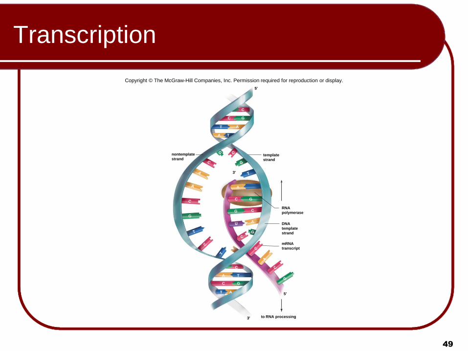
Transcription
Copyright © The McGraw-Hill Companies, Inc. Permission required for reproduction or display.
nontemplate
strand template
strand
5'
C
C G
T A
A T
G
G
G
A
C
C
A
G
RNA
polymerase
DNA
template
strand
mRNA
transcript
C
A T
C G
T A
to RNA processing
3'
3'
5'

Animation
Please note that due to differing
operating systems, some animations
will not appear until the presentation is
viewed in Presentation Mode (Slide
Show view). You may see blank slides
in the “Normal” or “Slide Sorter” views.
All animations will appear after viewing
in Presentation Mode and playing each
animation. Most animations will require
the latest version of the Flash Player,
which is available at
http://get.adobe.com/flashplayer.

Animation
51
Please note that due to differing
operating systems, some animations
will not appear until the presentation is
viewed in Presentation Mode (Slide
Show view). You may see blank slides
in the “Normal” or “Slide Sorter” views.
All animations will appear after viewing
in Presentation Mode and playing each
animation. Most animations will require
the latest version of the Flash Player,
which is available at
http://get.adobe.com/flashplayer.

52
RNA Polymerase
© Oscar L. Miller/Photo Researchers, Inc.
Copyright © The McGraw-Hill Companies, Inc. Permission required for reproduction or display.
a. 200m
b.
spliceosome
DNA
RNA
polymerase
RNA
transcripts

53
Processing Messenger RNA
Pre-mRNA, is modified before leaving the
eukaryotic nucleus.
Modifications to ends of primary transcript:
Cap of modified guanine on 5′ end
The cap is a modified guanine (G) nucleotide
Helps a ribosome where to attach when translation begins
Poly-A tail of 150+ adenines on 3′ end
Facilitates the transport of mRNA out of the nucleus
Inhibits degradation of mRNA by hydrolytic
enzymes.

54
Processing Messenger RNA
Pre-mRNA, is composed of exons and introns. The exons will be expressed,
The introns, occur in between the exons. Allows a cell to pick and choose which exons will go into a
particular mRNA
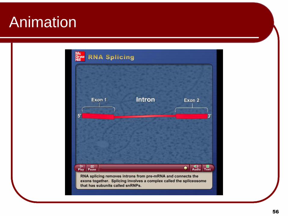
RNA splicing:
Primary transcript consists of:
Some segments that will not be expressed (introns)
Segments that will be expressed (exons)
Performed by spliceosome complexes in nucleoplasm
Introns are excised
Remaining exons are spliced back together
Result is mature mRNA transcript

55
RNA Splicing
In prokaryotes, introns are removed by
“self-splicing”—that is, the intron itself has
the capability of enzymatically splicing itself
out of a pre-mRNA
Animation
56
Please note that due to differing
operating systems, some animations
will not appear until the presentation is
viewed in Presentation Mode (Slide
Show view). You may see blank slides
in the “Normal” or “Slide Sorter” views.
All animations will appear after viewing
in Presentation Mode and playing each
animation. Most animations will require
the latest version of the Flash Player,
which is available at
http://get.adobe.com/flashplayer.

57
Messenger RNA Processing
Copyright © The McGraw-Hill Companies, Inc. Permission required for reproduction or display.
exon
intron intron
exon exon
DNA
transcription
exon
intron intron
exon exon
5' 3'
pre-mRNA
exon exon exon
intron intron cap poly-A tail 5' 3'
exon exon exon
spliceosome
cap poly-A tail
pre-mRNA
splicing
intron RNA
5' 3'
cap poly-A tail
mRNA
nuclear pore
in nuclear envelope
nucleus
cytoplasm
5' 3'

Animation
Please note that due to differing
operating systems, some animations
will not appear until the presentation is
viewed in Presentation Mode (Slide
Show view). You may see blank slides
in the “Normal” or “Slide Sorter” views.
All animations will appear after viewing
in Presentation Mode and playing each
animation. Most animations will require
the latest version of the Flash Player,
which is available at
http://get.adobe.com/flashplayer.
59
Functions of Introns
As organismal complexity increases; Number of protein-coding genes does not keep pace
But the proportion of the genome that is introns increases
Humans: Genome has only about 25,000 coding genes
Up to 95% of this DNA genes is introns
Possible functions of introns: More bang for buck
Exons might combine in various combinations
Would allow different mRNAs to result from one segment of DNA
Introns might regulate gene expression
Exciting new picture of the genome is emerging

60
Steps in Gene Expression: Translation
tRNA molecules have two binding sites
One associates with the mRNA transcript
The other associates with a specific amino acid
Each of the 20 amino acids in proteins associates with one or more of 64 species of tRNA
Translation
An mRNA transcript migrates to rough endoplasmic reticulum
Associates with the rRNA of a ribosome
The ribosome “reads” the information in the transcript
Ribosome directs various species of tRNA to bring in their specific amino acid “fares”
tRNA specified is determined by the code being translated in the mRNA transcript

61
tRNA
tRNA molecules come in 64 different kinds
All very similar except that
One end bears a specific triplet (of the 64
possible) called the anticodon
Other end binds with a specific amino acid type
tRNA synthetases attach correct amino acid to the
correct tRNA molecule
All tRNA molecules with a specific anticodon
will always bind with the same amino acid

Animation
Please note that due to differing
operating systems, some animations
will not appear until the presentation is
viewed in Presentation Mode (Slide
Show view). You may see blank slides
in the “Normal” or “Slide Sorter” views.
All animations will appear after viewing
in Presentation Mode and playing each
animation. Most animations will require
the latest version of the Flash Player,
which is available at
http://get.adobe.com/flashplayer.

63
Structure of tRNA
Copyright © The McGraw-Hill Companies, Inc. Permission required for reproduction or display.
amino
acid
leucine
3
5
Hydrogen
bonding
anticodon
mRNA
5'
codon
3'
b.
anticodon end
amino acid end

64
Ribosomes
Ribosomal RNA (rRNA):
Produced from a DNA template in the nucleolus
Combined with proteins into large and small ribosomal
subunits
A completed ribosome has three binding sites to
facilitate pairing between tRNA and mRNA
The E (for exit) site
The P (for peptide) site, and
The A (for amino acid) site

65
Ribosomal Structure and Function
Copyright © The McGraw-Hill Companies, Inc. Permission required for reproduction or display.
Courtesy Alexander Rich
large subunit
small subunit
a. Structure of a ribosome
tRNA binding
sites
outgoing
tRNA
3 5
mRNA
b. Binding sites of ribosome
polypeptide incoming
tRNA
incoming
tRNA
c. Function of ribosomes d. Polyribosome
66
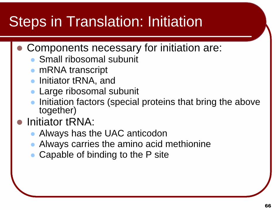
Steps in Translation: Initiation
Components necessary for initiation are: Small ribosomal subunit mRNA transcript Initiator tRNA, and Large ribosomal subunit Initiation factors (special proteins that bring the above
together)
Initiator tRNA: Always has the UAC anticodon Always carries the amino acid methionine Capable of binding to the P site

67
Steps in Translation: Initiation
Small ribosomal subunit attaches to mRNA
transcript
Beginning of transcript always has the START
codon (AUG)
Initiator tRNA (UAC) attaches to P site
Large ribosomal subunit joins the small
subunit

Animation
Please note that due to differing
operating systems, some animations
will not appear until the presentation is
viewed in Presentation Mode (Slide
Show view). You may see blank slides
in the “Normal” or “Slide Sorter” views.
All animations will appear after viewing
in Presentation Mode and playing each
animation. Most animations will require
the latest version of the Flash Player,
which is available at
http://get.adobe.com/flashplayer.

69
Steps in Translation: Initiation
Copyright © The McGraw-Hill Companies, Inc. Permission required for reproduction or display.
A small ribosomal subunit
binds to mRNA; an initiator
tRNA pairs with the mRNA
start codon AUG. The large ribosomal subunit
completes the ribosome.
Initiator tRNA occupies the
P site. The A site is ready
for the next tRNA.
Initiation
Met
amino acid methionine
initiator tRNA
mRNA
small ribosomal subunit
3'
5'
P site A site E site
Met
large ribosomal subunit
U A C
A U G
start codon 5' 3'

70
Steps in Translation: Elongation
“Elongation” refers to the growth in length of the polypeptide
RNA molecules bring their amino acid fares to the ribosome Ribosome reads a codon in the mRNA
Allows only one type of tRNA to bring its amino acid
Must have the anticodon complementary to the mRNA codon being read
Joins the ribosome at it’s A site
Methionine of initiator is connected to amino acid of 2nd tRNA by peptide bond

71
Steps in Translation: Elongation
Second tRNA moves to P site (translocation)
Spent initiator moves to E site and exits
Ribosome reads the next codon in the mRNA
Allows only one type of tRNA to bring its amino acid
Must have the anticodon complementary to the mRNA codon
being read
Joins the ribosome at it’s A site
Dipeptide on 2nd amino acid is connected to amino acid
of 3nd tRNA by peptide bond
Animation
Please note that due to differing
operating systems, some animations
will not appear until the presentation is
viewed in Presentation Mode (Slide
Show view). You may see blank slides
in the “Normal” or “Slide Sorter” views.
All animations will appear after viewing
in Presentation Mode and playing each
animation. Most animations will require
the latest version of the Flash Player,
which is available at
http://get.adobe.com/flashplayer.

73
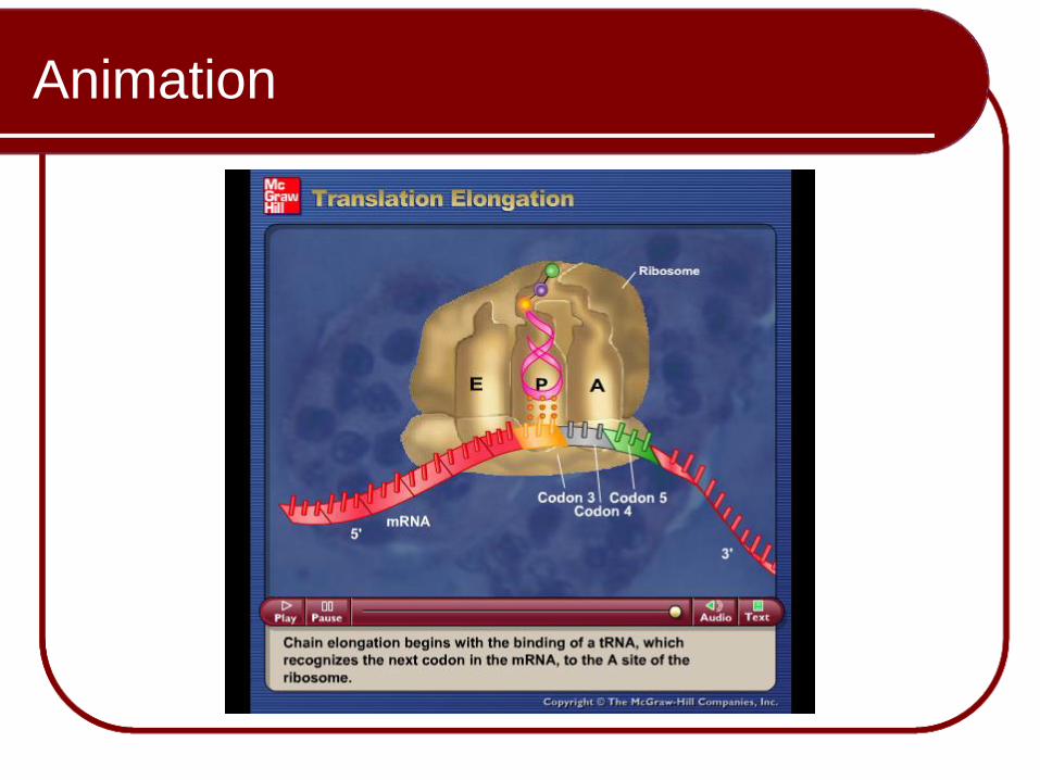
Steps in Translation: Elongation
Copyright © The McGraw-Hill Companies, Inc. Permission required for reproduction or display.
A tRNA–amino acid
approaches the
ribosome and binds
at the A site.
Two tRNAs can be at a
ribosome at one time;
the anticodons are
paired to the codons.
Peptide bond formation
attaches the peptide
chain to the newly
arrived amino acid.
The ribosome moves forward; the
“empty” tRNA exits from the E site;
the next amino acid–tRNA complex
is approaching the ribosome.
1 2 3 4
Elongation
peptide
bond
Met
Ala
Trp
Ser
Val
U
A
C A
U G G A C
3 3
C G
anticodon
tRNA
asp
U
Met
Ala
Trp
Ser
Val
U
A
C A
U G G A C
C U G
Asp
6
U
A
C A
U G G A C
C U G
Met
Val
Asp
Ala
Trp
Ser
peptide
bond
6 3
G A C
C U G
A U G A C C
Met
Val
Asp
Ala
Trp
Ser Thr
6 3

74
Steps in Translation: Termination
Previous tRNA moves to P site
Spent tRNA moves to E site and exits
Ribosome reads the STOP codon at the end of the mRNA UAA, UAG, or UGA
Does not code for an amino acid
Polypeptide is released from last tRNA by release factor
Ribosome releases mRNA and dissociates into subunits
mRNA read by another ribosome

Animation
Please note that due to differing
operating systems, some animations
will not appear until the presentation is
viewed in Presentation Mode (Slide
Show view). You may see blank slides
in the “Normal” or “Slide Sorter” views.
All animations will appear after viewing
in Presentation Mode and playing each
animation. Most animations will require
the latest version of the Flash Player,
which is available at
http://get.adobe.com/flashplayer.

76
Steps in Translation: Termination
Copyright © The McGraw-Hill Companies, Inc. Permission required for reproduction or display.
Termination
The release factor hydrolyzes the bond
between the last tRNA at the P site and
the polypeptide, releasing them. The
ribosomal subunits dissociate.
3
5
The ribosome comes to a stop
codon on the mRNA. A release
factor binds to the site.
U A
U A U G A
stop codon 5' 3'
Asp
Ala
Trp
Val Glu
release factor

Animation
77
Please note that due to differing
operating systems, some animations
will not appear until the presentation is
viewed in Presentation Mode (Slide
Show view). You may see blank slides
in the “Normal” or “Slide Sorter” views.
All animations will appear after viewing
in Presentation Mode and playing each
animation. Most animations will require
the latest version of the Flash Player,
which is available at
http://get.adobe.com/flashplayer.

78
Summary of Gene Expression (Eukaryotes)
Copyright © The McGraw-Hill Companies, Inc. Permission required for reproduction or display.
TRANSCRIPTION
1. DNA in nucleus serves
as a template for mRNA.
2. mRNA is processed
before leaving the nucleus.
mRNA
pre-mRNA
DNA
introns
3. mRNA moves into
cytoplasm and
becomes associated
with ribosomes.
TRANSLATION
mRNA large and small
ribosomal subunits
5
3'
nuclear pore
4. tRNAs with
anticodons carry
amino acids
to mRNA.
5
peptide
codon
ribosome
3 U A
A U
C G
5 C C G G
G C G
C G
C G
U A
U A
U A
U A
6. During elongation, polypeptide
synthesis takes place one
amino acid at a time. 7. Ribosome attaches to rough
ER. Polypeptide enters lumen,
where it folds and is
modified.
8. During termination, a
ribosome reaches a stop
codon; mRNA and
ribosomal subunits
disband.
5. During initiation, anticodon-codon
complementary base pairing begins
as the ribosomal subunits come
together at a start codon.
amino
acids
anticodon
tRNA
3'

Animation
Please note that due to differing
operating systems, some animations
will not appear until the presentation is
viewed in Presentation Mode (Slide
Show view). You may see blank slides
in the “Normal” or “Slide Sorter” views.
All animations will appear after viewing
in Presentation Mode and playing each
animation. Most animations will require
the latest version of the Flash Player,
which is available at
http://get.adobe.com/flashplayer.

80
Structure of Eukaryotic Chromosome
Contains a single linear DNA molecule, but is composed of more than 50% protein.
Some of these proteins are concerned with DNA and RNA synthesis,
Histones, play primarily a structural role
Five primary types of histone molecules
Responsible for packaging the DNA DNA double helix is wound at intervals around a
core of eight histone molecules (called nucleosome)
Nucleosomes are joined by “linker” DNA.

81
Structure of Eukaryotic Chromosome
Copyright © The McGraw-Hill Companies, Inc. Permission required for reproduction or display.
a. Nucleosomes (“beads on a string”)
b. 30-nm fiber
c. Radial loop domains
d. Heterochromatin
e. Metaphase chromosome
1. Wrapping of DNA
around histone proteins.
4. Tight compaction of radial
loops to form heterochromatin.
3. Loose coiling into radial
loops.
2. Formation of a three-dimensional
zigzag structure via histone H1 and
other DNA-binding proteins.
5. Metaphase chromosome forms
with the help of a protein
scaffold.
2 nm
1 nm
300 nm
1,400 nm
700 nm
30 nm
DNA
double helix
histones
histone H1
nucleosome
euchromatin
Animation
Please note that due to differing
operating systems, some animations
will not appear until the presentation is
viewed in Presentation Mode (Slide
Show view). You may see blank slides
in the “Normal” or “Slide Sorter” views.
All animations will appear after viewing
in Presentation Mode and playing each
animation. Most animations will require
the latest version of the Flash Player,
which is available at
http://get.adobe.com/flashplayer.
83
Review
Genetic Material
Transformation
DNA Structure
Watson and Crick
DNA Replication
Prokaryotic versus Eukaryotic
Replication Errors
Transcription
Translation
Structure of Eukaryotic Chromosome

Sylv
ia S
. Ma
der
Copyright © The McGraw Hill Companies Inc. Permission required for reproduction or display
PowerPoint® Lecture Slides are prepared by Dr. Isaac Barjis, Biology Instructor
BIOLOGY 10th Edition
Molecular Biology of the Gene
Chapter 12: pp. 211 - 232
84
Copyright © The McGraw-Hill Companies, Inc. Permission required for reproduction or display.
b.
3.4 nm
2 nm
0.34 nm
G C
A
T
T A
P
P
P
P
C G
G
C
Sugar hydrogen bonds
sugar-phosphate
backbone