Embed Size (px)
Citation preview
Biology Chapter 1
Biology as a science
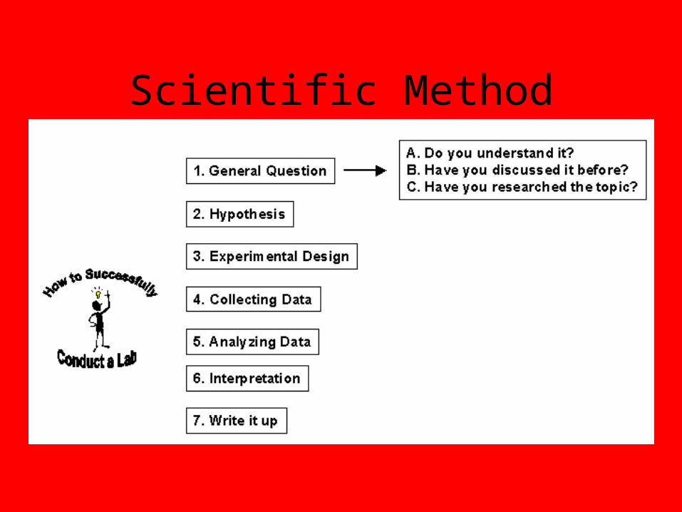
Scientific Method


Scientific Method

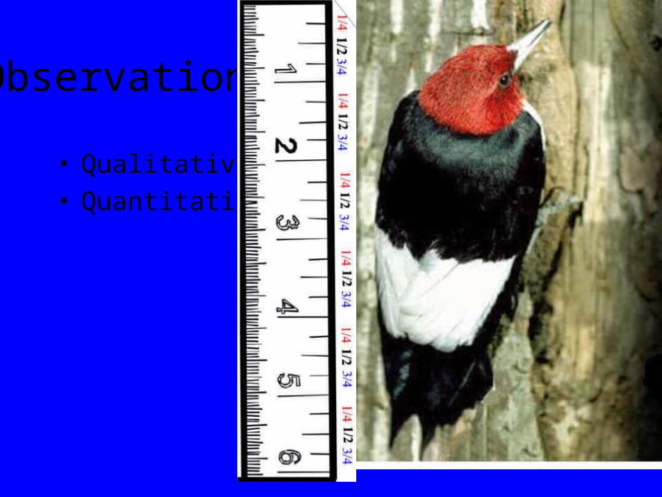
Observations
• Qualitative• Quantitative
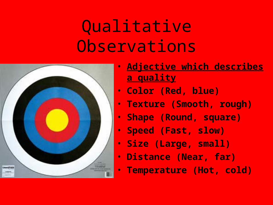
Qualitative Observations
• Adjective which describes a quality
• Color (Red, blue)
• Texture (Smooth, rough)
• Shape (Round, square)
• Speed (Fast, slow)
• Size (Large, small)
• Distance (Near, far)
• Temperature (Hot, cold)

Quantitative Observations
• Numerical measurements which describe:• Color (700 nM)• Texture (50 grit sandpaper)• Shape (25 cm diameter circle)• Speed ( 75 m.p.h.)• Size (150 m)• Distance (130 miles)• Temperature (37.5 C)
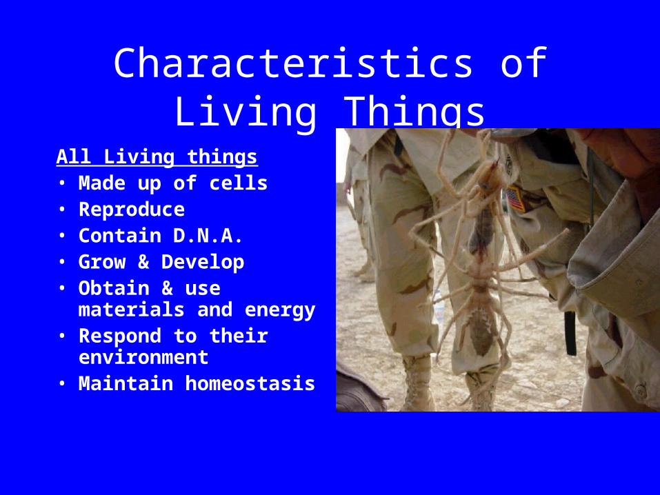
Characteristics of Living Things
All Living things• Made up of cells• Reproduce• Contain D.N.A.• Grow & Develop• Obtain & use materials
and energy• Respond to their
environment• Maintain homeostasis
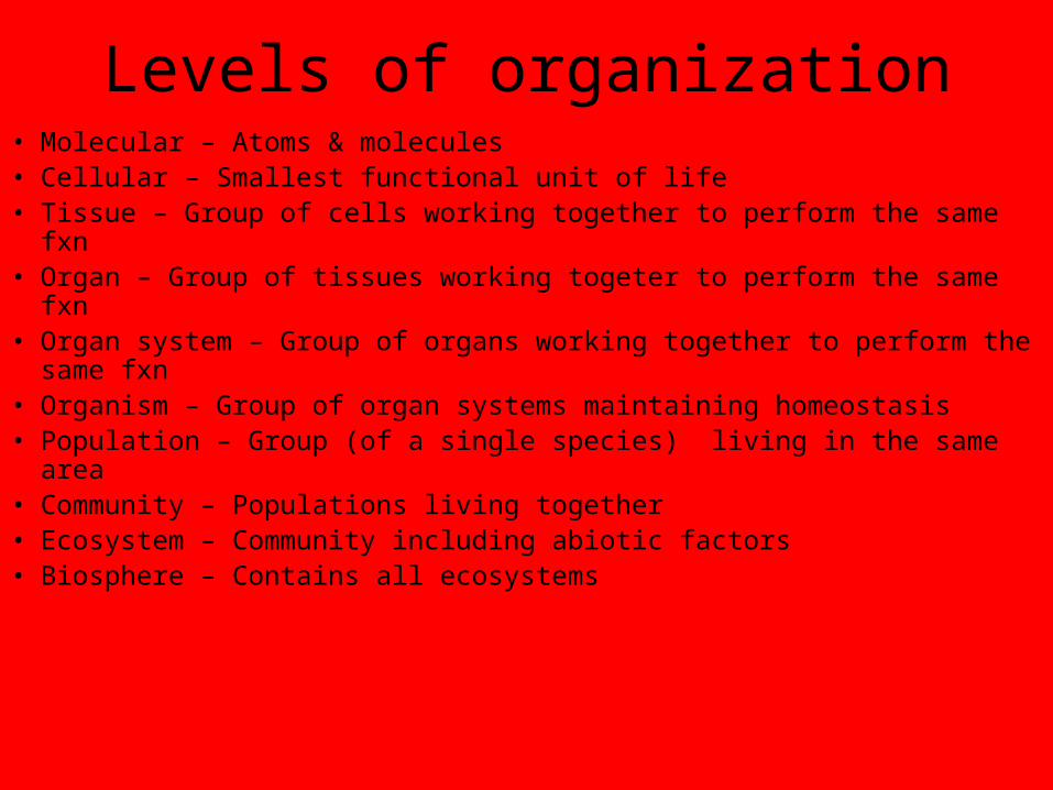
Levels of organization• Molecular – Atoms & molecules• Cellular – Smallest functional unit of life• Tissue – Group of cells working together to perform the same fxn• Organ – Group of tissues working togeter to perform the same fxn• Organ system – Group of organs working together to perform the
same fxn• Organism – Group of organ systems maintaining homeostasis• Population – Group (of a single species) living in the same area• Community – Populations living together• Ecosystem – Community including abiotic factors• Biosphere – Contains all ecosystems


Compound Microscope Parts & Fxn’s
• Occular – viewing eyepiece• Coarse adjustment – Rough focus• Fine adjustment – Fine focus• High power objective (400X)• Low objective (100X)• Scanning objective (40X)• Stage – holds slide up against stage clips• Stage clips – holds slide down on stage• Diaphragm – controls amount of light entering slide• Lamp – light source

Power of magnification • The relative enlargement of the specimen
when seen through the microscope. The power of magnification can be calculated by multiplying the power of the eye piece lens by the power of the objective lens.

Inversion
• The reversal of the specimen image by the microscope lenses. A specimen that appears upside down when being viewed is actually right-side up on the slide. Moving the specimen to the right causes its image to move to the left likewise, moving it down causes it to move upward.
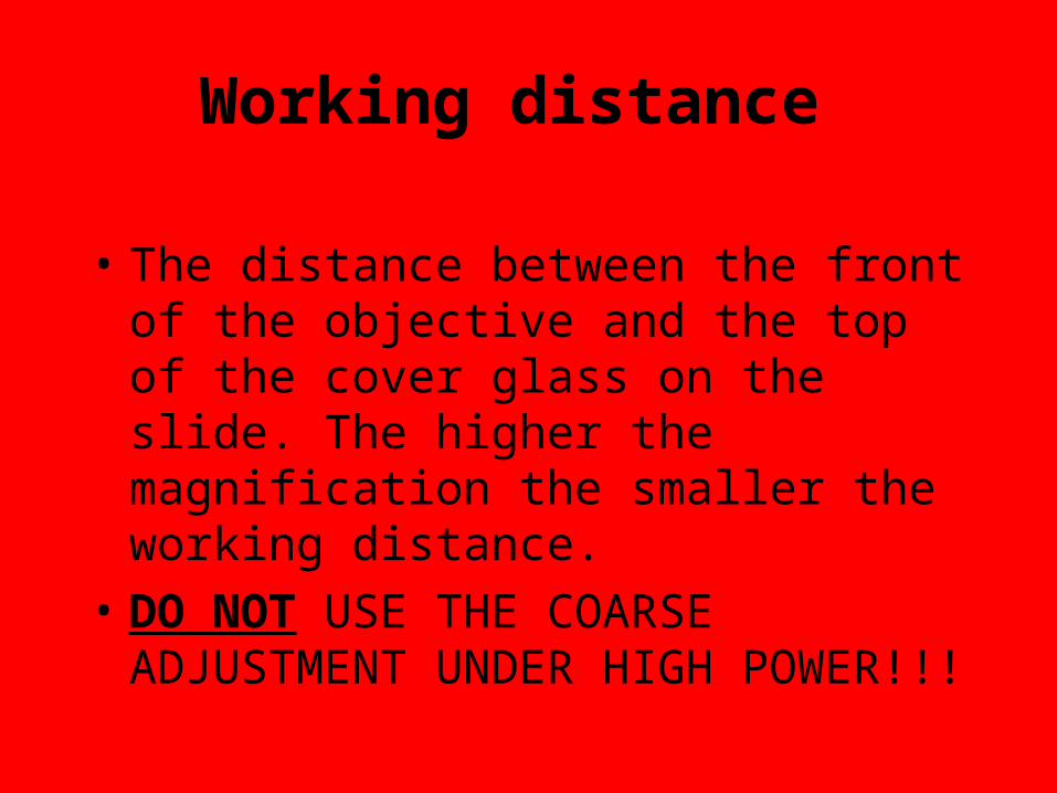
Working distance
• The distance between the front of the objective and the top of the cover glass on the slide. The higher the magnification the smaller the working distance.
• DO NOT USE THE COARSE ADJUSTMENT UNDER HIGH POWER!!!

Resolution (Resolving Power)
• The least distance between two points or lines at which they are seen as two, rather than a single blur. The greater the numerical aperture the greater the resolution.

Depth of focus
• The thickness of a specimen which may be seen in focus at one time. The greater the power of magnification the lesser the depth of focus.

Field of vision
• The surface area which can be seen when looking through the light microscope. The area decreases with increasing power of magnification.
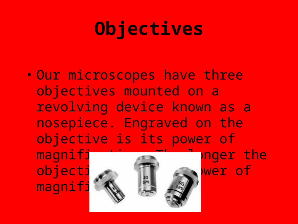
Objectives
• Our microscopes have three objectives mounted on a revolving device known as a nosepiece. Engraved on the objective is its power of magnification. The longer the objective the more power of magnification.

Diaphragm
• A device under the stage of a microscope that can regulate the amount of light reaching a specimen. The more power of magnification the more the diaphragm is opened.

Power of Magnification
• Definition - The relative enlargement of the specimen when seen through the microscope.
• Calculation - The power of magnification can be calculated by multiplying the power of the eye piece lens by the power of the objective lens.
• Power of magnification = (Power of the eyepiece lens) X (Power of the objective lens)

Parfocal
• Once the specimen is focused on low power, you never have to use the course adjustment knob to focus on the next higher power.
Oil Immersion Lens
• For maximum magnification such as looking at bacteria or white blood cells. It can not be used without a drop of special oil placed between the slide and the objective.

Rules for Handling the Microscope
• Always carry the microscope with one hand under the base and the other grasping the arm.
• Keep both eyes open when looking through the eyepiece.
• Keep the stage clean and dry.• Do not remove parts of the microscope.• Use only lens paper when cleaning lenses.• Always begin focusing with the lowest power objective.• Always look from the side when changes lenses• After completing your work, place the microscope on
the lowest power objective.• Always return the microscope where you found it & as
you found it
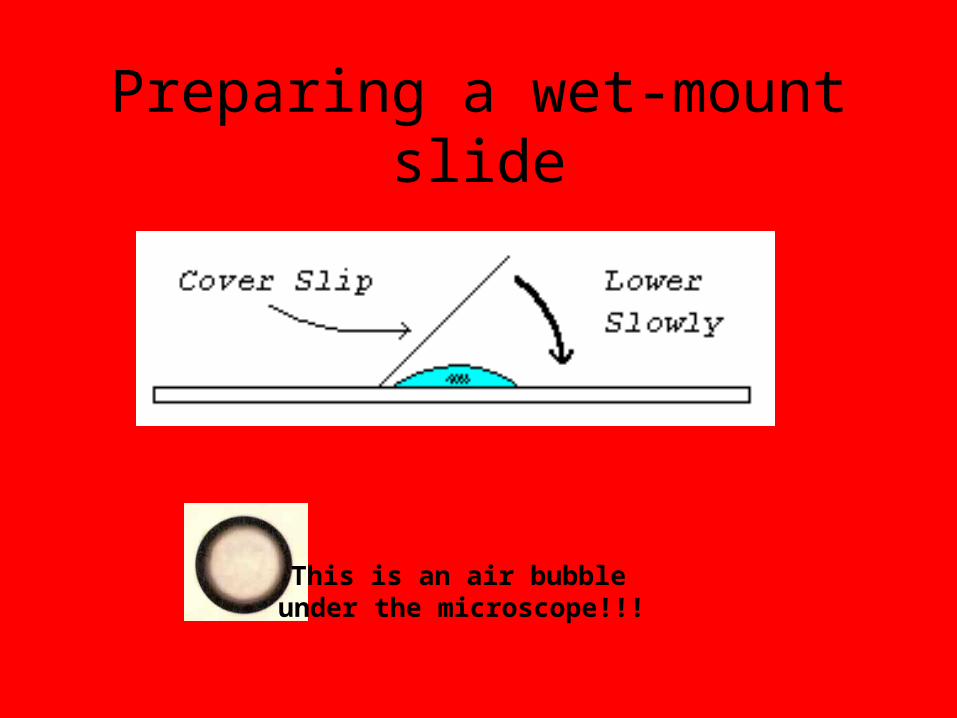
Preparing a wet-mount slide
This is an air bubble under the microscope!!!


![Biology scientific method[10]](https://img.pdfslide.us/doc/110x75/558e565a1a28ab2a508b4685/biology-scientific-method10.jpg)