Embed Size (px)
Citation preview
conference papers
J. Appl. Cryst. (2007). 40, s217–s222 Grossmann � Biological solution scattering s217
Journal of
AppliedCrystallography
ISSN 0021-8898
Received 16 August 2006
Accepted 2 February 2007
# 2007 International Union of Crystallography
Printed in Singapore – all rights reserved
Biological solution scattering: recent achievementsand future challenges
J. Gunter Grossmann
CCLRC Daresbury Laboratory, Molecular Biophysics Group, Warrington WA4 4AD, UK. Correspondence e-mail:
In the post-genomic age it is apparent that as structures of larger
macromolecules and their complexes are investigated, structure–function
investigations are often confronted with the necessity to apply a portfolio of
tools for biochemical and biophysical characterization. A survey of the
published literature over the last decade reveals that publications in the area
of structural biology employing neutron or X-ray scattering as one of their
techniques tripled since 1995. Yet, taken as a whole, the contribution from small-
angle scattering (SAS) to research papers dealing with structural analyses is still
only of the order of 1% in 2005 (for comparison, less than 0.5% in 1995).
Nevertheless, the last few years saw stimulating biological applications and
analysis procedures which emphasize the growing potential of SAS applications
for the structural studies of macromolecules in solution. The usage of SAS
largely consists of low-resolution reconstructions of molecules with partial or
without presumption of structural details, consistency analysis of high-resolution
crystallographic structures and their corresponding low-resolution models
determined in the solution state, and rigid-body refinement of multi-subunit
assemblies including complexes and full-length multidomain proteins. Comple-
mentary structural information obtained from SAS in conjunction with data
acquired by protein crystallography, NMR, molecular dynamics or computa-
tional docking provides a means to link low- and high-resolution models
essential for the elucidation of biomolecular organization, interactions and
function. The capabilities as well as limitations of determining low-resolution
structures of multidomain proteins, macromolecular complexes and assemblies
are highlighted in three examples. The first example is the characterization of
the conformation of the PDZ region of SAP97, a multidomain protein involved
in the regulation and localization of membrane receptor molecules. The second
example is the characterization of the structural features of the TIM10 complex,
an escort molecule for mitochondrial inner-membrane proteins, and the third
example is the description of the shape and pH-induced conformational
transition of a full-length bacterial potassium channel. The latter two in
particular benefited from neutron scattering with contrast variation by using H–
D labelling of the macromolecular complex or the solvent, respectively.
1. Introduction
Solution X-ray and neutron scattering are complimentary techniques
that can provide the size and shape of macromolecules with mole-
cular masses ranging from a few kilodaltons to several million
daltons. Even though scattering techniques do not have the high-
resolution capability of protein crystallography (PX) or NMR, they
are much more widely applicable to biological systems as long as
monodisperse solutions are at hand. Thus, even intrinsically
unstructured proteins can be evaluated by the scattering method, and
very large complexes are also amenable. Despite being low-resolu-
tion methods, the ability to provide molecular dimensions and
envelopes is especially important for establishing global information
about partially folded, disordered or conformationally mobile states
of macromolecules under physiological conditions, such as those that
occur in protein folding, multidomain molecules or multi-subunit
complexes. In these cases, crystallization is challenging if not
impossible and NMR studies give local rather than global structural
information. The past decade has seen the appearance of data-
analysis techniques which enable the reconstruction of molecular
shapes from scattering data [see e.g. Svergun & Koch (2003) for a
review]. Svergun & Stuhrmann (1991) were the first to show that it is
feasible to obtain meaningful three-dimensional density maps from
one-dimensional scattering data. Their remarkable procedure is
capable of selecting an ensemble of consistent shapes from a large
number of possible solutions. Several other approaches have since
been tested successfully (Chacon et al., 1998; Svergun, 1999; Walther
et al., 2000; Svergun et al., 2001, Heller et al., 2002); all of them treat
the scatterer as a system with a fixed number of connected scattering
centres (an assembly of spheres) that form a more-or-less compact
particle with a distinct solvent boundary. Disjointed centres or
mixtures of different particles therefore do not meet the criteria for a
biologically meaningful shape reconstruction. It is this approach of ab
initio shape reconstruction in conjunction with scattering profile
simulations and molecular modelling which has had a significant
impact on the use of solution scattering techniques recently, as more
valuable three-dimensional details can be extracted compared to
results that merely hinge on quantities such as the radius of gyration,
the maximum molecular dimension or on simple ellipsoidal or
spherical conformational outlines. The currently available ATSAS
software package developed and expanded by Svergun and cowor-
kers (Konarev et al., 2006) is popular with a large number of SAS
users and is reaching a status similar to the software suite for PX
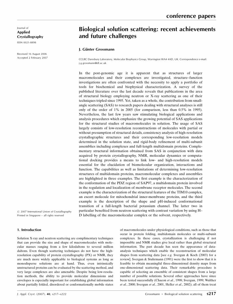
(CCP4) a decade ago. Whether the threefold increase of published
SAS papers in the area of structural biology (see Fig. 1) over the last
decade is a result of these advances remains an open question.
Regardless of the relatively low overall contribution (~1%) to
structural studies, the significant contributions from a growing
amount of SAS investigations can not be ignored.
The era of structural genomics results in an ever increasing archive
of experimentally determined biological macromolecular three-
dimensional structures at high resolution. Yet, despite the yearly
growth of total structures in the Protein Data Bank (PDB), statistics
show that 63% (85%) of structures deposited in the PDB (37 556
structures as of 4 July 2006) have molecular masses up to 50 kDa
(100 kDa). Consequently, the representation of proteins consisting of
one or a few domains seems to be adequate, whereas large macro-
molecules and complexes still indicate a challenge for structural
genomics efforts. Inherent structural flexibility and the transient
nature of complex formation represent significant barriers for high-
resolution studies. Low-resolution information from SAS on macro-
molecules in solution can still provide valuable structure–function
information not only in terms of ab initio shape determination but
also in view of rigid-body modelling taking into account the knowl-
edge of a wealth of structures from small proteins and protein
domains. In this context, it is worthwhile to briefly mention a few
topical developments that are likely to provide stimuli for future
directions of SAS applications. Petoukhov & Svergun (2005) describe
methods to construct models of macromolecules and complexes from
atomic information of subunits or domains against SAS data. The
procedures allow, for instance, the addition of missing polypeptide
segments (e.g. linkers between domains), exploitation of molecular
symmetry and the consideration of information deduced from other
techniques such as distance constraints between distinct residues. The
latter is an essential validation criterion to select among multiple
solutions that are otherwise in harmony with the experimental data.
In a study of the Ras-specific nucleotide exchange factor SOS,
Sondermann et al. (2005) employed X-ray scattering data to validate
their computational docking models for this multidomain protein.
The results were corroborated by other techniques. Similarly,
Mattinen et al. (2002) showed that residual dipolar coupling infor-
mation can reduce orientational degeneracy of shapes from multi-
domain proteins obtained by rigid-body modelling. This idea was
taken forward significantly by Grishaev et al. (2005) who used SAS
data as a constraint to complement high-resolution NMR structure
determination. Considering the huge computational costs of fitting
SAS data at each step of molecular dynamics and energy minimiza-
tion during NMR structure refinement, a coarse-grain method was
applied to decrease the structural complexity of amino acids (defi-
nition of ‘globs’ and associated correction factors to rectify the
discrepancy between atomic and ‘glob’ scattering) and a reduced
number of experimental data points to be fitted were taken into
account. This led to a considerable reduction in the CPU time needed
for the cross-validation of experimental and simulated scattering
data. An analogous approach was reported recently by Wu et al.
(2005), who used the X-ray scattering profile as a soft constraint to
guide the folding of small helical proteins by computational methods.
In the following, three specific examples are presented in which the
interplay between solution X-ray and neutron scattering and atomic
structure information was exploited to extend low-resolution models
towards pseudo high-resolution structures. Achievements are also
discussed in the light of limitations and ensuing experimental and
computational studies.
2. Materials and methods
For sample preparation and explicit experimental details the reader is
referred to the published literature given in the sections below.
Specifically, Tim9, one of the two essential translocases for inner-
membrane proteins (Tim9 and Tim10) that form part of the TIM10
complex, has been expressed in deuterated form at the ILL–EMBL–
PSB Deuteration Laboratory in Grenoble, France. Subsequently, a
functional complex was formed between deuterated Tim9 and
hydrogenated Tim10, purified and dialysed against buffers of 40 and
100% D2O/H2O mixtures for neutron scattering studies. Neutron
experiments were performed at instrument D22 (Institut Laue–
Langevin, Grenoble, France). X-ray scattering experiments were
carried out at beamline 2.1 of the UK Synchrotron Radiation Source
at Daresbury. Concerning data reduction, this was carried out with
software provided at the experimental facilities and for the subse-
quent analysis the ATSAS program package (Konarev et al., 2006)
was exploited. Molecular dynamics (MD) calculations on detergent
and detergent–protein systems have been performed according to
procedures as described e.g. in Bond & Sansom (2003) or Patargias et
conference papers
s218 Grossmann � Biological solution scattering J. Appl. Cryst. (2007). 40, s217–s222
Figure 1Upwards trend in the number of published SAS papers in the field of structuralbiology over the last 15 years. The results are displayed in absolute (main graph)and relative (inset) figures. The survey was performed with a search of the ISI Webof KnowledgeSM publications database. For further comparison (not included in theabove diagram), the number of papers with contributions from electron microscopy(EM) is almost four times the number of SAS publications in 2005, whereas adecade ago it was eight times the number of SAS publications. Moreover, theoverall contributions of EM to structural biology papers decreased from 4% (in1995) to 3% (in 2005).
al. (2005). Theoretical scattering patterns of the resulting MD
structures have been compared with experimental profiles.
3. Results and discussion
3.1. Conformational features of the PDZ region of SAP97
SAP97 is a member of the membrane-associated guanylate kinase
(MAGUK) family of proteins consisting of 911 residues, made up of
three PDZ domains (~100 amino acids each) and a ‘fused’ SH3-GK
domain. All these domains are common sites for protein–protein
interactions, suggesting that SAP97 functions as a scaffold bringing
together different proteins (e.g. ion channels) for information
transfer. Structural knowledge so far relied on individual PDZ or
SH3-GK domains; yet, the function of the protein involves all
domains acting in concert. Even though long regions of flexibility are
present in SAP97, the full-length protein was suggested to have a
rather compact conformation in contrast to ‘beads on a string’. X-ray
scattering experiments were initiated to characterize the three SAP97
PDZ domains, in particular double and triple PDZ domain constructs
(PDZ12: residues 218 to 406 and PDZ123: residues 218 to 577). The
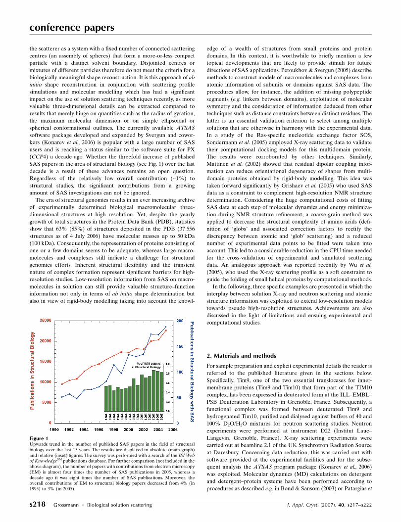
results reveal that PDZ12 is clearly smaller than PDZ123 (Rg = 23.6�
0.1 A, Dmax = 75 � 3 versus 33.3 � 0.1 and 115 � 6 A, respectively).
Ab initio shape calculations of the double domain show a dumbbell-
shaped molecule with two distinct and apparently non-interacting
domains; the triple domain is elongated, comprising three distinct
components (Fig. 2). Nevertheless, considering the long linker
between the second and third PDZ domain and an additional C-
terminal segment, the scattering results still point to a rather compact
shape for the triple domain. It was not expected to see the third PDZ
domain in such close proximity to the other two domains. Rigid-body
modelling allowed exploration of the spatial arrangement of domains
together with plausible conformations for the mobile linker sections
(Fig. 2). NMR data are consistent with the unstructured nature of the
linker and C-terminal regions, and also confirm little short-range
contacts among the globular PDZ domains (Goult et al., 2007). It is
remarkable that an extensive linker segment would keep the third
PDZ domain in close proximity to the first and second PDZ domains.
Therefore the unstructured linker is likely to induce transient inter-
actions of PDZ3 with PDZ1 or PDZ2 which not only bring the PDZ
domains closer together but may also assist in the organization and
orientation of SAP97 domains and/or their interaction partners.
Future experiments will concentrate on full-length SAP97 and its
interaction complex with target proteins.
3.2. Structural characterization of the TIM10 chaperone complex
Protein import into the mitochondrion is mediated by specialized
multi-protein machineries called translocases in both the outer and
inner membrane. The TIM10 complex negotiates the aqueous divide
between outer and inner membranes and thus specifically chaperones
the inner-membrane insertion of hydrophobic proteins like the ADP/
ATP carrier (AAC) that delivers ATP to the rest of the cell. The
TIM10 complex is made of two proteins, Tim9 (89 residues) and
Tim10 (90 residues). Despite their primarily helical secondary
structure, their homodimeric association and four cysteine residues
with the characteristic twin CX3C motif, they only share a sequence
identity of 21%. The functional TIM10 chaperone particle (60 kDa),
which consists of three Tim9 and three Tim10 protomers, does not
require ATP and as such is expected to adopt quite a distinct and
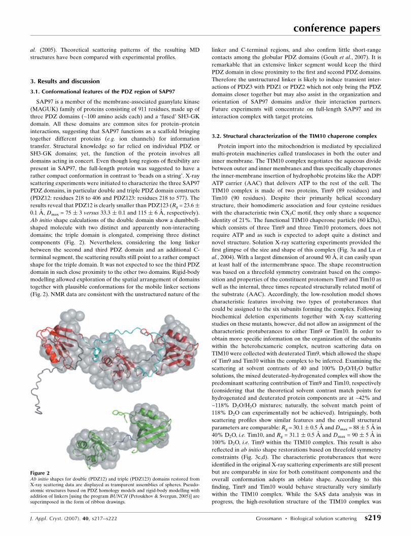
novel structure. Solution X-ray scattering experiments provided the
first glimpse of the size and shape of this complex (Fig. 3a and Lu et
al., 2004). With a largest dimension of around 90 A, it can easily span
at least half of the intermembrane space. The shape reconstruction
was based on a threefold symmetry constraint based on the compo-
sition and properties of the constituent protomers Tim9 and Tim10 as
well as the internal, three times repeated structurally related motif of
the substrate (AAC). Accordingly, the low-resolution model shows
characteristic features involving two types of protuberances that
could be assigned to the six subunits forming the complex. Following
biochemical deletion experiments together with X-ray scattering
studies on these mutants, however, did not allow an assignment of the
characteristic protuberances to either Tim9 or Tim10. In order to
obtain more specific information on the organization of the subunits
within the heterohexameric complex, neutron scattering data on
TIM10 were collected with deuterated Tim9, which allowed the shape
of Tim9 and Tim10 within the complex to be inferred. Examining the
scattering at solvent contrasts of 40 and 100% D2O/H2O buffer
solutions, the mixed deuterated–hydrogenated complex will show the
predominant scattering contribution of Tim9 and Tim10, respectively
(considering that the theoretical solvent contrast match points for
hydrogenated and deuterated protein components are at ~42% and
~118% D2O/H2O mixtures; naturally, the solvent match point of
118% D2O can experimentally not be achieved). Intriguingly, both
scattering profiles show similar features and the overall structural
parameters are comparable: Rg = 30.1� 0.5 A and Dmax = 88� 5 A in
40% D2O, i.e. Tim10, and Rg = 31.1 � 0.5 A and Dmax = 90 � 5 A in
100% D2O, i.e. Tim9 within the TIM10 complex. This result is also
reflected in ab initio shape restorations based on threefold symmetry
constraints (Fig. 3c,d). The characteristic protuberances that were
identified in the original X-ray scattering experiments are still present
but are comparable in size for both constituent components and the
overall conformation adopts an oblate shape. According to this
finding, Tim9 and Tim10 would behave structurally very similarly
within the TIM10 complex. While the SAS data analysis was in
progress, the high-resolution structure of the TIM10 complex was
conference papers
J. Appl. Cryst. (2007). 40, s217–s222 Grossmann � Biological solution scattering s219
Figure 2Ab initio shapes for double (PDZ12) and triple (PDZ123) domains restored fromX-ray scattering data are displayed as transparent assemblies of spheres. Pseudo-atomic structures based on PDZ homology models and rigid-body modelling withaddition of linkers [using the program BUNCH (Petoukhov & Svergun, 2005)] aresuperimposed in the form of ribbon drawings.
solved and published (Webb et al., 2006) providing clarification of
some of the structural peculiarities raised by the scattering results. So,
the complex has a new architecture consisting of a six-bladed �-
propeller, in which the alternate arrangement of Tim9 and Tim10
subunits produces a central ring with pseudo-sixfold symmetry. The
propeller blades extend from this core like tentacles, breaking the
symmetrical layout due to their inherent flexibility. The latter and the
non-compactness of the overall structure in solution complicate a
consistent ab initio shape reconstruction, as can be seen from a
comparison with shapes based on three- and sixfold symmetry
constraints (Fig. 3a,b). Yet there is an excellent agreement consid-
ering the evaluation of simulated and experimental scattering profiles
(Fig. 3e). Further investigations concerning the interaction with and
escorting of precursor proteins will capitalize on the TIM10 structure.
3.3. Shape analysis and simulations of the bacterial potassium
channel KcsA
The bacterial potassium channel KcsA from Streptomyces lividans
was used as a model system for structural and functional analysis of
membrane protein channels, as it has proven extraordinarily acces-
sible to structural studies (Sansom et al., 2002). The successful
isolation and purification of membrane proteins from their native
state requires the substitution of the lipid bilayer by micelle-forming
detergent molecules. Screening a series of detergents revealed that
decyl-�-d-maltopyranoside (DM) not only solubilized KcsA well but
also maintained the structural integrity of the channel tetramer in
solution (Zimmer, 2003). Given the number of potassium channel
structures available, it provides a unique opportunity to understand
the functional properties of a gated, ion-selective channel at the
molecular level. Even though a mechanism of gating in the KcsA
channel is beginning to emerge from a variety of techniques, the
conference papers
s220 Grossmann � Biological solution scattering J. Appl. Cryst. (2007). 40, s217–s222
Figure 3Shape reconstructions for the TIM10 complex based on three- (a) and sixfold (b) symmetry constraints compared with the superimposed ribbon model from the recentlysolved high-resolution structure. Shapes for Tim9 (c) and Tim10 (d) within the complex deduced from neutron contrast studies are also displayed, as well as the scatteringprofile simulation (e) calculated using the TIM10 crystal structure with added missing residues (f) restored by BUNCH and yielding a goodness-of-fit value of � = 1.82.
Figure 4Proposed ribbon structure (left) and experimental shape (right) of full-lengthKcsA. The sphere model corresponds to the reconstructed shape from SANS dataat maximum contrast (in 100% D2O). The red space-filling model depicts thecrystal structure of truncated KcsA that is imbedded in the membrane.
structure of the full-length channel (including the C-terminal
domain) has so far been elusive. The cytoplasmic C-terminal domain
has been shown to play a critical role in the pH-dependent gating
process (Cortes et al., 2001). The bacterial K+ channel KcsA solubi-
lized in DM was analysed by neutron and X-ray small-angle solution
scattering (Zimmer et al., 2006). Before the experiments, samples
were dialysed for several hours in a 30 kDa cut-off membrane at
room temperature against dialysis buffer containing 5 mM detergent
[corresponding to about twice the critical micelle concentration
(CMC) of DM, where the CMC refers to the total concentration of
detergent that corresponds to the maximum possible concentration of
detergent monomers in solution]. The C-terminally truncated version
of KcsA, amenable for crystallographic studies, was compared with
the full-length channel. Analysing the scattering data in terms of
radius of gyration reveals differences between both KcsA species of
up to 13.2 A. Equally, the real-space distance distribution identifies a
40–50 A extension of full-length KcsA compared to its C-terminally
truncated counterpart. The X-ray and neutron scattering data are
amenable for molecular shape reconstruction of full-length KcsA.
The molecular envelopes calculated for full-length KcsA display an
hourglass-shaped structure of the C-terminal domain (Fig. 4). The C-
terminus extends the membrane spanning region of KcsA by 54 to
70 A with a central constriction 10 to 30 A wide. SAS was also used to
characterize the KcsA channel under conditions favouring its open
conformation. The solution scattering at pH 5.0 of full-length KcsA
shows characteristics of a dumbbell-shaped macromolecular struc-
ture, originating from dimerization of the tetrameric K+ channel.
Since C-terminally truncated KcsA measured under the same low pH
conditions remains tetrameric, oligomerization of full-length KcsA
seems to proceed via structurally changed C-terminal domains. The
determined maximum dimensions of the newly formed complex
increase by 50 to 60%. Shape reconstruction of the pseudo-octameric
complex indicates the pH-induced conformational reorganization of
the intracellular domain (Zimmer et al., 2006). It is interesting to note
that the dimerization of the detergent-solubilized KcsA K+ channel at
low pH most certainly is due to an in vitro effect. However, the
observed dimerization is very likely stabilizing a conformation of the
KcsA channel which in vivo might be achieved by the relevant
interaction partner(s).
As a result of the close resemblance between restored shape and
structural prediction for full-length KcsA (Fig. 4), which was under-
lined by simple scattering profile simulations (Zimmer et al., 2006),
more detailed simulations on KcsA were initiated based on MD
calculations of membrane proteins and their interactions with
detergents (Bond et al., 2005; Bond & Sansom, 2006). As a first step,
the simulation of a detergent micelle was attempted and compared to
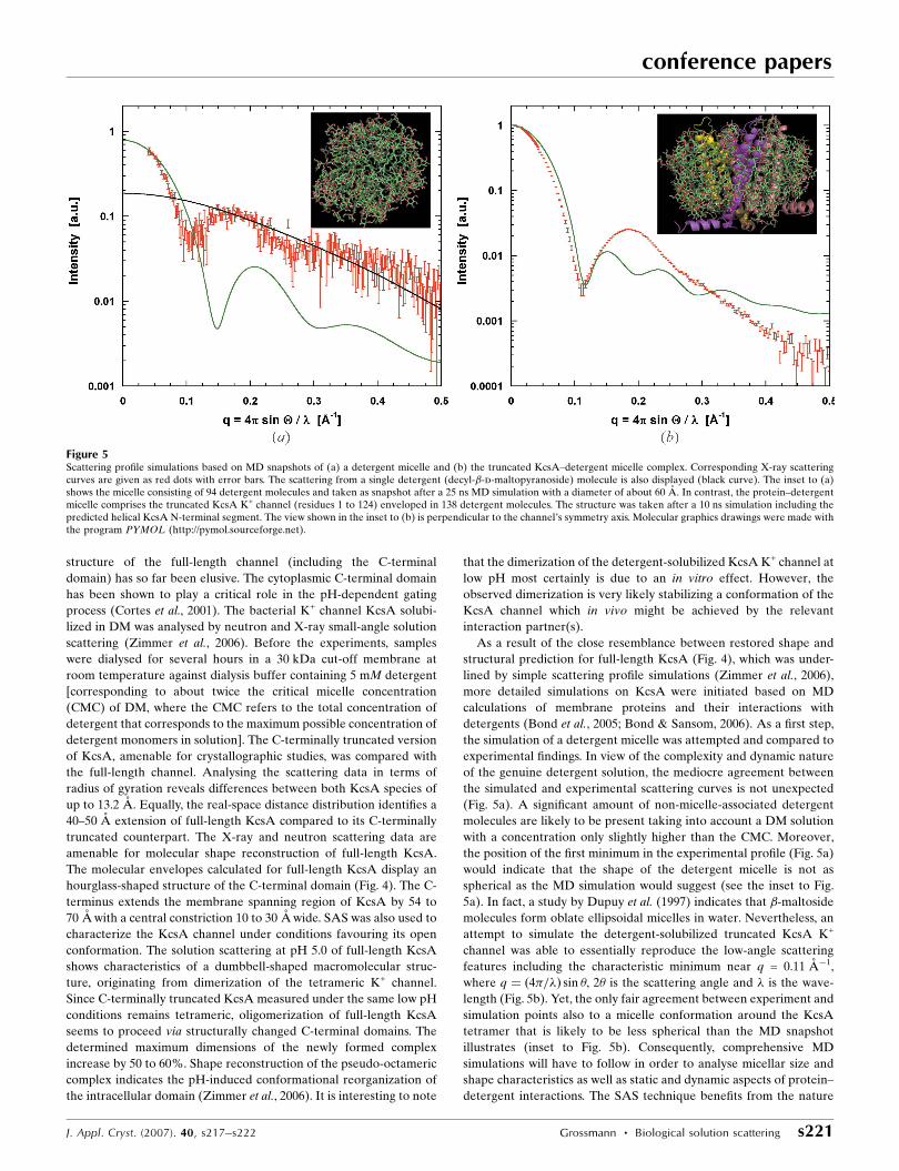
experimental findings. In view of the complexity and dynamic nature
of the genuine detergent solution, the mediocre agreement between
the simulated and experimental scattering curves is not unexpected
(Fig. 5a). A significant amount of non-micelle-associated detergent
molecules are likely to be present taking into account a DM solution
with a concentration only slightly higher than the CMC. Moreover,
the position of the first minimum in the experimental profile (Fig. 5a)
would indicate that the shape of the detergent micelle is not as
spherical as the MD simulation would suggest (see the inset to Fig.
5a). In fact, a study by Dupuy et al. (1997) indicates that �-maltoside
molecules form oblate ellipsoidal micelles in water. Nevertheless, an
attempt to simulate the detergent-solubilized truncated KcsA K+
channel was able to essentially reproduce the low-angle scattering
features including the characteristic minimum near q = 0.11 A�1,
where q ¼ ð4�=�Þ sin �, 2� is the scattering angle and � is the wave-
length (Fig. 5b). Yet, the only fair agreement between experiment and
simulation points also to a micelle conformation around the KcsA
tetramer that is likely to be less spherical than the MD snapshot
illustrates (inset to Fig. 5b). Consequently, comprehensive MD
simulations will have to follow in order to analyse micellar size and
shape characteristics as well as static and dynamic aspects of protein–
detergent interactions. The SAS technique benefits from the nature
conference papers
J. Appl. Cryst. (2007). 40, s217–s222 Grossmann � Biological solution scattering s221
Figure 5Scattering profile simulations based on MD snapshots of (a) a detergent micelle and (b) the truncated KcsA–detergent micelle complex. Corresponding X-ray scatteringcurves are given as red dots with error bars. The scattering from a single detergent (decyl-�-d-maltopyranoside) molecule is also displayed (black curve). The inset to (a)shows the micelle consisting of 94 detergent molecules and taken as snapshot after a 25 ns MD simulation with a diameter of about 60 A. In contrast, the protein–detergentmicelle comprises the truncated KcsA K+ channel (residues 1 to 124) enveloped in 138 detergent molecules. The structure was taken after a 10 ns simulation including thepredicted helical KcsA N-terminal segment. The view shown in the inset to (b) is perpendicular to the channel’s symmetry axis. Molecular graphics drawings were made withthe program PYMOL (http://pymol.sourceforge.net).
of these simulations, which will help to interpret the experimental
data, but at the same time emerges as an appreciated experimental
tool to provide feedback for the theoretical observations.
4. Conclusions
Considering the substantial advances in scattering-data-analysis
methods for biological molecules in solution over the last decade,
more complex and larger macromolecules can be meaningfully
investigated with SAS. In view of the wealth of known high-resolu-
tion protein domain structures, the focus of scattering experiments
will increasingly move from ab initio shape determination to rigid-
body modelling of full-length multidomain proteins and multi-
subunit complexes. Concomitantly, neutron contrast variation (in
particular in the form of selectively labelled protein subunits),
distance constraints between specific molecular surface areas (e.g.
deduced from NMR or mutagenesis studies) and computer simula-
tions will help to further reduce ambiguities so as to arrive at
biologically relevant and unique results. Along with findings from
other techniques, the collective information will permit crucial
insights into challenging questions of structural biology to be gained,
such as the origins of aberrant protein structure–function relation-
ships and their consequences in human diseases. Moreover, with the
continuing enhancements in high-performance computing, SAS data
ought to become a convenient assessment and a viable constraint for
molecular dynamics simulations. These advancements are expected
to raise the profile of SAS, pushing the percentage of publications in
structural biology with contributions from SAS towards the 5%
margin.
The solution scattering investigations would not have been
possible without the enthusiasm, interest and encouragement of
several collaborators and colleagues: Professor Lu-Yun Lian and her
group at Liverpool University (PDZ domains), Dr Kostas Tokatlidis
and his group at the University of Crete (TIM10 chaperone complex)
and Drs Declan Doyle and Jochen Zimmer at Oxford University
(bacterial potassium channel, KcsA). The KcsA study initiated MD
simulations of protein–micelle interactions in Professor Mark
Sansom’s group (with Drs Peter Bond and Zara Sands) at Oxford
University. My appreciation also goes to Dr Peter Timmins at the ILL
for his support, not only during neutron scattering experiments, and
Dr Michael Haertlein and the staff at the ILL–EMBL–PSB
Deuteration Laboratory in Grenoble, which enabled the labelling of
the TIM10 complex. Last but not least I would like to thank my
colleagues at the Daresbury SRS as well as the support and funding
from CCLRC Daresbury Laboratory.
References
Bond, P. J., Cuthbertson, J. & Sansom, M. S. P. (2005). Biochem. Soc. Trans. 33,910–912.
Bond, P. J. & Sansom, M. S. P. (2003). J. Mol. Biol. 329, 1035–1053.Bond, P. J. & Sansom, M. S. P. (2006). J. Am. Chem. Soc. 128, 2697–2704.Chacon, P., Moran, F., Diaz, J. F., Pantos, E. & Andreu, J. M. (1998). Biophys. J.
74, 2760–2775.Cortes, D. M., Cuello, L. G. & Perozo, E. (2001). J. Gen. Physiol. 117, 165–180.Dupuy, C., Auvray, X., Petipas, C., Rico-Lattes, I. & Lattes, A. (1997).
Langmuir, 13, 3965–3967.Goult, B. T., Rapley, J. D., Golovanova, M., Sampson, L. J., Dart, C., Kitmitto,
A., Grossmann, J. G., Leyland, M. L. & Lian, L.-Y. (2007). Biochemistry.Submitted.
Grishaev, A., Wu, J., Trewhella, J. & Bax, A. (2005). J. Am. Chem. Soc. 127,16621–16628.
Heller, W. T., Abusamhadneh, E., Finley, N., Rosevear, P. R. & Trewhella, J.(2002). Biochemistry, 41, 15654–15663.
Konarev, P. V., Petoukhov, M. V., Volkov, V. V. & Svergun, D. I. (2006). J. Appl.Cryst. 39, 277–286.
Lu, H., Golovanov, A. P., Alcock, F., Grossmann, J. G., Allen, S., Lian, L.-Y. &Tokatlidis, K. (2004). J. Biol. Chem. 279, 18959–18966.
Mattinen, M.-L., Paakkonen, K., Ikonen, T., Craven, J., Drakenberg, T.,Serimaa, R., Waltho, J. & Annila, A. (2002). Biophys. J. 83, 1177–1183.
Patargias, G., Bond, P. J., Deol, S. S. & Sansom, M. S. P. (2005). J. Phys. Chem.B109, 575–582.
Petoukhov, M. V. & Svergun, D. I. (2005). Biophys. J. 89, 1237–1250.Sansom, M. S. P., Shrivastava, I. H., Bright, J. N., Tate, J., Capener, C. E. &
Biggin, P. C. (2002). Biochim. Biophys. Acta, 1565, 294–307.Sondermann, H., Nagar, B., Bar-Sagi, D. & Kuriyan, J. (2005). Proc. Natl Acad.
Sci. USA, 102, 16632–16637.Svergun, D. I. (1999). Biophys. J. 76, 2879–2886.Svergun, D. I. & Koch, M. H. J. (2003). Rep. Prog. Phys. 66, 1735–1782.Svergun, D. I., Petoukhov, M. V. & Koch, M. H. J. (2001). Biophys. J. 80, 2946–
2953.Svergun, D. I. & Stuhrmann, H. B. (1991). Acta Cryst. A47, 736–744.Walther, D., Cohen, F. E. & Doniach, S. (2000). J. Appl. Cryst. 33, 350–363.Webb, C. T., Gorman, M. A., Lazarou, M., Ryan, M. T. & Gulbis, J. M. (2006).
Mol. Cell, 21, 123–133.Wu, Y., Tian, X., Lu, M., Chen, M., Wang, Q. & Ma, J. (2005). Structure, 13,
1587–1597.Zimmer, J. (2003). PhD Thesis, Department of Biochemistry, University of
Oxford.Zimmer, J., Doyle, D. A. & Grossmann, J. G. (2006). Biophys. J. 90, 1752–1766.
conference papers
s222 Grossmann � Biological solution scattering J. Appl. Cryst. (2007). 40, s217–s222


![Evaluation of a spectrally resolved scattering microscope · sis of biological tissue [12]. The contrast is given only by the scattering, so the technique is marker-free and no further](https://img.pdfslide.us/doc/110x75/60e2f3b7a2c35741795224cc/evaluation-of-a-spectrally-resolved-scattering-microscope-sis-of-biological-tissue.jpg)